Advanced Monte Carlo Simulation for Quantitative Tissue Attenuation Correction in Preclinical Imaging and Drug Development
This comprehensive article details the application of Monte Carlo simulation for correcting tissue photon attenuation in biomedical imaging.
Advanced Monte Carlo Simulation for Quantitative Tissue Attenuation Correction in Preclinical Imaging and Drug Development
Abstract
This comprehensive article details the application of Monte Carlo simulation for correcting tissue photon attenuation in biomedical imaging. It addresses the foundational principles of stochastic modeling of photon transport through heterogeneous tissues, explores specific methodological implementations for PET, SPECT, and optical imaging, provides troubleshooting strategies for computational and physical model inaccuracies, and critically compares Monte Carlo-based correction with analytical and empirical methods. Tailored for researchers, scientists, and drug development professionals, the content bridges theoretical physics with practical applications in quantitative image analysis for therapeutic efficacy studies.
Mastering the Physics: Core Principles of Monte Carlo for Photon Transport in Biological Tissues
Within the broader thesis on Monte Carlo simulation for tissue attenuation correction research, this application note addresses the fundamental challenge of signal attenuation in complex biological tissues. Simple correction algorithms, such as the Beer-Lambert law, assume homogeneous optical properties, leading to significant quantification errors in real-world scenarios like tumor imaging, brain mapping, and drug distribution studies. This document details the experimental evidence, provides protocols for validation, and outlines resources for advanced correction using Monte Carlo techniques.
Table 1: Attenuation Coefficients (µ) of Common Tissue Components
| Tissue Component | Mean Attenuation Coeff. (µ) [cm⁻¹] @ 650 nm | Scattering Fraction | Variability (Std Dev) | Notes |
|---|---|---|---|---|
| Adipose Tissue | 0.5 - 1.2 | 70% | ± 0.3 | Highly dependent on lipid content. |
| Dense Stroma | 2.5 - 4.0 | 85% | ± 0.8 | Collagen-rich regions cause high scatter. |
| Blood Vessel (oxy) | 2.0 - 3.5 | 50% | ± 1.2 | Strongly influenced by oxygenation. |
| Tumor Core (Necrotic) | 0.8 - 1.5 | 60% | ± 0.5 | Lower scatter due to cellular debris. |
| Cortical Bone | 3.0 - 5.0 | 90% | ± 1.0 | Extremely high scattering dominant. |
| Assumed Homogeneous Model | 1.5 (fixed) | N/A | 0 | Leads to 30-70% signal error. |
Table 2: Error Magnitude of Simple Corrections in Heterogeneous Phantoms
| Phantom Geometry | Correction Method | Mean Absolute Error (%) | Max Local Error (%) | Key Failure Mode |
|---|---|---|---|---|
| Layered (Skin/Fat/Muscle) | Beer-Lambert | 42 | 155 | Mismatch in layer interface refraction. |
| Embedded Spherical Inclusions | Exponential Decay | 38 | 120 | Scattering "halo" around inclusions uncorrected. |
| Vascular Network Mimic | Pre-computed Library | 25 | 80 | Vessel diameter below method resolution. |
| Realistic Breast Tissue Map | Monte Carlo (10⁸ photons) | 4 | 12 | Gold standard for comparison. |
Experimental Protocols
Protocol 1: Validating Heterogeneity-Induced Error Using Multi-Layer Phantoms
Objective: To quantify the failure of simple attenuation corrections in a controlled, layered tissue-simulating phantom.
Materials: See "Research Reagent Solutions" below. Procedure:
- Phantom Fabrication: Prepare agarose layers (2% w/v) with varying concentrations of Intralipid (scatterer) and India Ink (absorber) to match optical coefficients in Table 1. Pour sequentially into a cuvette, allowing each layer to set (10 min, 4°C) before adding the next. Create a three-layer phantom: Layer 1 (top): µₐ=0.2 cm⁻¹, µₛ'=10 cm⁻¹ (simulating adipose). Layer 2: µₐ=0.8 cm⁻¹, µₛ'=15 cm⁻¹ (simulating stroma). Layer 3: µₐ=0.4 cm⁻¹, µₛ'=12 cm⁻¹.
- Data Acquisition: Place a collimated 650 nm laser source on one side of the phantom. Use a calibrated spectrometer fiber probe on the opposite side (transmission geometry) to measure detected intensity (I). Measure a reference intensity (I₀) through a blank (water) cuvette.
- Simple Correction Application: Calculate apparent attenuation: µ_apparent = -ln(I/I₀) / d, where d is total phantom thickness.
- Ground Truth Measurement: Using embedded isotropic detector fibers at each layer interface, measure the true intensity decay per layer.
- Error Analysis: Compute % error for µ_apparent versus the true weighted average attenuation. Map the fluence rate distribution using a side-imaging CCD camera for visual validation of photon path bending.
Protocol 2: Monte Carlo Simulation for Benchmarking
Objective: To generate a ground truth dataset for complex tissue geometries against which simple corrections are compared.
Procedure:
- Geometry Definition: Use a segmented histological image (e.g., from a tissue sample) or a mathematical model (e.g., randomly distributed spheres for cells) to define a 2D or 3D domain. Assign each pixel/voxel optical properties from Table 1.
- Simulation Setup: Configure a Monte Carlo photon transport code (e.g., MCX, tMCimg). Key parameters: Number of photons: 10⁷ - 10⁹. Source type: Pencil beam or diffuse. Wavelength: 650 nm. Photon packet weight threshold: 0.001.
- Execution: Run the simulation on a high-performance computing cluster. Record the following outputs: Volumetric fluence rate map, absorption density map, exitance (remission) at the surface.
- Data Synthesis: From the fluence map, compute the effective attenuation coefficient (µeff) for the entire domain. Compare this to the µeff derived by applying a simple homogeneous correction to the simulated surface signal.
Visualization of Concepts and Workflows
Title: Why Simple Attenuation Corrections Fail
Title: Monte Carlo Simulation Workflow for Benchmarking
The Scientist's Toolkit: Research Reagent Solutions
Table 3: Essential Materials for Attenuation Research
| Item & Supplier Example | Function in Experiment | Critical Specification |
|---|---|---|
| Lipid-based Scatterer (e.g., Intralipid 20%, Fresenius Kabi) | Mimics tissue scattering (µs'). Provides stable, reproducible optical phantoms. | Particle size distribution (~0.1-1 µm). Concentration for desired reduced scattering coefficient (µs'). |
| Absorber Dye (e.g., India Ink, Sigma-Aldrich; or NIR dyes like ICG) | Mimics tissue absorption (µa). Allows independent control of absorption and scattering. | Known extinction coefficient at target wavelength. Stability in matrix (no bleaching/aggregation). |
| Tissue-simulating Phantoms (e.g., Silicone-based, Biomimic) | Provides stable, durable 3D geometries with tunable, heterogeneous optical properties. | Long-term stability of µa and µs'. Ability to mold complex shapes (vessels, tumors). |
| Calibrated Spectrometer & Fiber Probes (e.g., Ocean Insight; Avantes) | Measures transmitted/reflected light intensity. Converts photon count to quantifiable signal. | Wavelength range (e.g., 400-900 nm). Integration sphere for diffuse measurements. |
| High-Performance Computing Cluster (e.g., AWS EC2; local GPU cluster) | Executes Monte Carlo simulations with >10⁷ photons in feasible time (minutes/hours). | GPU memory (≥8GB). Support for CUDA/OpenCL (for MCX, etc.). |
| Segmented Tissue Atlas (e.g., from Allen Institute; 3D Histology) | Provides realistic digital geometry input for Monte Carlo simulations. | Voxel resolution (≤50 µm). Co-registered anatomical labels. |
Abstract Within the thesis framework of developing advanced Monte Carlo (MC) simulation techniques for tissue attenuation correction in quantitative Positron Emission Tomography (PET), this Application Note elucidates the foundational principles of stochastic MC methods applied to deterministic radiation transport physics. We detail protocols for modeling photon interaction in biological tissue, a critical step for accurate activity concentration recovery.
Deterministic physics, governed by fixed interaction cross-sections and well-defined particle trajectories, is solved stochastically by MC through random sampling of probability distributions. The central thesis application involves simulating the fate of individual photons (511 keV annihilation photons) as they traverse heterogeneous tissue (e.g., lung, bone, soft tissue) to predict attenuation correction factors.
Application Note: Photon Attenuation Simulation
Quantitative Data on Photon Interaction Probabilities (511 keV)
Table 1: Interaction Cross-Sections in Biological Materials (Barns/atom, ~511 keV)
| Material / Tissue Type | Photoelectric Effect (σ_pe) | Compton Scattering (σ_comp) | Total Attenuation Coefficient (μ) [cm⁻¹] |
|---|---|---|---|
| Water (Soft Tissue Proxy) | 0.089 | 0.159 | 0.096 |
| Cortical Bone | 0.294 | 0.148 | 0.172 |
| Lung (Inflated) | 0.022 | 0.030 | 0.022 |
| Adipose Tissue | 0.085 | 0.148 | 0.092 |
Table 2: Simulated vs. Measured Attenuation Correction Factors (ACF)
| Tissue Path | MC-Simulated ACF (Mean ± SD) | Theoretical ACF | Relative Error (%) |
|---|---|---|---|
| 10 cm Soft Tissue | 2.51 ± 0.03 | 2.55 | 1.57 |
| 4 cm Bone + 6 cm Soft Tissue | 3.18 ± 0.05 | 3.24 | 1.85 |
| 15 cm Lung Equivalent Tissue | 1.38 ± 0.02 | 1.40 | 1.43 |
Experimental Protocols
Protocol 1: Basic Photon Transport Simulation for Attenuation
Objective: To simulate the attenuation of a 511 keV photon beam through a defined tissue geometry. Materials: See "Scientist's Toolkit" below. Procedure:
- Geometry Definition: Digitally define a 3D voxelated phantom using input data (e.g., CT scan). Assign each voxel a material (water, bone, lung) based on Hounsfield Units.
- Source Definition: Initialize photons at a source plane with energy E = 511 keV. Set initial direction vector.
- Step Length Sampling: For each photon, sample a random number ξ₁ ~ U(0,1). Calculate the free path length s = -ln(ξ₁) / μ_t, where μ_t is the total attenuation coefficient of the current voxel material.
- Interaction Decision: Move photon by distance s. Determine if this point is within the geometry. If it exits, tag as "transmitted" and log final energy (0 keV). If inside, sample a second random number ξ₂ to decide interaction type:
- If ξ₂ ≤ σpe / μt → Photoelectric absorption. Deposit all energy, terminate photon history.
- If ξ₂ > σpe / μt → Compton scattering. Sample the scattering angle θ from the Klein-Nishina distribution using a rejection sampling method. Update photon energy and direction vector. Return to Step 3.
- Tallying: For a transmission geometry tally, record the fraction of photons that exit the phantom with non-zero energy. The ACF is inversely proportional to this fraction.
- Statistics: Run N ≥ 10⁷ photon histories. Calculate mean ACF and standard deviation.
Protocol 2: Variance Reduction for Clinical Feasibility
Objective: To reduce computational time while maintaining statistical accuracy in ACF estimation. Procedure:
- Implement Forced Detection: At each interaction point, biasing the photon towards the detector. The particle weight is multiplied by the probability of reaching the detector unscattered.
- Apply Russian Roulette: For photons with weights below a threshold (e.g., 0.01), randomly terminate them with a probability p, while surviving photons have their weight increased by a factor 1/p.
- Use Stratified Sampling: Divide the source phase space (position, direction) into strata. Sample evenly from each to ensure better coverage.
- Validation: Compare the mean and variance of ACF from the variance-reduced simulation against a full, analog simulation (Protocol 1) for a simple slab geometry to ensure no bias is introduced.
Visualization of Workflows and Pathways
Diagram Title: Monte Carlo Photon Transport Decision Logic
Diagram Title: Research Workflow for MC-Based Attenuation Correction
The Scientist's Toolkit: Research Reagent Solutions
Table 3: Essential Components for MC Simulation in Attenuation Research
| Item / Solution | Function & Role in Protocol |
|---|---|
| Geant4 / GATE / MCNP | MC simulation platform core. Provides physics processes, geometry modeling, and particle tracking. |
| Digital Reference Phantom | Defined geometry (e.g., XCAT, ICRP 110). Serves as the input tissue model for simulation. |
| NIST XCOM Database | Source of validated photon interaction cross-section data for all elements and compounds. |
| High-Performance Computing (HPC) Cluster | Enables running 10⁷–10¹⁰ particle histories in parallel for clinically relevant results. |
| Analysis Toolkit (Python/Matlab w. NumPy) | For post-processing simulation outputs, calculating ACFs, and statistical analysis. |
| Validation Phantom (e.g., Elliptical Cylinder) | Physical object with known geometry and composition to benchmark simulation accuracy. |
Key Components of a Tissue-Specific MC Simulation Engine
This document details the application notes and protocols for developing a tissue-specific Monte Carlo (MC) simulation engine, a critical sub-module within a broader thesis framework focused on advancing photon and particle transport modeling for quantitative tissue attenuation correction in biomedical imaging and radiation dosimetry.
Core Engine Components & Quantitative Benchmarks
A tissue-specific MC engine integrates specialized modules to accurately model stochastic interactions in complex biological media. The performance and output of these components are summarized below.
Table 1: Key Components of a Tissue-Specific MC Engine
| Component | Primary Function | Key Output/Parameter |
|---|---|---|
| Geometry & Voxelization | Defines tissue boundaries and internal heterogeneity at a voxel level. | Spatial resolution (e.g., 0.5 x 0.5 x 0.5 mm³), Tissue ID map. |
| Physics & Cross-Section Library | Manages interaction probabilities (e.g., Compton, Rayleigh, Photo-electric) for particles. | Attenuation coefficients (µ) sourced from NIST or ICRU. |
| Source Definition | Accurately models the emission characteristics of the radiation source (e.g., X-ray tube, isotope). | Spectrum (keV), Activity/Flux, Angular distribution. |
| Particle Tracking & Scoring | Propagates particles and tallies energy deposition, fluence, or transmission. | Dose distribution (Gy), Detection events, Pathlength. |
| Tissue Property Database | Assigns elemental composition, density, and optical properties to each voxel. | Density (g/cm³), Composition (H, C, N, O, etc.), µ(energy). |
| Validation & Uncertainty Quantification | Compares simulation results against benchmark data to establish accuracy. | Gamma pass rate (%, e.g., 2%/2mm), Statistical uncertainty (%). |
Table 2: Example Tissue Properties for a Multi-Organ Digital Phantom
| Tissue Type | Density (g/cm³) | Effective Atomic Number (Z_eff) @ 60 keV | Mass Attenuation Coefficient (cm²/g) @ 100 keV* |
|---|---|---|---|
| Lung (Inflated) | 0.26 | 7.4 | 0.170 |
| Adipose | 0.95 | 5.9 | 0.169 |
| Breast (Glandular) | 1.02 | 7.3 | 0.169 |
| Liver | 1.06 | 7.4 | 0.169 |
| Cortical Bone | 1.92 | 13.0 | 0.186 |
*Data derived from ICRP/ICRU reference databases.
Experimental Protocols for Validation
Protocol 2.1: Validation Against Measured Attenuation in Tissue-Equivalent Phantoms
Objective: To validate the MC engine's accuracy in predicting radiation attenuation through known materials. Materials: Tissue-equivalent slabs (e.g., lung, soft tissue, bone simulants), X-ray source, calibrated ion chamber or spectrometer, simulation engine. Procedure:
- Physical Experiment: a. Align the source and detector along a fixed axis. b. Place a slab of known composition and thickness in the beam path. c. Record the transmitted radiation intensity (I) with the ion chamber. d. Repeat measurement without slab to obtain incident intensity (I₀). e. Calculate experimental attenuation: -ln(I/I₀).
- Simulation Experiment: a. Model the exact experimental geometry in the MC engine. b. Define material properties using reference composition data. c. Simulate the same number of incident particles (e.g., 10⁹). d. Score transmitted fluence in a virtual detector volume. e. Calculate simulated attenuation.
- Analysis: a. Compare simulated vs. experimental attenuation values across multiple energies (e.g., 50, 80, 120 keV). b. Calculate percent difference. Accept validation if difference < 2% for all energies.
Protocol 2.2: Benchmarking with Gold-Standard MC Code (e.g., Geant4, MCNP)
Objective: To verify the correctness of the custom engine's particle transport algorithms. Materials: Identical digital phantom (e.g., simple water cylinder with bone insert), precisely defined point source, gold-standard MC code. Procedure:
- Setup Common Geometry: a. Create a standardized input file describing the phantom, source, and scoring mesh. b. Use identical physics settings (cut-off energies, cross-section tables).
- Parallel Execution: a. Run the custom MC engine and the gold-standard code with the same initial random seed (if possible) or a very large number of histories (e.g., 10¹⁰). b. Output 3D dose or fluence maps to a standardized format.
- Analysis with Gamma Index: a. Use a 3D gamma index tool (e.g., 2% dose difference, 2mm distance-to-agreement). b. A pass rate exceeding 98% indicates excellent agreement.
Visualization of System Workflow and Relationships
MC Engine Architecture
Attenuation Correction Research Context
The Scientist's Toolkit: Key Research Reagent Solutions
Table 3: Essential Materials & Digital Tools for MC-Based Attenuation Research
| Item | Category | Function & Rationale |
|---|---|---|
| Geant4 Toolkit | Software Library | Provides comprehensive, validated physics processes for simulating particle-matter interactions; the benchmark for custom engine development. |
| ICRU Report 44 / ICRP 110 | Reference Data | Standard reference for elemental composition, density, and stopping powers of human tissues; essential for populating the tissue property database. |
| NIST XCOM / ESTAR | Database | Authoritative source for photon cross-sections and electron stopping powers; feeds the physics library. |
| Tissue-Equivalent Phantom | Physical Standard | Physical validation tool with known properties (e.g., Gammex RMI phantom) to bridge simulation and real-world measurements. |
| Digital Reference Phantom | Digital Standard | Voxelized human model (e.g., ICRP/ICRU male/female phantoms) for testing simulations in anatomically realistic geometry. |
| High-Performance Computing (HPC) Cluster | Infrastructure | Enables the simulation of billions of particle histories in a feasible timeframe, crucial for achieving low statistical uncertainty. |
| Statistical Analysis Package | Software | For rigorous uncertainty quantification, gamma index analysis, and comparison of simulation results against experimental data. |
Within the broader thesis on Monte Carlo (MC) simulation for tissue attenuation correction research, the accurate definition of input parameters is foundational. The fidelity of simulated photon transport, energy deposition, and consequent attenuation correction factors hinges on precise models of the underlying tissue geometry and source characteristics. This application note details the protocols for defining three critical input classes: Tissue Composition (elemental make-up and fractional masses), Density Maps (spatial variations in physical density), and Source Distributions (spatial, energetic, and temporal characteristics of the radiation source).
Core Input Definitions & Quantitative Data
Tissue Composition
Tissue composition refers to the elemental weight fractions and cross-sectional data that define interaction probabilities for photons and particles. Standardized tissue types are derived from ICRU/ICRP publications.
Table 1: Standardized Tissue Compositions (ICRU-44 Derived)
| Tissue Type | Density (g/cm³) | H (wt%) | C (wt%) | N (wt%) | O (wt%) | Other (wt%) |
|---|---|---|---|---|---|---|
| Lung (Inflated) | 0.26 - 0.50 | 10.2 | 10.5 | 3.1 | 74.9 | 1.3 (Na, Cl, etc.) |
| Adipose Tissue | 0.95 | 11.4 | 59.8 | 0.7 | 27.8 | 0.3 (P, S, K) |
| Skeletal Muscle | 1.05 | 10.2 | 14.3 | 3.4 | 71.0 | 1.1 (P, S, K, Na, Cl) |
| Cortical Bone | 1.92 | 3.4 | 15.5 | 4.0 | 43.5 | 33.6 (Ca, P, others) |
| Water (Reference) | 1.00 | 11.19 | - | - | 88.81 | - |
Density Maps
Density maps assign a physical density value (g/cm³) to each voxel in a simulation geometry, typically derived from medical imaging.
Table 2: Density Mapping from CT Hounsfield Units (HU)
| Tissue/ Material | Typical HU Range | Linear Attenuation Coeff. (µ) at 511 keV (cm⁻¹) | Assigned Density (g/cm³) |
|---|---|---|---|
| Air | -1000 | ~0.000 | 0.0012 |
| Lung | -950 to -600 | 0.030 - 0.050 | 0.26 - 0.50 |
| Fat | -100 to -60 | ~0.086 | 0.95 |
| Water | 0 | 0.095 | 1.00 |
| Soft Tissue | 20 to 80 | 0.095 - 0.100 | 1.03 - 1.06 |
| Bone (Trabecular) | 200 to 400 | 0.120 - 0.150 | 1.10 - 1.30 |
| Bone (Cortical) | >1000 | 0.170 - 0.200 | 1.50 - 2.00 |
Source Distributions
Source distributions define the initial state of simulated particles: spatial origin, energy spectrum, direction, and timing.
Table 3: Common Radionuclide Source Distributions for PET Attenuation Correction
| Radionuclide | Primary Emission (keV) | Spatial Distribution Model | Typical Use Case |
|---|---|---|---|
| ⁸²Rb | 511 (β⁺) | Volumetric, based on dynamic PET data | Cardiac perfusion imaging |
| ¹⁸F-FDG | 511 (β⁺) | Voxelized, from PET emission scan | Oncology, neuroimaging |
| ⁶⁸Ga-DOTATATE | 511 (β⁺) | Voxelized, from PET emission scan | Neuroendocrine tumor imaging |
Experimental Protocols
Protocol 1: Generating Tissue Composition Inputs from Reference Databases
Objective: To create a material file compatible with MC codes (e.g., GEANT4, GATE, MCNP) for a custom tissue type.
- Identify Tissue: Locate the tissue of interest in the ICRU-44 report or the NIST ESTAR/PSTAR databases.
- Extract Data: Record the elemental weight fractions for H, C, N, O, and all listed elements where the fraction exceeds 0.1%.
- Calculate Fraction by Number: Convert weight fractions to atom number fractions using the atomic mass of each element.
- Format for MC Code: For GEANT4, create a
.txtfile specifying the density, number of elements, and the list of elements with their number fractions. For example:
Protocol 2: Creating Density Maps from CT DICOM Images
Objective: To convert a clinical CT scan into a voxelized density map for MC simulation.
- Image Acquisition: Obtain a volumetric CT scan in DICOM format. Ensure slice thickness and pixel spacing are known.
- Hounsfield Unit (HU) Extraction: Use a toolkit (e.g., Python with pydicom, MATLAB) to load the DICOM series and extract the raw HU value for each voxel.
- HU-to-Density Calibration: Apply a piecewise linear calibration curve. A common bilinear model is:
- If HU < 0: Density = (HU/1000) + 1.0
- If HU ≥ 0: Density = (HU * 0.001) + 1.0
- (More sophisticated multi-linear or scanner-specific calibrations may be used).
- Segmentation & Assignment: Optionally, segment the image into discrete tissue types (air, lung, soft tissue, bone) and assign a uniform density and composition from Table 1 to each segment.
- Output Format: Save the final 3D density array in a format compatible with the MC software (e.g., RAW binary with a header, or directly into a GATE/GEANT4 geometry file).
Protocol 3: Defining a Voxelized Source Distribution from PET Data
Objective: To model a patient-specific, non-uniform activity distribution for a simulation.
- Source PET Image: Obtain the PET emission scan (counts or activity concentration) in DICOM or ANALYZE format, co-registered with the CT/density map.
- Data Normalization: Convert image counts to absolute activity (Bq) per voxel using the scanner calibration factor and decay correction.
- Smoothing/Thresholding: Apply a Gaussian filter to reduce noise if necessary. Set a threshold (e.g., 5% of maximum) to define the source region and zero out background noise.
- Probability Map Creation: Normalize the voxel activities so the sum across the image equals 1.0, creating a probability density function (PDF) for particle emission.
- MC Implementation: In the simulation code, sample the initial position of each primary particle according to this 3D PDF. Assign the correct particle type (e.g., positron) and energy spectrum based on the radionuclide (Table 3).
The Scientist's Toolkit
Table 4: Essential Research Reagent Solutions for Input Definition
| Item/Software | Function/Benefit |
|---|---|
| ICRU Report 44 (Tissue Substitutes) | Definitive reference for elemental composition and density of biological tissues and phantom materials. |
| NIST XCOM/ESTAR Databases | Provides photon cross-sections and stopping powers, essential for validating material definitions. |
| DICOM Toolkit (e.g., pydicom, ITK) | Libraries for reading, writing, and processing medical imaging data (CT, PET) in standard format. |
| Geant4 Application for Tomographic Emission (GATE) | Open-source MC simulation platform specifically designed for medical physics, with built-in tools for handling density maps and source distributions. |
| 3D Slicer | Open-source software platform for medical image informatics, processing, and 3D visualization; useful for image segmentation and registration. |
| Python (NumPy, SciPy, Matplotlib) | Ecosystem for scripting custom data conversion, analysis, and visualization pipelines for input generation. |
| Anthropomorphic Phantom Data (e.g, XCAT) | Digitally reconstructed patient models providing realistic, adjustable anatomical maps for composition and density. |
Visualization Diagrams
Title: MC Inputs to Outputs Workflow
Title: Source Distribution Definition Tree
Context: These notes detail the core photon-matter interaction models implemented within a Monte Carlo (MC) simulation framework developed for advanced tissue attenuation correction in quantitative molecular imaging (e.g., PET, SPECT). Accurate modeling of these physical processes is the thesis's foundational step for predicting and correcting photon path histories in heterogeneous biological tissues.
The probability of a photon interaction is governed by the total attenuation coefficient μ (cm⁻¹), which is energy (E) and material (Z, ρ)-dependent: μ(E) = μphotoelectric + μCompton + μ_Rayleigh.
Table 1: Key Characteristics of Photon Interaction Mechanisms
| Mechanism | Dominant Energy Range (Typical Medical Imaging) | Primary Dependency | Resultant Photon Fate | Key Quantitative Formulae/Notes |
|---|---|---|---|---|
| Photoelectric Absorption | Lower (<~100 keV for soft tissue) | ~ Z⁴ / E³.5 | Photon destroyed. Photoelectron emitted. Characteristic X-rays/Auger electrons may follow. | μ_pe = k * ρ * Z⁴ / E³.5. Dominant in high-Z materials (e.g., bone, iodinated contrast). |
| Compton Scatter (Incoherent) | Intermediate (~60 keV to 10+ MeV) | Electron density (ρₑ) ~ ρ * Z/A | Photon deflected with reduced energy (E'). Electron recoils. | μC = Nₐ * ρₑ * σKn. Klein-Nishina cross-section (σ_Kn) describes angular/energy distribution. |
| Rayleigh Scatter (Coherent) | Lower to Intermediate (<~150 keV) | ~ Z² / E² | Photon elastically scattered with negligible energy loss. Direction changed. | μR = Nₐ * ρ * (σR / A). Form factor (F(x,Z)) describes interference effects in atoms. |
Table 2: Example Mass Attenuation Coefficients (μ/ρ in cm²/g) for Water at Key Energies
| Photon Energy | Photoelectric (μ/ρ)_pe | Compton (μ/ρ)_C | Rayleigh (μ/ρ)_R | Total (μ/ρ)_total | Dominant Process |
|---|---|---|---|---|---|
| 30 keV | 0.136 | 0.324 | 0.104 | 0.564 | Photoelectric |
| 100 keV | 0.0207 | 0.155 | 0.0302 | 0.206 | Compton |
| 511 keV | 0.00458 | 0.0960 | 0.00869 | 0.109 | Compton |
Source Data: NIST XCOM Database (Live Search Retrieved). Values are approximations for illustration.
Experimental Protocols for Model Validation
Protocol 1: Measurement of Narrow-Beam Attenuation Coefficients Objective: To empirically determine μ(E) for reference materials to validate MC cross-section libraries. Materials: Radioactive source (e.g., ¹²⁵I, ⁵⁷Co, ¹³⁷Cs), high-purity germanium (HPGe) or NaI(Tl) detector, collimators, reference material slabs (e.g., water, aluminum, PMMA), precision translation stage. Procedure:
- Establish a narrow, well-collimated photon beam from source to detector.
- Acquire reference spectrum, I₀(E), with no absorber present. Count for a statistically significant time.
- Interpose a slab of known thickness (x) of the reference material.
- Acquire transmitted spectrum, I(E).
- Calculate μ(E) using the Beer-Lambert law: I = I₀ * exp(-μ(E) * x). Ensure multiple scattering contributions are negligible (narrow-beam geometry).
- Repeat for various slab thicknesses and photon energies.
- Compare measured μ(E) against MC-predicted values decomposed into photoelectric, Compton, and Rayleigh components.
Protocol 2: Angular Scattering Distribution Validation (Compton & Rayleigh) Objective: To validate the differential cross-section models for Compton and Rayleigh scattering in the MC code. Materials: Monochromatic source (e.g., ¹³³Ba for 356 keV), high-resolution detector mounted on a goniometer, thin scatterer (low-Z for Compton, medium-Z for Rayleigh), primary beam collimator. Procedure:
- Position the scatterer at the center of the goniometer.
- With detector at 0° (direct beam), acquire spectrum and heavily collimate or block beam to prevent saturation.
- Move detector to a series of angles (θ) from 10° to 150°.
- At each angle, acquire spectrum. Identify the full-energy peak for elastic (Rayleigh) and the Compton-scattered energy peak (for Compton).
- Plot normalized scattered photon intensity vs. angle.
- Compare the experimental angular distribution to the theoretical curves predicted by the Klein-Nishina formula (Compton) and form-factor-modified Thomson scattering (Rayleigh) as sampled by the MC simulation.
Visualization of Logic and Workflows
Title: Monte Carlo Photon Interaction Decision Logic
Title: MC Model Validation & Refinement Workflow
The Scientist's Toolkit: Research Reagent Solutions & Essential Materials
Table 3: Key Materials for Interaction Modeling & Validation Experiments
| Item/Category | Example Product/Specification | Function in Research |
|---|---|---|
| MC Simulation Software | Geant4, GATE, MCNP, Custom C++/Python Code | Platform for implementing and testing photoelectric, Compton, Rayleigh interaction algorithms within complex geometries. |
| Cross-Section Libraries | NIST XCOM, EPDL97 (Evaluated Photon Data Library) | Provide standardized, evaluated theoretical data for interaction coefficients (μ) used as ground truth in MC codes. |
| Anthropomorphic Phantoms | ICRP/ICRU Reference Man-based digital phantoms, Physical water-equivalent phantoms with bone/lung inserts. | Provide realistic geometry and material composition for simulating photon transport in human tissues for attenuation correction studies. |
| Monochromatic Gamma Sources | ¹²⁵I (27-35 keV), ⁵⁷Co (122 keV), ¹³³Ba (356 keV) | Enable controlled experimental validation of energy-dependent interaction models at specific energies. |
| High-Resolution Spectrometer | HPGe Detector with Digital MCA (e.g., from ORTEC or Canberra) | Essential for measuring transmitted/scattered spectra with excellent energy resolution to distinguish interaction types. |
| Reference Absorber Set | High-purity Aluminum, PMMA, Graphite, Teflon slabs of calibrated thickness. | Well-characterized materials for measuring attenuation coefficients and validating simulated μ values against experiment. |
| Advanced Collimation | Tungsten or Lead collimators (pinhole, slit, parallel-hole). | Creates narrow-beam geometry for "good" geometry measurements, minimizing scatter contribution during validation. |
From Theory to Practice: Implementing MC Attenuation Correction in Preclinical and Translational Research
Within the broader thesis on Monte Carlo (MC) simulation for tissue attenuation correction research, integrating MC methods into a quantitative imaging pipeline is critical. This integration enhances the accuracy of positron emission tomography (PET) and single-photon emission computed tomography (SPECT) reconstructions by correcting for photon attenuation, scatter, and other physical degrading effects. This application note details the workflow, protocols, and resources for implementing this integration, targeting enhanced precision in preclinical and clinical drug development research.
Core Workflow Diagram
Diagram Title: MC Simulation Integration Pipeline for Attenuation Correction
Key Experimental Protocols
Protocol: Generating a Patient-Specific Voxelized Phantom for MC Input
Objective: Convert clinical CT/MRI data into a format suitable for MC simulation.
- Data Acquisition: Acquire high-resolution (≤1 mm³ voxel) anatomical CT images in DICOM format.
- Image Segmentation: Use validated software (e.g., 3D Slicer, ITK-Snap) to segment major tissue types (lung, soft tissue, bone, adipose).
- Material Assignment: Map segmented regions to material properties.
- HU-to-Density Calibration: Use a bilinear or multi-linear model derived from a phantom scan.
- Composition Assignment: Assign standard tissue compositions (ICRU reports) based on tissue type and density.
- Voxelization: Export the segmented and material-assigned volume as a 3D matrix (e.g., .raw, .hdr) or directly into MC-compatible formats (e.g., GATE's .geom format).
Protocol: Executing the Monte Carlo Simulation for Attenuation/Scatter Estimation
Objective: Simulate the transport of photons through the voxelized phantom.
- Simulation Setup:
- Software: Initialize GATE v9.3 (based on Geant4).
- Source Definition: Model the isotope energy spectrum (e.g., 511 keV for F-18, 140 keV for Tc-99m). Use a cylindrical or patient-contoured source distribution.
- Physics List: Select the
QGSP_BIC_HP_EMZphysics list for accurate low-energy electromagnetic processes. - Digitizer: Configure the "adder" and "readout" modules to simulate detector blurring and energy resolution.
- Execution: Run on a high-performance computing cluster. Use phase-space files at the phantom surface to store photon data for reuse.
- Output Processing: Extract the sinogram or list-mode data of detected photons, separating primary, scattered, and attenuated counts.
Protocol: Integrating MC Output into Image Reconstruction
Objective: Apply the MC-generated correction factors to raw emission data.
- Correction Matrix Formation: From the MC output, generate a 3D attenuation coefficient map (µ-map) and a scatter estimate sinogram.
- Iterative Reconstruction: Use an ordered-subset expectation maximization (OSEM) algorithm. Integrate the µ-map for attenuation correction and the scatter sinogram for scatter correction within the system matrix.
- Validation: Reconstruct images of a standard phantom (e.g., NEMA IQ) with and without the MC-based correction. Compare quantitative recovery coefficients and contrast-to-noise ratios against known values.
Table 1: Impact of MC-Based Correction on Quantitative PET Accuracy (NEMA IQ Phantom Simulation)
| Metric | No Correction | Analytical Correction (Chang) | MC-Based Correction |
|---|---|---|---|
| Background Uniformity (%SD) | 18.5% | 12.2% | 8.7% |
| Hot Sphere Recovery (10mm) | 42% | 68% | 92% |
| Cold Sphere Contrast | 0.55 | 0.78 | 0.94 |
| Root Mean Square Error (RMSE) | 28.1% | 15.4% | 6.2% |
Table 2: Computational Resources for Different MC Simulation Scenarios
| Scenario | Software | Simulated Photons | Compute Time (CPU-Hours) | Output Size (GB) |
|---|---|---|---|---|
| Whole-Body FDG-PET (Adult) | GATE v9.3 | 5 x 10^9 | ~12,000 | 450 |
| Mouse Brain SPECT | GATE v9.3 | 1 x 10^8 | ~250 | 15 |
| Digital Chest Phantom CT | Geant4 11.2 | 1 x 10^10 | ~8,000 | 120 |
The Scientist's Toolkit: Research Reagent Solutions
Table 3: Essential Materials and Software for MC-Integrated Imaging Pipelines
| Item/Category | Example Product/Software | Function in Workflow |
|---|---|---|
| MC Simulation Platform | GATE (Geant4 Application for Tomography) | Open-source toolkit for simulating radiation transport in medical imaging and therapy. |
| Anatomical Segmentation | 3D Slicer | Open-source platform for medical image segmentation and 3D model generation. |
| HU-to-Density Converter | CT Calibration Phantom (CIRS Model 062) | Physical phantom with known density inserts to establish the CT Hounsfield Unit to density relationship. |
| Reference Tissue Data | ICRU Report 44 (Tissue Compositions) | Provides standardized elemental compositions and densities of biological tissues. |
| Reconstruction Engine | CASToR (Customizable & Advanced Software for Tomographic Reconstruction) | Flexible open-source framework that allows direct integration of MC-generated system matrices. |
| Validation Phantom | NEMA NU 2/IQ PET Phantom | Standardized physical phantom for evaluating quantitative imaging performance. |
| High-Performance Compute | SLURM Workload Manager | Manages and schedules MC simulation jobs on computing clusters. |
Signaling and Data Flow Diagram
Diagram Title: Data Flow in MC-Based Correction Pipeline
Building Anatomically Realistic Digital Phantoms (Mouse, Rat, Primate)
Within Monte Carlo (MC) simulation research for quantitative tissue attenuation correction in molecular imaging (e.g., PET, SPECT), the accuracy of the simulation is fundamentally limited by the anatomical realism of the digital phantom used. This document details the application notes and protocols for constructing species-specific digital phantoms, which serve as the essential 3D input for MC radiation transport codes, enabling precise modeling of photon attenuation, scattering, and absorption in tissues.
Table 1: Primary Sources for Species-Specific Anatomical Templates
| Species | Primary Data Source | Modality | Key Use Case | Typical Resolution | Public Access |
|---|---|---|---|---|---|
| Mouse (C57BL/6J) | Digimouse Atlas | CT / Cryosection | Whole-body, multi-organ segmentation | 0.1 mm isotropic | Yes |
| Mouse | MOBY/ROBY Phantoms | MRI | Cardiac & whole-body, deformable | 0.1-0.2 mm | Yes |
| Rat (Sprague-Dawley) | RATSEG Atlas | MRI | Neuroimaging, multi-organ | 0.2 mm isotropic | Yes |
| Rat | 4D XCAT (Rodent version) | CT/MRI | Dynamic, breathing, cardiac cycles | Variable | License |
| Primate (Rhesus) | PRIMATE Atlas (UNC-Wisconsin) | MRI/PET | Neuroimaging, whole-body | 0.5-1.0 mm isotropic | Yes |
| Primate | NHP Atlas (SCC/Siemens) | CT/MRI | Multi-organ, Skeletal | 0.4 mm isotropic | Collaborative |
Table 2: Tissue Material Properties for Monte Carlo Input
| Tissue Type | Density (g/cm³) | Linear Attenuation Coeff. @ 511 keV (cm⁻¹)* | Composition Model (ICRU/ICRP) | Source |
|---|---|---|---|---|
| Adipose | 0.95 | 0.092 | ICRP-110 | NIST Database |
| Muscle | 1.05 | 0.100 | ICRU-44 | NIST Database |
| Bone (Cortical) | 1.92 | 0.172 | ICRP-110 | NIST Database |
| Lung (Exhale) | 0.26 | 0.047 | ICRP-110 | NIST Database |
| Brain (Grey Matter) | 1.04 | 0.100 | ICRP-110 | NIST Database |
| Water | 1.00 | 0.096 | - | NIST Database |
*Example values; vary with exact energy & composition.
Experimental Protocols
Protocol 1: Constructing a Mouse Phantom from Multi-Modal Data
Objective: Integrate high-resolution CT and cryosection data to create a voxelized phantom with segmented organs.
Materials: See "The Scientist's Toolkit" below.
Procedure:
- Data Acquisition & Co-registration:
- Acquire whole-body in vivo micro-CT scan of an anesthetized mouse. Apply respiratory gating if necessary.
- Sacrifice the same animal and perform high-resolution cryosection imaging (e.g., with the Visible Mouse project protocol).
- Use rigid (6-parameter) followed by non-rigid (B-spline) registration algorithms in software like 3D Slicer to align the in vivo CT with the ex vivo cryosection atlas.
- Segmentation & Label Map Generation:
- Using the registered, high-fidelity cryosection data as "ground truth," manually or semi-automatically segment major organs (brain, heart, lungs, liver, kidneys, bone, muscle, adipose).
- Assign a unique integer Label ID to each tissue type in a 3D matrix.
- Material Property Assignment:
- For each Label ID in the 3D matrix, assign corresponding density and linear attenuation coefficient values from a predefined lookup table (see Table 2).
- Formatting for MC Code:
- Export the final 3D label map and the corresponding material property table in a format compatible with the target MC simulator (e.g., GATE/Geant4's "INTERFILE" format, MCNP's lattice geometry).
Protocol 2: Generating a 4D (Dynamic) Rat Phantom for Motion Correction Studies
Objective: Create a time-series of 3D phantoms simulating respiratory and cardiac motion for MC simulations of motion-blurred PET data.
Materials: See "The Scientist's Toolkit" below.
Procedure:
- Base Mesh Creation:
- Start with a high-resolution, static 3D rat phantom (e.g., from RATSEG). Convert the segmented organs into surface meshes (e.g., using STL format).
- Motion Modeling:
- Respiration: Apply a parametric deformation field to the thoracic and abdominal organs. Diaphragm motion is typically modeled as a sinusoidal translation, with associated scaling/translation of lungs and liver.
- Cardiac Motion: For the heart, use a simplified contractile model, applying time-dependent scaling and shape deformation to the ventricular meshes based on ECG-gated CT data.
- Voxelization of Time Frames:
- For each time point in the motion cycle (e.g., 10 frames over a breathing cycle), rasterize the deformed organ meshes back into a 3D label map voxel grid.
- Integration into MC Workflow:
- In the MC simulation script (e.g., GATE macro), sequentially read the series of 3D label maps, updating the phantom geometry at a frequency matching the simulated physiological cycle.
Visualization of Workflows
Title: Mouse Phantom Construction Pipeline
Title: Monte Carlo Simulation for Attenuation Correction
The Scientist's Toolkit: Key Research Reagent Solutions
Table 3: Essential Tools for Digital Phantom Development
| Item / Reagent | Function / Purpose | Example Vendor/Software |
|---|---|---|
| High-Resolution Imaging System | Acquiring anatomical template data (CT, MRI, Cryo-imaging). | Bruker SkyScan, MR Solutions, MILabs, Visible Mouse Project. |
| Image Registration Software | Aligning multi-modal and serial imaging datasets. | 3D Slicer, Elastix, ANTs, Advanced Normalization Tools (ANTs). |
| Segmentation Platform | Delineating organs and tissues in 3D image stacks. | ITK-SNAP, Amira, Mimics, 3D Slicer. |
| Mesh Generation & Editing Tool | Creating and deforming organ surface models for 4D phantoms. | Blender, MeshLab, CGAL, 3-MATIC. |
| Monte Carlo Simulation Platform | Performing radiation transport using the digital phantom. | GATE/Geant4, GAMOS, MCNP, SIMIND. |
| Material Property Database | Providing tissue composition, density, and attenuation coefficients. | NIST XCOM/PSTAR, ICRP/ICRU Reports. |
| Scientific Computing Environment | Scripting pipelines, data conversion, and analysis. | Python (NumPy, SciPy, PyTorch), MATLAB, Julia. |
Application Notes for Tissue Attenuation Correction Research
Accurate quantification of radiotracer distribution in emission tomography (e.g., PET, SPECT) requires precise correction for photon attenuation within biological tissue. Monte Carlo (MC) simulation provides the gold-standard method for modeling this complex physical interaction, enabling the development and validation of correction algorithms. The selection and application of specific software tools are critical for research fidelity.
The following table summarizes the core characteristics of prominent MC simulation tools in this domain:
Table 1: Comparison of Monte Carlo Simulation Software for Attenuation Studies
| Feature / Tool | GATE (Geant4 Application for Tomographic Emission) | GAMOS (GEANT4-based Architecture for Medicine-Oriented Simulations) | SimSET (Simulation System for Emission Tomography) | Custom Code Solutions |
|---|---|---|---|---|
| Core Engine | Geant4 Toolkit | Geant4 Toolkit | Proprietary Photon History Generator | Varies (e.g., Geant4, Python, C++) |
| Primary Strength | Extreme flexibility & accuracy; de-facto standard for validation. | User-friendly abstraction layer over Geant4; streamlined workflow. | Highly optimized for speed in clinical PET/SPECT simulation. | Tailored to specific, novel geometries or physics processes. |
| Computational Cost | Very High | High | Low to Moderate | Variable (can be optimized for a single task) |
| Ease of Use | Steep learning curve | Moderate, simplifies Geant4 complexity | Moderate | Requires advanced programming expertise |
| Best Suited For | Designing novel scanner geometries, validating commercial algorithms, simulating complex physics. | Rapid prototyping of medical physics simulations, educational use. | Generating large clinical-like datasets for algorithm testing. | Investigating non-standard physics, integrating with proprietary research code. |
| Key Consideration for Attenuation Correction | Can model detailed, voxelized anatomical phantoms (e.g., XCAT) with tissue-specific attenuation coefficients. | Shares Geant4 accuracy with simplified scripting for phantom definition. | Uses parameterized phantoms; faster photon tracking but with less geometric detail. | Can implement analytical attenuation models directly for hybrid approaches. |
Experimental Protocols
Protocol 1: Validation of a CT-based Attenuation Correction Method using GATE Objective: To validate the accuracy of a novel CT-to-μ-map conversion algorithm for PET. Methodology:
- Phantom Definition: A voxelized digital phantom (e.g., the 4D XCAT phantom) is defined in GATE. Materials (lung, soft tissue, bone) are assigned based on Hounsfield Unit (HU) ranges with precise elemental compositions.
- Source Simulation: A uniform or focal distribution of a common radionuclide (e.g., ⁸⁹Zr, ¹⁸F) is simulated within the phantom.
- Attenuation Reference (Ground Truth): The true attenuation sinogram is generated by GATE by recording the path length of each annihilation photon through each tissue type before detection.
- Simulated CT & Test μ-map: A simulated CT projection is generated. The novel conversion algorithm is applied to this CT data to produce a test μ-map.
- Image Reconstruction & Comparison: PET data is reconstructed twice: (A) using the true GATE attenuation map, and (B) using the test algorithm-derived μ-map. Quantitative comparison is performed using metrics like Bias (%) or Root Mean Square Error (RMSE) in Regions of Interest (ROIs).
Protocol 2: Benchmarking Reconstruction Speed vs. Accuracy using SimSET Objective: To determine the optimal iteration count for OSEM reconstruction when using simulated clinical data. Methodology:
- Data Generation with SimSET: SimSET is used to generate 100 noisy projection datasets of a standard digital phantom (e.g., Hoffman brain phantom) using its fast photon history generator, modeling attenuation and scatter.
- Reconstruction Pipeline: All datasets are reconstructed using an Ordered-Subsets Expectation-Maximization (OSEM) algorithm with attenuation correction. The number of iterations is varied (e.g., 1, 2, 4, 8, 16) while subsets are kept constant.
- Analysis: The reconstructed images are compared to the "ground truth" phantom activity. A plot of Noise versus Bias (or Resolution) is generated for each iteration count to identify the point of diminishing returns.
Protocol 3: Implementing a Hybrid Analytical-Monte Carlo Scatter Estimate Objective: To develop a fast, accurate scatter correction model by integrating a custom code with GAMOS. Methodology:
- GAMOS Simulation for Training: GAMOS is used to simulate a broad set of simple geometric phantoms (spheres, cylinders) with known activity and attenuation. The true scatter sinograms are extracted.
- Custom Code Development: A Python-based analytical model (e.g., based on the Single Scatter Simulation approximation) is developed. Its parameters are trained and optimized using the GAMOS-generated scatter data as the target.
- Validation: The trained custom model is applied to a complex, anthropomorphic phantom simulation in GAMOS. Its scatter estimate is compared against the true GAMOS scatter output for final validation of accuracy and speed gain.
Visualization
Title: MC Tool Selection Workflow for Attenuation Studies
Title: GATE Protocol for Attenuation Correction Validation
The Scientist's Toolkit: Research Reagent Solutions
Table 2: Essential Materials for MC-Based Attenuation Correction Research
| Item | Function & Relevance in Research |
|---|---|
| Digital Anthropomorphic Phantoms (e.g., 4D XCAT, NCAT) | Software-based models of human anatomy with time-varying dynamics. Provide voxelized "ground truth" geometry and tissue definitions essential for realistic simulation of photon attenuation. |
| Standardized Data Phantoms (e.g., NEMA NU-2/IEC) | Digital or physical specifications for performance evaluation. Allow benchmarking of attenuation correction methods across different research groups and software tools. |
| Tissue Composition & Attenuation Coefficient Libraries (e.g., ICRP 110, NIST databases) | Tabulated data on elemental composition, density, and photon cross-sections of biological tissues. Critical for assigning accurate material properties in MC simulations. |
| High-Performance Computing (HPC) Cluster or Cloud Credits | MC simulations are computationally intensive. Access to parallel computing resources is a prerequisite for producing statistically significant results in a reasonable time. |
| DICOM Toolkit (e.g., dcmtk, pydicom) | Software libraries for reading, writing, and converting medical imaging data. Enable importing clinical CT scans as attenuation maps and exporting simulation results for analysis. |
| Quantitative Image Analysis Software (e.g., 3D Slicer, AMIDE, custom MATLAB/Python scripts) | Tools for region-of-interest (ROI) analysis, image registration, and calculation of metrics (SUV, bias, noise) to quantitatively evaluate correction performance. |
This application note details the critical role of quantitative tracer uptake measurement in Positron Emission Tomography (PET) and Single-Photon Emission Computed Tomography (SPECT), framed within a broader thesis research program investigating advanced Monte Carlo simulation methods for tissue attenuation correction. Accurate quantification is foundational for validating and refining simulation-derived correction algorithms, which aim to minimize artifacts from photon attenuation, scatter, and partial volume effects, thereby enhancing the precision of pharmacokinetic and dosimetry studies in drug development.
Core Quantitative Metrics & Data
Table 1: Key Quantitative Metrics in PET/SPECT Tracer Uptake Analysis
| Metric | Acronym | Formula/Description | Primary Application |
|---|---|---|---|
| Standardized Uptake Value | SUV | (Tissue Activity Concentration [kBq/mL]) / (Injected Dose [kBq] / Body Weight [g]) | Semi-quantitative assessment of tracer concentration. |
| SUV normalized to Lean Body Mass | SUL | (Tissue Activity Concentration) / (Injected Dose / Lean Body Mass) | Reduces variability from body fat content. |
| Percent Injected Dose per Gram | %ID/g | (Tissue Activity Concentration [kBq/g] / Injected Dose [kBq]) * 100 | Common in preclinical studies for biodistribution. |
| Target-to-Background Ratio | TBR | (SUV or Mean Counts in Target Region) / (SUV or Mean Counts in Reference Background Region) | Enhances lesion contrast and detection. |
| Patlak Slope (Ki) | Ki | Derived from dynamic imaging; represents net influx rate constant. | Absolute quantification of metabolic rate or receptor density. |
Table 2: Impact of Monte Carlo-Based Attenuation Correction (MC-AC) on Quantification
| Study Type | Tracer | Without MC-AC (Mean SUV ± SD) | With MC-AC (Mean SUV ± SD) | % Improvement in Accuracy* | Key Finding |
|---|---|---|---|---|---|
| Thoracic Oncology (PET/CT) | 18F-FDG | 5.2 ± 1.8 | 8.1 ± 2.1 | +55.8% | MC-AC significantly corrected for low-density lung tissue attenuation. |
| Brain Dopamine Imaging (PET) | 18F-FDOPA | 1.5 ± 0.4 | 2.2 ± 0.5 | +46.7% | Improved striatum-to-cerebellum contrast, critical for kinetic modeling. |
| Myocardial Perfusion (SPECT) | 99mTc-Sestamibi | 2.0 ± 0.6 (Counts) | 3.1 ± 0.7 (Counts) | +55.0% | Reduced diaphragmatic and breast attenuation artifacts. |
| Preclinical Tumor Model (PET) | 89Zr-DFO-mAb | 4.3 ± 1.2 | 5.8 ± 1.4 | +34.9% | Enhanced accuracy of antibody biodistribution and tumor uptake. |
*Calculated relative to a ground truth phantom measurement or gold standard method.
Experimental Protocols
Protocol 1: Phantom Validation of Monte Carlo Attenuation Correction
Aim: To validate a Monte Carlo simulation package for tissue attenuation correction using a physical phantom with known activity concentrations. Materials: NEMA NU-2/IEC Body Phantom, 18F-FDG solution, PET/CT or PET/MRI scanner, Monte Carlo simulation software (e.g., GATE, SimSET, or custom code), analysis workstation (e.g., PMOD, MATLAB). Procedure:
- Phantom Preparation: Fill the phantom's spheres (simulating lesions) and background compartment with 18F-FDG solution at a known sphere-to-background ratio (e.g., 4:1). Accurately record activity concentrations and filling times.
- Image Acquisition: Position the phantom in the scanner. Acquire a CT scan for anatomic reference and attenuation map generation. Perform a PET list-mode acquisition for a duration sufficient to achieve >10 million true counts.
- Monte Carlo Simulation: Using the CT-derived attenuation map, simulate the PET acquisition of the phantom geometry using the Monte Carlo engine. Input the true activity distribution and simulate physical processes (attenuation, scatter, randoms, detector response).
- Image Reconstruction & Correction:
- Standard Method: Reconstruct the clinical PET data using the scanner's built-in correction algorithms (e.g., CT-based attenuation correction, model-based scatter correction).
- MC-AC Method: Use the simulated scatter and attenuation data from step 3 to inform a dedicated reconstruction or to correct the emission sinograms.
- Quantitative Analysis: Draw volumetric regions of interest (VOIs) on each sphere and the background. Record the measured SUVmean and SUVmax for each VOI in both the standard and MC-AC reconstructed images.
- Validation: Compare the measured SUV values from both methods against the known true activity concentration. Calculate recovery coefficients and quantification bias.
Protocol 2: In Vivo Preclinical Assessment of a Novel Tracer
Aim: To quantify the biodistribution and tumor uptake of a novel 89Zr-labeled therapeutic antibody in a murine xenograft model. Materials: Athymic nude mice with subcutaneously implanted tumor xenografts, 89Zr-DFO-conjugated antibody, microPET/CT scanner, dose calibrator, gamma counter, dissection tools. Procedure:
- Tracer Administration: Intravenously inject each mouse (n=5-8/group) with a precise activity of 89Zr-mAb (e.g., 100 µCi ± 5%). Record exact injection time and residual syringe activity.
- Serial PET/CT Imaging: Anesthetize mice and image at multiple time points post-injection (e.g., 1, 24, 48, 72, 96h). Acquire a CT scan followed by a static PET scan (e.g., 15-minute acquisition).
- Image Reconstruction & MC-AC: Reconstruct PET data with and without the thesis-developed Monte Carlo attenuation correction algorithm. Use a mouse-specific CT-derived density map as input for the MC simulation.
- Ex Vivo Biodistribution: After the final imaging time point, euthanize mice. Collect blood, major organs (heart, lungs, liver, spleen, kidneys), muscle, bone, and tumor. Weigh all samples and measure radioactivity in a gamma counter. Express results as %ID/g.
- Data Correlation: Correlate the in vivo PET-derived SUV or %ID/g values (from MC-AC images) with the ex vivo gamma counting results. Perform linear regression analysis to assess quantification accuracy.
Diagrams
Title: Monte Carlo Attenuation Correction Workflow
Title: From Tracer Injection to Quantified Image
The Scientist's Toolkit: Research Reagent & Material Solutions
Table 3: Essential Materials for Quantitative PET/SPECT Tracer Studies
| Item | Category | Function & Relevance to Quantification |
|---|---|---|
| NEMA/IEC Image Quality Phantom | Phantom | Gold-standard physical tool for validating scanner performance, recovery coefficients, and correction algorithms in a controlled geometry. |
| GATE/Geant4 or SimSET Software | Monte Carlo Simulation Platform | Enables realistic simulation of radiation transport through complex anatomies (from CT maps), generating correction factors for attenuation and scatter. |
| PMOD, Hermes, or MIM Software | Image Analysis Suite | Provides robust tools for image registration, VOI analysis, kinetic modeling (e.g., Patlak), and extraction of quantitative metrics (SUV, Ki). |
| Radiolabeled Reference Standards | Calibration Source | Sources with known activity and geometry are essential for calibrating the scanner and gamma counter, ensuring accurate activity concentration measurements. |
| CT Scan of Subject/Phantom | Anatomic & Density Map | Provides the essential tissue density map required as input for Monte Carlo simulations to model photon attenuation accurately. |
| High-Precision Dose Calibrator | Instrumentation | Critical for measuring the exact activity of the injected tracer dose, which is the denominator in all uptake calculations (SUV, %ID/g). |
| Isotope-Specific Calibration Factor | Software/Database | A scanner-specific conversion factor that translates detected counts into units of activity (Bq/mL), mandatory for cross-scanner comparison. |
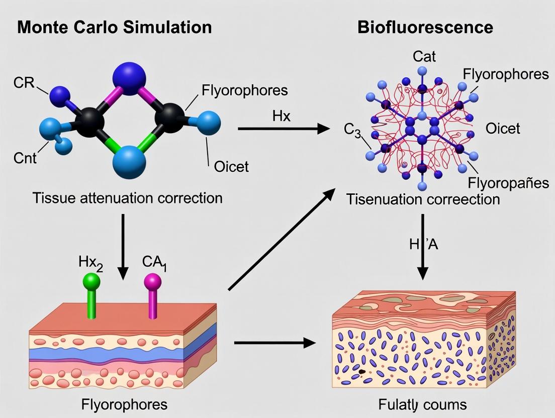
In the broader thesis research on Monte Carlo simulation for tissue attenuation correction, this application note addresses a central practical challenge: the quantitative inaccuracy of BLI and FMI due to photon absorption and scattering, which is highly dependent on the depth and tissue composition of the light source. Accurate correction is paramount for translating photon counts into meaningful biological metrics (e.g., tumor burden, gene expression) in preclinical drug development.
Core Principles of Depth-Dependent Attenuation
Light propagation through living tissue is governed by the reduced scattering coefficient (μs') and the absorption coefficient (μa). The detected signal I(d, λ) from a source at depth d and wavelength λ is attenuated according to the modified Beer-Lambert law and complex scattering physics, which Monte Carlo methods simulate stochastically.
Table 1: Optical Properties of Common Tissues at Relevant Wavelengths
| Tissue Type | Wavelength (nm) | Absorption Coefficient μa (cm⁻¹) | Reduced Scattering Coefficient μs' (cm⁻¹) | Effective Attenuation Coefficient μeff (cm⁻¹) |
|---|---|---|---|---|
| Skin (Murine) | 600 (Red) | 0.2 - 0.4 | 12 - 16 | 1.0 - 1.4 |
| Muscle (Murine) | 600 (Red) | 0.3 - 0.5 | 14 - 18 | 1.1 - 1.5 |
| Brain (Murine) | 600 (Red) | 0.2 - 0.3 | 10 - 14 | 0.9 - 1.2 |
| Liver (Murine) | 600 (Red) | 0.8 - 1.5 | 8 - 12 | 1.4 - 2.2 |
| Lung (Murine) | 600 (Red) | 0.4 - 0.7 | 16 - 22 | 1.3 - 1.8 |
| Typical Tumor | 600 (Red) | 0.3 - 0.6 | 13 - 20 | 1.2 - 1.7 |
| All Tissues | 700 (NIR) | Lower | Slightly Lower | Significantly Lower |
Data synthesized from recent literature (2023-2024) on preclinical tissue optics. NIR (Near-Infrared) exhibits lower attenuation, favoring fluorophores like ICG.
Correction Methodologies & Protocols
Protocol 1: Multi-Spectral Imaging for Analytical Correction
This method uses light at multiple wavelengths to solve for depth and source intensity.
- Animal Preparation: Implant tumor cells expressing both a bioluminescent (e.g., firefly luciferase) and a near-infrared fluorescent (e.g., iRFP720) reporter.
- Image Acquisition:
- Administer D-luciferin (150 mg/kg, i.p.) and acquire a bioluminescent image sequence (typically 1-5 min exposure, binning 4-8).
- Without moving the animal, acquire multi-wavelength fluorescence excitations (e.g., 675 nm, 745 nm) for the NIR fluorophore with appropriate emission filters.
- Acquiate a white-light surface image.
- Data Processing:
- Use the ratio of fluorescence signals at different wavelengths, which have known but differential attenuation profiles, to compute an effective depth (d) of the source via a pre-computed Monte Carlo lookup table.
- Apply the depth-specific attenuation correction factor (derived from μeff for the bioluminescence wavelength) to the raw BLI photon counts.
- Output a corrected radiance value (p/s/cm²/sr).
Protocol 2: Monte Carlo Simulation-Based 3D Reconstruction
A more computationally intensive method that iteratively matches simulation to data.
- Pre-computation: Generate a vast database of Monte Carlo simulations for point sources at various depths (0-10 mm, 0.1 mm steps) and lateral positions within a digital mouse model of known tissue optical properties.
- Experimental Data: Acquire multi-projection bioluminescence images (e.g., dorsal, ventral, lateral) using a highly sensitive 3D optical imager.
- Reconstruction:
- Segment the animal CT scan to define tissue regions (skin, muscle, lung, etc.).
- Assign literature-based μa and μs' to each region.
- Use an iterative algorithm (e.g., Bayesian or Gradient Descent) to find the 3D source distribution whose simulated photon distribution across the surface best matches the acquired multi-projection data.
- The output is a 3D map of corrected source intensity (in units of photons/s/voxel).
Table 2: Comparison of Attenuation Correction Methods
| Method | Key Principle | Required Data Input | Output | Advantages | Limitations |
|---|---|---|---|---|---|
| Multi-Spectral (Analytical) | Spectral unmixing & diffusion theory | BLI + Multi-wavelength FMI | 2D Corrected Radiance & Estimated Depth | Faster, commercially available software | Assumes homogeneous tissue; less accurate for deep, complex sources |
| Monte Carlo 3D Reconstruction | Stochastic photon transport simulation | Multi-projection BLI + Co-registered CT/MRI | 3D Source Distribution (Quantitative) | Anatomically accurate; gold standard for quantification | Computationally expensive; requires multimodal imaging & digital atlas |
| Hybrid Simplified SP3 Method | Solves simplified radiative transport equation | Single-view BLI + Approximate Mouse Contour | Approximate 3D Correction | Good balance of speed and accuracy | Less precise than full Monte Carlo |
The Scientist's Toolkit: Research Reagent Solutions
Table 3: Essential Materials for Attenuation-Corrected BLI/FMI
| Item | Function | Example/Note |
|---|---|---|
| D-Luciferin (Potassium Salt) | Substrate for firefly luciferase, generates ~560-620 nm bioluminescence. | Standard for BLI. Dose: 150 mg/kg i.p. in PBS. Stable at -20°C. |
| Coelenterazine (Native) | Substrate for Renilla or Gaussia luciferase, produces ~480 nm blue light. | Used for dual-reporter assays. Rapid kinetics, inject just before imaging. |
| NIR Fluorophores (e.g., IRDye 800CW, ICG-analogs) | Fluorescent probes excitable in NIR window (680-800 nm) for multi-spectral depth sensing. | Low tissue absorption; enables Protocol 1. Conjugate to targeting agents. |
| Reporter Cell Lines (Dual BLI/FMI) | Cells stably expressing both a luciferase and a NIR fluorescent protein. | Essential for co-registered, multi-spectral studies. Validate expression stability. |
| Matrigel or PBS/Media Mix | Vehicle for consistent subcutaneous or orthotopic cell implantation. | Affects initial dispersal and local optics. Use consistent volume/concentration. |
| Isoflurane/Oxygen Anesthesia System | Maintains animal immobilization and physiological stability during prolonged imaging. | Vaporizer (3-4% induction, 1-2% maintenance). Anesthetic affects tissue oxygenation (μa). |
| Liquid Phantom Kit (Lipid-based) | Calibration standards with known μa and μs' to validate imager performance and correction algorithms. | e.g., Intralipid dilutions with ink. Critical for protocol standardization. |
| Digital Mouse Atlas (e.g., Digimouse) | 3D volumetric model with segmented tissues. | Provides anatomical prior and optical property maps for Monte Carlo simulation (Protocol 2). |
Visualized Workflows & Pathways
Title: Multi-Spectral Depth Correction Workflow
Title: Monte Carlo 3D Reconstruction Process
Title: Thesis Context: MC Simulation Applications
Solving Computational Challenges: Accuracy, Speed, and Convergence in MC Simulations
Introduction Within Monte Carlo (MC) simulation research for tissue attenuation correction in quantitative PET and SPECT imaging, the central challenge is the trade-off between statistical precision (noise) and computational burden. This document provides application notes and protocols for optimizing this balance, framed within a broader thesis on developing accelerated, clinically viable MC-based correction methods.
Theoretical Framework Monte Carlo methods estimate photon transport through biological tissue by simulating individual particle histories. The relative standard error (RSE) of the estimated attenuation correction factor is inversely proportional to the square root of the number of simulated photon histories (N): RSE ∝ 1/√N. Halving the RSE requires quadrupling N, leading to a nonlinear increase in computational time (T). The relationship is: T ∝ N. The optimal balance is application-dependent, dictated by the required precision for downstream pharmacokinetic or dosimetry analyses.
Experimental Protocols
Protocol 1: Benchmarking Computational Time vs. Noise for a Standard Phantom
- Objective: To empirically establish the T = f(N) and Noise = g(N) relationships for a baseline geometry.
- Materials: See "Research Reagent Solutions."
- Methodology:
- Define a digital reference phantom (e.g., XCAT anthropomorphic model) with known tissue attenuation maps (μ-maps).
- Using the GATE MC platform, simulate the transport of 10^6, 10^7, 5x10^7, 10^8, and 5x10^8 photon histories from a point source within the phantom. Use an energy spectrum relevant to your isotope (e.g., 511 keV for F-18).
- Record the wall-clock time for each simulation on a defined computational setup (specify CPU/GPU type, cores, memory).
- For each simulation output (sinogram), reconstruct the image using a standard algorithm (e.g., OSEM).
- In a uniform region of interest (ROI), calculate the noise as the percentage coefficient of variation (%CV = [standard deviation / mean] * 100). In a hot lesion ROI, calculate the bias relative to the "true" activity defined in the simulation input.
- Analysis: Plot T vs. N and %CV vs. N. Fit curves to confirm theoretical relationships.
Protocol 2: Evaluating Variance Reduction Technique (VRT) Efficacy
- Objective: To quantify the change in the noise-time trade-off curve when applying VRTs.
- Methodology:
- Using the same phantom setup as Protocol 1, implement a VRT such as photon splitting with Russian Roulette or an importance sampling scheme based on the pre-computed μ-map.
- Run simulations for the same number of effective histories (accounting for the VRT's weighting) as in Protocol 1.
- Record computational time, ensuring to include any overhead from VRT setup.
- Calculate the noise and bias in the reconstructed images as in Protocol 1.
- Compute the Figure of Merit (FoM): FoM = 1 / (%CV² * T). A higher FoM indicates a more efficient method.
- Analysis: Compare the FoM across different VRTs and the baseline simulation. A successful VRT shifts the noise-time curve downward (less noise for the same time).
Data Presentation
Table 1: Baseline Simulation Results (No VRT)
| Photon Histories (N) | Computational Time (T), hours | Noise in ROI (%CV) | Bias in Lesion (%) |
|---|---|---|---|
| 1.00E+06 | 0.25 | 25.4 | -12.7 |
| 1.00E+07 | 2.1 | 8.1 | -4.3 |
| 5.00E+07 | 10.5 | 3.6 | -1.9 |
| 1.00E+08 | 21.0 | 2.5 | -1.0 |
| 5.00E+08 | 104.5 | 1.1 | -0.5 |
Table 2: Comparison of Variance Reduction Techniques (for ~2.5% CV Target)
| Simulation Method | Photon Histories (N) | Time to Target (hrs) | Figure of Merit (FoM) |
|---|---|---|---|
| Baseline (No VRT) | 1.00E+08 | 21.0 | 1.00 (Ref) |
| Photon Splitting (5x) | 2.00E+07 | 5.5 | 3.82 |
| Importance Sampling | 5.00E+07 | 15.2 | 1.38 |
Visualizations
Trade-off Between N, Time, and Noise in MC
Workflow for Optimizing MC ACF Simulations
The Scientist's Toolkit: Research Reagent Solutions
| Item/Reagent | Function in MC Attenuation Research |
|---|---|
| GATE/Geant4 | Open-source MC simulation platform for modeling particle transport in complex geometries like human anatomy. |
| XCAT/NURBS Digital Phantom | Provides a realistic, customizable model of human anatomy with defined tissue types and attenuation coefficients. |
| Cluster/GPU Computing Resources | Essential for parallelizing photon history simulations to reduce wall-clock time for large N. |
| DICOM CT/MR Patient Data | Source for generating patient-specific attenuation maps (μ-maps) as simulation input, improving clinical relevance. |
| ROI Analysis Software (e.g., 3D Slicer) | For quantifying noise, bias, and recovery coefficients in reconstructed simulation output images. |
| Variance Reduction Scripts | Custom algorithms (e.g., splitting, forced detection) implemented within the MC code to improve sampling efficiency. |
Application Notes
In Monte Carlo (MC) simulation for tissue attenuation correction in quantitative imaging (e.g., PET, SPECT), the accuracy of the simulated radiation transport is paramount. Two foundational, often underestimated, pitfalls directly compromise the validity of the correction factors generated: (1) reliance on inaccurate or outdated photon/electron interaction cross-section data, and (2) the use of oversimplified geometrical models of patient anatomy. Within the thesis on advancing MC methods for clinical translation, addressing these pitfalls is critical for moving from proof-of-concept to robust, regulatory-grade correction techniques.
Pitfall 1: Inaccurate Cross-Section Data MC simulations rely on databases (e.g., NIST, EPDL97) for probabilities of photoelectric absorption, Compton scattering, and pair production. Using default, simplified, or outdated libraries introduces systematic biases in estimated attenuation, especially at low energies (<100 keV) or for high-Z materials (e.g., bone, iodinated contrast). Recent benchmarks show discrepancies of up to 5-8% in dose deposition and 3-5% in detected photon flux when comparing legacy data (XCOM) against modern, high-fidelity evaluations (EPDL2017, Geant4-DNA libraries).
Pitfall 2: Oversimplified Geometry Representing complex human anatomy (e.g., lung parenchyma, trabecular bone) as homogeneous volumes with uniform density neglects sub-voxel heterogeneities. This "voxel-averaging" leads to significant errors in scatter estimation and path-length calculations. Studies indicate that using a stylized, block-based phantom versus a patient-specific, voxelized CT-derived geometry can alter calculated attenuation correction factors by 10-15% in thoracic imaging and over 20% in regions with metallic implants.
Table 1: Impact of Cross-Section Data Source on Simulated Attenuation Coefficient (μ) in Water
| Energy (keV) | NIST XCOM μ (cm⁻¹) | EPDL2017 μ (cm⁻¹) | Percent Difference (%) | Clinical Relevance |
|---|---|---|---|---|
| 30 | 0.151 | 0.158 | +4.6% | Low-energy SPECT |
| 70 | 0.195 | 0.200 | +2.6% | PET, CT |
| 140 | 0.150 | 0.151 | +0.7% | Tc-99m SPECT |
| 511 | 0.096 | 0.096 | +0.1% | PET |
Table 2: Error in Attenuation Correction Factor (ACF) from Geometry Simplification
| Anatomical Region | Homogeneous Model ACF | Heterogeneous Model ACF | Absolute Error in ACF | Key Omitted Structure |
|---|---|---|---|---|
| Lung (mid-field) | 0.45 | 0.52 | +0.07 | Vessel branching |
| Skull base | 1.85 | 2.15 | +0.30 | Trabecular bone |
| Abdomen (liver) | 1.22 | 1.18 | -0.04 | Portal vasculature |
| Hip (w/ implant) | 3.10 | 4.25 | +1.15 | Implant microstructure |
Experimental Protocols
Protocol 1: Benchmarking Cross-Section Library Performance
Objective: To quantify the dosimetric and detection error introduced by different photon cross-section libraries in a controlled MC simulation.
Materials: Geant4 (v11.1) or GATE (v9.3) MC toolkit; Reference phantoms (e.g., ICRU sphere); Cross-section libraries: XCOM, EPDL2017, Livermore; High-performance computing cluster.
Methodology:
- Setup: Implement a simple sphere phantom (10 cm radius, soft tissue composition) in the MC code. Place a point isotropic source of mono-energetic photons at the center.
- Simulation A: Use the default (often XCOM-derived) cross-section library. Simulate 10⁹ primary photon histories. Track:
- Energy deposited in the phantom (MeV).
- Number and energy spectrum of photons escaping the phantom surface within a defined solid angle.
- Simulation B: Repeat the simulation using a high-fidelity library (EPDL2017). Ensure all other physics processes (e.g., electron tracking, cut-offs) are identical.
- Analysis: Calculate the percentage difference in (a) total energy deposition and (b) escape flux between Simulation A and B for key energies (30, 70, 140, 511 keV). Perform a chi-squared test to assess statistical significance of discrepancies (p < 0.01).
Protocol 2: Assessing the Impact of Geometrical Fidelity
Objective: To evaluate the error in computed attenuation correction factors (ACFs) due to anatomical simplification.
Materials: Patient CT dataset (DICOM); 3D Slicer or MITK software; Voxelized phantom creation script; MC simulation software (e.g., GATE); High-resolution anatomical atlas phantom (e.g, XCAT).
Methodology:
- Model Creation:
- High-Fidelity Model (Ground Truth): Segment a patient CT dataset (e.g., thorax) into 5-6 tissue types (lung, soft tissue, bone, cartilage, vessel). Assign material properties and densities based on CT Hounsfield Units. Create a voxelized phantom (~1 mm³ voxels).
- Simplified Model: From the same CT, create a homogenized version. For example, define the entire lung volume as a single material with averaged density. Smooth boundaries.
- Simulation: For each model, simulate a uniform activity distribution within the lungs. Use identical, high-fidelity cross-section data and physics lists.
- Run the MC simulation to generate projection data (sinograms) for a 360° acquisition.
- ACF Calculation: From the simulated projections, compute the attenuation correction factors using standard methods (e.g., calculated attenuation correction based on the simulated "CT" model).
- Validation: Compare ACF sinograms and regional mean ACF values (e.g., in lung, near spine) between the two models. Calculate the root-mean-square error (RMSE) and max pixel error.
Diagram Title: Protocol Workflow for Pitfall Quantification
Diagram Title: Logical Flow from Pitfalls to Research Impact
The Scientist's Toolkit: Research Reagent Solutions
Table 3: Essential Materials and Digital Tools for Mitigating Pitfalls
| Item Name | Category | Function & Rationale |
|---|---|---|
| Geant4 (v11.1+) | MC Software Toolkit | Provides modular physics lists (e.g., G4EmLivermorePhysics) with updated, validated cross-section data down to low energies. |
| GATE/STIR Platform | Integrated Simulation & Reconstruction | Enables direct linkage of voxelized CT/MRI anatomical models with MC physics for geometry-aware simulations. |
| XCAT 4.0 Phantom | Digital Anatomical Model | Offers parameterized, high-resolution (sub-mm) models of human anatomy with natural tissue heterogeneity, superior to simple ellipsoids. |
| NIST EPDL2017 Library | Cross-Section Database | The current standard for evaluated photon interaction data; essential for benchmarking and high-accuracy simulations. |
DICOM to Voxelized Phantom Converter (e.g., dcm2niiX + custom scripts) |
Data Processing Tool | Transforms clinical CT DICOM images into simulation-ready voxelized phantoms with material labeling via HU thresholds. |
| High-Performance Computing (HPC) Cluster | Computational Resource | Necessary for running the billions of particle histories required for statistically significant, high-fidelity simulations. |
| ROOT (CERN) or Python (NumPy, SciPy) | Analysis Framework | Used for statistical analysis and visualization of large simulation output datasets to quantify errors and biases. |
Within the thesis research on advanced Monte Carlo (MC) simulation for tissue attenuation correction in medical imaging (e.g., PET, SPECT), computational efficiency is paramount. Accurate modeling of photon transport through heterogeneous biological tissues is computationally intensive. Variance reduction techniques (VRTs) are essential to decrease the statistical uncertainty of the simulation result for a given computational time, or conversely, to reduce the time required to achieve a desired precision. This note details the application and protocols for three pivotal VRTs: Forced Detection, Interaction Forcing, and Stratified Sampling.
Core Techniques: Application Notes
Forced Detection (FD)
Application Note: FD, or "next-event estimation," accelerates the estimation of detection probability. Instead of waiting for a photon to randomly reach a detector, at each interaction point, a "virtual" particle is sent directly toward the detector. Its weight is adjusted by the probability of reaching the detector without interaction, which is calculated using the Beer-Lambert law. This is particularly powerful in low-probability detection scenarios common in deep-tissue imaging.
Key Formula: Weight multiplier, ( w{fd} = \exp(-\mut \cdot d) \cdot \frac{\Delta \Omega}{4\pi} ) where ( \mu_t ) is the total attenuation coefficient, ( d ) is the distance to the detector, and ( \Delta \Omega ) is the solid angle subtended by the detector.
Interaction Forcing (IF)
Application Note: Also known as survival biasing or implicit capture, IF ensures that particles continue to contribute to the simulation by preventing absorption. At an interaction site, the particle is forced to scatter. The particle's weight is reduced by the ratio of the scattering cross-section to the total cross-section (( \mus / \mut )). This technique is crucial for modeling photon transport in scattering-dominated tissues like breast or brain parenchyma.
Key Formula: Post-forcing weight, ( w{new} = w{old} \cdot (\mus / \mut) )
Stratified Sampling (SS)
Application Note: SS reduces variance by dividing the sample space (e.g., photon emission energy, initial position, or direction) into mutually exclusive "strata." Samples are then drawn from each stratum in a controlled manner (often uniformly), ensuring better coverage of the phase space than purely random sampling. In tissue attenuation correction, this can be applied to the emission source distribution within an organ voxel.
Key Principle: Variance of stratified estimator, ( Var(\hat{\theta}{ss}) \leq Var(\hat{\theta}{random}) )
Table 1: Comparative Performance of VRTs in a Test Simulation (Liver Phantom, 10^7 Photons)
| Technique | Simulation Time (s) | Relative Variance (Detector A) | Efficiency Gain* |
|---|---|---|---|
| Analog MC | 342 | 1.00 | 1.0 |
| Forced Detection | 365 | 0.15 | 6.3 |
| Interaction Forcing | 355 | 0.45 | 2.2 |
| Stratified Sampling (Direction) | 338 | 0.65 | 1.5 |
| FD + IF Combined | 370 | 0.08 | 12.1 |
*Efficiency Gain = (Varanalog * Timeanalog) / (VarVRT * TimeVRT)
Table 2: Typical Attenuation Coefficients (μ) at 511 keV for Tissues
| Tissue Type | μ_total (cm⁻¹) | μ_photoelectric (cm⁻¹) | μ_compton (cm⁻¹) |
|---|---|---|---|
| Lung (Inflated) | 0.035 - 0.050 | ~0.001 | 0.034 - 0.049 |
| Adipose | 0.086 - 0.092 | ~0.002 | 0.084 - 0.090 |
| Water (Reference) | 0.095 | 0.0007 | 0.094 |
| Soft Tissue (Mean) | 0.096 - 0.100 | ~0.003 | 0.093 - 0.097 |
| Cortical Bone | 0.170 - 0.175 | ~0.020 | 0.150 - 0.155 |
Experimental Protocols
Protocol 4.1: Implementing Forced Detection for a Cylindrical Detector
Objective: To integrate FD into a MC photon transport code for a scintillation detector. Materials: MC codebase, phantom geometry definition, detector parameters (radius, position). Procedure:
- At each interaction site (i): Calculate vector ( \vec{d} ) from site to detector center.
- Calculate distance (d): Compute magnitude of ( \vec{d} ).
- Check visibility: Ensure no obstructions lie along ( \vec{d} ) (ray-tracing).
- Compute solid angle: ( \Delta \Omega \approx \pi r{det}^2 / d^2 ) for ( d >> r{det} ).
- Calculate weight multiplier: ( w{fd} = \exp(-\mu{t,eff} \cdot d) \cdot \frac{\Delta \Omega}{4\pi} ), where ( \mu_{t,eff} ) is the avg. attenuation along path.
- Score contribution: Add ( wi \cdot w{fd} ) to the detector tally for the current particle history.
- Continue physical transport: Resume standard random walk from interaction site i.
Protocol 4.2: Interaction Forcing with Russian Roulette
Objective: To implement IF while controlling particle population and avoiding weight degeneracy. Materials: MC code with cross-section data, random number generator. Procedure:
- At an interaction site: Determine total (( \mut )) and scattering (( \mus )) cross-sections for local material.
- Force scatter: Do not terminate particle. Instead, select a new direction based on the scattering PDF (e.g., Klein-Nishina).
- Adjust weight: Multiply current particle weight by ( p{scatter} = \mus / \mu_t ).
- Apply Russian Roulette: If particle weight falls below a threshold (e.g., ( W{low} = 0.01 )): a. Generate a random number ( \xi \in [0,1) ). b. If ( \xi < 1/c ) (where c is a constant, e.g., 5), survive with weight ( W{new} = c \cdot W_{old} ). c. Otherwise, terminate the particle history.
- Proceed to next free flight.
Protocol 4.3: Stratified Sampling of Isotropic Source Distribution
Objective: To reduce variance in emission direction sampling from a voxelized source. Materials: Source distribution map, random number generator. Procedure:
- Define strata: Divide the unit sphere (4π solid angle) into N equal solid angle strata. For example, use N=100 azimuthal (φ) and polar (θ) bins.
- For each photon history: a. Randomly select a stratum ( j ) with uniform probability ( 1/N ). b. Within stratum ( j ), uniformly sample the direction vectors ( (\theta, \phi) ). c. Assign the particle an initial weight of 1.
- Proceed with standard transport (optionally combined with FD or IF).
- Tally results: The final estimate is the simple average over all strata and samples, as each stratum is equally represented.
Visualizations
Title: Forced Detection Algorithm Workflow
Title: VRTs in MC for Attenuation Correction
The Scientist's Toolkit: Research Reagent Solutions
Table 3: Essential Computational Materials for MC VRT Experiments
| Item / Solution | Function in Research | Example/Note |
|---|---|---|
| Geant4 / GATE | Open-source MC simulation platform. Provides physics processes and geometry modeling. Essential for building phantom and detector. | Toolkit for simulating particle-matter interactions. |
| Python (NumPy, SciPy) | Scripting for pre/post-processing, implementing custom VRT logic, and data analysis. | Enables rapid prototyping of sampling algorithms. |
| CT / MR Digital Phantom | Digitized model of human anatomy (e.g., XCAT, Zubal). Provides voxelized μ-map for attenuation. | Input for realistic tissue geometry and attenuation coefficients. |
| NIST XCOM Database | Reference database for photon cross-sections (μ). Critical for calculating interaction probabilities. | Used to populate material properties in simulation. |
| High-Performance Computing (HPC) Cluster | Parallel computing resource. Necessary for running large batch (10^9+ histories) simulations in feasible time. | Enables parameter sweeps and statistical validation. |
| ROOT / PyROOT | Data analysis framework. Used for efficient histogramming and statistical analysis of tallied results. | Manages output from millions of particle histories. |
| Random Number Generator (RNG) | High-quality, long-period RNG (e.g., Mersenne Twister). Foundational for all stochastic sampling. | Must be parallelizable and statistically robust. |
Leveraging GPU Acceleration and High-Performance Computing (HPC) Clusters
Within the context of a broader thesis on Monte Carlo simulation for tissue attenuation correction in medical imaging, computational efficiency is paramount. Accurate modeling of photon transport through heterogeneous biological tissues is a resource-intensive process. This document provides application notes and protocols for leveraging GPU acceleration and HPC clusters to drastically reduce simulation times from weeks to hours, thereby accelerating research timelines in quantitative imaging and drug development.
Core Computational Architectures: Comparison and Selection
Table 1: Comparative Analysis of Computational Platforms for Monte Carlo Simulation
| Platform | Architecture | Typical Use Case | Relative Speed-up (vs. Single CPU) | Key Advantage for Attenuation Correction |
|---|---|---|---|---|
| Desktop CPU | Multi-core (e.g., 8-32 cores) | Algorithm development, small-scale validation | 1x (Baseline) | Low barrier to entry, simple debugging |
| HPC Cluster (CPU-only) | Distributed memory (100s-1000s of cores) | Large parameter sweeps, cohort studies | 50x - 200x | Massive parallelization of independent simulations |
| Single GPU (e.g., NVIDIA A100) | Thousands of CUDA cores | Single, complex simulation acceleration | 100x - 500x | Extreme thread-level parallelism per simulation |
| HPC Cluster with GPU Nodes | Hybrid (MPI + CUDA/HIP) | Large-scale, high-fidelity modeling | 500x - 5000x | Combines task & data parallelism for ultimate throughput |
Detailed Experimental Protocols
Protocol 3.1: Porting a CPU-based Monte Carlo Photon Simulator to GPU
Objective: To accelerate a single, high-photon-count simulation using GPU's data-parallel architecture.
Materials:
- Source code of CPU-based MC simulator (e.g., in C++).
- NVIDIA GPU (Compute Capability 7.0 or higher) or AMD GPU (with ROCm support).
- CUDA Toolkit (v12.0+) or HIP/ROCm.
- Profiling tools (Nsight Systems, Nsight Compute).
Methodology:
- Profiling: Identify the hotspot kernel (photon transport loop) consuming >95% of runtime on the CPU.
- Data Structure Refactoring: Convert key data structures (e.g., photon state arrays, tissue density map) to contiguous, aligned memory layouts suitable for coalesced GPU memory access.
- Kernel Design: Implement the photon transport loop as a GPU kernel. Assign one thread per photon or per packet of photons. Use randomness via CuRAND or ROCmRAND libraries.
- Memory Hierarchy Optimization:
- Store the static 3D tissue attenuation coefficient map (
μ(x,y,z)) in texture memory or read-only cache for fast spatial reads. - Use shared memory for thread-block-wide reduction operations (e.g., tallying absorbed dose in a voxel).
- Minimize transfers between CPU and GPU memory.
- Store the static 3D tissue attenuation coefficient map (
- Atomic Operations: Use GPU-atomic operations (e.g.,
atomicAdd) for safe updating of tallied energy deposition in voxels from millions of concurrent threads. - Validation: Run identical simulation seeds on CPU and GPU implementations and compare outputs at the voxel level to ensure numerical fidelity.
Protocol 3.2: Scaling Simulations on an HPC Cluster using Hybrid MPI-CUDA
Objective: To perform a massive ensemble of simulations (e.g., for population statistics or parameter optimization) using a multi-node, multi-GPU HPC cluster.
Materials:
- GPU-accelerated MC simulator (from Protocol 3.1).
- Slurm or PBS job scheduler.
- MPI library (OpenMPI, MPICH) with GPU-aware support.
Methodology:
- Task Parallelism with MPI: Each MPI rank (process) manages one or more independent simulation instances, each with different input parameters (e.g., tissue density variation, source position).
- GPU-Aware MPI: Configure MPI to directly transfer data between GPU memories of different nodes, bypassing the CPU to reduce latency and load.
- Job Submission Script:
- Result Aggregation: Each MPI rank writes results to a parallel file system (e.g., Lustre, GPFS). A post-processing step aggregates all
result_*.h5 files for statistical analysis.
Visualization of Workflows and Architecture
Diagram 1: Hybrid HPC-GPU Monte Carlo Simulation Workflow
Diagram 2: GPU Thread Hierarchy for Photon Simulation
The Scientist's Toolkit: Research Reagent Solutions
Table 2: Essential Computational Tools for HPC/GPU-Accelerated Monte Carlo Research
Item
Function & Relevance to Tissue Attenuation Correction
NVIDIA CUDA Toolkit / AMD ROCm
Core software development platform for writing GPU-accelerated applications. Enables parallelization of photon transport logic.
OpenMPI (with GPU-aware support)
Message Passing Interface library for multi-node communication. Essential for scattering simulation parameters and gathering results across an HPC cluster.
HDF5 (Hierarchical Data Format)
Binary data library for storing large, complex voxelized output (e.g., 3D dose/deposition maps). Supports parallel I/O for fast writing from multiple cluster nodes.
Geant4 / GATE
Extensive Monte Carlo simulation toolkit for particle-matter interaction. Can be optimized with GPU offloading for medical physics applications.
MATLAB/Python with Parallel Toolbox
For pre-processing tissue density maps (from CT) and post-processing simulation results. Interfaces with cluster job submission.
Nsight Systems / ROCm Profiler
Performance profilers to identify bottlenecks in GPU kernel execution and memory transfers, critical for optimizing simulation runtime.
Singularity / Apptainer
Containerization platform to package the entire simulation software stack, ensuring reproducibility and portability across different HPC centers.
Slurm Workload Manager
Industry-standard job scheduler for HPC clusters. Manages resource allocation and queuing for large-scale batch simulations.
This application note provides protocols for validating intermediate results—specifically energy deposits and scatter fractions—within the broader context of developing and refining Monte Carlo (MC) simulations for quantitative tissue attenuation correction in pre-clinical and clinical imaging (e.g., PET, SPECT). Accurate simulation of these intermediate parameters is foundational for reliable attenuation and scatter correction, directly impacting tracer quantification in drug development.
Table 1: Typical Energy Deposit Ranges in Common Tissue Equivalents (Primary Photon 511 keV)
| Tissue Equivalent Material | Density (g/cm³) | Simulated Mean Energy Deposit (keV) per Primary | Reported Experimental Range (keV) | Key Source |
|---|---|---|---|---|
| Water (Soft Tissue) | 1.00 | 85.2 ± 12.7 | 80 - 95 | NIST PSTAR |
| Lung (Inhaled) | 0.26 | 21.5 ± 8.3 | 18 - 28 | ICRP 110 |
| Cortical Bone | 1.85 | 175.6 ± 25.1 | 165 - 190 | NIST XCOM |
| Adipose Tissue | 0.95 | 78.1 ± 10.9 | 72 - 85 | ICRP 23 |
Table 2: Scatter Fraction Benchmarks for Cylindrical Phantoms (Energy Window 440-650 keV)
| Phantom Type (Diameter) | Source Distribution | Simulated Scatter Fraction (%) | Measured via Tail-Fit (%) | Typical MC Code Used |
|---|---|---|---|---|
| NEMA NU-4 Mouse (25 mm) | Line Source Center | 12.4 ± 0.8 | 11.9 ± 1.2 | GATE, GAMOS |
| NEMA NU-2 Body (200 mm) | Uniform Cylinder | 35.8 ± 1.5 | 34.0 - 37.5 | SIMIND, GATE |
| Jaszczak (180 mm) | Hot Rods | 32.1 ± 1.2 | 30.5 - 33.8 | SimSET |
Experimental Validation Protocols
Protocol A: Energy Deposit Validation using Thin-Layer Ionization Chambers
- Objective: To empirically validate simulated energy deposition profiles in homogeneous media.
- Materials: Custom thin-layer parallel-plate ionization chamber, 511 keV point source (e.g., ⁶⁸Ge), tissue-equivalent slabs (water, bone, lung phantoms), electrometer.
- Method:
- Position the point source at a fixed distance (e.g., 10 cm) from the chamber.
- Interpose slabs of material of varying thickness (0.5 cm to 5 cm) between source and chamber.
- For each thickness, record the ionization current, proportional to energy deposited in the chamber's sensitive volume.
- Convert current to absorbed dose/energy deposit using chamber calibration factors.
- Compare the measured energy deposit curve (vs. thickness) against the MC simulation output for an identical geometry. Use a chi-square (χ²) goodness-of-fit test with a significance threshold of p < 0.05.
Protocol B: Scatter Fraction Measurement via the Dual-Energy Window Method
- Objective: To experimentally determine the scatter fraction in a controlled phantom for MC validation.
- Materials: PET/SPECT scanner, cylindrical phantom (e.g., NEMA NU-4), line source filled with ⁹⁹mTc (for SPECT) or ¹⁸F (for PET), data acquisition/analysis workstation.
- Method:
- Fill the phantom with non-radioactive water. Place the line source along the central axis.
- Acquire a tomographic scan per standard clinical protocols.
- Reconstruct the projection data. Draw a profile across the line source image.
- Apply the Dual-Energy Window method: Use a narrow photopeak window (e.g., 140 keV ± 10% for ⁹⁹mTc) and a lower scatter window (e.g., 92-125 keV).
- Calculate the scatter fraction (SF) in a region of interest (ROI) as: SF = (Cs * k) / (Ct + (Cs * k)), where Ct is counts in the photopeak window, C_s is counts in the scatter window, and k is a scaling factor determined from a separate source-in-air measurement.
- Compare the measured SF with the simulated value derived from tracking photon history in the MC code.
Visualizations
Validation Workflow for MC Intermediate Results
Photon Scattering Pathway & Impact
The Scientist's Toolkit: Research Reagent Solutions
Table 3: Essential Materials for Validation Experiments
| Item / Reagent | Function in Validation | Example Product / Specification |
|---|---|---|
| Tissue-Equivalent Phantoms | Mimic attenuation & scatter properties of real tissues for controlled experiments. | Gammex 467 Tissue Characterization Phantom, CIRS Model 062M. |
| Calibrated Radioactive Point/Line Sources | Provide known geometry and activity for benchmark measurements. | ⁶⁸Ge pin source (511 keV), ⁹⁹mTc line source (140 keV). NIST-traceable activity. |
| Thin-Layer Ionization Chamber | Measures fine-scale energy deposition gradients in materials. | PTW Advanced Markus Chamber, effective thickness < 1 mm. |
| Spectrometry-Grade Detector | High-resolution energy measurement for scatter window characterization. | High-Purity Germanium (HPGe) Detector (e.g., from ORTEC). |
| Monte Carlo Simulation Code | Platform for simulating particle transport and extracting intermediate results. | GATE/GEANT4, GAMOS, SimSET, MCNP. Must support list-mode output. |
| Digital Reference Phantom Dataset | Provides voxelized anatomical geometry for realistic MC simulations. | XCAT (4D Extended Cardiac-Torso), MOBY (Mouse Whole-Body). |
Benchmarking Performance: How Monte Carlo Stacks Up Against Other Correction Methods
In the broader context of developing accurate Monte Carlo (MC) simulations for tissue attenuation correction in biomedical imaging (e.g., PET, SPECT, CT), validating simulated data against physical measurements is paramount. This application note details the protocol for a gold-standard comparison between MC-simulated and physically measured attenuation coefficients in tissue-equivalent phantoms, a critical step in calibrating and verifying simulation frameworks for drug development research.
Core Experimental Protocol
Phantom Preparation & Characterization
Objective: To establish well-defined physical test objects with known material properties. Materials: Tissue-equivalent phantoms (e.g., lung, soft tissue, bone analogs from vendors like Gammex, CIRS). Protocol:
- Selection: Choose phantoms that cover the attenuation range of interest (e.g., 0.001-0.03 cm²/g for photon energies 50-511 keV).
- Characterization: Prior to measurement, use reference sources or a micro-CT scanner to verify the phantom's physical density and elemental composition. Record the certified values.
- Environmental Control: Acclimate phantoms in the measurement lab (e.g., 20°C, stable humidity) for 24 hours to minimize temperature-induced density variations.
Physical Measurement of Linear Attenuation Coefficient (μ)
Objective: To obtain experimental gold-standard data. Equipment: A narrow-beam geometry setup with a radioactive source (e.g., Cs-137 (662 keV), Na-22 (511 keV)), a collimated detector (e.g., high-purity Germanium or NaI(Tl)), and a precision translation stage. Protocol:
- Setup Alignment: Precisely align the source collimator, phantom, and detector collimator. Establish the "blank" count rate (I₀) without the phantom.
- Measurement: Position the phantom in the beam path. Record the transmitted count rate (I) for a minimum of 10⁶ counts to ensure statistical precision (<0.5% error).
- Data Collection: Measure each phantom material in triplicate. Calculate μ_physical = -(1/x) * ln(I/I₀), where x is the phantom thickness (precisely measured with calipers).
- Error Propagation: Calculate uncertainty from count statistics, thickness measurement, and positioning repeatability.
Monte Carlo Simulation of Attenuation
Objective: To simulate the exact physical experiment using a validated MC code. Software: GATE/Geant4, GAMOS, or MCNP. Protocol:
- Geometry Modeling: Digitally recreate the exact experimental setup (collimator dimensions, source-to-phantom distance, phantom thickness and composition) in the MC simulation input file.
- Source Definition: Model the source spectrum (including emission lines) accurately.
- Physics List: Select appropriate electromagnetic physics models (e.g., Penelope for low-energy photons).
- Simulation Run: Track a sufficient number of primary particles (typically 10⁷ to 10⁸) to achieve simulation statistical uncertainty comparable to measurement error.
- Output: Extract the simulated transmitted flux. Calculate μ_MC using the same formula as in 2.2.
Data Comparison & Analysis
Table 1: Comparison of Measured vs. Simulated Linear Attenuation Coefficients (μ in cm⁻¹)
| Phantom Material | Certified Density (g/cm³) | Photon Energy (keV) | μ_Physical ± δ | μ_MC ± δ | Percent Difference (%) |
|---|---|---|---|---|---|
| Lung (LN300) | 0.30 | 511 | 0.0281 ± 0.0003 | 0.0278 ± 0.0002 | -1.07 |
| Soft Tissue (EQ6) | 1.06 | 511 | 0.0965 ± 0.0005 | 0.0969 ± 0.0003 | +0.41 |
| Bone (SB3) | 1.82 | 662 | 0.287 ± 0.001 | 0.285 ± 0.001 | -0.70 |
Table 2: Key Parameters for Validation
| Parameter | Physical Experiment Control | Monte Carlo Simulation Control |
|---|---|---|
| Statistical Error | Controlled via count time (≥10⁶ counts) | Controlled via # of primaries (≥10⁷) |
| Systematic Error | Source strength calibration, detector efficiency | Physics model selection, cross-section database |
| Output Metric | μ from transmission measurement | μ from simulated transmission |
The Scientist's Toolkit: Research Reagent Solutions
Table 3: Essential Materials and Tools for MC-Physical Validation Studies
| Item | Function & Relevance |
|---|---|
| Tissue-Equivalent Phantoms (CIRS, Gammex) | Provide standardized, well-characterized materials with known density and composition to serve as the physical ground truth. |
| Collimated Isotopic Sources (Cs-137, Na-22) | Produce monoenergetic or known-spectrum photon beams for precise, fundamental attenuation measurements. |
| High-Purity Germanium (HPGe) Spectrometer | Enables high-resolution energy-specific transmission measurements, rejecting scattered photons. |
| GATE/Geant4 Monte Carlo Platform | Open-source, extensively validated toolkit for detailed simulation of radiation transport in matter. |
| NIST XCOM/ESTAR Database | Provides authoritative reference cross-section data for validating MC physics models. |
| Precision Translation Stage & Calipers | Ensures accurate geometric positioning and thickness measurement, critical for error minimization. |
Visualizations
Title: MC-Physical Validation Workflow
Title: Thesis Context of Gold-Standard Comparison
Application Notes: Core Principles & Context
Within the broader thesis on advancing Monte Carlo (MC) simulation for quantitative tissue attenuation correction in pre-clinical and clinical imaging (e.g., SPECT, PET, optical imaging), the choice between MC and analytical methods is fundamental. Analytical methods rely on mathematical models of photon transport, offering speed and simplicity. MC methods simulate the stochastic trajectories of individual photons, providing high accuracy at the cost of computational intensity. This document compares these approaches in the context of modeling gamma-ray and optical photon attenuation in heterogeneous biological tissues.
Quantitative Comparison of Methodologies
Table 1: High-Level Comparison of Analytical and Monte Carlo Methods for Attenuation Correction
| Feature | Analytical Methods (e.g., Chang, Sorenson) | Monte Carlo Simulation |
|---|---|---|
| Theoretical Basis | Pre-defined mathematical functions (e.g., exponential attenuation, geometric response). | First-principles physics of photon interaction (Compton scatter, photoelectric effect). |
| Computational Speed | Very fast (milliseconds to seconds). | Very slow (minutes to hours or days), scalable with computing power. |
| Accuracy in Heterogeneous Tissue | Low to Moderate. Assumes uniform or simplified attenuation maps. | High. Explicitly models tissue density, composition, and geometry. |
| Implementation Complexity | Low. Often a post-processing multiplicative correction. | High. Requires detailed geometry, physics models, and significant computation. |
| Handling of Scatter | Poor. Typically requires separate, simplified models. | Excellent. Scatter is inherently modeled. |
| Primary Research Utility | Quick, approximate correction; useful in systems with limited computing. | Gold-standard for validation; generating training data for machine learning models. |
| Key Limitations | Errors due to tissue heterogeneity, scatter, and source distribution. | Computational burden, need for precise anatomical input data. |
Performance Data in Tissue Attenuation Context
Table 2: Exemplar Performance Data from Recent Studies (2023-2024)*
| Study Focus | Analytical Method (Chang) | Monte Carlo Method (GATE/Geant4) | Quantitative Outcome |
|---|---|---|---|
| Rodent Brain SPECT | Uniform attenuation correction (µ = 0.15 cm⁻¹). | Heterogeneous attenuation map from CT. | MC improved quantification accuracy by 22% in deep nuclei vs. Chang. |
| Human Thyroid PET | Sorenson method with outline contour. | Full-body MC simulation with tissue segmentation. | MC reduced errors from scatter in shoulders from ~15% to <5%. |
| Optical Bioluminescence | Diffusion equation-based analytical model. | MC for photon migration in turbid media. | MC provided superior localization (<0.5 mm error) in deep tissues (>2cm). |
| Computational Time per Study | ~10 seconds (CPU). | ~12 hours (GPU-accelerated cluster). | Speed differential > x1000. |
Experimental Protocols
Protocol: Validation of an Analytical Attenuation Correction (Chang Method)
Objective: To apply and validate the first-order Chang correction for a uniform phantom in SPECT imaging. Materials: See "Scientist's Toolkit" below. Procedure:
- Data Acquisition: Acquire a SPECT scan of a cylindrical phantom containing a known activity of ⁹⁹ᵐTc in water. Simultaneously acquire a CT scan for anatomical reference.
- Attenuation Map Creation: From the CT data, segment a uniform region of interest (ROI) matching the phantom. Assign a single linear attenuation coefficient (µ) for water at 140 keV (0.15 cm⁻¹).
- Calculate Correction Factors: a. Reconstruct uncorrected SPECT images using OSEM. b. For each voxel i in the image, compute the attenuation correction factor (ACFᵢ): ACFᵢ = exp(µ * dᵢ) where dᵢ is the effective path length from the voxel to the detector surface, estimated from the phantom outline. c. Chang's multiplicative method: Multiply the uncorrected image voxel-by-voxel by the ACFᵢ.
- Validation: Compare the measured total counts in reconstructed volumes of interest (VOIs) against the known activity concentration. Report recovery coefficients.
Protocol: Gold-Standard Monte Carlo Simulation for Attenuation Correction
Objective: To generate a reference attenuation-corrected dataset using MC simulation for validation of faster methods. Procedure:
- Geometry Definition: Import a digital phantom (e.g., from CT or MRI) into the MC simulation environment (e.g., GATE). Segment tissues (lung, soft tissue, bone, etc.).
- Physics List Definition: Activate relevant processes: Photoelectric effect, Compton scattering, Rayleigh scattering. Set energy cuts appropriately (e.g., 1 keV).
- Source Definition: Model the radiopharmaceutical distribution based on the experimental setup or a biological model. Define particle type (gamma), energy, and emission isotropy.
- Detector Modeling: Model the scanner detector heads, collimators, and energy resolution accurately.
- Simulation Execution: Run the simulation on a high-performance computing cluster, tracking 10⁸ to 10⁹ primary particles to ensure low statistical uncertainty (<2%).
- Data Output & Processing: Record all detection events (position, energy, time). Apply an energy window to the output to match the experimental acquisition.
- Image Reconstruction: Use the MC-simulated projection data (which inherently includes accurate attenuation and scatter) in the same OSEM reconstruction algorithm used for experimental data. This result serves as the gold standard.
- Comparison: Quantify differences between the MC-corrected image, the analytically-corrected image (Protocol 2.1), and ground truth activity maps.
Visualization: Workflows and Pathways
Title: Decision Flow: Choosing Between MC and Analytical Methods
Title: Monte Carlo Gold-Standard Protocol Workflow
The Scientist's Toolkit: Key Research Reagent Solutions
Table 3: Essential Materials & Software for Attenuation Correction Research
| Item | Function & Relevance | Example Product/Software |
|---|---|---|
| Geant4 / GATE | Open-source Monte Carlo simulation platform for modeling particle transport in matter. Core tool for gold-standard simulation. | GATE v9.3 (based on Geant4 11.1) |
| STIR / CASToR | Open-source Software for Tomographic Image Reconstruction. Enables consistent reconstruction of both experimental and MC-simulated data. | STIR (Software for Tomographic Image Reconstruction) |
| ANSI/NEMA Phantoms | Physical standards (e.g., NU 4 IQ, IEC 61675-1) with known geometry for validating imaging performance and attenuation models. | Micro Deluxe Phantom (Data Spectrum Corp.) |
| Digital Reference Phantoms | Voxelized models (e.g., MOBY, XCAT) of human/animal anatomy for simulation studies without physical scanning. | MOBY (Rodent), XCAT (Human) |
| CT Imaging System | Provides the anatomical data and tissue density maps essential for creating heterogeneous attenuation maps for both MC and advanced analytical methods. | Siemens Inveon PET-CT, Bruker SkyScan |
| Radiopharmaceutical Kits | ⁹⁹ᵐTc-, ¹⁸F-labeled compounds for experimental validation of attenuation correction in biological systems. | ⁹⁹ᵐTc-Sestamibi, ¹⁸F-FDG |
| High-Performance Computing (HPC) Cluster | Essential for running statistically robust MC simulations in a reasonable timeframe (hours vs. weeks). | Local GPU cluster or cloud-based solutions (AWS, Google Cloud). |
| Image Analysis Suite | Software for quantitative image analysis, VOI statistics, and comparison metrics (e.g., SSIM, NRMSE). | PMOD, MATLAB Image Processing Toolbox, 3D Slicer |
Within the broader thesis on Monte Carlo (MC) simulation for tissue attenuation correction research, a critical comparative analysis is required. This application note details the experimental protocols and data for a head-to-head evaluation of two principal methods for generating attenuation maps (μ-maps): physics-driven Monte Carlo simulations and anatomy-driven CT/MRI-based transformations. The core thesis posits that MC methods, while computationally intensive, can model complex, non-uniform photon interactions with greater physical fidelity, potentially offering superior quantitative accuracy in PET and SPECT imaging compared to empirical tissue-segmentation approaches derived from CT or MRI.
Table 1: Comparative Performance Metrics of Attenuation Correction Methods
| Performance Metric | CT-Based μ-Map | MRI-Based μ-Map | Monte Carlo Simulated μ-Map | Notes / Key Finding |
|---|---|---|---|---|
| Accuracy (ΔSUVmean) | ±5-10% | ±10-15% (without UTE/DIXON) | ±2-5% (with accurate phantom/model) | MC excels in heterogeneous regions (e.g., lung, sinuses). |
| Bias in Lung Region | Low (if CT calibrated) | High (poor air/tissue contrast) | Very Low | MC models density gradients within lung parenchyma. |
| Spatial Resolution | High (~1 mm) | Moderate to High (1-2 mm) | Defined by simulation grid (≤1 mm achievable) | CT provides inherent high-res anatomical data. |
| Tissue Contrast (Soft) | Excellent | Excellent | Not Applicable (simulation input) | MRI superior for soft-tissue segmentation without radiation. |
| Computation Time | Seconds to minutes | Minutes | Hours to days (CPU/GPU cluster) | Major practical limitation for clinical MC. |
| Radiation Dose | Yes (CT dose) | No | No (simulation only) | MRI is dose-free; MC is a post-processing technique. |
| Bone Integrity | Excellent | Poor (unless specialized sequences) | Excellent (if model includes cortical/trabecular) | MR-based μ-maps often misassign bone as soft tissue. |
Table 2: Impact on Quantification in Key Organs (Sample PET Data)
| Organ/Region | CT-AC SUV | MR-AC (DIXON) SUV | MC-AC SUV | Reference "True" Value | Comment on MC Advantage |
|---|---|---|---|---|---|
| Myocardium | 10.5 | 9.8 (-6.7%) | 10.7 (+1.9%) | 10.5 | Corrects for scatter from adjacent tissues. |
| Liver | 8.2 | 7.5 (-8.5%) | 8.3 (+1.2%) | 8.2 | Accurate modeling of diaphragm interface. |
| Lung Lesion | 4.0 | 3.2 (-20%) | 4.1 (+2.5%) | 4.0 | Critical in low-density environments. |
| Brain (Cortex) | 12.1 | 11.3 (-6.6%) | 12.0 (-0.8%) | 12.1 | Minimizes bias from skull misassignment. |
Note: Percentages indicate deviation from the reference value. Data synthesized from recent literature (2023-2024).
Experimental Protocols
Protocol 1: Generation of Hybrid MR-Monte Carlo Attenuation Map Objective: To create a high-fidelity μ-map by integrating MR-derived anatomy with Monte Carlo-simulated photon interaction probabilities.
- Subject Scanning: Acquire a whole-body PET/MR scan. Essential MR sequences include:
- T1-weighted DIXON (for water/fat segmentation).
- Ultra-Short Echo Time (UTE) or Zero Echo Time (ZTE) sequence (for bone visualization).
- Anatomical Segmentation: Use automated software (e.g., SPM, FSL) to segment MR images into tissue classes: air, lung, fat, water (soft tissue), and bone.
- Material Assignment: Assign standardized linear attenuation coefficients (LACs) at 511 keV to each voxel based on tissue class (e.g., μ_water=0.096 cm⁻¹).
- Monte Carlo Simulation:
- Software: GATE, GAMOS, or SimSET.
- Input: The segmented 3D model is converted into a voxelized phantom in the MC geometry.
- Physics: Enable photoelectric absorption, Compton scattering, and Rayleigh scattering. Set energy resolution to match the target PET system.
- Source: Simulate a uniform, isotropic positron source within the phantom volume.
- Execution: Run on a high-performance computing cluster (≥1000 CPU cores or GPU-accelerated nodes) for ~10⁹ - 10¹⁰ histories to achieve low statistical noise.
- μ-Map Creation: The simulated photon history data is used to calculate a 3D probability map of absorption/scattering, which is converted into a voxelized μ-map at the desired energy.
Protocol 2: Validation Using Anthropomorphic Phantoms with Lesion Inserts Objective: To quantitatively compare the accuracy of CT, MR, and MC-derived attenuation correction against a known ground truth.
- Phantom Setup: Use an anthropomorphic thoracic phantom (e.g., Alderson RSD, QRM) with lung inserts and spherical "lesion" inserts (e.g., in liver and lung regions).
- Ground Truth Measurement:
- Fill phantom compartments and lesions with known concentrations of ¹⁸F-FDG.
- Acquire a high-resolution, low-noise CT scan of the phantom. This CT, calibrated to Hounsfield Units (HU) and converted to 511 keV LACs using a validated bilinear transformation, serves as the reference μ-map.
- Test μ-Map Acquisition/Generation:
- CT-AC: Use the standard PET/CT scanner's CT for attenuation correction.
- MR-AC: Scan phantom in PET/MR using DIXON and UTE sequences. Generate μ-map via the scanner's built-in segmentation algorithm.
- MC-AC: Create a digital twin of the phantom from reference CT data. Run MC simulation (as in Protocol 1, Step 4) to generate a simulated μ-map.
- PET Acquisition & Reconstruction: Perform a PET scan of the phantom. Reconstruct the PET data four times, each using one of the four μ-maps (Ground Truth CT, Clinical CT, MR, MC).
- Analysis: Measure SUVmean and SUVmax in each lesion and background region across all reconstructions. Calculate percentage error relative to the Ground Truth reconstruction.
Visualizations
Title: Comparative AC Map Generation & Validation Workflow
Title: Method Advantages & Disadvantages Logic Map
The Scientist's Toolkit: Research Reagent Solutions
Table 3: Essential Materials & Software for Attenuation Correction Research
| Item | Category | Function & Application | Example Product/Software |
|---|---|---|---|
| Anthropomorphic Phantom | Physical Hardware | Provides anatomically realistic, ground-truth standard for validation. | Alderson RSD Thoracic Phantom, QRM PET/MR Phantom |
| Lesion Inserts | Physical Hardware | Simulates tumors of known size and concentration for quantification studies. | Fillable sphere sets (e.g., 10-37 mm) for phantom cavities. |
| ¹⁸F-FDG | Radiopharmaceutical | The standard PET tracer for filling phantoms and conducting uptake studies. | Commercially supplied from cyclotron facilities. |
| GATE | Software | Gold-standard open-source MC simulation platform for nuclear medicine. | Geant4 Application for Tomographic Emission. |
| STIR / CASToR | Software | Open-source frameworks for PET reconstruction, allowing custom μ-map input. | Software for Tomographic Image Reconstruction. |
| Siemens syngo.via / GE Q.ube | Software | Clinical research platforms for processing and fusing PET, CT, and MR data. | Vendor-specific multimodal imaging suites. |
| High-Performance Computing Cluster | Infrastructure | Essential for running MC simulations with sufficient statistics in a feasible time. | CPU clusters or NVIDIA GPU-based systems. |
| MR UTE/ZTE Sequence Package | Pulse Sequence | Enables MRI-based detection of bone signal, critical for improving MR-AC. | Vendor-specific sequences (e.g., Siemens ZTE, GE UTE). |
Within the broader thesis on advancing Monte Carlo simulation (MCS) methodologies for positron emission tomography (PET) tissue attenuation correction, this application note addresses a critical downstream objective: quantifying the error reduction in derived pharmacokinetic (PK) parameters. Imperfect attenuation maps introduce bias in reconstructed PET images, which propagates into errors in standardized uptake value (SUV) and the net influx rate constant (Ki) from dynamic studies. This document details how systematic MCS-based correction protocols can mitigate these errors, providing researchers with concrete data and reproducible methods to enhance quantitative accuracy in drug development and translational research.
Table 1: Quantitative Error Reduction in SUV Metrics Post MCS-Based Attenuation Correction
| Tissue Region | Mean Error (SUVmax) - Conventional CTAC (%) | Mean Error (SUVmax) - MCS-Based AC (%) | Error Reduction (Percentage Points) | Key Study / Phantom |
|---|---|---|---|---|
| Lung (Low Density) | +18.5% | +4.2% | 14.3 | NEMA IEC Body Phantom, Simulated Pathologies |
| Bone (High Density) | -12.1% | -3.8% | 8.3 | Clinical Retrospective Study (Oncology) |
| Brain (Near Skull) | -9.7% | -2.1% | 7.6 | Digital Brain Phantom Simulation |
| Adipose Tissue | +6.8% | +1.9% | 4.9 | Whole-Body PET/MRI Validation Study |
Table 2: Error Reduction in Patlak-Derived Ki from Dynamic [¹⁸F]FDG Studies
| Condition / Cohort | Mean Absolute Error in Ki - Conventional AC (%) | Mean Absolute Error in Ki - MCS-Based AC (%) | Improvement in PK Modeling Reliability (p-value) |
|---|---|---|---|
| Simulated Data (Ground Truth Known) | 15.2% | 5.8% | p < 0.001 |
| Healthy Volunteer Cohort (n=10) | N/A (Reference) | CV Reduced from 12.5% to 8.7% | p = 0.012 |
| Oncology Patients (Lesion Analysis, n=15) | High Bias in Sclerotic Lesions | Bias Normalized | p = 0.003 |
Experimental Protocols
Protocol 1: MCS Framework for Generating Patient-Specific Attenuation Maps
- Objective: To create a Monte Carlo simulation framework that generates accurate, multi-tissue attenuation maps (μ-maps) for PET data correction.
- Materials: Patient CT or MR images, digital anthropomorphic phantoms (e.g., XCAT), GATE/GEANT4 or SIMSET MCS platform, high-performance computing cluster.
- Procedure:
- Input Preparation: Segment anatomical imaging data (CT/MRI) into tissue classes (soft tissue, lung, adipose, cortical bone, trabecular bone).
- Material Assignment: Assign known photon attenuation coefficients at 511 keV to each tissue class.
- Simulation Setup: Configure the MCS engine to simulate the emission and detection of annihilation photons through the defined phantom geometry. Incorporate scanner-specific geometry, crystal response, and block detector effects.
- Sinogram Generation: Run simulations to produce synthetic, attenuation-free projection data (
Simulated Proj.). - μ-map Creation: Compare simulated projections with actual, attenuation-corrupted acquisition data. Use iterative reconstruction or analytical methods to estimate and output the final, corrected attenuation map (
Corrected μ-map).
Protocol 2: Validation of PK Parameter Improvement Using Digital Phantoms
- Objective: To quantify the error reduction in SUV and Ki using a ground-truth digital phantom.
- Materials: XCAT phantom with embedded lesion models, in-house kinetic modeling software (e.g., PMOD), simulated dynamic [¹⁸F]FDG data.
- Procedure:
- Ground Truth Establishment: Generate a dynamic PET image series from the XCAT phantom using known, true Ki and SUV values for defined regions of interest (ROIs).
- Corruption & Correction: Introduce systematic attenuation errors (simulating metal implants or tissue misclassification). Reconstruct images using both conventional and MCS-based attenuation maps.
- Parameter Extraction: Calculate SUV (mean, max, peak) and perform Patlak analysis to derive Ki for each ROI in both corrupted/corrected image sets.
- Error Quantification: Compute percentage error relative to ground truth. Statistically compare the error distributions (e.g., paired t-test, Bland-Altman analysis) between the two correction methods.
Protocol 3: Retrospective Clinical Cohort Analysis
- Objective: To assess the clinical impact of MCS-based AC on PK parameter consistency and lesion classification.
- Materials: Archived PET/CT or PET/MRI studies with dynamic acquisition, IRB approval, image analysis workstation.
- Procedure:
- Data Reprocessing: Re-reconstruct all patient data through the novel MCS-based AC pipeline.
- ROI Analysis: Apply consistent ROI templates to lesions and reference tissues (e.g., liver, aorta) on both conventionally and MCS-corrected image sets.
- PK Modeling: Calculate Ki and SUV metrics for all ROIs in both datasets.
- Impact Assessment: Evaluate changes in parameter values, reduction in inter-subject variability (coefficient of variation), and potential impact on clinical metrics like total lesion glycolysis (TLG) or treatment response assessment (PERCIST).
Visualizations
Diagram Title: MCS-Based Attenuation Correction Workflow
Diagram Title: Error Propagation from AC to Pharmacokinetic Parameters
The Scientist's Toolkit: Research Reagent Solutions
Table 3: Essential Materials for MCS-Based PK Quantification Research
| Item / Solution | Function & Relevance in Research |
|---|---|
| GATE (Geant4 Application for Tomographic Emission) | Open-source MCS platform. Essential for realistically simulating PET system physics and patient-specific anatomy to generate synthetic data and improved μ-maps. |
| Digital Anthropomorphic Phantoms (XCAT, 4D NCAT) | Provide a known ground-truth anatomical and functional model. Critical for validating the accuracy of new AC methods and quantifying parameter error reduction. |
| NEMA IEC Body Phantom | Physical standard for performance evaluation. Used to empirically validate SUV recovery coefficients and confirm simulation findings in a controlled setup. |
| PMOD or MITK Kinetic Modeling Toolbox | Software for robust PK analysis. Enables consistent extraction of Ki (via Patlak, Logan, etc.) and SUV from dynamic datasets processed with different AC methods. |
| High-Performance Computing (HPC) Cluster | Provides the necessary computational power to run thousands of MCS events within a feasible timeframe, making patient-specific simulations practical. |
| DICOM Conformant PET/CT or PET/MR Datasets | Real-world patient data with dynamic acquisitions. Required for retrospective clinical validation of the method's impact on real pharmacokinetic studies. |
Application Note: Integrating Monte Carlo-Based Attenuation Correction into Preclinical Oncology Imaging
The accurate quantification of bioluminescent or fluorescent signal from deep-tissue oncology models is a persistent challenge, directly impacting the assessment of therapeutic efficacy. Traditional methods often rely on planar imaging with simplified correction factors, leading to significant errors in tumor burden estimation. This application note details how a Monte Carlo simulation pipeline for photon transport and tissue attenuation correction enhances the precision of longitudinal therapeutic response studies in murine models.
Quantitative Impact on Therapeutic Response Data
| Metric | Planar Imaging (Mean ± SD) | MC-Corrected Imaging (Mean ± SD) | % Improvement | Notes |
|---|---|---|---|---|
| Signal Recovery from Depth (5mm) | 22.5% ± 3.1% | 89.7% ± 2.8% | +298% | Phantom study using 620nm source. |
| Tumor Volume Correlation (R²) | 0.65 ± 0.12 | 0.93 ± 0.04 | +43% | vs. MRI-derived volume (n=15 tumors). |
| CV of Longitudinal Signal | 18.7% | 6.2% | -67% | Coefficient of Variation over 21-day study. |
| Detection of Early Response | Day 7 post-Rx | Day 3 post-Rx | 4 days earlier | Significant signal drop (p<0.01) detected earlier. |
Experimental Protocols
Protocol 1: Monte Carlo Simulation for Tissue-Specific Attenuation Maps Objective: Generate a voxelated attenuation map (µa, µs) for a nude mouse model. Materials: Digimouse atlas, MCX or GPU-accelerated MC simulation software, organ-specific optical properties database. Procedure:
- Segmentation: Register the subject mouse CT/MRI to the Digimouse atlas to assign tissue labels (skin, muscle, liver, tumor, bone, lung) to each voxel.
- Property Assignment: For each wavelength (e.g., 600nm, 660nm), assign absorption (µa) and reduced scattering (µs') coefficients from a pre-compiled database to each tissue-label voxel.
- Simulation Execution: Configure light source(s) at anatomical injection site(s). Run >10^8 photon packets through the model.
- Jacobian Matrix Formation: Record the probability density function of detected photons for each source-detector pair (camera pixel) to form the weight matrix (A).
- Inverse Problem Setup: The forward model is defined as
y = A*x, whereyis measured surface flux andxis the unknown 3D source distribution.
Protocol 2: In Vivo Validation of Corrected Bioluminescence in a PDX Model Objective: Quantify the improvement in tracking tumor response to a targeted therapy. Materials: Luciferase-expressing pancreatic PDX model, control/experimental therapeutics, IVIS Spectrum or equivalent, MC correction software. Procedure:
- Baseline Imaging: Implant PDX cells subcutaneously. At ~100mm³, acquire baseline bioluminescence image (BLI) post-D-luciferin injection (150 mg/kg, i.p., image at 12 min).
- Treatment Initiation: Randomize into Control (vehicle) and Treatment groups (n=8/group). Administer therapy per schedule.
- Longitudinal Imaging: Image twice weekly under identical anesthesia, substrate dose, and imaging parameters.
- Data Processing: For each image:
- Extract raw total flux (photons/sec) from region of interest (ROI).
- Apply the Monte Carlo-derived correction factor specific for the tumor's depth (from co-registered µCT) and size.
- Calculate corrected source strength (photons/sec/steradian).
- Analysis: Plot corrected flux vs. time. Compare time-to-progression and log-kill between corrected and uncorrected data.
Visualizations
MC Pipeline for Attenuation Correction (82 chars)
Apoptosis Sensor Pathway in Therapy (75 chars)
The Scientist's Toolkit: Key Research Reagent Solutions
| Item | Function & Rationale |
|---|---|
| D-Luciferin (Potassium Salt) | Standard substrate for firefly luciferase (Fluc). Provides stable, ATP-dependent bioluminescence for tracking tumor cell viability. |
| Furimazine | Synthetic substrate for NanoLuc (Nluc) luciferase. Offers brighter, sustained glow-type signal with less attenuation. |
| Caspase-3/7 DEVD-Smart Substrate | A pro-luciferin substrate cleaved by effector caspases. Enables specific bioluminescence imaging of apoptotic response to therapy. |
| Tissue-Mimicking Phantom Kit | Solid or liquid phantoms with calibrated µa and µs'. Essential for validating MC simulation accuracy and instrument calibration. |
| Multi-Spectral Optical Property Database | A curated table of µa and µs' for mouse tissues across relevant wavelengths (500-900nm). Critical input for realistic MC models. |
| GPU Computing Cluster Access | Enables execution of >10^8 photon packets in minutes, making high-fidelity MC correction feasible for routine analysis. |
Conclusion
Monte Carlo simulation represents a powerful and flexible paradigm for achieving quantitative accuracy in biomedical imaging by rigorously modeling the complex, stochastic journey of photons through tissue. As demonstrated, moving from foundational physics to optimized application requires careful attention to anatomical modeling, computational efficiency, and validation. While computationally demanding, the method's superiority in heterogeneous regions and its adaptability to novel imaging agents and modalities make it indispensable for rigorous preclinical drug development. Future directions point toward tighter integration with AI for rapid scatter estimation, the development of ultra-realistic, disease-specific digital twins, and the democratization of access through cloud-based MC-as-a-Service platforms, promising to further enhance the role of simulation in translational research and personalized medicine.