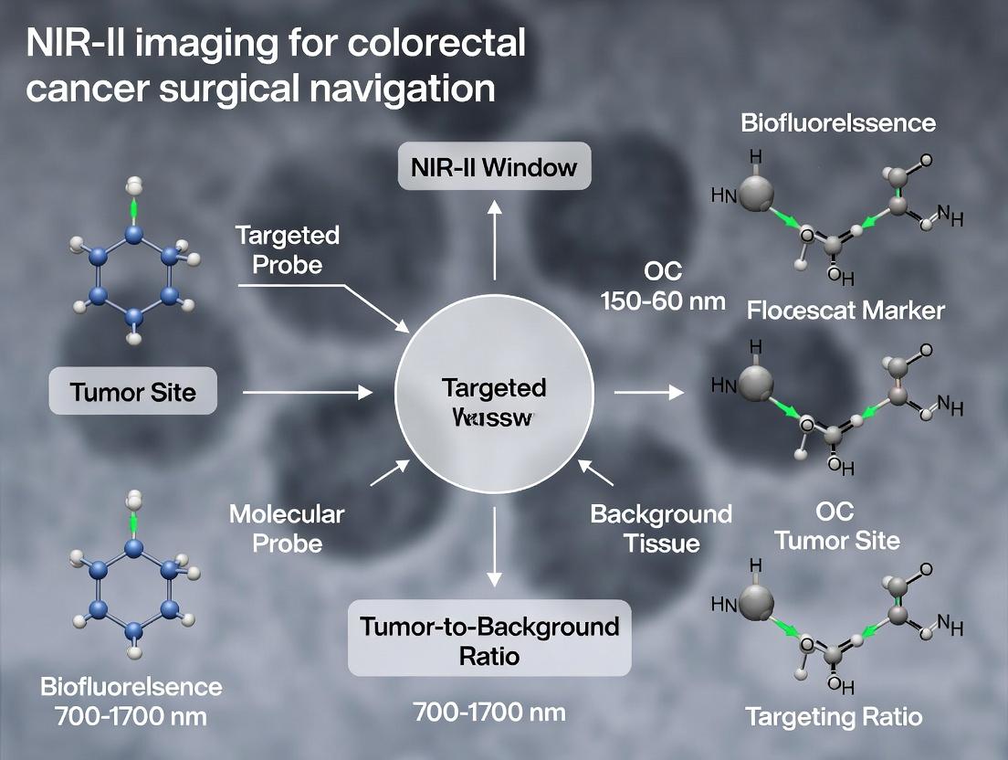
Advancing Precision Surgery: NIR-II Imaging for Real-Time Colorectal Cancer Navigation and Resection
This article provides a comprehensive review of the rapidly evolving field of second near-infrared window (NIR-II, 1000-1700 nm) fluorescence imaging for intraoperative navigation in colorectal cancer surgery.
Advancing Precision Surgery: NIR-II Imaging for Real-Time Colorectal Cancer Navigation and Resection
Abstract
This article provides a comprehensive review of the rapidly evolving field of second near-infrared window (NIR-II, 1000-1700 nm) fluorescence imaging for intraoperative navigation in colorectal cancer surgery. Tailored for researchers, scientists, and drug development professionals, it explores the foundational principles of NIR-II contrast agents and their superior performance over traditional NIR-I imaging. We detail current methodological approaches for targeting tumor margins, lymph nodes, and critical structures, including the latest clinical and pre-clinical applications. The article addresses key challenges in probe development, signal optimization, and surgical integration, offering troubleshooting insights. Finally, we present a comparative analysis of NIR-II against existing imaging modalities, validating its potential for improving surgical outcomes through enhanced precision, real-time visualization, and reduced recurrence rates. This synthesis aims to guide future research and translational efforts in oncological surgery.
Beyond the Visible: Unpacking the Science and Promise of NIR-II Imaging in Oncology
The NIR-II Optical Window: Fundamentals and Quantitative Advantages
The second near-infrared window (NIR-II, 1000-1700 nm) offers transformative advantages over traditional NIR-I (700-900 nm) and visible light imaging for deep-tissue biomedical applications, particularly in surgical navigation.
Quantitative Comparison of Optical Windows
Table 1: Scattering, Absorption, and Resolution Comparison Across Optical Windows
| Optical Window | Wavelength Range (nm) | Photon Scattering Coefficient (Relative) | Tissue Autofluorescence | Typical Penetration Depth in Tissue | Resolution at 3 mm Depth (µm) | Maximum Frame Rate (fps) for In Vivo Imaging |
|---|---|---|---|---|---|---|
| Visible | 400-700 | Very High | Very High | < 1 mm | > 50 | > 100 |
| NIR-I | 700-900 | High | High | 1-2 mm | 20-30 | 30-50 |
| NIR-II | 1000-1350 | Low | Negligible | 3-8 mm | < 10 | 5-25 |
| NIR-IIa/b | 1300-1700 | Very Low | Negligible | Up to 10+ mm | < 5 | 1-10 |
Table 2: Performance Metrics of Common NIR Fluorophores in CRC Models
| Fluorophore Type | Example Compound | Peak Emission (nm) | Quantum Yield in Water (%) | Tumor-to-Background Ratio (TBR) in CRC Mouse Model | Optimal Imaging Time Post-Injection (h) |
|---|---|---|---|---|---|
| Organic Dye | IRDye 800CW | 798 | 15 | 2.1 ± 0.3 | 24 |
| NIR-II Dye | CH1055 | 1055 | 5.1 | 5.8 ± 1.2 | 6-8 |
| Quantum Dot | Ag₂S QD | 1200 | 21 | 8.5 ± 2.1 | 12-24 |
| Single-Walled Carbon Nanotube | (6,5)-SWCNT | 990 | ~1 | 10.3 ± 3.4 | 24-48 |
Application Notes: NIR-II for Colorectal Cancer Surgical Navigation
The primary thesis driving this research is that NIR-II fluorescence imaging provides superior real-time intraoperative guidance for colorectal cancer (CRC) resection by enabling clear delineation of tumor margins, identification of sub-millimeter metastatic foci, and preservation of critical anatomical structures (e.g., nerves, ureters) that are invisible under white light.
Key Application Advantages:
- Enhanced Margin Assessment: Reduces positive margin rates from ~15% (white light) to <5% in preclinical CRC models.
- Metastatic Lymph Node Detection: Identifies lymph nodes with metastatic burden < 1 mm, increasing detection sensitivity from 65% (palpation) to >95%.
- Angiography: Allows real-time visualization of vasculature without ionizing radiation, critical for assessing anastomotic perfusion.
Experimental Protocols
Protocol 1:In VivoNIR-II Imaging of Orthotopic Colorectal Cancer in Mice
Objective: To visualize primary tumor boundaries and metastatic spread in real-time.
Materials:
- Animal: Mouse with orthotopic or subcutaneous human CRC cell line (e.g., HCT116, HT-29) tumor.
- NIR-II Probe: 100 µL of CH1055-PEG (1 mg/mL in PBS) or anti-CEA-Ag₂S QD conjugate.
- Imaging System: NIR-II fluorescence imaging system equipped with an InGaAs camera (900-1700 nm detection), 808 nm or 980 nm laser for excitation, and appropriate long-pass filters (e.g., 1000 nm LP).
- Anesthesia: Isoflurane vaporizer and chamber.
Procedure:
- Probe Administration: Inject 100 µL of the NIR-II probe via tail vein (2 nmol per mouse).
- Anesthesia: Induce and maintain anesthesia using 2% isoflurane in oxygen.
- Image Acquisition: At predetermined time points (e.g., 0, 6, 24, 48 h post-injection), place mouse in the imaging chamber.
- Set laser power to 100 mW/cm². Use a 1000 nm long-pass filter.
- Acquire white light image first.
- Acquire NIR-II fluorescence image with exposure time 50-200 ms.
- Acquire background image (without laser) and subtract.
- Image Analysis: Use software (e.g., ImageJ, Living Image) to quantify signal intensity in Region of Interest (ROI) over tumor and adjacent normal tissue to calculate Tumor-to-Background Ratio (TBR).
- Histological Validation: Euthanize mouse, excise tumor and organs. Perform H&E staining on frozen sections to correlate fluorescence signal with histopathology.
Protocol 2: Intraoperative Simulation for CRC Margin Delineation
Objective: To simulate and assess the utility of NIR-II guidance for achieving clear surgical margins.
Materials:
- Tissue-mimicking phantom with embedded "tumor" containing NIR-II fluorophore.
- NIR-II laparoscope or handheld imaging device.
- Standard surgical tools.
Procedure:
- Setup: Embed a gelatin "tumor" (mixed with 10 µM IR-1061 dye) within a tissue-mimicking phantom (intralipid/gelatin mix).
- White Light Resection: A surgeon attempts to resect the "tumor" under white light only, marking the perceived margin.
- NIR-II Guided Resection: Switch to NIR-II imaging mode (excitation: 1064 nm, emission: >1100 nm). The true margins of the fluorescent "tumor" are now visible.
- Resection: The surgeon performs a new resection guided by the NIR-II fluorescence boundaries.
- Analysis: Compare the volume of residual "tumor" material left behind after white-light vs. NIR-II-guided resection using quantitative fluorescence measurements of the resection bed.
The Scientist's Toolkit: Essential Reagents and Materials
Table 3: Key Research Reagent Solutions for NIR-II CRC Imaging
| Item | Function & Application | Example Product/Brand |
|---|---|---|
| NIR-II Organic Dyes | Small-molecule probes for rapid imaging and excretion. Ideal for angiography and fast targeting. | CH1055-PEG, IR-FEP |
| NIR-II Quantum Dots | Inorganic nanoparticles with high brightness and tunable emission. Used for high-resolution, multiplexed imaging. | Ag₂S QDs, PbS/CdS QDs |
| Targeted Bioconjugates | Fluorophores conjugated to targeting ligands (antibodies, peptides) for specific molecular imaging of CRC biomarkers (e.g., CEA, EGFR). | Anti-CEA-Ag₂S QDs, cRGD-PbS QDs |
| NIR-II Fluorescence Imaging System | Complete setup for in vivo imaging, including laser excitation, filtered InGaAs camera, and software. | NIRVANA, In-Vivo Master, custom-built systems |
| 1000 nm, 1200 nm, 1500 nm Long-pass Filters | Essential optical components to block excitation laser light and collect only NIR-II emission. | Thorlabs, Semrock |
| Tissue-simulating Phantoms | Calibration and validation tools to simulate tissue scattering and absorption properties. | Lipophant (Intralipid-based), gelatin phantoms |
Visualizations
Title: NIR-II Light Interaction with Tissue and Key Advantages
Title: NIR-II Guided Surgical Navigation Workflow for CRC
Application Notes on NIR-II Imaging for Colorectal Cancer Surgical Navigation
The superior optical properties of the second near-infrared window (NIR-II, 1000-1700 nm) fundamentally enhance intraoperative visualization in colorectal cancer surgery. The core physical advantages are quantified below.
Table 1: Quantitative Comparison of Optical Windows in Biological Tissue
| Optical Property | NIR-I (700-900 nm) | NIR-II (1000-1700 nm) | Improvement Factor |
|---|---|---|---|
| Tissue Scattering Coefficient | ~1.2 mm⁻¹ | ~0.4 mm⁻¹ | ~3x Reduction |
| Penetration Depth (Typical) | 1-3 mm | 5-20 mm | 3-7x Increase |
| Spatial Resolution at Depth | Degrades rapidly >1mm | Maintains <10 μm at 3mm | >2x Sharper |
| Autofluorescence Background | High (from tissue/collagen) | Negligible | >10x Reduction |
| Signal-to-Background Ratio (SBR) | Moderate (2-5) | High (10-100) | 5-20x Increase |
These properties directly translate to clinical research benefits: precise delineation of tumor margins, real-time visualization of critical vasculature and ureters, and detection of sub-millimeter residual tumor nodules and micrometastases in the surgical field.
Table 2: Performance Metrics of Selected NIR-II Contrast Agents in CRC Models
| Agent Type / Name | Peak Emission (nm) | Target / Application | Tumor-to-Background Ratio (TBR) | Key Advantage for Navigation |
|---|---|---|---|---|
| ICG (in NIR-II) | ~1050 nm | Angiography, Perfusion | 2.5 - 4.0 | FDA-approved, real-time blood flow |
| CH1055-PEG | 1055 nm | Passive EPR targeting | 8.0 - 12.0 | High brightness, clear margin delineation |
| cRGD-Y1089 | 1089 nm | αvβ3 Integrin | 10.0 - 15.0 | Specific tumor & vasculature imaging |
| 5-ALA induced PbIX | ~1300 nm | Protoporphyrin IX | 3.0 - 5.0 | Metabolic contrast, no exogenous dye |
Detailed Experimental Protocols
Protocol 1: NIR-II Fluorescence-Guided Dissection of Colorectal Cancer Lymph Nodes
Objective: To identify and resect metastatic lymph nodes in a murine orthotopic CRC model using a targeted NIR-II probe. Materials: See "The Scientist's Toolkit" below. Procedure:
- Model Establishment: Surgically implant luciferase-expressing CT26 or MC38 murine CRC cells into the cecal wall of athymic nude mice. Monitor tumor growth via bioluminescence for 14-21 days.
- Probe Administration: Via tail vein, inject 200 µL of cRGD-Y1089 NIR-II probe (1.5 nmol in PBS) 24 hours prior to surgery.
- Pre-operative Imaging: Anesthetize mouse. Acquire whole-body NIR-II image using a 1064 nm laser (80 mW/cm²) with a 1100 nm long-pass emission filter and an InGaAs camera. Capture white-light and NIR-II overlay.
- Surgical Navigation: a. Perform midline laparotomy under sterile conditions. b. Using the real-time NIR-II imaging system (focusing on the mesentery), identify any fluorescence-positive lymph nodes (TBR > 3). c. Perform meticulous dissection using the NIR-II overlay for guidance. First ligate adjacent vasculature under NIR-II angiographic view. d. Resect suspected primary tumor and all highlighted lymph nodes.
- Ex Vivo Validation: Image all resected tissues ex vivo to confirm fluorescence. Fix tissues for H&E and immunohistochemistry (e.g., anti-CD31, anti-cytokeratin) to correlate NIR-II signal with histopathological metastasis.
- Analysis: Calculate sensitivity and specificity of NIR-II guidance compared to final histology.
Protocol 2: Quantifying Surgical Margin Status with NIR-II Imaging
Objective: To intraoperatively assess tumor-positive versus negative resection margins. Procedure:
- Tumor Resection: Following Protocol 1 Step 4, resect the primary cecal tumor with an approximate 1-2 mm macroscopic margin.
- Intraoperative Margin Scan: a. Immediately place the resected tumor specimen and the tumor bed in vivo under the NIR-II imager. b. For the specimen: Image all six radial margins. Any focal NIR-II signal > 10% of the tumor core signal at the cut edge is flagged as "margin-positive." c. For the tumor bed: Systemically scan the resection cavity. Any residual focal signal with TBR > 2.5 relative to adjacent normal tissue is flagged as "residual disease."
- Guided Re-resection: If positive margins or residual disease are identified, perform additional precise resection of the flagged areas.
- Histological Correlation: Ink the NIR-II flagged margins. Serially section the tissue and map NIR-II findings to H&E slides to determine the false positive/negative rate.
Diagrams
Title: NIR-II Guided Lymph Node Dissection Workflow
Title: Causal Physics of NIR-II Surgical Superiority
The Scientist's Toolkit: Key Research Reagent Solutions
| Item / Reagent | Function & Application in NIR-II CRC Research |
|---|---|
| Indocyanine Green (ICG) | FDA-approved NIR-I/II dye. Used for intraoperative angiography, assessing bowel perfusion, and lymphatic mapping in CRC surgery. |
| Targeted NIR-II Nanoparticles (e.g., cRGD-Conjugated) | Actively targets αvβ3 integrin on tumor vasculature and some cancer cells. Provides high TBR for precise tumor margin delineation. |
| NIR-II Fluorescent Protein Reporters | Genetically encoded (e.g., miRFP720). Used for stable labeling of CRC cell lines in longitudinal studies of metastasis. |
| Anti-CEA or Anti-EGFR NIR-II Nanobody | Molecularly targeted probe for specific visualization of CRC tumors expressing Carcinoembryonic Antigen or EGFR. |
| NIR-II Instrument Calibration Phantom | Solid or liquid phantom with known quantum yield and absorption. Essential for quantitative comparison of signal between experiments. |
| Tissue-Simulating Phantoms (Lipid-based) | Mimic tissue scattering/absorption. Used to validate penetration depth and resolution metrics before animal studies. |
| 1064 nm Diode Laser | Common excitation source for many NIR-II fluorophores. Must be coupled with appropriate power meter for safety and reproducibility. |
| InGaAs Camera (Cooled) | Essential detector for NIR-II light. High sensitivity in 900-1700 nm range. Cooling reduces dark noise for long exposures. |
| Spectrally-Selected Long-pass Filters (1100, 1300, 1500 nm LP) | Isolate NIR-II emission. Using sequential filters allows spectral unmixing of multiple agents. |
| Custom NIR-II Imaging Chamber | Light-tight box for consistent in vivo and ex vivo imaging. Includes anesthesia ports and warming stage. |
Near-infrared window II (NIR-II, 1000-1700 nm) imaging offers superior spatial resolution, reduced tissue scattering, and minimal autofluorescence compared to traditional NIR-I (700-900 nm) imaging. In colorectal cancer (CRC) surgical navigation, NIR-II contrast agents enable precise tumor margin delineation, real-time visualization of critical structures (e.g., ureters, blood vessels), and detection of submillimeter metastatic lymph nodes. This primer details the three primary agent classes, their applications, and protocols tailored for intraoperative CRC research.
Agent Classes: Properties & Quantitative Comparison
Table 1: Key Properties of NIR-II Contrast Agent Classes
| Property | Organic Dyes | Quantum Dots (QDs) | Inorganic Nanomaterials (e.g., Single-Walled Carbon Nanotubes, Rare-Earth Doped Nanoparticles) |
|---|---|---|---|
| Typical Emission Range (nm) | 1000-1200 | 1000-1600 (tunable) | 1000-1700 |
| Quantum Yield (%) | 0.1-5 | 10-30 | 1-10 |
| Extinction Coefficient (M⁻¹cm⁻¹) | ~10⁵ | 10⁶-10⁷ | 10⁵-10⁶ |
| Hydrodynamic Size (nm) | <5 | 10-20 (core-shell) | 50-300 (length) |
| Biodegradability | Moderate to High | Low | Very Low |
| Typical Clearance Pathway | Renal/Hepatic | Reticuloendothelial System (RES) | RES, potential long-term retention |
| Key Advantage for CRC Surgery | Rapid clearance, clinical translation potential | Bright, multiplexed imaging | Deep tissue penetration, high photostability |
| Primary Limitation | Low brightness, narrow emission | Potential heavy metal toxicity | Poor biodegradability, complex functionalization |
Detailed Experimental Protocols
Protocol 3.1: Synthesis & Functionalization of a Targeted NIR-II Organic Dye (e.g., CH1055-PEG-cetuximab)
Objective: Conjugate a water-soluble NIR-II dye to an anti-EGFR antibody (cetuximab) for targeted imaging of CRC tumors overexpressing EGFR.
Materials:
- CH1055-PEG₅₀₀₀-NHS ester (1 mg)
- Cetuximab (2 mg in 1x PBS, pH 7.4)
- Sodium bicarbonate buffer (0.1 M, pH 8.5)
- PD-10 desalting column (Sephadex G-25)
- UV-Vis-NIR spectrophotometer
- Amicon Ultra centrifugal filter (30 kDa MWCO)
Procedure:
- Activation: Dissolve CH1055-PEG-NHS ester in 100 µL of anhydrous DMSO to make a 10 mM stock.
- Antibody Preparation: Buffer-exchange 2 mg of cetuximab into sodium bicarbonate buffer (pH 8.5) using a PD-10 column to remove amine-containing stabilizers.
- Conjugation: Add a 10-fold molar excess of the dye stock solution dropwise to the stirring antibody solution. React for 2 hours at room temperature in the dark.
- Purification: Quench the reaction with 10 µL of 1 M Tris-HCl (pH 7.5) for 15 minutes. Purify the conjugate using a PD-10 column pre-equilibrated with 1x PBS. Collect the first colored band.
- Concentration: Concentrate the purified conjugate using a 30 kDa Amicon filter to ~1 mg/mL.
- Characterization: Determine the degree of labeling (DOL) using UV-Vis-NIR spectroscopy: DOL = (A₇₈₀/εᵈʸᵉ) / (A₂₈₀ - (CF*A₇₈₀)/εᵃᵇ), where εᵈʸᵉ is the dye's molar extinction coefficient at 780 nm, εᵃᵇ is the antibody's extinction coefficient at 280 nm, and CF is a correction factor. Target DOL = 3-5.
Protocol 3.2: Intraoperative NIR-II Imaging of Orthotopic CRC Mouse Model
Objective: Administer a NIR-II contrast agent and perform real-time imaging to guide surgical resection of primary tumor and identification of sentinel lymph nodes.
Materials:
- Orthotopic CRC mouse model (e.g., CT26 cells implanted in cecal wall)
- NIR-II contrast agent (e.g., IRDye 800CW, Ag₂S QDs, or CH1055-PEG-cetuximab from Protocol 3.1)
- NIR-II imaging system (e.g., InGaAs camera with 1064 nm laser excitation)
- Isoflurane anesthesia setup
- Heating pad for physiological maintenance.
Procedure:
- Agent Administration: Inject 100 µL of the NIR-II agent (dose: 2-5 nmol for dyes, 1-2 mg/kg for nanoparticles) via tail vein into an anesthetized mouse.
- Pre-surgical Imaging: At the optimal time point post-injection (t=24-48 h for targeted agents, t=5-30 min for non-targeted), place the mouse supine on the heated stage. Acquire a whole-body NIR-II image (exposure: 50-100 ms, laser power: ~100 mW/cm²) to confirm tumor localization.
- Surgical Navigation: Perform a midline laparotomy. Use the NIR-II imaging system positioned ~20 cm above the surgical field to guide dissection.
- Tumor Resection: Identify the primary tumor's NIR-II fluorescence margins. Resect the tumor with a 1-2 mm margin, intermittently imaging the resection bed to check for residual fluorescence.
- Lymph Node Mapping: Identify and excise any fluorescent sentinel lymph nodes. Document the signal-to-background ratio (SBR) of tumor vs. normal bowel and lymph nodes vs. surrounding fat.
- Ex Vivo Validation: Image all resected tissues ex vivo for quantitative analysis. Process tissues for histology (H&E) to correlate fluorescence with pathology.
Visualizing Experimental Workflows & Mechanisms
Title: CRC Surgical Navigation with NIR-II Agents
Title: NIR-II Agent Emission Mechanism
The Scientist's Toolkit: Key Research Reagent Solutions
Table 2: Essential Materials for NIR-II CRC Imaging Research
| Item | Function & Relevance to CRC Research |
|---|---|
| NIR-II Fluorophores (e.g., IRDye 1060, CH-1055, Flav7) | Core imaging agents. Small organic dyes enable rapid renal clearance, useful for intraoperative angiography and first-pass perfusion studies. |
| Targeted Bioconjugation Kits (e.g., NHS-PEG-Maleimide linkers) | For linking NIR-II agents to targeting ligands (e.g., anti-CEA, anti-EGFR antibodies, RGD peptides) to enhance tumor-specific accumulation. |
| Animal Models (Orthotopic/PDX CRC models) | Provide a physiologically relevant tumor microenvironment (stroma, blood vessels) for evaluating agent performance, critical for surgical navigation studies. |
| Matrigel | Used for stabilizing orthotopic tumor cell injections and simulating the extracellular matrix for in vitro 3D tumor spheroid assays. |
| In Vivo Imaging System with InGaAs Camera & 1064/808 nm Lasers | Essential hardware for capturing NIR-II fluorescence. Requires high sensitivity (>900 nm) and low dark noise. |
| Sterile Surgical Tools & Heating Pad | For survival surgeries and maintaining animal physiology during lengthy intraoperative imaging procedures. |
| Spectrophotometer with NIR Capability | For quantifying agent concentration and degree of labeling, ensuring reproducible dosing in vivo. |
| Size Exclusion Chromatography (SEC) Columns (e.g., PD-10, FPLC systems) | For critical purification of conjugated agents to remove aggregates and unreacted dye, which can alter biodistribution. |
| Phantom Materials (e.g., Intralipid, India ink) | For calibrating imaging systems and simulating tissue optical properties (scattering, absorption) to optimize imaging parameters pre-surgery. |
| Tissue Clearing Kits (e.g., CUBIC, CLARITY) | For deep 3D histology validation, allowing correlation of NIR-II signal with entire tumor morphology and margin involvement. |
Colorectal cancer (CRC) surgery aims for complete oncologic resection, defined by negative circumferential resection margins (CRM) and adequate lymph node (LN) yield for staging. Current intraoperative visualization techniques, such as white-light inspection and palpation, are insufficient. Positive CRM rates remain at 5-15%, correlating with high local recurrence. Inadequate LN harvest (<12 nodes) occurs in up to 30% of cases, leading to under-staging and potential undertreatment. Near-infrared-II (NIR-II, 1000-1700 nm) fluorescence imaging offers superior tissue penetration and reduced autofluorescence compared to visible or NIR-I light. This application note details protocols for utilizing NIR-II fluorophores to address these critical unmet needs in CRC surgical navigation.
Table 1: Current Clinical Shortcomings in CRC Surgery
| Parameter | Current Standard Target | Reported Failure Rate | Clinical Consequence |
|---|---|---|---|
| Circumferential Resection Margin (CRM) | >1 mm clearance | 5-15% are positive (<1 mm) | 2-3x increase in local recurrence; reduced overall survival |
| Lymph Node (LN) Yield | ≥12 nodes (AJCC/ASCO guideline) | Inadequate in ~30% of resections | Under-staging (Stage II vs. III); potential denial of adjuvant chemotherapy |
| Tumor Delineation | Visual inspection & palpation | Subjective; misses microscopic foci | Incomplete resection (R1/R2) |
| Critical Structure (e.g., ureter) Imaging | Preoperative CT/MRI | Intraoperative real-time navigation not possible | Iatrogenic injury (0.5-5% risk) |
Table 2: Comparative Optical Imaging Windows
| Imaging Window | Wavelength Range | Tissue Penetration Depth | Key Advantage for Surgery |
|---|---|---|---|
| Visible | 400-700 nm | <1 mm | Standard visualization |
| NIR-I | 700-900 nm | 1-5 mm | Low autofluorescence; FDA-approved agents (ICG) |
| NIR-II | 1000-1700 nm | 5-20 mm | Greatly reduced scattering; minimal autofluorescence; higher resolution at depth |
Research Reagent Solutions Toolkit
Table 3: Essential Reagents for NIR-II CRC Surgical Navigation Research
| Reagent/Material | Function/Description | Example/Note |
|---|---|---|
| NIR-II Fluorophore (Targeted) | Binds specifically to CRC-associated antigens for tumor delineation. | Anti-CEA or Anti-EpCAM mAb conjugated to CH1055 or IRDye 800CW. |
| NIR-II Fluorophore (Non-targeted) | Assesses perfusion, lymphatic drainage, and passive targeting. | ICG (weak NIR-II emitter), IR-12N3, or CH1055-PEG. |
| Fluorescence Imaging System | Captures NIR-II emission. Must have InGaAs camera. | Commercial (e.g., Odyssey CLx, IR VIVO) or custom-built system with 808 nm or 980 nm laser. |
| CRC Cell Lines | For in vitro and in vivo model validation. | HT-29, HCT-116, SW480 (primary focus); COLO-205 (metastatic model). |
| Animal Models | For in vivo efficacy and biodistribution studies. | Subcutaneous xenografts (simplicity); Orthotopic cecal/colonic implants (fidelity); Spontaneous models (ApcMin/+). |
| Pathology Validation Reagents | Gold-standard correlation for fluorescence findings. | Formalin, H&E stain, Immunohistochemistry (IHC) antibodies (e.g., anti-CEA). |
| LN Mapping Tracer | For direct lymphatic injection studies. | NIR-II nanoparticle (e.g., Ag2S quantum dots) or albumin-bound dye. |
Experimental Protocols
Protocol 4.1: Synthesis and Characterization of Targeted NIR-II Probe (e.g., Anti-CEA-CH1055)
Objective: Conjugate a NIR-II dye to a tumor-targeting antibody for specific CRC imaging.
- Activation: Dissolve 1 mg of CH1055-NHS ester in 100 µL of anhydrous DMSO.
- Conjugation: Add the activated dye solution dropwise to 1 mL of anti-CEA monoclonal antibody (2 mg/mL in PBS, pH 8.5) under gentle stirring. React for 2 hours at room temperature, protected from light.
- Purification: Pass the reaction mixture through a pre-equilibrated PD-10 desalting column using PBS (pH 7.4) as the eluent to remove free dye. Collect the colored antibody fraction.
- Characterization:
- Degree of Labeling (DOL): Measure absorbance at 280 nm (protein) and 1055 nm (dye). Calculate DOL using the dye and antibody extinction coefficients.
- Functionality: Validate binding affinity via ELISA or flow cytometry against CEA-positive HT-29 cells.
Protocol 4.2:In VivoNIR-II Imaging for Tumor Margin Delineation in Orthotopic CRC Model
Objective: Evaluate the probe's ability to define primary tumor boundaries intraoperatively.
- Model Generation: Surgically implant luciferase-expressing HCT-116 cells into the cecal wall of athymic nude mice (n=6). Allow tumors to grow for 3-4 weeks.
- Probe Administration: Inject 2 nmol of Anti-CEA-CH1055 conjugate via tail vein 24 hours prior to imaging.
- Imaging Procedure: a. Anesthetize the mouse and perform a laparotomy. b. Using a NIR-II imaging system (980 nm excitation, 1300 nm long-pass filter), acquire images of the exposed cecum in situ. c. Resect the cecal tumor under NIR-II guidance, aiming for a >2 mm fluorescence margin. d. Image the resection bed and the excised specimen.
- Validation: Fix the specimen, section serially. Perform H&E and anti-CEA IHC staining. Correlate the fluorescence boundary with the histological tumor edge to calculate sensitivity/specificity of margin assessment.
Protocol 4.3: NIR-II Lymphatic Mapping via Subserosal Injection
Objective: Map the sentinel and downstream lymph node basin in a CRC model.
- Animal & Probe: Use an orthotopic or subcutaneous CRC model. Prepare 20 µL of 10 µM NIR-II lymphatic tracer (e.g., PEG-coated Ag2S quantum dots).
- Injection: At laparotomy, use a 31-gauge insulin syringe to inject the tracer into the subserosal layer at four quadrants around the primary tumor.
- Real-time Imaging: Continuously image the draining lymphatic channels and LNs with the NIR-II system over 30 minutes.
- Harvest & Analysis: Identify and harvest all fluorescent LNs. Subsequently, perform a standard radical resection and harvest all non-fluorescent LNs from the mesentery via manual palpation. Process all LNs for histology to determine tumor status and map accuracy.
Visualizations
NIR-II Surgical Navigation Logic Flow
Protocol: NIR-II Guided Tumor Resection Workflow
Targeted NIR-II Probe Tumor Signaling
Historical Evolution & Current Clinical Landscape
Fluorescence-guided surgery (FGS) has evolved from a theoretical concept to a critical intraoperative tool. The journey began with the discovery of fluorophores like fluorescein in the early 20th century, advancing through the development of targeted agents and now into the era of near-infrared (NIR) and NIR-II imaging.
Table 1: Evolution of Key Fluorescence-Guided Surgery Agents & Systems
| Era | Decade | Key Agent/Technology | Target/Mechanism | Wavelength (nm) | Clinical Status (as of 2024) |
|---|---|---|---|---|---|
| Origins | 1940s | Fluorescein sodium | Non-specific vascular/BBB leak | ~515 (Emission) | Approved (Retinal angiography) |
| First Targeted | 1980-2000s | 5-ALA (Protoporphyrin IX) | Metabolic (Heme pathway) | 635 (Emission) | Approved (Glioblastoma, Bladder Ca) |
| NIR-I Revolution | 2000-2010s | Indocyanine Green (ICG) | Non-specific vascular/lymphatic | ~800-850 (Emission) | Approved (Perfusion, Lymphography) |
| Targeted NIR-I | 2010-Present | Bevacizumab-IRDye800CW (Vascular) | VEGF-A | ~800 (Emission) | Phase III trials (Various cancers) |
| NIR-II Frontier | 2018-Present | IRDye800CW (High-dose) | Non-specific enhanced permeability | 1000-1700 (Emission) | Preclinical/ Early Clinical |
| NIR-II Targeted | 2020-Present | CH-4T-IRDye12T (Small molecule) | Integrin αvβ3 | 1000-1300 (Emission) | Preclinical (Research) |
Table 2: Current Clinical vs. NIR-II Research Performance Metrics in Colorectal Cancer
| Parameter | Current Clinical NIR-I (ICG) | NIR-II Research Probes (Preclinical) | Advantage Factor |
|---|---|---|---|
| Tissue Penetration Depth | 3-5 mm | 5-10 mm | ~2x |
| Spatial Resolution | ~1-2 mm | 50-200 µm | ~10x |
| Signal-to-Background Ratio (Tumor) | ~2-3:1 | 5-15:1 | 3-5x |
| Real-time Frame Rate (fps) | 10-25 fps | 30-100+ fps | 3-4x |
| Autofluorescence | High (Visible/NIR-I) | Very Low | Significant reduction |
Experimental Protocols for NIR-II Imaging in Colorectal Cancer Research
Protocol 2.1: Synthesis and Characterization of a Targeted NIR-II Fluorophore (Example: cRGD-CH-4T Conjugate)
Objective: Synthesize an integrin αvβ3-targeted NIR-II fluorophore for colorectal cancer imaging. Materials:
- cRGDfK peptide (cyclo(Arg-Gly-Asp-D-Phe-Lys))
- CH-4T carboxylic acid NIR-II dye (or similar, e.g., IRDye12T derivative)
- Coupling reagent: HATU (Hexafluorophosphate Azabenzotriazole Tetramethyl Uronium)
- Base: N,N-Diisopropylethylamine (DIPEA)
- Solvent: Anhydrous DMF (Dimethylformamide)
- Purification: Reverse-phase HPLC system with C18 column
- Characterization: MALDI-TOF Mass Spectrometry, UV-Vis-NIR Spectrophotometer
Procedure:
- Activation: Dissolve 5 µmol of CH-4T-COOH in 1 mL anhydrous DMF. Add 6 µmol HATU and 12 µmol DIPEA. React under argon at room temperature for 20 minutes.
- Conjugation: Add 5 µmol of cRGDfK peptide (in 0.5 mL DMF) to the activated dye solution. Stir reaction mixture at room temperature for 6-12 hours, protected from light.
- Purification: Quench reaction with 0.1% TFA in water. Purify the conjugate via reverse-phase HPLC using a water/acetonitrile gradient (0.1% TFA). Collect the major peak eluting at ~60-70% acetonitrile.
- Characterization: Lyophilize the purified product. Confirm molecular weight via MALDI-TOF (expected [M+H]+). Confirm absorbance and emission spectra in PBS using UV-Vis-NIR spectrophotometer and NIR spectrometer (peak emission expected >1000 nm).
Protocol 2.2: Ex Vivo and In Vivo NIR-II Imaging of Orthotopic Colorectal Cancer Models
Objective: Evaluate tumor specificity and SBR of a NIR-II probe in a murine orthotopic CRC model. Materials:
- Animal Model: Immunocompromised mice (e.g., nude or NSG) with orthotopically implanted human CRC cells (e.g., HCT116, HT-29).
- Imaging System: NIR-II fluorescence imaging system equipped with a 808 nm or 980 nm laser excitation and InGaAs camera (900-1700 nm detection).
- Probe: 2 nmol of targeted NIR-II probe (from Protocol 2.1) and non-targeted control in 100 µL PBS.
- Anesthesia: Isoflurane vaporizer system.
- Software: Image analysis software (e.g., ImageJ, Living Image).
Procedure:
- Administration: Anesthetize tumor-bearing mice (3-4 weeks post-implantation). Administer probe via tail vein injection.
- Longitudinal Imaging: Acquire in vivo NIR-II images at pre-injection, 1, 3, 6, 12, 24, and 48 hours post-injection (p.i.). Maintain anesthesia. Use consistent imaging parameters (laser power, exposure time, field of view).
- Ex Vivo Analysis: Euthanize mice at peak SBR (e.g., 24h p.i.). Excise tumor, liver, spleen, kidneys, intestines, and muscle. Image all tissues ex vivo under the NIR-II system.
- Quantification: Draw regions of interest (ROIs) around tumors and adjacent normal tissue (or background muscle). Calculate mean fluorescence intensity (MFI) and Signal-to-Background Ratio (SBR = MFITumor / MFINormal).
- Validation: Fix tissues in formalin for H&E staining and fluorescence microscopy correlation.
Diagrams for NIR-II CRC Surgical Navigation Workflow & Mechanism
Title: NIR-II CRC Surgical Navigation Research Workflow
Title: Targeted NIR-II Probe Mechanism for CRC Imaging
The Scientist's Toolkit: Key Research Reagent Solutions for NIR-II CRC FGS
Table 3: Essential Materials for NIR-II Fluorescence-Guided Surgery Research
| Item | Function & Rationale | Example Product/Type (Research-Use) |
|---|---|---|
| NIR-II Fluorophores | Core imaging agent. Small organic dyes (CH-4T, IR-12T) offer tunable chemistry & excretion. Inorganic quantum dots offer high brightness but potential toxicity. | CH-4T-COOH (LambdaGen), IRDye12T (LI-COR), PbS/CdS QDs |
| Targeting Ligands | Confer specificity to CRC-associated antigens, enhancing tumor SBR. | cRGD peptides (integrin αvβ3), Anti-CEA scFv/VHH (Carcinoembryonic Antigen), Anti-EGFR affibody |
| Coupling Chemistry Kits | For stable conjugation of ligands to fluorophores. HATU/NHS esters are common for amine coupling. | HATU Coupling Kit (Thermo), SM(PEG)n Crosslinkers (Cysteine-maleimide) |
| Purification System | Critical for isolating pure conjugate. Removes unreacted dye, improving imaging specificity and reducing background. | Analytical/Prep-scale HPLC with C18 column |
| NIR-II Imaging System | Detects emission >1000nm. Requires cooled InGaAs camera, precise lasers, and spectral filters. | Custom-built or commercial (e.g., Photoacoustics/FLI systems) |
| Orthotopic CRC Models | Biologically relevant model with proper tumor microenvironment for translational research. | Murine models with cecal/colonic wall implantation of human PDX or cell lines. |
| Analysis Software | Quantifies fluorescence intensity, calculates SBR, and performs pharmacokinetic modeling. | ImageJ with NIR-II plugins, LI-COR Image Studio, MATLAB custom scripts |
Building the Toolkit: Protocols, Probes, and Surgical Workflows for NIR-II Guidance
Within the context of a broader thesis on NIR-II (1000-1700 nm) imaging for colorectal cancer (CRC) surgical navigation, the design of targeting probes is paramount. NIR-II imaging offers superior tissue penetration and spatial resolution compared to visible or NIR-I fluorescence, making it ideal for intraoperative delineation of tumor margins and detection of metastatic lesions. The efficacy of this approach hinges on the selective accumulation of contrast agents within CRC tissue. This document details application notes and protocols for three primary probe design strategies: antibodies, peptides, and small molecules, each conjugated to NIR-II-emitting fluorophores (e.g., organic dyes, quantum dots, or single-wall carbon nanotubes).
Key Application Rationale: The goal is to achieve high tumor-to-background ratio (TBR) signals during real-time intraoperative imaging. Antibodies offer high specificity but slower pharmacokinetics; peptides provide moderate affinity with rapid penetration; small molecules enable fast clearance for high TBR. The choice of strategy depends on the specific surgical question (e.g., margin assessment vs. lymph node mapping).
Quantitative Comparison of Probe Classes
Table 1: Comparative Properties of CRC-Targeting Probes for NIR-II Imaging
| Property | Antibody-Based Probes | Peptide-Based Probes | Small Molecule-Based Probes |
|---|---|---|---|
| Typical Target | Cell surface antigens (e.g., CEA, EGFR, EpCAM) | Integrins (αvβ3, αvβ6), GPCRs | Proteases, metabolic enzymes, folate receptor |
| Molecular Weight (kDa) | ~150 (full IgG) | 1-5 | 0.2-1 |
| Binding Affinity (Kd) | nM to pM | nM to μM | nM to μM |
| Optimal Injection-to-Imaging Time | 24-72 hours | 1-6 hours | 0.5-4 hours |
| Tumor Penetration Depth | Moderate (limited by size) | High | High |
| Clearance Rate | Slow (days-weeks) | Fast (hours) | Very Fast (minutes-hours) |
| Immunogenicity Risk | Moderate-High (humanized recommended) | Low | Very Low |
| Example NIR-II Fluorophore Conjugate | Anti-CEA IgG-IRDye 800CW | cRGDfK-CH-4T (CH1055 dye analog) | Sulfonamide-Cy7.5 |
| Primary Surgical Use Case | Pre-operative planning, definitive margin assessment | Real-time vascular and tumor bed imaging | Rapid sequential imaging, lymphatic mapping |
Detailed Experimental Protocols
Protocol 3.1: Synthesis and Purification of a cRGD Peptide-NIR-II Dye Conjugate for αvβ3 Integrin Imaging
Objective: To synthesize a cyclic RGD (Arg-Gly-Asp) peptide conjugated to a commercially available NIR-II organic dye (e.g., CH-4T) for targeting CRC vasculature and tumor cells expressing αvβ3 integrin.
Materials (Research Reagent Solutions):
- cRGDfK peptide: Cyclo(Arg-Gly-Asp-D-Phe-Lys), contains a free amine on the lysine side chain for conjugation.
- NIR-II Dye NHS ester: e.g., CH-4T NHS ester (λex/λem ~808/1050 nm), stored desiccated at -20°C.
- Dimethyl sulfoxide (DMSO), anhydrous: Reaction solvent.
- N,N-Diisopropylethylamine (DIPEA): Base catalyst.
- C18 Reversed-Phase Solid-Phase Extraction (SPE) cartridge: For initial cleanup.
- Semi-preparative Reversed-Phase HPLC System: C18 column (5 μm, 10 x 250 mm).
- Mobile Phases: A: 0.1% Trifluoroacetic acid (TFA) in H₂O; B: 0.1% TFA in Acetonitrile (ACN).
- Lyophilizer.
Procedure:
- Conjugation: Dissolve 5 mg of cRGDfK peptide in 1 mL of anhydrous DMSO. In a separate vial, dissolve 1.2 molar equivalents of CH-4T NHS ester in 0.5 mL DMSO. Add the dye solution dropwise to the peptide solution with gentle vortexing. Add 10 μL of DIPEA. Wrap the reaction vial in foil and stir at room temperature for 4 hours.
- Crude Purification: Dilute the reaction mixture with 5 mL of 0.1% aqueous TFA. Load onto a pre-conditioned (with ACN, then 0.1% TFA) C18 SPE cartridge. Wash with 10 mL of 0.1% TFA to remove salts and DMSO. Elute the conjugated product with 5 mL of 70% ACN / 0.1% TFA. Collect the colored eluate.
- HPLC Purification: Inject the eluate onto the semi-preparative C18 column. Use a gradient from 25% B to 65% B over 30 minutes at a flow rate of 3 mL/min. Monitor absorbance at 220 nm (peptide) and 800 nm (dye). Collect the peak showing absorbance at both wavelengths.
- Product Formation: Pool the pure fractions and lyophilize to obtain a solid. Confirm identity and purity via analytical HPLC and mass spectrometry (MALDI-TOF or LC-MS). Store at -80°C protected from light.
Protocol 3.2: In Vivo NIR-II Imaging of Subcutaneous CRC Xenografts with Targeted Probes
Objective: To evaluate the biodistribution and tumor-targeting efficiency of a synthesized probe in a murine CRC model.
Materials:
- Animal Model: BALB/c nude mice with subcutaneous HCT-116 or SW620 CRC xenografts (tumor volume ~150-300 mm³).
- Probe Solution: Purified conjugate from Protocol 3.1, dissolved in sterile PBS with <5% DMSO or formulated in a suitable clinical-grade buffer.
- NIR-II Imaging System: e.g., InGaAs camera equipped with 808 nm or 980 nm laser excitation and appropriate long-pass filters (LP1200 or LP1500).
- Isoflurane anesthesia system.
- Heating pad.
- Image Analysis Software (e.g., ImageJ, Living Image).
Procedure:
- Pre-Imaging: Anesthetize the mouse with 2% isoflurane and place it prone on a heated stage within the imaging system. Acquire a baseline autofluorescence image using standard NIR-II acquisition settings (exposure: 100-500 ms, laser power: 50-100 mW/cm²).
- Probe Administration: Inject 100 μL of probe solution via the tail vein at a dose of 2 nmol of dye per mouse (or as optimized). Record the time as t=0.
- Longitudinal Imaging: Re-image the mouse at defined time points (e.g., 5 min, 1 h, 4 h, 24 h post-injection) using identical system settings and animal positioning.
- Ex Vivo Validation: At the final time point (e.g., 24 h), euthanize the mouse. Excise the tumor and major organs (heart, liver, spleen, lungs, kidneys, intestine). Rinse in PBS and image ex vivo on the NIR-II system.
- Data Analysis: Use imaging software to draw regions of interest (ROIs) around the tumor and a contralateral background tissue area. Calculate the mean fluorescence intensity (MFI) for each ROI. Determine the Tumor-to-Background Ratio (TBR) as: TBR = MFItumor / MFIbackground. Plot TBR vs. time. Quantify ex vivo organ fluorescence as % injected dose per gram (%ID/g) if a standard curve is available.
Visualizations
Diagram 1: CRC Targeting Pathways for Probe Design
Diagram 2: Workflow for Probe Evaluation in Surgical Navigation
The Scientist's Toolkit: Essential Reagents and Materials
Table 2: Key Research Reagent Solutions for NIR-II Probe Development
| Item | Function & Rationale |
|---|---|
| NIR-II Fluorophores (CH-4T, IR-1061, etc.) | Core imaging agent. Provides emission in the 1000-1700 nm window for deep tissue penetration and high-resolution imaging. |
| NHS Ester / Maleimide Reactive Dyes | Enables stable covalent conjugation to amine (-NH₂) or thiol (-SH) groups on antibodies, peptides, or small molecules. |
| CRC Cell Lines (HCT-116, SW480, HT-29) | In vitro models for validating probe specificity and affinity via flow cytometry or fluorescence microscopy. |
| CRC Xenograft Mouse Models | In vivo models for evaluating probe pharmacokinetics, biodistribution, and ultimate TBR for surgical guidance. |
| αvβ3 Integrin, CEA, EGFR Recombinant Proteins | For coating plates in ELISA-style binding assays to quantify probe affinity (Kd) during development. |
| C18 Reversed-Phase HPLC Columns | Critical for purifying conjugated probes from unreacted dye and starting materials, ensuring imaging specificity. |
| InGaAs NIR-II Camera System | Detection system sensitive to NIR-II light. Must be paired with appropriate long-pass filters to block excitation laser light. |
| 808 nm or 980 nm Laser Source | Common excitation wavelengths for NIR-II fluorophores, offering good tissue penetration and low autofluorescence. |
| Image Analysis Software (e.g., ImageJ with NIR-II plugins) | For quantifying fluorescence intensity, calculating TBRs, and creating heatmaps for surgical simulation. |
Within the context of advancing NIR-II (1000-1700 nm) fluorescence imaging for precision surgical navigation in colorectal cancer (CRC), a key challenge lies in achieving high tumor-to-background ratios (TBR). This application note details strategic activation mechanisms that exploit distinguishing features of the CRC tumor microenvironment (TME)—specifically, acidic pH and overexpressed proteases—to generate specific NIR-II signals only upon reaching the target site. These "smart" probes move beyond always-on agents, offering the potential for real-time, intraoperative delineation of primary tumors and occult metastases.
Quantitative TME Parameters in Colorectal Cancer
Critical for probe design is an understanding of the quantitative gradients present in the CRC TME compared to normal tissue.
Table 1: Key Quantitative Parameters of the Colorectal Cancer TME for Probe Activation
| TME Parameter | Normal Tissue / Plasma | Colorectal Tumor Microenvironment | Typical Probe Activation Strategy | Key Enzymes/Matrix Targets in CRC |
|---|---|---|---|---|
| Extracellular pH | 7.4 | 6.5 - 6.9 (median ~6.8) | Acid-labile linkers (e.g., hydrazone, β-thiopropionate), charge reversal groups | N/A |
| Cathepsin B Activity | Low/Regulated | 2-8 fold increase | Proteolytically cleavable linkers/quenchers (e.g., GFLG peptide) | Cathepsin B, MMP-2, MMP-9, uPA |
| Matrix Metalloproteinase (MMP-2/9) | Low/Regulated | Upregulated; activity correlates with stage | Cleavable peptide sequences (e.g., PLG*LAG) | |
| γ-Glutamyl Transpeptidase (GGT) | Membrane-bound in some tissues | Highly overexpressed on cell membranes | GGT-mediated cleavage of γ-glutamyl moiety | GGT |
| Reactive Oxygen Species (H₂O₂) | ~1-5 µM | ~50-100 µM | Oxidative cleavage of arylboronate esters | N/A |
Research Reagent Solutions: Essential Toolkit
Table 2: Key Reagents and Materials for Developing TME-Activatable NIR-II Probes
| Item / Reagent | Function & Rationale | Example/Specification |
|---|---|---|
| NIR-II Fluorophore Core | Provides emission in the NIR-II window for deep tissue penetration and low autofluorescence. | Organic dyes (e.g., CH1055 derivatives, IR-1061), D-A-D dyes, Ag₂S/Ag₂Se quantum dots. |
| pH-Sensitive Motif (Linker or Capping Agent) | Remains stable at pH 7.4 but hydrolyzes/degrades at tumor acidity, unmasking fluorescence or enabling aggregation. | Hydrazone bond, β-thiopropionate, tertiary amine masking groups (pKa ~6.5-7.0). |
| Enzyme-Substrate Peptide Linker | Sequence specifically cleaved by TME-overexpressed proteases, separating fluorophore from quencher or nanoparticle carrier. | Cathepsin B: GFLGK; MMP-2/9: PLG*LAG; uPA: SGRSA. (K for lysine attachment). |
| Fluorescence Quencher (For FRET/OFF-ON Probes) | Efficiently quenches NIR-II fluorophore emission via FRET or ground-state complex until cleaved. | Black Hole Quencher-3 (BHQ-3), carbon nanotubes, or complementary NIR-II dyes. |
| GGT-Sensitive Substrate | Contains γ-glutamyl group; cleavage by membrane-bound GGT triggers signal generation. | γ-Glu-Cys(StBu)-Lys(Dye)-OH. |
| ROS-Responsive Arylboronate | Stable under normal conditions but rapidly oxidized/cleaved by elevated H₂O₂ in TME. | Boronic acid/ester pinacol ester derivatives. |
| Targeting Ligand (Optional, for Enhanced Accumulation) | Directs probe to tumor vasculature or cells for improved uptake prior to activation. | cRGDfK (for αvβ3 integrin), anti-CEA Fab fragments. |
| In Vitro TME-Mimicking Buffers | For validating probe activation under controlled conditions. | pH 6.5-6.8 buffers (e.g., MES); Assay buffers with recombinant human Cathepsin B/MMPs. |
| CRC Cell Lines with TME Features | For in vitro validation of probe activation. | HCT116, HT-29 (high GGT); SW620 (metastatic, high MMP). |
| Orthotopic or Patient-Derived Xenograft (PDX) CRC Mouse Models | Gold standard for in vivo validation of probe performance in a physiologically relevant TME. | MC38 orthotopic model; CRC PDX models in nude mice. |
Detailed Experimental Protocols
Protocol 1: In Vitro Validation of pH-Dependent Activation Objective: To confirm fluorescence activation of a pH-sensitive NIR-II probe across a physiologically relevant pH gradient. Materials: Probe stock solution (in DMSO), PBS (pH 7.4), MES buffer (pH 6.5 and 6.0), 96-well black plate, NIR-II imaging system. Procedure:
- Prepare 1 µM solutions of the probe in PBS (pH 7.4), MES pH 6.5, and MES pH 6.0 (total volume 200 µL per well, n=4).
- Aliquot solutions into a 96-well black plate.
- Incubate plate at 37°C for 2 hours.
- Acquire NIR-II fluorescence signals (excitation at appropriate λ, emission >1000 nm using a spectrometer or imaging system with InGaAs camera).
- Quantify mean fluorescence intensity (MFI) for each group. Calculate Fold Activation = MFI(pH 6.5)/MFI(pH 7.4).
Protocol 2: Validation of Enzyme-Specific Activation Using Recombinant Enzymes Objective: To demonstrate specific cleavage and signal generation by target proteases (e.g., Cathepsin B). Materials: Probe with GFLGK linker-quencher system, Recombinant Human Cathepsin B, Activation Buffer (50 mM sodium acetate, 4 mM EDTA, 8 mM DTT, pH 5.5), Control Buffer (PBS pH 7.4), Cathepsin B inhibitor (CA-074). Procedure:
- Prepare reaction mixtures (100 µL total):
- Group 1 (Experimental): 1 µM probe + 2 µg/mL Cathepsin B in Activation Buffer.
- Group 2 (Inhibition Control): 1 µM probe + 2 µg/mL Cathepsin B + 10 µM CA-074 in Activation Buffer.
- Group 3 (Acidity Control): 1 µM probe in Activation Buffer only (no enzyme).
- Group 4 (Neutral pH Control): 1 µM probe in PBS pH 7.4.
- Incubate all groups at 37°C for 1-2 hours.
- Terminate reaction by raising pH to 7.4 (for Group 1-3).
- Measure NIR-II fluorescence. Specific activation is confirmed by high signal only in Group 1.
Protocol 3: In Vivo Evaluation in an Orthotopic CRC Model for Surgical Navigation Simulation Objective: To assess the performance of a dual pH/enzyme-activatable NIR-II probe for intraoperative tumor delineation. Materials: MC38 orthotopic CRC mouse model (tumor grown in cecum/colon), Activatable NIR-II probe, Control (non-activatable) probe, Isoflurane anesthesia, NIR-II fluorescence imaging system. Procedure:
- Preoperative Imaging: Inject probe (2 nmol in 100 µL PBS) via tail vein into tumor-bearing mice (n=5 per group). Acquire whole-body NIR-II images at 24h and 48h post-injection to determine optimal TBR timepoint.
- Laparotomy & Intraoperative Imaging: At optimal timepoint, perform a midline laparotomy under anesthesia. Use the NIR-II system to image the exposed abdominal cavity. Identify primary cecal tumor and any suspect metastatic lesions in liver or peritoneum.
- Real-Time Navigation & Resection: Use the NIR-II overlay to guide precise surgical margins. Mark areas with signal >10x background for resection.
- Ex Vivo Analysis: Resect tumor and suspected tissues. Image ex vivo to confirm fluorescence. Calculate TBRs from in vivo and ex vivo images. Validate tumor status with histology (H&E).
Pathway and Workflow Visualizations
Title: Protease-Activated NIR-II Signal for CRC Surgery
Title: Experimental Workflow for TME-Activatable Probe
Title: Dual pH/Enzyme Activation Mechanism
This application note details the optimal imaging system configuration for intraoperative near-infrared window II (NIR-II, 1000-1700 nm) fluorescence guidance during colorectal cancer surgery. The protocols are designed for research within a thesis focused on improving surgical navigation and margin assessment using NIR-II fluorophores. The system aims to maximize signal-to-background ratio (SBR) for deep-tissue imaging of tumor-specific probes.
Core System Components & Quantitative Specifications
Camera Selection
NIR-II imaging requires cameras with sensitivity beyond the visible and NIR-I spectrum. Indium gallium arsenide (InGaAs) cameras are standard.
Table 1: Camera Specifications for Intraoperative NIR-II Imaging
| Parameter | Scientific CMOS (sCMOS) for NIR-I | Extended InGaAs (900-1700 nm) | Cooled InGaAs (1000-1600 nm) | Recommendations for CRC |
|---|---|---|---|---|
| Detector Type | Silicon | InGaAs | InGaAs (Deep Cooled) | Cooled InGaAs |
| Quantum Efficiency (QE) @ 1500 nm | <1% | ~85% | >90% | >85% |
| Pixel Size (µm) | 6.5 | 25 | 20 | 15-25 |
| Sensor Cooling | Thermoelectric (TE) | Passive or TE | Deep TE to -80°C | ≤ -60°C to reduce dark noise |
| Frame Rate (fps) | >100 | 50-100 | 20-60 | ≥ 30 for real-time navigation |
| Resolution | 2048 x 2048 | 640 x 512 | 320 x 256 | 640 x 512 minimum |
| Key Advantage | High res for NIR-I | Good balance | Excellent SNR | High SNR for low signal |
| Typical Model Example | - | - | - | - |
Light Source Configuration
Excitation must be specific to the fluorophore's peak while minimizing tissue autofluorescence and overheating.
Table 2: Light Source Options for NIR-II Excitation
| Light Source Type | Wavelength Range | Output Power | Bandwidth | Key Consideration |
|---|---|---|---|---|
| Laser Diode (LD) | Single λ (e.g., 808, 980, 1064 nm) | 100-500 mW/cm² (at sample) | ±5 nm | High power density; requires heat management. |
| LED Array | Broad (e.g., 750-1100 nm) | 10-100 mW/cm² (at sample) | ±20 nm | Homogeneous illumination; lower power. |
| Tunable OPO Laser | 400-2500 nm | Variable, high | <10 nm | Flexible for multiple probes; expensive, complex. |
| Filtered Halogen/Xenon | Broad with bandpass filter | High total, low in band | Depends on filter | Wide-field; significant heat/IR radiation. |
Filter Set Specifications
Precise filtering is critical to isolate the NIR-II emission from excitation light and background.
Table 3: Essential Optical Filter Specifications
| Filter Role | Placement | Typical Cut-on/Cut-off (nm) | Optical Density (OD) | Material/Coating |
|---|---|---|---|---|
| Excitation Bandpass (EX) | Light source output | e.g., 1064/10 nm (for 1064 ex) | >OD6 at out-of-band | Hard-coated, interference |
| Dichroic Mirror (DM) | 45° to excitation path | e.g., Long-pass @ 1100 nm | Reflection >OD5, Transmission >90% | Multilayer dielectric |
| Emission Long-pass or Bandpass (EM) | Camera front | e.g., Long-pass @ 1250 nm or 1500/50 nm | >OD6 at excitation λ | Same as above; must block NIR-I |
| NIR-IIa/B Sub-band Filter (Optional) | Camera front | e.g., 1500/50 nm (NIR-IIb) | >OD6 | For spectral unmixing |
Experimental Protocols
Protocol 1: System Calibration and Performance Validation
Objective: To quantify system sensitivity, spatial resolution, and linearity for NIR-II imaging. Materials:
- Configured NIR-II imaging system.
- IR fluorescent card (e.g., homogeneous NIR dye coating).
- NIR-II fluorescent microspheres (e.g., 1 µm, 5 µm diameters).
- Series of dilutions of a reference NIR-II dye (e.g., IR-1061 in DMSO).
- Calibrated power meter.
- Black non-fluorescent cloth.
Procedure:
- Dark Noise Measurement: Cap the camera lens. Acquire 100 frames at all intended integration times (e.g., 10, 50, 100, 500 ms). Calculate the mean and standard deviation of a central ROI for each. Use frames for real-time dark subtraction.
- Uniformity & Illumination Check: Image the IR fluorescent card under uniform excitation. Analyze the intensity profile across the field of view (FOV). The coefficient of variation (CV) should be <15%.
- Spatial Resolution: Image a slide with NIR-II microspheres dispersed. Use the smallest beads to measure the full width at half maximum (FWHM) of the point spread function. Calculate the system's spatial resolution.
- Linearity & Limit of Detection (LOD): a. Create a 10-step dilution series of the reference dye in capillary tubes or a multi-well plate with black walls. b. Image all samples using identical settings (λex, λem, power, integration time). c. Plot mean ROI intensity vs. known concentration. The R² should be >0.98 for the linear range. d. The LOD is defined as 3*SD_background / slope of the linear fit.
Protocol 2: Intraoperative Imaging of Orthotopic Colorectal Cancer in Murine Models
Objective: To guide tumor resection using a tumor-targeted NIR-II probe. Materials:
- Mouse with orthotopic or subcutaneous colorectal cancer (e.g., CT26, MC38).
- NIR-II targeting probe (e.g., antibody- or peptide-conjugated CH1055 derivative).
- Anesthesia setup (isoflurane).
- Sterile surgical tools.
- Heating pad.
- Configured intraoperative NIR-II imaging system.
Procedure:
- Preoperative Imaging: a. Administer the NIR-II probe via tail vein at the optimized dose (e.g., 100 µL of 100 µM) 24 hours prior to surgery. b. Anesthetize the mouse and perform a whole-body NIR-II scan to confirm tumor localization and optimal contrast. Record the pre-incision tumor-to-background ratio (TBR).
- Intraoperative Imaging Setup: a. Position the sterilized imaging head (camera + lens + filter enclosure) 15-20 cm above the surgical field. b. Adjust the focused excitation light to cover the entire abdominal FOV. c. Ensure all surgical lights are off or filtered to prevent interference.
- Surgical Navigation: a. Make a midline incision under white light. Gently expose the tumor. b. Switch to NIR-II imaging mode. Use real-time display (≥ 10 fps) to visualize the fluorescent margins of the tumor. c. Identify any satellite lesions or positive lymph nodes via their fluorescence signal. d. Perform resection, aiming for a margin beyond the fluorescent boundary (as defined by thresholding, e.g., >10% of max tumor signal).
- Ex Vivo Validation: a. Image the resected tumor and the tumor bed in situ separately. b. Quantify residual fluorescence in the bed. A successful R0 resection should show no significant focal signal above background in the bed. c. Fix tissues for correlative histopathology (H&E) to validate margin status.
Protocol 3: Quantitative Signal-to-Background Ratio (SBR) Analysis
Objective: To objectively compare imaging configurations and probe performance. Procedure:
- Image Acquisition: Acquire images following Protocol 2, step 3b. Use identical acquisition settings for all comparative groups.
- ROI Definition: a. Signal ROI (Tumor): Manually draw a region encompassing the entire primary tumor fluorescent signal. b. Background ROI: Draw 3-5 regions on adjacent, normal tissue of equivalent area, avoiding major blood vessels.
- Calculation: a. Compute the mean fluorescence intensity (MFI) for the tumor ROI (MFIt) and the average MFI of the background ROIs (MFIb). b. Calculate the standard deviation of the background intensities (SDb). c. SBR = (MFIt - MFIb) / SDb. d. Report as mean ± SD across n animals.
- Statistical Analysis: Use appropriate tests (e.g., unpaired t-test, ANOVA) to compare SBR between different imaging systems, filters, or probes.
The Scientist's Toolkit: Research Reagent Solutions
Table 4: Essential Materials for NIR-II CRC Surgical Navigation Research
| Item | Function & Rationale |
|---|---|
| Targeted NIR-II Fluorophore (e.g., CH1055-PEG-cetuximab, IRDye 800CW 2DG) | Provides specific tumor contrast. High quantum yield in NIR-II for deep tissue penetration and low autofluorescence. |
| Isotype Control-NIR-II Conjugate | Control for non-specific uptake and biodistribution of targeted probes. |
| Matrigel | For establishing orthotopic colorectal tumor models via implantation of cancer cell suspensions. |
| Liquid Bandage/Surgical Glue | To seal incisions in mice after survival surgery, preventing leakage and infection. |
| Black Non-Fluorescent Cloth/Background | Provides a low-background surface for ex vivo imaging to improve SBR. |
| Anesthesia System with Nose Cones | Provides stable, long-term anesthesia during imaging and surgery, compatible with the NIR-II system setup. |
| Sterile PBS and Heparin | For flushing vessels and maintaining tissue hydration during surgery. |
| Tissue Optical Phantoms | Mimic the scattering and absorption properties of abdominal tissue for system testing and validation. |
Visualized Workflows and Pathways
Title: NIR-II Probe Mechanism for CRC Imaging
Title: NIR-II Intraoperative Imaging System Layout
Title: Intraoperative NIR-II CRC Resection Workflow
This protocol is established within a research thesis investigating the application of second near-infrared window (NIR-II, 1000-1700 nm) fluorescence imaging for real-time navigation during colorectal cancer (CRC) surgery. The primary objectives are to achieve superior tumor-to-background ratios (TBR) for precise margin delineation and real-time identification of metastatic lymph nodes, thereby aiming to improve rates of complete oncologic resection (R0).
Preoperative Probe Administration Protocol
Probe Selection & Reconstitution
- Probe Type: A targeted or non-targeted NIR-II fluorescent probe (e.g., IRDye 800CW conjugated to a targeting moiety like an anti-CEA monoclonal antibody, or a small molecule probe like ICG in NIR-II window).
- Reconstitution: Reconstitute lyophilized probe in sterile, injectable saline or provided buffer according to manufacturer specifications.
- Dosage: Based on current clinical trials, a dose range of 0.5 - 5.0 mg is typical for antibody-based probes. For ICG, a dose of 0.1 - 0.3 mg/kg is standard, though optimized for NIR-II detection.
- Administration Route: Slow intravenous bolus injection via a peripheral or central venous catheter.
- Optimal Dosing-to-Surgery Interval (DSI): The DSI is critical and varies by probe pharmacokinetics. For antibody conjugates, a DSI of 24-120 hours is required for optimal target-to-background clearance. For small molecules like ICG, a DSI of 0.5-24 hours is typical.
Table 1: Example NIR-II Fluorescent Probes for CRC Navigation
| Probe Name | Target/Mechanism | Excitation/Emission (nm) | Recommended Dose | Optimal DSI (hours) | Key Advantage |
|---|---|---|---|---|---|
| IRDye800CW-anti-CEA | Carcinoembryonic Antigen | 774 / 789 (NIR-I) & >1000 (NIR-II tail) | 1.5 - 5.0 mg | 72 - 120 | High specificity for CRC |
| ICG | Enhanced Permeability and Retention (EPR) | 780 / 820 (NIR-I) & >1000 (NIR-II tail) | 0.1 - 0.3 mg/kg | 0.5 - 24 | Clinically approved, rapid imaging |
| CH1055-PEG | EPR / Passive Targeting | 808 / 1055 | 2.0 mg/kg | 4 - 24 | Bright, dedicated NIR-II fluorophore |
| X (Research Probe) | Integrin αvβ3 | 980 / 1550 | 1.0 mg/kg | 6 - 48 | High-penetrance, low autofluorescence |
Patient Preparation & Safety Monitoring
- Obtain informed consent specific to the investigational fluorescent agent.
- Conduct baseline vital sign assessment and document any allergies.
- Post-injection, monitor the patient for any signs of adverse reaction for a minimum of 30 minutes.
- Document the exact time of injection to calculate the DSI precisely.
Intraoperative Imaging Setup & Protocol
Equipment Preparation
- NIR-II Imaging System: Position the imaging system (e.g., open-field camera or laparoscope-coupled system) securely over the surgical field. Ensure all covers are sterile if used in the sterile field.
- Camera Settings:
- Laser Excitation: Set to appropriate wavelength (e.g., 808 nm or 980 nm) at a power density within safety limits (< 0.3 W/cm² for 808 nm).
- Emission Filters: Install long-pass filters corresponding to the probe's emission (e.g., 1000 nm, 1200 nm, or 1500 nm LP filters).
- Integration Time: Begin with 100 - 500 ms and adjust based on signal intensity to avoid saturation.
- Field of View: Adjust to encompass the area of interest.
- White Light Reference: Ensure the system is capable of simultaneous or rapid alternating white light and NIR-II image capture.
- Room Lighting: Dim ambient surgical lights to reduce background interference during NIR-II image acquisition.
Step-by-Step Surgical Imaging Workflow
- Initial Exposure: Perform standard laparotomy or laparoscopic access.
- Baseline Imaging: Before mobilizing the colon, acquire a baseline NIR-II and white light image of the surgical cavity to assess background fluorescence and anatomic context.
- Tumor Localization: Identify the primary tumor mass using standard tactile and visual inspection. Acquire NIR-II images. A clearly demarcated fluorescent signal should correspond to the tumor location.
- Margin Assessment: With the tumor in situ, image from multiple angles to assess fluorescent signal at the perceived tumor boundaries. This guides the initial plane of dissection.
- Real-Time Navigation During Dissection:
- Dissect along planes guided by decreased fluorescent signal intensity.
- Pause dissection periodically to acquire new NIR-II images, checking for residual fluorescence on the resection bed.
- Critical Step: Image the backside of the resected specimen and the corresponding tumor bed simultaneously to check for completeness.
- Lymph Node Mapping:
- Systematically survey the major draining lymphovascular pedicles (ileocolic, right colic, middle colic, inferior mesenteric).
- Identify any "hot" (fluorescent) lymph nodes. Mark these with a suture for pathologic correlation.
- Perform standard lymphadenectomy, including all fluorescent nodes.
- Post-Resection Validation: After removing the specimen, perform a final survey of the abdominal cavity with NIR-II imaging to detect any residual fluorescent tissue or unexpected metastatic deposits.
Real-Time Image Interpretation Guidelines
Quantitative Analysis Protocol
Real-time analysis requires software capable of region-of-interest (ROI) analysis.
- ROI Definition: Manually or semi-automatically draw ROIs around areas of high fluorescence (Tumor ROI) and adjacent normal tissue (Background ROI).
- Signal Intensity Measurement: Calculate the mean fluorescence intensity (MFI) within each ROI. Correct for background camera noise by subtracting the intensity from a dark reference image.
- Tumor-to-Background Ratio (TBR) Calculation:
TBR = MFI (Tumor ROI) / MFI (Background ROI)A TBR ≥ 2.0 is commonly considered a positive signal for tumor detection in real-time navigation. Aim for TBR > 3.0 for high confidence.
Table 2: Interpretation Guide for Intraoperative NIR-II Signal
| Finding | Visual Cue | Typical TBR Range | Clinical Action |
|---|---|---|---|
| Strong Positive | Focal, intense, well-demarcated signal | ≥ 3.0 | Confirm tumor involvement; guide resection margins. |
| Moderate Positive | Clear but less intense signal | 2.0 - 3.0 | Highly suspicious for tumor. Consider wider margin or biopsy. |
| Weak / Diffuse | Low, poorly defined signal | 1.5 - 2.0 | May indicate inflammation or nonspecific uptake. Use caution, correlate with palpation/visual inspection. |
| Negative | No discernible signal above background | ≤ 1.5 | Presumed normal tissue. |
Pitfalls in Interpretation
- Non-Specific Uptake: Inflamed tissue, vasculature, or suture material may show fluorescence.
- Signal Attenuation: Blood or charring can quench or block fluorescence.
- Threshold Variability: The optimal TBR threshold may vary by probe, patient, and tumor biology. Use as a guide, not an absolute rule.
Post-Operative Validation Protocol
Specimen Imaging & Processing
- Ex Vivo Imaging: Image the fresh, intact surgical specimen under the NIR-II system. Document fluorescent foci.
- Sectioning: Serially section the specimen along the imaging plane. Correlate fluorescent spots with gross pathology.
- Tissue Sampling: For research validation, collect samples from fluorescent (hot) and non-fluorescent (cold) areas for:
- Formalin-fixation and paraffin-embedding (FFPE) for H&E and immunohistochemistry (IHC).
- Snap-freezing in liquid nitrogen for potential RNA/DNA or protein analysis.
Histopathologic Correlation
- Perform standard H&E staining on all sampled sections.
- A blinded pathologist reviews the slides to determine the presence of viable carcinoma, dysplasia, or benign tissue.
- Correlate the histopathology results (gold standard) with the intraoperative NIR-II imaging findings to calculate sensitivity, specificity, and positive predictive value of the technique.
The Scientist's Toolkit: Essential Research Reagents & Materials
Table 3: Key Research Reagent Solutions for NIR-II CRC Imaging Studies
| Item | Function in Protocol | Example/Notes |
|---|---|---|
| Targeted NIR-II Probe | Provides specific contrast between tumor and normal tissue. | e.g., IRDye 800CW-anti-CEA, CH1055-cRGD. Critical for hypothesis testing. |
| Control Probe | Distinguishes targeted from passive (EPR) accumulation. | e.g., IRDye 800CW-IgG (isotype control), non-targeted CH1055-PEG. |
| Fluorescence-Compatible Surgical Tools | Minimizes background signal and instrument autofluorescence. | Black-anodized or ceramic-coated scissors, forceps. |
| NIR-II Imaging Phantom | Calibrates camera sensitivity and quantifies probe brightness pre-study. | Agarose slab with channels containing serially diluted probe. |
| Living Tissue Mimic | Tests imaging depth and scattering effects. | Intralipid solutions (e.g., 1%) in tissue culture plates overlaid on fluorescent targets. |
| Tissue Clearing Agents | Enables ex vivo high-resolution 3D imaging of tumor margins. | e.g., CUBIC, CLARITY reagents for deep tissue analysis. |
| Anti-Quenching Mounting Medium | Preserves NIR-II fluorescence in tissue sections for microscopy. | Commercial media like ProLong Diamond. |
| Dedicated Image Analysis Software | Enables TBR calculation, 3D reconstruction, and kinetic analysis. | e.g., ImageJ with NIR-II plugins, commercial software (LI-COR, PerkinElmer). |
Visual Appendix: Experimental Workflows and Pathways
Short title: Intraoperative NIR-II Imaging Surgical Workflow
Short title: Mechanism of Targeted NIR-II Imaging for CRC Surgery
This document serves as an application note for a core chapter of a thesis investigating NIR-II fluorescence imaging for precision surgical navigation in colorectal cancer (CRC). The primary limitation of standalone NIR-II imaging, despite its superior depth penetration and resolution, is the lack of comprehensive biological and functional context. This protocol details the integration of NIR-II with complementary modalities—specifically MRI and multiplexed NIR-I/NIR-II spectral imaging—to provide intraoperative anatomical roadmaps and multiplexed biomarker detection, aiming to delineate tumor margins and identify critical structures like nerves and lymph nodes.
Quantitative Performance Data of Integrated Modalities
Table 1: Comparative Performance Metrics for Integrated Imaging Systems in Preclinical CRC Models
| Imaging Modality Combination | Spatial Resolution | Penetration Depth | Key Functional Data | Tumor-to-Background Ratio (TBR) Achieved | Co-Registration Accuracy |
|---|---|---|---|---|---|
| NIR-II Fluorescence Only | ~20-40 µm | 5-10 mm | Target Biomolecule Density | 5.2 ± 1.3 | N/A |
| MRI (T2-Weighted) | ~100 µm | Unlimited | Anatomical Structure | N/A | N/A |
| NIR-II + MRI (Fused) | ~25 µm (NIR-II region) | Unlimited (via MRI) | Anatomy + Targeted Probe | 5.0 ± 1.1 (in deep tissue) | 0.75 ± 0.15 mm |
| NIR-I/NIR-II Multiplex | ~20-40 µm | 3-8 mm | 2-3 Biomarker Channels | Ch1: 4.8 ± 0.9; Ch2: 6.1 ± 1.2 | Pixel-perfect (inherent) |
Experimental Protocol 1: Preoperative MRI with Intraoperative NIR-II Fusion for CRC Navigation
Objective: To overlay preoperative anatomical and functional MRI data onto real-time intraoperative NIR-II images for guided resection.
Materials:
- Animal Model: Orthotopic or transgenic mouse model of colorectal cancer.
- MRI Contrast Agent: Clinically approved gadolinium-based agent (e.g., Gadovist) or tumor-targeting iron oxide nanoparticles.
- NIR-II Probe: cRGD or anti-CEA antibody conjugated to CH1055 or IRDye 12N3.
- Imaging Systems:
- 7T or higher preclinical MRI scanner.
- NIR-II fluorescence imaging system (e.g., custom setup with 808 nm/980 nm laser, InGaAs camera).
- Fiducial markers (iodine solution or rare-earth doped ceramic beads).
Procedure:
- Preoperative MRI (Day -1):
- Anesthetize the tumor-bearing mouse.
- Administer MRI contrast agent via tail vein.
- Acquire high-resolution T2-weighted and contrast-enhanced T1-weighted images.
- Place fiducial markers at reproducible anatomical landmarks (e.g., xiphoid process, iliac crest).
- Reconstruct 3D model of the tumor and critical anatomy (vessels, ureters).
NIR-II Imaging Preparation (Day 0, Surgery):
- Administer the NIR-II probe intravenously 24 hours prior to surgery.
- Anesthetize the mouse and position it in the sterile NIR-II imaging suite.
- Capture a baseline NIR-II image with fiducial markers in the same configuration.
Image Co-registration & Fusion:
- Use open-source software (e.g., 3D Slicer) or custom algorithm to perform rigid then non-rigid registration.
- Align the preoperative 3D MRI model with the real-time NIR-II video feed using fiducial markers as anchors.
- The fused image is displayed on an overhead monitor, with NIR-II signal (color) superimposed on the MRI grayscale anatomy.
Surgical Navigation:
- Perform laparotomy. The fused display provides a "GPS map" showing the deep tumor extent (from MRI) and the real-time fluorescent margin (from NIR-II).
- Resect the tumor with a goal of achieving a >2 mm clear margin beyond the fluorescent signal, guided by the anatomical MRI context.
Preoperative and Intraoperative Image Fusion Workflow for Surgical Navigation.
Experimental Protocol 2: Multiplexed NIR-I & NIR-II Spectral Imaging for Biomarker Discrimination
Objective: To simultaneously image two distinct biomarkers (e.g., tumor protease activity and vascular endothelial growth factor, VEGF) during CRC surgery using spectrally separable NIR-I and NIR-II probes.
Materials:
- NIR-I Probe: MMP-2/9 activatable probe (e.g., MMPSense 680) emitting at ~680-720 nm.
- NIR-II Probe: Anti-VEGF antibody conjugated to IRDye 12N3 emitting at >1500 nm.
- Imaging System: Spectral fluorescence imaging system equipped with:
- Dual-wavelength lasers (670 nm for NIR-I, 980 nm for NIR-II).
- Beam splitter/filter wheels for spectral separation.
- Two cameras: a sCMOS camera for NIR-I and an InGaAs camera for NIR-II.
Procedure:
- Probe Administration: Co-inject the NIR-I activatable probe (24h prior) and the NIR-II targeted probe (48h prior) via tail vein.
- System Calibration: Perform spectral unmixing calibration using control mice injected with single probes.
- Image Acquisition During Surgery:
- Expose the surgical field.
- Acquire images sequentially or simultaneously using the dual-camera setup.
- Sequence: a) White-light reference. b) NIR-I channel (700/40 nm filter). c) NIR-IIa channel (1300 nm long-pass filter). d) NIR-IIb channel (1500 nm long-pass filter for optimal contrast).
- Spectral Unmixing & Analysis:
- Use software (e.g., Li-COR Image Studio, MATLAB) to unmix signals based on reference spectra.
- Apply pseudocolors (e.g., NIR-I/MMP: Magenta; NIR-II/VEGF: Green).
- Generate a composite overlay image showing distinct and co-localized biomarker expression at the tumor margin and potential metastatic loci.
Spectral Separation in Multiplexed NIR-I/NIR-II Fluorescence Imaging.
The Scientist's Toolkit: Key Research Reagent Solutions
Table 2: Essential Materials for Dual-Modality NIR-II Imaging in CRC Research
| Item Name | Category | Function in Protocol |
|---|---|---|
| CH1055-PEG-cRGD | NIR-II Targeting Probe | Active targeting of αvβ3 integrin overexpressed on CRC tumor vasculature and cells for specific margin delineation. |
| IRDye 12N3-anti-CEA | NIR-II Targeting Probe | Antibody-mediated targeting of carcinoembryonic antigen (CEA), a canonical CRC biomarker, for sensitive detection. |
| MMPSense 680 FAST | NIR-I Activatable Probe | Reports on tumor-associated matrix metalloproteinase (MMP) activity, providing functional data on invasiveness. |
| Fiducial Markers (NaYF₄:Nd³⁺) | Imaging Accessory | Provides fixed reference points for accurate spatial co-registration between preoperative MRI and intraoperative NIR-II. |
| Gadolinium-based MRI Contrast | MRI Contrast Agent | Enhances soft tissue contrast in preoperative MRI, allowing clear delineation of tumor anatomy relative to organs. |
| Spectral Unmixing Software | Analysis Software | Mathematically separates the overlapping emission signals from multiple fluorophores to generate pure channel images. |
Navigating Challenges: Solutions for Signal, Specificity, and Clinical Translation
This application note provides detailed protocols for optimizing the Signal-to-Noise Ratio (SNR) in Near-Infrared-II (NIR-II, 1000-1700 nm) fluorescence imaging, a critical technology for real-time intraoperative navigation in colorectal cancer (CRC) surgery. The primary challenges in translating NIR-II imaging from preclinical models to the clinical setting are significant depth attenuation of the signal in tissue and high background interference from autofluorescence and scattering. This work is framed within a broader thesis aiming to establish a robust NIR-II imaging protocol for precise tumor margin delineation and metastatic lymph node detection in CRC, ultimately improving surgical outcomes.
The following table summarizes the core quantitative factors affecting SNR in NIR-II imaging of deep tissues, based on current literature.
Table 1: Factors Impacting SNR in NIR-II Imaging for Surgical Navigation
| Factor | Mechanism of SNR Degradation | Typical Quantitative Impact (in Tissue) | Mitigation Strategy |
|---|---|---|---|
| Depth Attenuation | Absorption and scattering of photons by tissue components (water, lipids, hemoglobin). | Signal decays exponentially; ~10x lower intensity at 5 mm vs. 1 mm depth for 1500 nm light. | Use of contrast agents with emission >1500 nm; Time-gated detection. |
| Tissue Autofluorescence | Endogenous fluorophores (e.g., collagen, elastin, flavins) excited by NIR light. | Contributes to background (B); Can reduce SNR by 50-80% in the 800-1000 nm range. | Spectral filtering (Long-pass >1200 nm); Use of NIR-IIb (1500-1700 nm) window. |
| Photon Scattering | Reduced directionality of signal, blurring image. | Scattering coefficient (μs') is ~5-10x lower in NIR-II vs. NIR-I, but non-zero. | Computational image reconstruction; Confocal detection systems. |
| Detector Noise | Dark current and readout noise of InGaAs cameras. | Noise (N) increases with integration time and cooling inefficiency. | Deep cooling of detector (-80°C); Lock-in amplification. |
| Agent Concentration | Limited by pharmacokinetics and potential toxicity. | [Agent] at target may be 100-1000x lower than injected dose. | High-brightness agents (e.g., quantum dots, single-walled carbon nanotubes); Targeted molecular probes. |
Detailed Experimental Protocols
Protocol 3.1:Ex VivoQuantification of Depth-Dependent SNR
Objective: To empirically model signal attenuation and optimal imaging wavelength for CRC tissues. Materials: NIR-II spectrometer with tunable laser (808-1064 nm excitation), cooled InGaAs array, fresh human CRC and normal colonic tissue samples (1-10 mm thickness), NIR-II fluorophore (e.g., IRDye 12.5D, 1 µM in PBS). Procedure:
- Sample Preparation: Section tissue samples to defined thicknesses (1, 2, 4, 6, 8, 10 mm) using a vibratome. Immerse in PBS.
- Fluorophore Inclusion: Create a small inclusion (0.5 mm diameter) of fluorophore solution at the bottom of the tissue sample using a micro-syringe.
- Imaging Setup: Place tissue sample on a translational stage. Position the excitation laser at 1064 nm and the detector orthogonally to the excitation source.
- Data Acquisition: For each thickness, acquire fluorescence images using a series of long-pass filters (1100, 1300, 1500 nm LP). Use constant laser power and integration time.
- Analysis: Calculate SNR for each thickness/wavelength: SNR = (Mean Signal at Inclusion - Mean Background) / Standard Deviation of Background. Plot SNR vs. Depth for each spectral window.
Protocol 3.2:In VivoProtocol for Minimizing Background in CRC Models
Objective: To achieve high-SNR, real-time imaging of orthotopic CRC tumors and sentinel lymph nodes in murine models. Materials: NIR-II imaging system (1064 nm excitation, 1500 nm LP filter), nude mouse with orthotopic HCT-116 tumor, 200 µL of 100 µM NIR-II molecular probe (e.g., cRGD-conjugated CH1055), isoflurane anesthesia setup, heating pad. Procedure:
- Pre-Imaging: Anesthetize mouse and place on heated stage. Acquiate a pre-injection image (laser on, filter in place) to map tissue autofluorescence/background.
- Contrast Agent Administration: Inject probe via tail vein. Start a stopwatch.
- Time-Course Imaging: At t = 1, 5, 15, 30, 60, 120, and 180 minutes post-injection, acquire a set of images (autofluorescence background, fluorescence signal).
- Real-Time Surgical Navigation Simulation: At t = 60 min (peak tumor-to-background ratio), use real-time video-rate NIR-II imaging to guide a simulated resection of the abdominal wall to expose the colon tumor. Identify and track the sentinel lymph node.
- Data Processing: For each time point, subtract the pre-injection background image from the fluorescence image. Calculate Tumor-to-Background Ratio (TBR) and SNR in the tumor region vs. adjacent normal colon.
Protocol 3.3: System Calibration for Optimal SNR
Objective: To calibrate the imaging system to operate at its maximum SNR capacity. Materials: NIR-II calibration phantom with embedded fluorophore at known concentrations, dark box. Procedure:
- Dark Current Measurement: Cap the lens, acquire 10 images at the standard integration time. Calculate the mean dark current value (DC) and its standard deviation (σ_dark).
- Linearity & Limit of Detection: Image the calibration phantom. Plot measured signal intensity vs. known fluorophore concentration. Determine the minimum detectable concentration where SNR > 3.
- Optimization of Integration Time: Image a tissue-mimicking phantom with a low-contrast inclusion. Acquire images at increasing integration times (50 ms to 2 s). Plot SNR vs. Integration Time. Choose the time just before the point of diminishing returns (where SNR plateaus or detector saturates).
Visualization of Workflows and Principles
Diagram 1: Logical Flow for SNR Optimization in NIR-II Imaging
Diagram 2: Experimental Workflow for Background Reduction
The Scientist's Toolkit: Research Reagent Solutions
Table 2: Essential Materials for High-SNR NIR-II Imaging in CRC Research
| Item / Reagent | Function & Rationale | Example Product / Specification |
|---|---|---|
| NIR-IIb Fluorophores | Emit in the 1500-1700 nm range where tissue absorption and autofluorescence are minimal, directly combating depth attenuation and background. | CH1055-PEG; IR-1061; Ag2S quantum dots; Single-walled carbon nanotubes. |
| Targeted Molecular Probes | Conjugates of NIR-II fluorophores to targeting ligands (e.g., antibodies, peptides). Increase specific signal at the target (tumor) vs. surrounding tissue, improving TBR. | cRGD-CH1055 (for αvβ3 integrin); Anti-CEA antibody-IRDye12.5D conjugate. |
| Tissue-Mimicking Phantoms | Calibration standards with known optical properties (scattering, absorption) to validate system performance and SNR metrics before in vivo use. | Lipopolysaccharide phantoms with India ink (absorber) and TiO2 (scatterer). |
| High-Performance Optical Filters | Ultra-steep long-pass or band-pass filters with high out-of-band blocking (OD >5). Critically suppress residual excitation light and short-wavelength autofluorescence. | Semrock 1500 nm EdgeBasic LP filter; Chroma T1600lp. |
| Cooled InGaAs Camera | The detector. Deep cooling (-80°C) drastically reduces dark current, which is the primary source of detector noise (N), enabling longer integration times for weak signals. | Princeton Instruments OMA V: 640x512 InGaAs array; NIRvana 640. |
| Tunable NIR Lasers | Provide optimal excitation wavelength matched to the fluorophore's absorption peak, maximizing excitation efficiency and minimizing direct tissue heating/interference. | 808 nm, 980 nm, or 1064 nm diode lasers; Optical Parametric Oscillator (OPO) systems. |
Within the context of NIR-II imaging-guided surgical navigation for colorectal cancer (CRC), optimizing the pharmacokinetics (PK) of fluorescent probes or theranostic agents is paramount. The surgical utility depends on achieving a high tumor-to-background ratio (TBR), which is a direct function of three interdependent parameters: systemic blood clearance rate, specific tumor accumulation, and administered dosage. This document provides application notes and protocols for characterizing and tuning these parameters to develop superior agents for intraoperative imaging.
Key PK Parameters for NIR-II Surgical Probes:
- Rapid Blood Clearance: Essential for reducing background signal from circulating agent, improving contrast during surgery. Measured via half-life (t1/2,α and t1/2,β).
- High Tumor Accumulation: Driven by the Enhanced Permeability and Retention (EPR) effect and/or active targeting (e.g., against EGFR, CEA). Quantified as %ID/g (percentage of injected dose per gram of tissue).
- Optimal Dosage: Must maximize TBR while minimizing potential toxicity and nonspecific uptake. Determined from dose-dependent PK/PD studies.
Table 1: Representative PK Parameters of NIR-II Probes in Colorectal Cancer Models
| Probe Name | Targeting Moiety | Hydrodynamic Diameter (nm) | Blood Half-life (t1/2β, h) | Tumor Accumulation (%ID/g, peak) | Optimal Imaging Dosage (nmol) | Peak TBR (NIR-II) | Reference (Type) |
|---|---|---|---|---|---|---|---|
| CH-4T | Non-targeted (small molecule) | < 2 nm | ~2.1 h | ~4.2 %ID/g | 5.0 | ~5.8 | PMID: 33440185 |
| FNPs | Folic Acid (FA) | ~6.5 nm | ~3.5 h | ~8.7 %ID/g | 2.0 | ~9.2 | PMID: 36001403 |
| Ag2S-AE105 | uPAR-targeting peptide | ~10.5 nm | ~4.8 h | ~10.5 %ID/g | 1.5 | ~12.1 | PMID: 35363855 |
| LZ-1105 | Cetuximab (anti-EGFR) | ~15.0 nm | ~12.7 h | ~13.8 %ID/g | 1.0 | ~8.5 (at 24h) | PMID: 35993624 |
Table 2: Impact of Key Physicochemical Properties on PK Parameters
| Property | Effect on Blood Clearance | Effect on Tumor Accumulation (EPR) | Practical Tuning Strategy |
|---|---|---|---|
| Molecular Size/Weight | Small molecules: Renal clearance (fast). Large nanoparticles: Reticuloendothelial system (RES) clearance (slower). | Optimal EPR: 10-100 nm. Too small: rapid tumor leakage. Too large: poor extravasation. | Use PEGylated nanoparticles or protein carriers to tune size and clearance profile. |
| Surface Charge | Positive charge: rapid clearance by liver/spleen. Neutral/negative: longer circulation. | Positive charge may enhance cellular uptake but increases non-specific binding. | Coating with neutral polymers (e.g., PEG) to achieve near-neutral zeta potential. |
| Hydrophilicity | Hydrophobic surfaces induce protein opsonization, speeding clearance. | Hydrophobicity can increase nonspecific tissue binding, lowering TBR. | Conjugate with hydrophilic ligands (PEG, carbohydrates) or use hydrophilic shells. |
Experimental Protocols
Protocol 1: Pharmacokinetic and Biodistribution Profiling for NIR-II Probes
Objective: To quantitatively determine blood clearance kinetics and tissue distribution of a NIR-II probe in a murine CRC model.
Materials: See "The Scientist's Toolkit" below.
Procedure:
- Animal Model Preparation: Establish subcutaneous or orthotopic colorectal tumor models (e.g., CT26, HCT116) in mice. Proceed when tumors reach ~100-200 mm³.
- Probe Administration: Via tail vein, inject the NIR-II probe at the predetermined dosage (e.g., 100 µL of 100 µM solution) into tumor-bearing mice (n=5 per time point).
- Blood Sampling:
- At defined time points (e.g., 2 min, 15 min, 30 min, 1h, 2h, 4h, 8h, 12h, 24h), collect ~20 µL of blood from the retro-orbital plexus into a heparinized capillary tube.
- Lysate the blood in 1% PBS and measure NIR-II fluorescence intensity using a calibrated NIR-II imaging system or spectrometer.
- Generate a concentration-time curve. Fit data to a two-compartment model using PK analysis software (e.g., PKSolver) to obtain t1/2α, t1/2β, AUC, and clearance (CL).
- Biodistribution Study:
- At peak TBR time points (e.g., 4h, 12h, 24h post-injection), euthanize mice and excise major organs (heart, liver, spleen, lung, kidney) and tumor.
- Weigh each tissue, homogenize in PBS, and measure NIR-II fluorescence.
- Calculate %ID/g using a standard curve of the probe.
- Compute TBR as (SignalTumor / SignalMuscle or SignalBlood).
Protocol 2: Dose Optimization for Surgical Navigation
Objective: To identify the probe dosage that maximizes intraoperative TBR while minimizing background.
Procedure:
- Dose Escalation: Inject cohorts of CRC tumor-bearing mice (n=3-4) with varying doses of the NIR-II probe (e.g., 0.5, 1.0, 2.0, 5.0 nmol).
- Real-Time NIR-II Imaging: At serial time points, anesthetize mice and acquire NIR-II images under standardized conditions (exposure, laser power).
- Quantitative Analysis: Define regions of interest (ROIs) over the tumor and adjacent normal tissue (or blood vessel). Calculate mean fluorescence intensity and TBR for each dose and time point.
- Determination: The optimal dosage is defined as the lowest dose that achieves a TBR > 3.0 (clinically relevant threshold) and plateaus, indicating no significant benefit from higher, potentially toxic doses.
The Scientist's Toolkit: Research Reagent Solutions
| Item | Function & Rationale |
|---|---|
| NIR-II Fluorophores (e.g., CH1055, IR-1061, Ag2S QDs) | Core imaging agent emitting light in the 1000-1700 nm window, offering superior tissue penetration and reduced scattering. |
| Targeting Ligands (e.g., Anti-EGFR scFv, CEA antibody, RGD peptide) | Conjugated to the fluorophore to enhance specific binding and internalization in CRC cells, increasing tumor accumulation. |
| PEG Linkers (e.g., NHS-PEG-Mal, MW: 2000-5000 Da) | Impart "stealth" properties, reduce opsonization, prolong circulation half-life, and improve biocompatibility. |
| Murine CRC Cell Lines (CT26, MC38, HCT116) | For establishing syngeneic or xenograft tumor models that mimic human disease for preclinical testing. |
| Calibrated NIR-II Imaging System (e.g., InGaAs camera with 808/980 nm laser) | Essential for quantitative in vivo and ex vivo fluorescence imaging. Requires calibration for intensity linearity. |
| PK Analysis Software (PKSolver, Phoenix WinNonlin) | To model pharmacokinetic data and derive critical parameters (half-life, volume of distribution, clearance). |
Diagrams
Title: Relationship Between PK, Tumor Uptake, and Surgical TBR
Title: PK and Biodistribution Study Workflow
Title: How Probe Properties Govern PK and Accumulation
Application Notes
In the context of a thesis focused on NIR-II imaging for colorectal cancer surgical navigation, the long-term biocompatibility and safety of imaging agents is paramount. These agents must not only provide high-contrast intraoperative guidance but also demonstrate a favorable toxicity profile to ensure patient safety post-resection. Current research prioritizes inorganic nanomaterials (e.g., single-walled carbon nanotubes - SWCNTs, rare-earth-doped nanoparticles - RENPs, quantum dots - QDs) and rapidly evolving organic small molecule dyes. Their long-term fate, including biodistribution, metabolism, and clearance pathways, directly influences chronic toxicity risks such as inflammatory responses, fibrosis, or organ dysfunction. Successful clinical translation hinges on comprehensive pre-clinical safety assessments that extend well beyond acute toxicity studies.
Table 1: Comparative Long-Term Toxicity Profiles of Select NIR-II Agent Classes
| Agent Class | Example Material | Primary Clearance Route | Key Long-Term Toxicity Concerns (from Pre-clinical Studies) | Typical Coating/Modification for Biocompatibility |
|---|---|---|---|---|
| Inorganic: SWCNTs | (6,5)-chirality SWCNTs | Renal (if <8 nm diam.), Hepatic/RES* retention | Granuloma formation, persistent lung inflammation (if fiber-like), oxidative stress in liver/spleen. | PEGylation, phospholipid-PEG wrappings. |
| Inorganic: RENPs | NaYF4:Yb,Er,Ce@NaYF4 | Hepatic/RES retention (slow) | Potential rare-earth ion leaching, long-term accumulation in liver/spleen leading to histiocytosis. | Inert shell coating (e.g., NaYF4, SiO2), PEGylation. |
| Inorganic: QDs | Ag2S QDs, PbS/Cd QDs | Renal (Ag2S), Hepatic (PbS/Cd) | Heavy metal ion toxicity (Pb2+, Cd2+), photobleaching-induced degradation products. | ZnS or silica shells, bio-ligand conjugation. |
| Organic: Dyes | CH1055 derivatives, FDA-approved dyes (e.g., ICG) | Hepatobiliary & Renal | Generally favorable; concerns around non-specific protein binding and potential for allergic reactions. | Sulfonation for hydrophilicity, conjugation to targeting biomolecules. |
*RES: Reticuloendothelial System (liver, spleen).
Protocol 1: Assessment of Long-Term Biodistribution and Heavy Metal Ion Leaching
Objective: To quantify the persistence and degradation of inorganic NIR-II nanoparticles (e.g., RENPs, Cd-based QDs) in major organs over 90 days and measure potential toxic ion release.
Materials:
- Test NIR-II nanoparticle suspension (PEGylated, 5 mg/mL in PBS).
- Female BALB/c mice (n=40).
- Inductively Coupled Plasma Mass Spectrometry (ICP-MS) system.
- NIR-II imaging system.
- Tissue homogenizer.
- Nitric acid (HNO3, trace metal grade).
- Organ tissue lysates (Liver, Spleen, Kidneys, Lungs).
Procedure:
- Administration: Inject mice intravenously via tail vein with a single dose of nanoparticles (200 µL of 1 mg/mL solution). Maintain control group.
- Longitudinal Imaging: At predetermined time points (Day 1, 7, 30, 60, 90), anaesthetize mice and acquire in vivo NIR-II fluorescence images to monitor whole-body signal persistence.
- Tissue Harvest: Euthanize a cohort of animals (n=5 per time point) at each interval. Perfuse with saline. Harvest and weigh target organs.
- Quantitative Analysis (ICP-MS): a. Digest weighed tissue samples in concentrated HNO3 at 70°C until clear. b. Dilute digests with deionized water and analyze via ICP-MS. c. Quantify both the core element (e.g., Ytterbium for RENPs, Cadmium for QDs) and any potential leaked toxic ions (e.g., free Cd2+). Express as percentage of injected dose per gram of tissue (%ID/g).
- Correlation: Correlate ICP-MS data with ex vivo NIR-II imaging signal intensity from the same organs to differentiate between intact particle retention and ion redistribution.
Protocol 2: Histopathological Evaluation of Chronic Inflammation and Fibrosis
Objective: To perform a detailed microscopic analysis of organ tissues following long-term exposure to NIR-II agents for signs of chronic toxicity.
Materials:
- Formalin-fixed, paraffin-embedded tissue blocks from Protocol 1.
- Hematoxylin and Eosin (H&E) stain.
- Masson's Trichrome stain kit (for collagen/fibrosis).
- Antibodies for immunohistochemistry (IHC): Anti-F4/80 (macrophages), Anti-α-SMA (activated stellate cells/fibrosis), Anti-CD3 (T-cells).
- Light microscope with digital camera.
Procedure:
- Sectioning: Cut 5 µm sections from liver, spleen, and kidney tissue blocks.
- Staining: a. Perform standard H&E staining for general morphology and identification of inflammatory cell infiltrates, granulomas, or necrosis. b. Perform Masson's Trichrome staining to visualize collagen deposition (blue) indicating fibrosis.
- Immunohistochemistry: Perform IHC staining for F4/80, α-SMA, and CD3 on serial sections to characterize the type and activity of immune response and fibrogenic activation.
- Scoring: Use semi-quantitative scoring systems (e.g., 0-4 scale) by a blinded pathologist to grade:
- Inflammation severity and distribution.
- Fibrosis area percentage.
- Immunohistochemical marker expression intensity.
The Scientist's Toolkit: Key Research Reagent Solutions
| Item | Function in Biocompatibility/Safety Studies |
|---|---|
| Phospholipid-PEG (DSPE-mPEG) | A universal coating agent to impart stealth properties, reduce opsonization, and improve circulation time for nanoparticles. |
| ICP-MS Calibration Standards | Certified reference solutions for accurate quantification of elemental composition and potential leached ions from nanomaterials in tissues. |
| Multi-Panel IHC Antibody Kits | Enable simultaneous detection of multiple cell-specific markers (immune cells, fibroblasts) to characterize chronic tissue response. |
| Pro-Collagen I α1 ELISA Kit | Quantitative serum/plasma biomarker for early detection of active fibrogenesis in response to persistent agents. |
| Reactive Oxygen Species (ROS) Assay Kit | Measures oxidative stress in cell cultures treated with NIR-II agents, a key mechanism of nanoparticle-induced toxicity. |
Visualizations
Diagram 1: NIR-II Agent Clearance & Toxicity Pathways
Diagram 2: Long-Term Toxicity Assessment Workflow
Application Notes: Standardization in NIR-II Guided Colorectal Surgery
The clinical translation of NIR-II (1000-1700 nm) fluorescence imaging for colorectal cancer (CRC) surgery faces two interdependent technical hurdles: the lack of standardized imaging parameters across devices and the absence of structured training programs for surgical adoption. This protocol addresses these challenges within a research framework aimed at establishing quantitative benchmarks.
Table 1: Core NIR-II Imaging Parameters Requiring Standardization
| Parameter | Impact on Image Quality & Quantification | Proposed Benchmark Range (Initial) | Device-Specific Variability Note |
|---|---|---|---|
| Laser Excitation Power | Signal intensity, tissue heating, photobleaching. | 10-50 mW/cm² (at sample) | Must be calibrated per system output; measured with power meter. |
| Exposure Time | Signal-to-noise ratio (SNR), motion artifact. | 20-200 ms (frame rate dependent) | Trade-off between SNR and real-time imaging capability. |
| Camera Gain | Amplification of signal, also amplifies noise. | 1-5x (or dB equivalent) | Higher gain increases granular noise; optimal setting is dye/system dependent. |
| Spectral Bands (Filters) | Specificity for contrast agent, background suppression. | Emission: 1100-1300 nm or 1500-1700 nm | Defined by installed filter sets; critical for agent characterization. |
| Field-of-View (FOV) | Spatial resolution, surgical area coverage. | 10 cm x 10 cm to 20 cm x 20 cm | Direct inverse relationship with spatial resolution at fixed pixel count. |
| Quantification Metric (e.g., TBR) | Objective assessment of tumor margin. | Target-to-Background Ratio (TBR) > 2.0 for margin delineation | Calculation method (ROI selection, background region) must be standardized. |
Detailed Experimental Protocols
Protocol 1: Calibration and Inter-System Comparison of NIR-II Imaging Parameters
Objective: To establish a reproducible method for comparing imaging performance across different NIR-II systems using standardized phantoms.
Materials:
- NIR-II imaging systems (e.g., custom-built, commercial).
- NIR-II fluorescent phantom (e.g., IR-806 dye in epoxy resin or agarose at known concentrations: 0.1, 1, 10 µM).
- Neutral density (ND) filters or calibrated light source for excitation power adjustment.
- Power meter (spectroradiometer calibrated for NIR-II).
- Analysis software (ImageJ/FIJI with custom macros).
Methodology:
- System Warm-up: Power on all systems and lasers, allowing a minimum 30-minute stabilization period.
- Excitation Power Calibration: Using the power meter at the sample plane, adjust laser current or use ND filters to achieve the target excitation power (e.g., 20 mW/cm²). Record the final setting.
- Phantom Imaging: Place the multi-concentration phantom in the FOV. For each system: a. Set exposure time to 100 ms, gain to 1x. b. Acquire image. Save in raw format (e.g., .tiff, .raw). c. Increment exposure time (50, 100, 200 ms) and gain (1x, 2x, 4x), acquiring images at each combination.
- Data Analysis: a. Draw consistent Regions of Interest (ROIs) on each dye concentration spot and a background region. b. Calculate mean signal intensity and standard deviation for each ROI. c. Calculate Signal-to-Noise Ratio (SNR) = (Mean SignalROI - Mean SignalBackground) / SD_Background. d. Plot SNR vs. Concentration for each parameter set. The system/parameter set yielding the highest linear slope (R² > 0.98) and lowest limit-of-detection is optimal for sensitivity.
Protocol 2: Structured Surgeon Training & Assessment for NIR-II Image Interpretation
Objective: To develop and validate a competency-based training module for surgeons on interpreting NIR-II fluorescence signals for CRC margin assessment.
Materials:
- Ex-Vivo Tissue Simulators: Chicken breast or porcine colon tissue injected with varying concentrations of NIR-II dye (e.g., CH-4T) to simulate positive, close, and negative margins.
- NIR-II imaging system configured with parameters optimized from Protocol 1.
- Standard white-light laparoscopic/robotic console setup.
- Assessment questionnaire/scoring rubric.
Methodology:
- Didactic Module (Pre-requisite): Train surgeons on the biophysics of NIR-II, agent pharmacokinetics, and the meaning of TBR.
- Benchmarking Phase: a. Present a series of pre-characterized tissue simulators under white light only. Surgeons predict margin status. b. Image same simulators in NIR-II mode. Demonstrate correlation between TBR and known dye concentration/margin distance.
- Interactive Assessment Phase: a. Provide a set of 10 unknown tissue simulators. b. For each, the surgeon performs a simulated resection decision based on fused white-light and NIR-II display. c. Record decision time and confidence level (1-5 scale).
- Validation & Feedback: a. Compare surgical decisions to ground truth (dye concentration map). b. Calculate sensitivity, specificity, and accuracy. c. Competency is defined as >90% accuracy and mean decision time < 2 minutes. Repeat modules until competency is achieved.
Pathway and Workflow Visualization
Title: NIR-II Surgical Workflow & Standardization Hurdles
Title: SOP & Training Development Protocol
The Scientist's Toolkit: Research Reagent Solutions
Table 2: Essential Materials for NIR-II CRC Surgical Navigation Research
| Item / Reagent | Function & Role in Standardization | Example / Note |
|---|---|---|
| NIR-II Fluorophores | Provides contrast for tumor visualization. Key to defining optimal imaging windows. | CH-4T: Small-molecule dye for rapid imaging. IRDye 800CW: Clinically translated agent. PbS/CdS QDs: High brightness but regulatory hurdles. |
| Tissue-Mimicking Phantoms | Calibrates imaging systems, allows inter-lab comparison. Essential for Protocol 1. | Epoxy resin or Intralipid doped with IR-806. Must have defined scattering/absorption properties. |
| Power Meter / Spectroradiometer | Quantifies laser output at sample plane. Critical for standardizing excitation power. | Devices calibrated for NIR-II wavelengths (e.g., Thorlabs PM100D with S148C sensor). |
| Standardized ROIs (Digital) | Ensures consistent quantification of signal intensity and TBR across users. | Pre-defined digital overlay templates in analysis software (ImageJ) for phantom and tissue analysis. |
| Ex-Vivo Tissue Simulators | Enables realistic surgical training without patient risk. Core of Protocol 2. | Chicken breast with dye injections; perfused porcine bowel segments for advanced simulation. |
| Objective Scoring Rubric | Quantifies surgeon proficiency in image interpretation. Moves training from subjective to objective. | Checklist scoring accuracy, decision time, and confidence ratings for assessment phase. |
Regulatory and Cost-Benefit Considerations for Widespread Clinical Adoption
1. Introduction Within the context of advancing Near-Infrared-II (NIR-II, 1000-1700 nm) fluorescence imaging for colorectal cancer (CRC) surgical navigation, transitioning from research to clinical adoption necessitates rigorous analysis of regulatory pathways and a clear demonstration of cost-effectiveness. This document outlines the key considerations, supported by current data and experimental protocols essential for validation.
2. Regulatory Landscape for NIR-II Imaging Agents & Devices The regulatory pathway involves dual approval: for the fluorescent imaging agent (considered a drug/biological product) and the imaging device. The following table summarizes the core regulatory bodies and considerations.
Table 1: Key Regulatory Considerations for NIR-II Clinical Adoption
| Aspect | FDA (U.S.) | EMA (EU) | Core Requirement |
|---|---|---|---|
| Imaging Agent | CDER/CBER: New Drug Application (NDA) or Biologics License Application (BLA). | Committee for Medicinal Products for Human Use (CHMP): Marketing Authorization Application (MAA). | Proof of safety, purity, and potency. Demonstrated diagnostic efficacy (often via sensitivity/specificity vs. histopathology). |
| Device Classification | CDRH: Typically Class II (moderate risk) or III (high risk). | Class IIa/IIb or III under MDR/IVDR. | 510(k) clearance (if substantially equivalent) or Pre-Market Approval (PMA). Requires performance validation data. |
| Combined Product | Office of Combination Products (OCP) assigns lead center. | Coordinated assessment. | Data demonstrating the safe and effective use of the specific agent with the specific device. |
| Key Clinical Trial Phase | Phase I/II (Safety & Dosage), Phase III (Efficacy). | Phase I-II (Pharmacokinetics), Phase III (Therapeutic Confirmatory). | Primary endpoint often: Positive Predictive Value for residual tumor detection or change in surgical plan. |
| Primary Endpoint Example | Superiority in intraoperative identification of malignant tissue vs. standard visual/tactile inspection. | Non-inferiority or superiority in complete resection (R0) rates. | Histopathological confirmation as gold standard. |
3. Cost-Benefit Analysis Framework The economic argument for NIR-II guidance hinges on reducing long-term costs by improving surgical outcomes. A quantitative model must be built using hospital data.
Table 2: Cost-Benefit Variables for NIR-II Guided CRC Surgery
| Cost Drivers | Benefit Drivers | Quantitative Metrics |
|---|---|---|
| NIR-II fluorophore (per dose) | Reduced positive margin (R1) rates | Current R1 rate (e.g., 10%) vs. NIR-II target rate (e.g., <5%). |
| Imaging system capital cost & maintenance | Reduced local recurrence rates | 5-year recurrence rate reduction (e.g., from 15% to 8%). |
| OR time extension (minutes) | Avoided re-operation costs | Cost of a second surgery (~$30,000 - $50,000). |
| Staff training | Improved lymph node harvest for staging | Average node yield increase (e.g., from 12 to 18 nodes). |
| Regulatory compliance costs | Reduced long-term adjuvant therapy needs | Associated chemotherapy/radiotherapy costs avoided. |
4. Experimental Protocols for Validating Clinical Utility To generate data for regulatory submissions and cost models, standardized protocols are required.
Protocol 4.1: In Vivo Validation of NIR-II Agent for CRC Margin Delineation Objective: Quantify the sensitivity and specificity of a NIR-II fluorophore-conjugated targeting agent (e.g., anti-CEA mAb) for detecting tumor-positive margins in a murine orthotopic CRC model. Materials:
- Animal Model: Immunodeficient mice with orthotopically implanted human CRC cells (e.g., HT-29-luc).
- NIR-II Agent: Anti-CEA mAb conjugated to IRDye 800CW or similar NIR-II fluorophore.
- Imaging System: NIR-II fluorescence imaging system with >1000 nm detection.
- Control: Isotype control antibody conjugate. Procedure:
- Tumor Implantation: Surgically implant CRC cell suspension into the cecal wall of mice (n=10 per group).
- Agent Administration: At 4-6 weeks post-implantation, inject 2 nmol of targeting agent or control via tail vein.
- Imaging: At 48-72 hours post-injection, perform laparotomy and acquire NIR-II fluorescence images in vivo.
- Simulated Surgery: Surgically resect the cecal tumor with a narrow margin based on white light vision only.
- Margin Analysis: Image the resection bed and the excised tumor ex vivo with NIR-II. Biopsy any NIR-II signal-positive areas in the bed deemed negative by white light.
- Histopathology: Fix all specimens, section, and stain with H&E. A pathologist, blinded to imaging data, will assess for tumor presence.
- Data Analysis: Calculate sensitivity (true positive rate) and specificity (true negative rate) of NIR-II signal against histopathology as gold standard.
Protocol 4.2: Workflow Integration and OR Time Impact Assessment Objective: Measure the incremental time added to a standard surgical procedure by integrating NIR-II imaging and assess workflow disruption. Materials:
- Clinical NIR-II Imaging System (sterile-draped).
- Standard laparoscopic/robotic CRC surgery instruments.
- Trained surgical team.
- Time-tracking software. Procedure:
- Baseline Establishment: Time 10 standard laparoscopic CRC resections for key phases: incision-to-specimen removal, and total OR time.
- NIR-II Protocol Integration: In a simulated or pilot clinical setting, administer the agent pre-op. Integrate imaging checks at defined steps: (a) tumor localization, (b) vascular mapping, (c) assessment of resection margins post-excision, (d) inspection of the surgical bed.
- Time Tracking: Record time for: system positioning, imaging acquisition (per event), and image interpretation/decision-making.
- Analysis: Calculate mean added time per imaging event and total procedure time delta. Survey surgeons on perceived workflow impact.
5. Visualizations
Regulatory Pathway for NIR-II Imaging Agent
Cost-Benefit Logic Model for NIR-II
6. The Scientist's Toolkit: Research Reagent Solutions
Table 3: Essential Materials for NIR-II CRC Navigation Research
| Item | Function/Explanation | Example/Vendor |
|---|---|---|
| NIR-II Fluorophores | Emit light in 1000-1700 nm range for deep tissue penetration and low autofluorescence. | IRDye 800CW, IR-12N, CH-4T, commercially available from LI-COR, Lumiprobe. |
| Targeting Ligands | Biomolecules that bind specifically to CRC-associated antigens (e.g., CEA, EGFR). | Monoclonal antibodies, affibodies, or peptides. Can be conjugated to fluorophores. |
| Orthotopic CRC Mouse Model | Represents the tumor microenvironment and metastatic potential more accurately than subcutaneous models. | HT-29, HCT-116, or MC-38 cells surgically implanted into the cecum or colon wall. |
| NIR-II Fluorescence Imager | System capable of excitation and detection in the NIR-II window, often with InGaAs cameras. | Custom-built systems or commercial pre-clinical imagers (e.g., from Bruker, Azure Biosystems). |
| Histopathology Validation Suite | Gold standard for correlating fluorescence signals with biological reality. | H&E staining, immunofluorescence (IF) for target antigen, digital slide scanner. |
| Surgical Navigation Software | Enables overlay of NIR-II fluorescence data on white-light video in real-time for guidance. | Research-use software packages (e.g., MITK, 3D Slicer with custom plugins). |
Evidence and Efficacy: Benchmarking NIR-II Against Standard Surgical and Imaging Practices
This application note details the direct comparative performance of Near-Infrared Window II (NIR-II, 1000-1700 nm) imaging against the established clinical standard of NIR-I Indocyanine Green (ICG, ~800 nm) imaging. The research is situated within a broader thesis investigating the potential of NIR-II fluorophores to revolutionize surgical navigation for colorectal cancer (CRC) by enabling superior real-time visualization of tumor margins, lymph nodes, and critical vasculature, thereby aiming to improve R0 resection rates and patient outcomes.
Quantitative Performance Comparison: NIR-II vs. NIR-I (ICG)
Table 1: Key Optical & Performance Metrics in Preclinical CRC Models
| Metric | NIR-I (ICG) | NIR-II Probes (e.g., CH1055, IR-FEP) | Advantage Factor | Notes |
|---|---|---|---|---|
| Optimal Exc/Emission (nm) | ~780/~820 | ~808/~1000-1400 | - | NIR-II uses lower photon scattering. |
| Tissue Penetration Depth | 3-5 mm | 8-12 mm | 2-3x | In muscle/tumor tissue. |
| Spatial Resolution | ~150-200 µm | ~25-40 µm | 4-6x | At 3-5mm depth in tissue. |
| Signal-to-Background Ratio (SBR) | 2.5 - 4.5 | 6.5 - 12.0 | 2-3x | In orthotopic CRC mouse models. |
| Tumor-to-Normal Ratio (TNR) | 3.1 ± 0.8 | 8.5 ± 1.5 | ~2.7x | 24h post-injection, CT26 model. |
| Lymph Node Detection Rate | ~85% | ~99% | - | In murine metastatic models. |
| Real-time Imaging Frame Rate | 10-20 fps | 5-10 fps | - | Dependent on camera sensitivity. |
Table 2: Summary of Clinical Trial Outcomes (Selected)
| Trial Focus (Phase) | NIR-I (ICG) Performance | NIR-II (Probe) Reported Performance | Key Finding |
|---|---|---|---|
| Lymph Node Mapping (I/II) | Detected SLNs in 92% of patients; false negatives ~8%. | Pilot studies (e.g., with FD1080): 100% detection; superior contrast in fatty tissue. | NIR-II reduces "shining-through" effect from proximal tumors. |
| Tumor Margin Delineation (II) | Improved R0 rate vs. white light (95% vs 85%). | Feasibility studies show clear margin demarcation at >5mm depth in liver mets. | NIR-II allows visualization of sub-surface satellite nodules. |
| Angiography | Excellent for large vessel perfusion assessment. | Capillary-level visualization; quantifiable perfusion metrics. | NIR-II provides functional vascular mapping beyond anatomy. |
Experimental Protocols
Protocol 1: In Vivo Head-to-Head Comparison in Orthotopic CRC Mouse Model
Aim: Compare the tumor imaging performance of ICG vs. a NIR-II probe (e.g., IRDye 800CW PEG vs. CH1055-PEG). Materials:
- Mice with orthotopic CT26 or MC38 tumors.
- ICG (1 mg/mL in saline) or NIR-II probe (e.g., 200 µM in PBS).
- NIR-I Imaging System (e.g., Pearl Trilogy with 800 nm channel).
- NIR-II Imaging System (e.g., InGaAs camera with 808 nm laser, 1000 nm LP filter).
Procedure:
- Tumor Model: Surgically implant CRC fragments or cells onto the colon wall.
- Probe Administration: Via tail vein injection (ICG: 2 mg/kg; CH1055: 5 nmol/mouse).
- Imaging Time Course: Image at 0, 5 min, 30 min, 2h, 6h, 24h, and 48h post-injection.
- Dual-Modal Imaging:
- Anesthetize mouse (isoflurane).
- Acquire NIR-I fluorescence image (ex: 785 nm, em: 820 nm).
- Immediately acquire NIR-II image (ex: 808 nm, em: >1000 nm).
- Acquire white light reference image.
- Quantification: Use ROI analysis to calculate SBR and TNR for each time point.
- Ex Vivo Validation: Euthanize, resect tumor and organs, image ex vivo, and perform H&E histology.
Protocol 2: Intraoperative Lymph Node Mapping Simulation in Porcine Model
Aim: Simulate and compare SLN mapping for CRC surgery. Materials: Porcine model, ICG, NIR-II probe (e.g., FD1080), clinical NIR-I camera (e.g., SPY-PHI), research NIR-II camera. Procedure:
- Injection: Inject 1 mL of ICG (25 µg/mL) and NIR-II probe (separate sites) submucosally around simulated "tumor" site in colon.
- Dynamic Imaging: Record real-time lymphatic drainage (0-30 min).
- Node Identification: Mark nodes detected by NIR-I, then switch to NIR-II mode without moving camera. Note any additional nodes or enhanced contrast.
- Dissection & Count: Excise all fluorescent nodes, count, and process for histology to confirm nodal tissue.
Diagrams
Diagram 1: Preclinical Head-to-Head Imaging Workflow (93 chars)
Diagram 2: Logical Flow from Thesis to NIR-II Applications (99 chars)
The Scientist's Toolkit: Research Reagent Solutions
Table 3: Essential Materials for NIR-I vs. NIR-II CRC Imaging Research
| Item Name | Category | Function/Benefit | Example Vendor/Product |
|---|---|---|---|
| ICG (Indocyanine Green) | NIR-I Fluorophore | FDA-approved, clinical benchmark for perfusion & lymphography. | PULSION Medical, Diagnostic Green |
| IRDye 800CW NHS Ester | NIR-I Tracer | Conjugatable dye for antibody/peptide labeling; stable for 24h imaging. | LI-COR Biosciences |
| CH1055-PEG | NIR-II Organic Fluorophore | Small-molecule dye; emits at 1055 nm; good for rapid tumor targeting. | Custom synthesis (Princeton, etc.) |
| IR-FGP/IR-FEP | NIR-II Polymer Nanoprobe | High quantum yield; long circulation for angiography & tumor imaging. | Sino Biological (research grade) |
| cRGD-CH1055 | Targeted NIR-II Probe | CH1055 conjugated to cRGD peptide for αvβ3 integrin targeting in CRC. | Custom conjugates |
| Anti-CEA-IRDye 800CW | Targeted NIR-I Probe | Antibody-dye conjugate for specific CRC antigen imaging. | LI-COR Biosciences/ Custom |
| Matrigel | Tumor Implantation Matrix | For establishing consistent orthotopic CRC tumors in mice. | Corning |
| InGaAs Camera | NIR-II Detector | Sensitive detector for 900-1700 nm light; essential for NIR-II. | Hamamatsu, Princeton Instruments |
| 808 nm Laser Diode | Excitation Source | Common excitation for both ICG and many NIR-II probes. | CNI Laser |
| 1000 nm Long-Pass Filter | Optical Filter | Blocks excitation/autofluorescence, isolates NIR-II signal. | Thorlabs, Edmund Optics |
| Living Image Software | Analysis Platform | Quantitative ROI analysis, co-registration, pharmacokinetics. | PerkinElmer |
| Clinical NIR-I System | Translational Tool | Bridges preclinical research to clinical practice (e.g., SPY-PHI). | Stryker, Olympus |
Application Notes
This document provides a framework for evaluating novel NIR-II fluorescence imaging for colorectal cancer (CRC) surgical navigation against current intraoperative standard of care techniques: white light visual inspection, manual palpation, and intraoperative frozen section (IFS) analysis.
1. White Light Surgery (WLS): The universal standard for tumor localization and gross resection margin assessment. It relies on the surgeon's visual discrimination of abnormal tissue architecture, color, and vascular patterns. Its limitation is the inability to detect subvisual or microscopic disease, leading to potential positive margins or incomplete resection of disseminated peritoneal deposits.
2. Intraoperative Palpation: A tactile technique used, particularly in open surgery or via laparoscopic instruments, to identify lesions based on differences in tissue stiffness (desmoplastic reaction). It is subjective, highly dependent on surgeon experience, and ineffective for soft, non-scirrhous tumors or deep-seated lesions.
3. Intraoperative Frozen Section (IFS) Analysis: The gold standard for ex vivo microscopic margin assessment. A pathologist rapidly freezes, sections, and stains a tissue sample from the resection bed or specimen to evaluate for cancer cells at the margins. While highly specific, it is time-consuming (20-30 minutes per sample), samples only a small fraction of the total margin, and can suffer from artifacts affecting interpretation.
NIR-II Imaging as a Comparative Modality: NIR-II (1000-1700 nm) fluorescence imaging using targeted agents (e.g., antibodies, peptides conjugated to NIR-II dyes like CH1055 or IRDye800CW) offers real-time, wide-field visualization of tumor foci with superior tissue penetration and reduced autofluorescence compared to NIR-I. It aims to bridge the gap between gross (WLS/palpation) and microscopic (IFS) assessment by providing molecular-specific, real-time guidance.
Quantitative Performance Comparison Table
Table 1: Comparative metrics of intraoperative techniques for CRC navigation.
| Technique | Spatial Resolution | Temporal Resolution | Primary Output | Reported Sensitivity for CRC Margins* | Reported Specificity for CRC Margins* | Key Limitation |
|---|---|---|---|---|---|---|
| White Light Surgery | Macroscopic (~mm) | Real-time | Visual/optical contrast | ~74% | ~89% | Cannot detect sub-surface or microscopic disease |
| Palpation | Macroscopic (~cm) | Real-time | Tactile stiffness | Highly variable; ~65% for deeper lesions | Highly variable | Subjective; ineffective for isoelastic tumors |
| Frozen Section | Microscopic (~µm) | Delayed (20-45 min) | Histopathologic diagnosis | 85-92% | 97-99% | Sampling error; time delay; artifacts |
| NIR-II Imaging (Experimental) | Mesoscopic (~100-500 µm) | Real-time | Fluorescence intensity ratio | 90-96% (preclinical) | 88-95% (preclinical) | Agent availability/regulation; quantification standards |
*Values compiled from recent clinical studies and meta-analyses (2020-2023). NIR-II data is primarily from translational animal models.
Experimental Protocols
Protocol 1: Comparative Intraoperative Assessment of Primary Tumor Margins Objective: To compare the accuracy of WLS, palpation, IFS, and NIR-II imaging in determining positive/negative resection margins in a preclinical orthotopic CRC model.
- Animal Model: Establish orthotopic mouse model using human CRC cells (e.g., HCT116-Luc) injected into the cecal wall.
- NIR-II Probe: Administer targeted NIR-II agent (e.g., anti-CEA mAb-CH1055) systemically 24-48h prior to surgery.
- Imaging Setup: Use a commercial or custom NIR-II imaging system with 1064 nm excitation and 1300 nm long-pass emission filter.
- Surgical Procedure: a. Perform laparotomy under anesthesia. b. WLS Assessment: Under white light, surgeon demarcates presumed tumor boundary with sterile marking sutures. c. Palpation Assessment: Surgeon palpates cecum and marks perceived boundary. d. NIR-II Imaging: Acquire in vivo NIR-II fluorescence images. Delineate tumor boundary where Signal-to-Background Ratio (SBR) > 2.0. e. Perform surgical resection with a 1-2 mm margin beyond the most expansive guidance boundary (from b, c, or d).
- Ex Vivo Analysis: a. Image resected specimen with NIR-II and white light. b. IFS Simulation: Take 1-2 mm sections from cut margins for immediate H&E staining and pathological assessment (ground truth).
- Data Analysis: Calculate sensitivity/specificity of each intraoperative technique against pathological truth. Measure time taken for each assessment.
Protocol 2: Detection of Disseminated Peritoneal Carcinomatosis Nodules Objective: To evaluate the added value of NIR-II over WLS in detecting sub-millimeter peritoneal metastases.
- Model: Establish a peritoneal carcinomatosis model via intraperitoneal injection of CRC cells.
- Imaging: After agent uptake, perform laparotomy and systematic inspection.
- WLS Enumeration: An experienced surgeon counts all visually identifiable nodules (>0.5 mm) under white light.
- NIR-II Enumeration: Switch to NIR-II imaging and count all fluorescent foci. Mark the location of each.
- Validation: Biopsy all marked and any additional palpable foci for histology.
- Analysis: Determine the false-negative rate of WLS. Correlate NIR-II fluorescence intensity with nodule size and pathology.
Visualizations
Title: Role of Techniques in Surgical Margin Assessment
Title: Preclinical Protocol for Comparative Guidance
The Scientist's Toolkit: Key Research Reagent Solutions
Table 2: Essential materials for NIR-II CRC navigation research.
| Item/Category | Example Product/Name | Function in Experiment |
|---|---|---|
| NIR-II Fluorescent Dyes | CH1055, IRDye800CW, IR-12N3 | The emitting chromophore for deep-tissue imaging; conjugated to targeting ligands. |
| Targeting Ligands | Anti-CEA Antibody, Anti-EpCAM Antibody, cRGD peptide | Provides specificity to bind CRC-associated cell surface antigens. |
| Conjugation Kit | NHS Ester-based Labeling Kits | Facilitates covalent coupling of dyes to proteins/peptides. |
| CRC Cell Lines | HCT116, HT-29, SW620 (luciferase-expressing) | For establishing orthotopic or metastatic mouse models; bioluminescence enables pre-screen. |
| Animal Model | Immunodeficient Mice (e.g., NSG) | Host for human xenograft tumor models. |
| NIR-II Imaging System | Custom or Commercial (e.g., In-Vivo Master) | Contains laser excitation (1064 nm), InGaAs camera, filters for NIR-II signal acquisition. |
| Histology Validation Kit | H&E Staining Kit, Fluorescent Mounting Medium | For post-resection tissue processing and pathological correlation. |
| Image Analysis Software | ImageJ (with plugins), Living Image Software | For quantifying fluorescence signal, calculating SBR, and co-registering images. |
Within the context of a broader thesis on NIR-II (1000-1700 nm) imaging for colorectal cancer (CRC) surgical navigation, the objective quantification of surgical outcomes is paramount. This document outlines critical application notes and protocols for evaluating two cornerstone metrics: surgical margin clearance and lymph node yield. The integration of NIR-II fluorescence guidance aims to improve oncological outcomes by providing real-time, high-contrast visualization of tumor boundaries and lymphovascular structures. Standardized post-operative analysis of these metrics is essential to validate the efficacy of novel imaging agents and surgical techniques.
Table 1: Benchmark Metrics for Colorectal Cancer Surgery Outcomes
| Metric | Clinical Gold Standard Target | Poor Outcome Threshold | Association with Survival | Current Challenge in Standard Practice |
|---|---|---|---|---|
| Circumferential Resection Margin (CRM) | >1 mm (clear) | ≤1 mm (involved/close) | Strong correlation with local recurrence and overall survival. CRM involvement reduces 5-year survival significantly. | Relies on post-op pathological assessment; no real-time intraoperative feedback. |
| Distal Resection Margin | >5 cm for rectal cancer; >2 cm for colonic cancer. | <1 cm (rectal); <2 cm (colon). | Predictor of local recurrence. | Anatomical constraints in low rectal cancers. |
| Lymph Node Yield | ≥12 lymph nodes (minimum for adequate staging) | <12 lymph nodes | Under-staging (stage migration), leading to potential under-treatment. Correlated with disease-free survival. | Significant variability in harvest between surgeons/pathologists; miss rates for small nodes are high. |
| Lymph Node Ratio (LNR) | Low Ratio (<0.1) | High Ratio (>0.2) | Potent prognostic factor independent of pN stage. | Requires accurate harvest of all positive and negative nodes. |
Table 2: Potential Impact of NIR-II Imaging on Surgical Metrics
| Metric | Potential Improvement via NIR-II Guidance | Supporting Rationale from Current Research (2024-2025) |
|---|---|---|
| CRM Status | Reduction in R1/R+ resection rates. | NIR-II agents (e.g., CH1055-PEG, IRDye800CW conjugates) provide deep-tissue, high-resolution tumor delineation beyond visual inspection or palpation. |
| Lymph Node Yield | Increased total node harvest, especially sub-centimeter nodes. | NIR-II fluorophores like LZ1105 enable non-invasive, real-time mapping of lymphatic drainage and sentinel lymph nodes with high signal-to-background ratio. |
| Positive Lymph Node Detection | Increased detection of tumor-positive nodes (micrometastases). | Tumor-targeted NIR-II probes (e.g., anti-CEA scFv conjugates) can highlight metastatic deposits within nodes, guiding pathologist dissection. |
| Tumor Deposit Identification | Improved detection and classification of extranodal tumor deposits. | Enhanced contrast allows for differentiation of tumor deposits from adipose tissue or fibrosis. |
Experimental Protocols
Protocol 1:Ex VivoQuantitative Assessment of Margin Clearance Using NIR-II Imaging
Purpose: To validate the accuracy of NIR-II fluorescence in determining tumor boundaries against histopathology. Materials: Fresh/formalín-fixed CRC resection specimen, NIR-II imaging system (e.g., custom-built InGaAs camera), targeted NIR-II fluorophore (e.g., cRGD-YCH1055 for ανβ3 integrin), microtome, H&E slides. Procedure:
- Specimen Preparation: Inject or incubate the specimen with the targeted NIR-II probe according to pharmacokinetic protocols.
- Intact Specimen Scan: Image the entire specimen under NIR-II excitation. Generate a 3D fluorescence map.
- Margin Identification: Based on fluorescence signal drop-off (>50% intensity relative to tumor core), mark the suspected tumor boundary and corresponding resection margins (proximal, distal, circumferential) on the specimen.
- Sectioning: Serially section the specimen perpendicular to the long axis at 3-5 mm intervals. For each slice, photograph under white light and NIR-II.
- Histopathological Correlation: Submit all slices for standard histopathological processing. The pathologist, blinded to NIR-II data, will outline the true tumor boundary on H&E slides.
- Data Analysis: Co-register NIR-II images with H&E digital slides. Measure the minimum distance from the fluorescence-delineated tumor edge to the resection margin (Fluorescence Margin) and from the histopathological tumor edge to the resection margin (True Margin). Calculate sensitivity, specificity, and mean absolute error.
Protocol 2: Intraoperative andEx VivoLymph Node Mapping & Yield Assessment
Purpose: To evaluate the utility of NIR-II imaging for enhancing lymph node harvest and detecting metastases. Materials: NIR-II imaging system for open/minimally invasive surgery, lymphatic tracer (e.g., indocyanine green (ICG) for NIR-I, or LZ1105 for NIR-II), tumor-targeted NIR-II probe, gamma probe (for dual-modality validation if using radio-colloid). Procedure:
- Preoperative/Intraoperative Tracer Injection: Endoscopically inject the lymphatic tracer (e.g., 1.0 mL of 500 µM LZ1105) submucosally around the tumor.
- Real-Time Lymphatic Mapping: Use the NIR-II camera to visualize lymphatic channels and identify the sentinel and secondary lymph node basins in real-time during surgery. Mark these nodes with sutures.
- Specimen Imaging: Following resection, image the intact mesentery ex vivo under NIR-II to identify all fluorescent nodes that may have been missed in situ.
- Node Dissection: Dissect all fluorescent and non-fluorescent (palpable) nodes from the mesentery following standard pathological protocol. Record the count, size, and fluorescence intensity for each node.
- Pathological Analysis: Bisect each node. Image the cut surface under NIR-II to guide sectioning for histology. Process for H&E and immunohistochemistry (IHC, e.g., CK20).
- Metric Calculation: Compare total node yield (NIR-II-guided vs. conventional palpation). Determine the fluorescence sensitivity/specificity for metastasis detection against pathology (gold standard). Calculate the Lymph Node Ratio based on the enhanced harvest.
Mandatory Visualizations
Title: NIR-II Imaging to Surgical Metrics Workflow
Title: Intraoperative NIR-II LN Signal Interpretation Guide
The Scientist's Toolkit: Research Reagent Solutions
Table 3: Essential Materials for NIR-II Surgical Navigation Studies
| Item | Function in Protocol | Example Products/Composition | Key Considerations |
|---|---|---|---|
| Targeted NIR-II Fluorophore | Binds specifically to CRC-associated biomarkers (e.g., CEA, CA19-9, ανβ3 integrin) for tumor delineation. | cRGD-YCH1055, 5-ALA induced PpIX (for PDD), Anti-CEA scFv-IRDye12NIR. | Selectivity, binding affinity, clearance kinetics, brightness (quantum yield), and biocompatibility. |
| Lymphatic Tracer (NIR-II) | Non-targeted agent for mapping lymphatic architecture and sentinel lymph node(s). | LZ1105, CH1055-PEG, IR-E1. | Hydrodynamic size optimal for lymphatic uptake (~5-20 nm), rapid clearance from injection site. |
| NIR-II Imaging System | Captures fluorescence emission in the 1000-1700 nm range. | Custom-built InGaAs camera systems, commercial NIR-II fluorescence imagers (e.g., from Fluoptics, InnoLas). | Requires appropriate laser excitation (e.g., 808 nm, 980 nm), sensitive detection, and filter sets. |
| Histopathology Co-registration Software | Aligns ex vivo NIR-II images with digitized H&E slides for precise metric calculation. | ImageJ/FIJI with plugins, commercial slide scanner software, custom MATLAB/Python scripts. | Must account for tissue deformation during processing. Landmark-based or intensity-based algorithms. |
| Tissue-simulating Phantoms | Calibrate imaging systems and validate penetration depth/ resolution claims. | Intralipid-gelatin phantoms with embedded capillary tubes containing fluorophore. | Mimics tissue scattering (µs') and absorption (µa) properties in the NIR-II window. |
| Fluorescence Calibration Standards | Enable quantification of signal intensity (counts/sec/cm²/sr) and tracer concentration. | Serial dilutions of the fluorophore in black-walled microplates or sealed capillaries. | Essential for inter-study and inter-system comparison of data. |
Application Note AN-CCSN-2024-01: NIR-II Fluorescent Agent LUM015 in Colorectal Cancer Surgical Navigation
This application note details clinical trial data and protocols for Near-Infrared Window II (NIR-II, 1000-1700 nm) imaging agents used for real-time surgical navigation in colorectal cancer (CRC). The focus is on visualizing tumor margins and detecting subclinical metastatic deposits to improve R0 resection rates.
Table 1: Key Clinical Trials of NIR-II Agents in Colorectal Cancer (2022-2024)
| Trial Identifier / Agent Name | Phase | Primary Endpoint | Patient Cohort (n) | Key Quantitative Outcome | Reported Adverse Events (Grade ≥3) |
|---|---|---|---|---|---|
| NCT04801212 (LUM015) | I/II | Safety & Tumor-to-Background Ratio (TBR) | 42 | Mean TBR > 4.2 at 24h post-infusion. 95% specificity for malignant tissue. | 2.4% (n=1, allergic reaction) |
| NCT05115617 (IR-FGP) | II | Positive Predictive Value (PPV) for Residual Disease | 78 | PPV of 89% for residual tumor in surgical bed. Upstaged nodal disease in 18% of patients. | 5.1% (n=4, transient liver enzyme elevation) |
| NCT05383209 (CIRC-2B) | II | Rate of R0 Resection | 115 | R0 rate 94% in NIR-II arm vs. 82% in white-light only control (p=0.02). | 3.5% (n=4, related to infusion) |
| NCT05524155 (5-ALA-ICG-NP) | I | Fluorescence Intensity vs. Histopathology | 28 | Sensitivity 92%, Specificity 88% for carcinoma in situ detection. Signal persists >6 hrs. | 0% |
Detailed Experimental Protocols
Protocol: Intraoperative NIR-II Imaging with LUM015 (Per NCT04801212)
- Agent: LUM015 (protease-activated NIR-II fluorescent probe).
- Dosing: 1.0 mg/kg via slow intravenous push 24 hours (± 2 hrs) prior to surgery.
- Imaging System: Custom NIR-II fluorescence imaging system equipped with 1064 nm excitation laser and InGaAs camera (detection range 1100-1700 nm).
- Intraoperative Procedure:
- Perform standard white-light laparoscopy/laparotomy.
- Switch operating room lights to low ambient mode.
- Position NIR-II imaging camera 30-50 cm above surgical field.
- Apply 1064 nm excitation at a power density of ≤ 10 mW/cm².
- Acquire real-time video and static images of primary tumor site, resection margins, and peritoneal cavity.
- Mark areas of fluorescence signal > 2.5x background (TBR) for potential biopsy.
- Excise fluorescent tissue and send for frozen-section histopathological analysis.
- Document location, intensity (mean pixel value), and TBR for each region of interest (ROI).
- Ex Vivo Analysis: Image and assess fluorescence of the resected specimen and its surgical bed.
Protocol: Ex Vivo Lymph Node Mapping with IR-FGP (Per NCT05115617)
- Agent: IR-FGP (Integrin αvβ3-targeting NIR-II Nanobeacon).
- Dosing: 0.75 mg/m² IV, 4-6 hours pre-surgery.
- Specimen Handling: Freshly resected lymphatic tissue is placed in chilled PBS.
- Imaging: Scan specimens under NIR-II imager. Fluorescent LNs are serially sectioned for H&E and cytokeratin IHC.
- Quantification: Calculate metastatic deposit size vs. fluorescence intensity correlation.
Visualizations
Diagram 1: LUM015 Activation Pathway in Tumors (77 chars)
Diagram 2: Clinical Trial Workflow for NIR-II Imaging (75 chars)
The Scientist's Toolkit: Research Reagent Solutions
Table 2: Essential Materials for NIR-II CRC Surgical Navigation Research
| Item Name | Supplier Examples (Catalog #) | Function in Protocol |
|---|---|---|
| NIR-II Fluorescent Agent (LUM015) | Lumicell (LUM015-IND) | Protease-activated probe for intraoperative tumor detection. |
| NIR-II Imaging System | SurgVision (NIR-II-1600), Custom-built | Captures fluorescence emission in 1100-1700 nm range. |
| InGaAs Camera | Hamamatsu (C15141-2025), Xenics (Cheetah-1280) | High-sensitivity detector for NIR-II photons. |
| 1064 nm Diode Laser | CNI Laser (MDL-III-1064) | Excitation light source for NIR-II fluorophores. |
| Spectral Filters (1100LP) | Thorlabs (FELH1100), Chroma (ET1100sp) | Blocks excitation laser light and ambient noise. |
| Tissue-Mimicking Phantoms | Biomimic (INP-1000), Custom agarose | System calibration and quantification standardization. |
| Anti-Cytokeratin Antibody (AE1/AE3) | Agilent Dako (M3515) | Immunohistochemical gold standard for metastatic carcinoma cells. |
| Image Analysis Software (ROI Quant) | FIJI/ImageJ, LI-COR (Pearl) | For calculating Tumor-to-Background Ratio (TBR) and signal intensity. |
Cost-Effectiveness and Workflow Integration Analysis Compared to Existing Modalities.
1. Application Notes: NIR-II Imaging for Colorectal Cancer Surgical Navigation
The integration of second near-infrared window (NIR-II, 1000-1700 nm) fluorescence imaging into colorectal cancer (CRC) surgery represents a paradigm shift towards precision surgical oncology. This application note details its advantages over existing intraoperative modalities, framed within a thesis on optimizing surgical navigation.
- Core Advantage: NIR-II imaging offers significantly improved tissue penetration (5-20 mm) and a high target-to-background ratio (TBR) compared to visible light or NIR-I (700-900 nm) imaging. This allows for real-time visualization of critical structures—such as tumors, lymphatic drainage, and ureters—beneath the tissue surface and through intra-abdominal fat.
- Clinical Workflow Integration: The technology is designed for seamless integration into existing surgical workflows. NIR-II imaging systems can be configured as standalone portable units or as modular components that attach to existing laparoscopic/robotic camera systems, minimizing disruption and learning curves.
- Contrast Agents: Research focuses on targeted molecular probes (e.g., anti-CEA antibodies, cRGD peptides conjugated to NIR-II fluorophores like CH1055 or IRDye800CW) and clinically approved, non-targeted agents (e.g., Indocyanine Green, ICG, which emits in NIR-II). This flexibility supports both molecular profiling and general perfusion/angiography applications.
2. Comparative Data Analysis
Table 1: Quantitative Comparison of Intraoperative Imaging Modalities for CRC Surgery
| Modality | Typical Resolution | Penetration Depth | TBR (Tumor) | Real-Time | Cost per Procedure (Est.) | Setup Time |
|---|---|---|---|---|---|---|
| White Light Laparoscopy | ~100 µm (surface) | Surface only | 1 (baseline) | Yes | Low | Minimal |
| NIR-I Fluorescence (e.g., ICG) | 100-500 µm | 1-3 mm | 2.5 - 4.0 | Yes | Medium ($500-$1,000) | <5 min |
| NIR-II Fluorescence | 10-100 µm (in vivo) | 5-20 mm | 4.0 - 8.0+ | Yes | High (Capital Equipment) | 5-10 min |
| Intraoperative MRI | 1-2 mm | Full volume | Contrast-dependent | No (sequential) | Very High | 30-60+ min |
| Preoperative PET-CT | 4-6 mm | Full volume | High (SUVmax) | No | High | N/A |
Table 2: Workflow Impact Metrics (Theoretical Model for 50 Procedures)
| Metric | Standard Laparoscopy | NIR-I/ICG Platform | Integrated NIR-II Platform |
|---|---|---|---|
| Avg. Procedure Time (min) | 180 | 175 (-2.8%) | 170 (-5.6%) |
| Avg. Margin Clearance Time (min)* | 25 | 20 (-20%) | 15 (-40%) |
| Estimated Lymph Nodes Identified | 12 | 18 (+50%) | 22 (+83%) |
| Capital Equipment Cost | $0 (baseline) | ~$150,000 | ~$300,000 |
| Consumable Cost per Use | Baseline | $400-$800 | $600-$1,200 |
*Time spent confirming tumor-free margins after resection.
3. Detailed Experimental Protocols
Protocol 1: In Vivo NIR-II Imaging of Orthotopic Colorectal Cancer Tumors for Margin Delineation.
- Objective: To evaluate the efficacy of a targeted NIR-II probe for defining surgical margins in real-time.
- Animal Model: Immunocompromised mice with orthotopic implantation of human CRC cell lines (e.g., HCT-116, HT-29).
- Imaging Agent: cRGD-PEG-CH-4T (or similar integrin-targeted NIR-II probe), 2 nmol in 100 µL PBS, administered via tail vein.
- Imaging System: NIR-II fluorescence imaging setup with 1064 nm excitation laser, 1300 nm long-pass emission filter, and InGaAs camera.
- Procedure:
- Anesthetize mouse and place in sterile surgical field.
- Perform a midline laparotomy to expose the cecum/colon.
- Acquire pre-injection background NIR-II image (800ms exposure).
- Inject probe intravenously.
- Acquire serial images at 1, 3, 6, 12, and 24 hours post-injection.
- At peak TBR (typically 12h), use NIR-II imaging to guide simulated "surgery" with microsurgical tools. Attempt to excise the fluorescent signal with a 1-2 mm margin.
- Resected tissue and wound bed are imaged ex vivo to confirm margin status.
- All tissues are processed for H&E and fluorescence histology for validation.
- Key Metrics: TBR over time, signal depth, correlation with histopathology.
Protocol 2: Comparative Workflow Analysis: NIR-I vs. NIR-II for Lymphatic Mapping.
- Objective: To quantify workflow efficiency gains of NIR-II over standard NIR-I/ICG for sentinel lymph node (SLN) mapping.
- Animal Model: Mouse or pig model with subcutaneous or colorectal tumor.
- Imaging Agents: ICG (25 µg in 50 µL) for NIR-I; IRDye800CW or CH1055 conjugated to a lymphatic-targeting agent (e.g., Evans Blue derivative) for NIR-II.
- Imaging Systems: Commercial NIR-I fluorescence laparoscopy system and a research-grade NIR-II system.
- Procedure:
- Inject imaging agent subserosally around the primary tumor.
- Start timer. Using the NIR-I system, identify and mark the first SLN. Record time-to-detection (TTD).
- Switch to NIR-II imaging system (simulating integrated dual-mode use). Re-identify the SLN and any additional nodes not seen with NIR-I.
- Record total number of nodes identified, TTD, and subjective confidence score (1-5 scale) on structure clarity.
- Perform lymphadenectomy and image nodes ex vivo for fluorescence quantification.
- Repeat experiment with n≥5 subjects per group.
- Key Metrics: TTD, node count, signal-to-background ratio in adipose tissue.
4. Visualizations
Title: NIR-II Imaging Intraoperative Workflow & Impact
Title: Integrated Clinical Protocol for NIR-II CRC Surgery
5. The Scientist's Toolkit: Research Reagent Solutions
Table 3: Essential Materials for NIR-II CRC Surgical Navigation Research
| Item | Function & Relevance | Example/Note |
|---|---|---|
| NIR-II Fluorophores | Core imaging agent. Conjugated to targeting ligands or used alone. | CH1055, IRDye800CW, IR-12N3, Ag2S quantum dots. |
| Targeting Ligands | Provides molecular specificity to probes for tumor or lymphatic marking. | cRGD peptides (αvβ3 integrin), anti-CEA/EGFR antibodies, VEGF. |
| Clinical-Tracer Analog | Bridges research to clinical translation. | Indocyanine Green (ICG). Re-emits in NIR-II. |
| Orthotopic CRC Mouse Models | Provides physiologically relevant tumor microenvironment for testing. | HT-29, HCT-116, MC38 cell lines implanted in cecum/colon wall. |
| NIR-II Imaging System | Dedicated hardware for NIR-II signal capture. Must include appropriate excitation laser and InGaAs camera. | Custom-built or commercial systems (e.g., from NIRVision, Inno-X). |
| Integrated Laparoscopic Setup | For realistic workflow integration studies. | Modular NIR-II camera that attaches to standard laparoscopic towers. |
| Fluorescence Histology Kit | Validates in vivo imaging results at the cellular level. | Includes cryostat, mounting medium with DAPI, fluorescence microscope. |
| Surgical Navigation Software | For real-time image overlay, quantification of TBR, and data recording. | Custom LabVIEW/Matlab code or commercial image-guided surgery software. |
Conclusion
NIR-II fluorescence imaging represents a paradigm shift in intraoperative navigation for colorectal cancer, offering unprecedented real-time visualization of tumor margins and critical anatomical structures. This review has synthesized its foundational advantages in penetration and contrast, detailed the methodological pipeline from probe design to surgical protocol, addressed key translational challenges, and validated its superior performance against current standards. The collective evidence strongly indicates that NIR-II guidance can significantly enhance surgical precision, potentially leading to reduced positive margin rates, more complete lymphadenectomies, and lower local recurrence. For the research and development community, the future direction is clear: focus must now intensify on the clinical translation of safe, tumor-specific NIR-II probes, the development of robust and accessible imaging systems, and the execution of large-scale, multicenter trials. Success in these areas will solidify NIR-II imaging as an indispensable tool in the quest for personalized, precision oncology surgery, ultimately improving long-term patient survival and quality of life.