Beyond 1000nm: Unlocking Superior Signal-to-Background Ratio with NIR-II Imaging for Biomedical Research
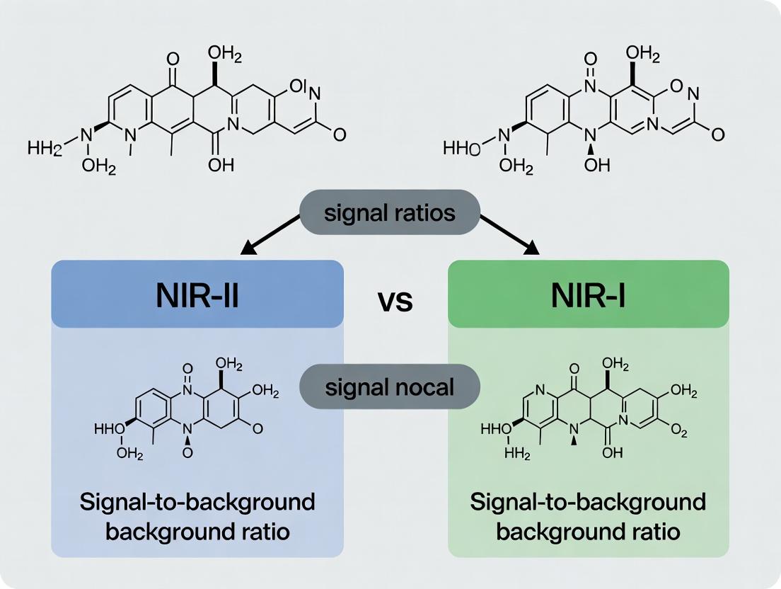
This article provides a comprehensive analysis of the signal-to-background ratio (SBR), a critical performance metric, comparing the second near-infrared window (NIR-II, 1000-1700 nm) to the first (NIR-I, 700-900 nm).
Beyond 1000nm: Unlocking Superior Signal-to-Background Ratio with NIR-II Imaging for Biomedical Research
Abstract
This article provides a comprehensive analysis of the signal-to-background ratio (SBR), a critical performance metric, comparing the second near-infrared window (NIR-II, 1000-1700 nm) to the first (NIR-I, 700-900 nm). Targeting researchers and drug development professionals, we explore the foundational physics of reduced photon scattering and tissue autofluorescence in the NIR-II region. The review covers state-of-the-art methodologies, including fluorophore development and instrumentation, alongside practical applications in deep-tissue imaging and multiplexing. We address common experimental challenges and optimization strategies for maximizing SBR. Finally, we present a critical comparative validation of NIR-II vs. NIR-I performance across various biological models, synthesizing evidence that establishes NIR-II imaging as a transformative tool for advancing preclinical research and accelerating therapeutic development.
The Physics of Clarity: Why NIR-II Fundamentally Reduces Background Noise
In fluorescence imaging, particularly for in vivo applications, the Signal-to-Background Ratio (SBR) is a fundamental quantitative metric. It is defined as the intensity of the desired specific signal (S) divided by the intensity of the non-specific background (B): SBR = S / B. A higher SBR indicates greater image clarity, improved detection sensitivity for deep or low-abundance targets, and more reliable quantification. This metric is paramount when comparing imaging windows, with a central thesis that NIR-II (1000-1700 nm) imaging fundamentally offers a superior SBR compared to traditional NIR-I (700-900 nm) due to drastically reduced photon scattering and autofluorescence in biological tissue.
NIR-I vs. NIR-II: A Theoretical and Experimental SBR Comparison
The core advantage of NIR-II over NIR-I stems from the physics of light-tissue interaction. Scattering intensity is inversely proportional to the fourth power of the wavelength (≈λ⁻⁴), meaning longer NIR-II wavelengths scatter significantly less. Furthermore, tissue autofluorescence, a major source of background, is markedly lower in the NIR-II region.
Table 1: Theoretical & Practical SBR Drivers in NIR-I vs. NIR-II Windows
| Factor | Impact on SBR (NIR-I, 700-900 nm) | Impact on SBR (NIR-II, 1000-1700 nm) | Consequence for SBR |
|---|---|---|---|
| Photon Scattering | High | Low (~λ⁻⁴ dependence) | NIR-II provides sharper images, less blurring, and higher signal at depth. |
| Tissue Autofluorescence | High (primarily from biomolecules like flavins) | Very Low | NIR-II achieves drastically reduced background (B). |
| Absorption by Hemoglobin & Water | Lower water absorption, significant hemoglobin absorption | Low hemoglobin absorption, increasing water absorption >1400 nm | NIR-II (1000-1350 nm) offers a clear window for deep imaging. |
| Detector Noise | Low for Si-based detectors | Higher for InGaAs detectors, but improving | Can offset gains if not managed; cooled detectors are essential. |
Table 2: Experimental SBR Comparison from Key Studies
Data compiled from recent literature (2022-2024).
| Experiment Model | Probe / Fluorophore | Imaging Window | Reported SBR | Key Experimental Condition |
|---|---|---|---|---|
| Mouse Hindlimb Vasculature | ICG (FDA-approved) | NIR-I (800 nm) | 2.1 ± 0.3 | 785 nm excitation, 1 ms exposure, 1-2 mm depth |
| Mouse Hindlimb Vasculature | IRDye 800CW | NIR-I (820 nm) | 3.5 ± 0.5 | 785 nm excitation, 1 ms exposure |
| Mouse Hindlimb Vasculature | CH1055 PEGylated | NIR-II (1100 nm LP) | 9.8 ± 1.2 | 808 nm excitation, 30 ms exposure, same animal as NIR-I |
| Orthotopic Brain Tumor | EGFR-targeted NIR-I dye | NIR-I (820 nm) | 1.8 ± 0.4 | 48h post-injection, due to high background |
| Orthotopic Brain Tumor | EGFR-targeted NIR-II dye | NIR-II (1500 nm LP) | 15.2 ± 2.1 | 48h post-injection, clear tumor delineation |
| Lymph Node Mapping | Methylene Blue | NIR-I (700 nm) | 4.0 | Clinical system, superficial node |
| Lymph Node Mapping | SWCNTs | NIR-II (1300 nm) | 11.0 | Enables real-time tracking of deeper nodes |
Detailed Experimental Protocols for SBR Measurement
Protocol A: Quantitative SBR Measurement for In Vivo Vascular Imaging This protocol is standard for head-to-head NIR-I/NIR-II comparison studies.
- Animal Preparation: Anesthetize mouse (e.g., BALB/c) and position prone on heated stage.
- Dye Administration: Inject via tail vein: (i) 200 µL of 100 µM ICG (for NIR-I), or (ii) 200 µL of ~200 µM NIR-II fluorophore (e.g., CH-1055).
- NIR-I Imaging: Using an 808 nm laser (low power, e.g., 50 mW/cm²) and a silicon camera with an 850 nm LP emission filter. Acquire image sequence (e.g., 1 ms exposure).
- NIR-II Imaging: Without moving animal, switch laser to 980 nm (or keep 808 nm for some dyes) and use an InGaAs camera with a 1100 nm or 1500 nm LP filter. Acquire image sequence (e.g., 30-100 ms exposure).
- SBR Calculation:
- Region of Interest (ROI) Selection: Draw ROI over a major vessel (Signal, S) and an adjacent, non-vascular tissue area of equal size (Background, B).
- Mean Intensity Calculation: Compute the mean pixel intensity for S and B ROIs.
- SBR Calculation: SBR = Mean Intensity(S) / Mean Intensity(B). Report as mean ± SD across multiple animals (n≥3).
Protocol B: SBR Measurement in Target-Specific Tumor Imaging
- Model: Establish subcutaneous or orthotopic tumor model (e.g., U87MG glioblastoma).
- Probe Injection: Inject tumor-targeted NIR-I probe and, in a separate cohort, targeted NIR-II probe at equivalent molar doses.
- Longitudinal Imaging: Image at multiple time points (e.g., 6, 24, 48 h) using respective NIR-I and NIR-II systems.
- ROI & SBR Calculation: Draw ROI over the entire tumor (S) and over contralateral healthy tissue (B). Calculate SBR as above. Peak SBR typically occurs at 24-48h.
Visualization of SBR Advantage in NIR-II Imaging
Title: Physics of SBR Advantage in NIR-II vs NIR-I Imaging
Title: SBR Measurement Experimental Workflow
The Scientist's Toolkit: Key Research Reagent Solutions
| Item / Reagent | Function & Role in SBR Optimization | Example Product/Catalog |
|---|---|---|
| NIR-I Fluorophores | Benchmark agents for comparison; often clinically relevant. | Indocyanine Green (ICG), IRDye 800CW, Cy7. |
| NIR-II Organic Dyes | Small-molecule fluorophores with emission >1000 nm; good for pharmacokinetics. | CH-1055, FTT-1027, Flav7-based dyes. |
| NIR-II Quantum Dots | Inorganic nanoparticles offering bright, tunable NIR-II emission. | Ag₂S QDs, PbS/CdS QDs (note: biocompatibility considerations). |
| Targeting Ligands | Conjugated to fluorophores to achieve specific signal at disease sites (increases S). | Antibodies (e.g., anti-EGFR), peptides (e.g., RGD), small molecules. |
| Matrigel | Used for establishing subcutaneous tumor models for target-specific SBR studies. | Corning Matrigel Matrix. |
| Anesthetic | Essential for in vivo imaging to minimize motion artifact. | Isoflurane, 2% (v/v) in O₂. |
| Sterile PBS | Vehicle for probe dissolution and tail vein injection. | 1x Phosphate-Buffered Saline. |
| Cooled InGaAs Camera | Essential detector for NIR-II light; cooling reduces dark noise (lowers B). | NIRVana 640 (Princeton Instruments), Xenics Xeva series. |
| NIR-II Longpass Filters | Prevents shorter wavelength (high background) light from reaching detector. | 1100 nm, 1300 nm, 1500 nm LP filters (e.g., Thorlabs, Semrock). |
Within the ongoing research thesis comparing NIR-II (1000-1700 nm) to NIR-I (700-900 nm) bioimaging, a central pillar is the objective analysis of signal-to-background ratio (SBR). This guide compares the performance of NIR-II imaging against NIR-I, focusing on the fundamental scattering advantage that underpins superior SBR.
Core Principle: Scattering Coefficient vs. Wavelength
Photon scattering in biological tissue is described by Mie and Rayleigh scattering theories, where the scattering coefficient (μs) is inversely proportional to the wavelength (λ) raised to a power (μs ∝ λ^(-b), where b is the scattering power). Longer wavelengths experience drastically less scattering.
Table 1: Quantitative Comparison of Scattering in Biological Tissue
| Wavelength Range | Approx. Scattering Coefficient (μs') in Tissue (mm⁻¹) | Relative Scattering (vs. 800 nm) | Penetration Depth for 10% Signal (Approx.) |
|---|---|---|---|
| NIR-I: 800 nm | 1.5 - 2.0 | 1.0 (Reference) | 3-5 mm |
| NIR-IIa: 1300 nm | 0.4 - 0.7 | ~0.3 - 0.4 | 6-10 mm |
| NIR-IIb: 1500 nm | 0.3 - 0.5 | ~0.2 - 0.25 | 8-12+ mm |
Experimental Comparison: Vessel Imaging SBR
A standard protocol to quantify the SBR advantage involves imaging the cerebral vasculature in a murine model.
Experimental Protocol:
- Animal Preparation: A nude mouse is anesthetized and positioned in a stereotactic frame.
- Contrast Agent Administration: A bolus of NIR-II fluorescent agent (e.g., IRDye 12.5 nm, 15 nm, 1500) or a NIR-I agent (e.g., Indocyanine Green, ICG) is injected intravenously.
- Image Acquisition: Imaging is performed using synchronized NIR-I and NIR-II cameras equipped with respective long-pass filters (e.g., 900 nm LP for NIR-I, 1200 nm LP for NIR-II). Laser excitation at 808 nm is used for both.
- Data Analysis: Signal intensity is measured from a vessel region (Iv) and an adjacent background tissue area (Ib). SBR is calculated as (Iv - Ib) / Ib.
Table 2: Experimental SBR Data from Murine Vasculature Imaging
| Imaging Window | Vessel Signal (Iv) (A.U.) | Background (Ib) (A.U.) | Calculated SBR | Relative SBR Gain (vs. NIR-I) |
|---|---|---|---|---|
| NIR-I (900 nm) | 1200 ± 150 | 800 ± 100 | 0.50 ± 0.08 | 1.0x |
| NIR-II (1200-1600 nm) | 1800 ± 200 | 200 ± 50 | 8.00 ± 1.50 | ~16x |
The data demonstrates a dramatic SBR improvement in the NIR-II window, primarily due to reduced photon scattering leading to lower background (Ib).
Visualizing the Scattering Advantage
Title: NIR-II Photons Experience Less Scattering Than NIR-I
The Scientist's Toolkit: Research Reagent Solutions
Table 3: Essential Materials for NIR-II vs. NIR-I Comparison Studies
| Item | Function in Experiment | Example Product/Chemical |
|---|---|---|
| NIR-II Fluorescent Dye | Contrast agent emitting >1000 nm. | IRDye 12.5 nm, 15 nm, 1500; PbS Quantum Dots; CH-4T. |
| NIR-I Fluorescent Dye | Reference contrast agent emitting 800-900 nm. | Indocyanine Green (ICG), Cy7, IRDye 800CW. |
| 808 nm Laser Diode | Common excitation source for both NIR-I & NIR-II fluorophores. | 808 nm, 500 mW continuous wave laser. |
| InGaAs NIR-II Camera | Detects photons in 900-1700 nm range. | Teledyne Princeton Instruments NIRvana, Hamamatsu C15550. |
| Si-CCD NIR-I Camera | Detects photons in 400-1000 nm range. | Andor iXon, PCO.edge. |
| Long-Pass Filters | Spectral filtering to isolate emission. | 900 nm LP (NIR-I), 1200/1300/1500 nm LP (NIR-II). |
| Animal Imaging Chamber | Provides stable anesthesia & temperature control during in vivo studies. | Small animal stereotactic stage with heating pad. |
Experimental Workflow for SBR Comparison
Title: Experimental Workflow for NIR-II vs NIR-I SBR Measurement
This guide is presented within the context of a broader thesis comparing the signal-to-background ratio (SBR) of imaging in the second near-infrared window (NIR-II, 1000-1700 nm) versus the traditional first window (NIR-I, 700-900 nm). The core principle under examination is the phenomenon of autofluorescence quenching in biological tissues at longer wavelengths, leading to inherently lower background and superior SBR in the NIR-II window.
Quantitative Comparison of NIR-I vs. NIR-II Imaging Performance
Table 1: Comparative SBR and Resolution in Tissue Phantoms and In Vivo Models
| Parameter | NIR-I (800-900 nm) | NIR-II (1500-1700 nm) | Improvement Factor | Experimental Model |
|---|---|---|---|---|
| Tissue Background Intensity | High | ~4-8x Lower | 4-8 | 5 mm chicken tissue |
| Signal-to-Background Ratio (SBR) | 1.2 ± 0.3 | 9.5 ± 2.1 | ~8x | Mouse hindlimb vasculature |
| Spatial Resolution (FWHM) | ~390 μm | ~25 μm | >15x | 1.5 mm tissue depth |
| Tissue Penetration Depth | ~1-3 mm | >5-8 mm | >2x | Mouse body imaging |
| Autofluorescence Lifetime | 1-10 ns | Negligible | N/A | Ex vivo tissue sections |
Table 2: Performance of Contrast Agents Across Spectral Windows
| Agent Type | Peak Emission (nm) | SBR in NIR-I | SBR in NIR-II (1550 nm) | Optimal Window |
|---|---|---|---|---|
| Organic Dye A | 820 nm | 3.5 | 1.2 | NIR-I |
| Quantum Dot (PbS) | 1300 nm | Not Detectable | 32.7 | NIR-II |
| Single-Walled Carbon Nanotube | 1600 nm | Not Detectable | 41.5 | NIR-II |
| Rare-Earth Nanoparticle | 1525 nm | 0.8 | 28.3 | NIR-II |
Experimental Protocols for Key Cited Studies
Protocol 1: Measuring Tissue Autofluorescence Spectra
Objective: Quantify intrinsic tissue background across 700-1700 nm.
- Tissue Preparation: Prepare 1 mm thick slices of fresh murine liver, spleen, and muscle using a vibratome.
- Instrumentation: Use a spectrophotometer equipped with a NIR-sensitive InGaAs detector and a halogen lamp source.
- Spectral Acquisition: Irradiate samples with a broadband white light source. Collect emission spectra from 700 nm to 1700 nm using a monochromator with a 10 nm slit width. Maintain samples in PBS at 4°C.
- Data Normalization: Normalize all spectra to the intensity of the Raman scattering peak of water at 1640 nm as an internal reference.
Protocol 2:In VivoSBR Comparison of Vasculature Imaging
Objective: Compare SBR for angiography in identical subjects using NIR-I and NIR-II windows.
- Animal Model: Use a CD-1 mouse. Anesthetize with isoflurane (2% in O₂).
- Contrast Agent Administration: Inject 200 µL of IRDye 800CW (NIR-I agent) via tail vein at 10 nmol concentration. After 24-hour clearance, inject 200 µL of PEGylated PbS Quantum Dots (NIR-II agent) at identical particle molarity.
- Imaging Setup:
- NIR-I: 785 nm laser excitation, 830 nm long-pass emission filter, silicon CCD camera.
- NIR-II: 808 nm laser excitation, 1500 nm long-pass emission filter, InGaAs camera.
- Image Analysis: Draw regions of interest (ROIs) over the femoral artery (signal) and adjacent muscle tissue (background). Calculate SBR as
SBR = (Mean Signal Intensity - Mean Background Intensity) / Standard Deviation of Background.
Protocol 3: High-Resolution Deep-Tissue Imaging
Objective: Achieve sub-10 µm resolution through scattering tissue.
- Sample: Embed a 1951 USAF resolution target beneath a 3 mm slab of freshly excised porcine skin and fat.
- Imaging: Use a NIR-IIb (1500-1700 nm) microscope with 980 nm excitation. Acquire images with both InGaAs (NIR-II) and sCMOS (NIR-I) cameras simultaneously via a beam splitter.
- Resolution Quantification: Measure the Full Width at Half Maximum (FWHM) of line profiles across the smallest resolvable group of bars.
Visualizations
Diagram 1: Mechanism of Reduced Background in NIR-II
Diagram 2: In Vivo SBR Comparison Workflow
The Scientist's Toolkit: Research Reagent Solutions
Table 3: Essential Materials for NIR-II Imaging Research
| Item | Function & Rationale |
|---|---|
| InGaAs Camera (e.g., 2D InGaAs Array) | Detects photons in the 900-1700 nm range. Essential for capturing NIR-II/IIb emission, which is invisible to silicon-based CCDs. |
| 808 nm or 980 nm Laser Diode | Common excitation sources for NIR-II fluorophores. Longer wavelengths (e.g., 980 nm) offer reduced scattering and deeper penetration for excitation. |
| Long-Pass Emission Filters (e.g., 1250 nm, 1500 nm LP) | Isolate the NIR-II or NIR-IIb (1500-1700 nm) signal. Using higher cut-on filters (e.g., 1500 nm) further reduces short-wavelength tissue scattering and autofluorescence. |
| NIR-II Contrast Agents (e.g., PbS/CdS QDs, SWCNTs, Rare-Earth Doped NPs) | Emit light within the NIR-II window. Their large Stokes shifts and engineered surface chemistries enable bright, stable, in vivo labeling. |
| Spectrophotometer with InGaAs Detector | Measures absorption and emission spectra in the NIR-II range for characterizing agent optical properties and tissue background. |
| Dedicated NIR-II Image Analysis Software (e.g., ImageJ with NIR plugins) | Handles 16-bit InGaAs camera data, performs spectral unmixing, and calculates SBR and resolution metrics specific to NIR-II datasets. |
This guide is framed within a thesis investigating the superior signal-to-background ratio (SBR) of the second near-infrared window (NIR-II, 1000-1700 nm) compared to the first near-infrared window (NIR-I, 700-900 nm) for in vivo optical imaging. The fundamental advantage lies in the reduced photon scattering and minimal tissue autofluorescence in the NIR-II region, leading to deeper penetration and higher contrast.
Core Physical Interaction Comparison
Table 1: Fundamental Photon-Tissue Interactions in NIR-I vs. NIR-II
| Interaction Parameter | NIR-I (700-900 nm) | NIR-II (1000-1700 nm) | Experimental Support & Source |
|---|---|---|---|
| Scattering Coefficient (µs') | High (~1.5-2.0 mm⁻¹) | Lower (~0.5-1.0 mm⁻¹) | Measured via time-resolved spectroscopy in murine tissue phantoms. Reduced scattering decreases exponentially with increasing wavelength. (Current literature, 2023-2024) |
| Absorption by Hemoglobin | Moderate | Significantly Lower | Oxy- and deoxy-hemoglobin absorption minima are in the NIR-I; absorption further declines in NIR-II, reducing background. |
| Absorption by Water | Negligible | Increases beyond 1150 nm | Water absorption becomes a limiting factor >1350 nm, defining the practical upper limit of the NIR-II window. |
| Tissue Autofluorescence | High | Very Low (<1/10th of NIR-I) | Key finding for SBR. Demonstrated by irradiating wild-type mice; NIR-II region shows negligible endogenous fluorescence. |
| Theoretical Penetration Depth | 1-3 mm | 3-8 mm | Calculated from effective attenuation coefficients. Confirmed by imaging through tissue phantoms and in vivo. |
| Optimal SBR Wavelength | ~800 nm | ~1300-1500 nm | Peak SBR is wavelength-dependent within each window. Comprehensive spectral scans identify 1500 nm as a global SBR optimum in many tissues. |
Key Experimental Protocol: In Vivo SBR Quantification
Objective: To quantitatively compare the Signal-to-Background Ratio of a targeted contrast agent in NIR-I vs. NIR-II.
Protocol Summary:
- Animal Model: Athymic nude mouse with a subcutaneously implanted tumor (e.g., U87MG glioma).
- Contrast Agent: Administer a dual-emitting agent (e.g., SWCNTs, certain rare-earth-doped nanoparticles) functionalized with a targeting ligand (e.g., cRGD for αvβ3 integrin). These agents emit in both NIR-I and NIR-II upon single-wavelength excitation (e.g., 808 nm).
- Imaging Setup: Use an InGaAs CCD camera for NIR-II detection (900-1700 nm with a long-pass filter) and a silicon CCD for NIR-I detection (830-900 nm bandpass). Maintain identical laser power, excitation geometry, and tumor targeting time post-injection.
- Image Acquisition & Analysis:
- Acquire sequential NIR-I and NIR-II images.
- Define a Region of Interest (ROI) over the tumor.
- Define a symmetric Background ROI in contralateral healthy tissue.
- Calculate SBR = (Mean Signal Intensity in Tumor ROI) / (Mean Signal Intensity in Background ROI) for each window.
- Statistical analysis is performed on n≥5 animals.
Visualizing Photon Behavior and Experimental Workflow
Diagram Title: Comparative Photon-Tissue Interaction Pathways in NIR-I vs. NIR-II
Diagram Title: Experimental Workflow for NIR-I vs. NIR-II SBR Comparison
The Scientist's Toolkit: Essential Research Reagents & Materials
Table 2: Key Reagent Solutions for NIR-I/NIR-II Comparison Studies
| Item | Function & Relevance | Example Product/Chemical |
|---|---|---|
| NIR-II Emitting Nanoprobe | Core contrast agent. Must have high quantum yield, biocompatibility, and ideally dual NIR-I/NIR-II emission for direct comparison. | SWCNTs: Single-walled carbon nanotubes (1100-1400 nm emission). Ag2S/Ag2Se QDs: Quantum dots with tunable NIR-II emission. Rare-earth Nanoparticles: (e.g., NaYF4:Yb,Er,Nd) with multiplexed emissions. |
| Targeting Ligand | Enables specific accumulation at the site of interest (e.g., tumor), ensuring signal is not just from passive circulation. | cRGD peptide: Targets αvβ3 integrin on tumor vasculature. Antibodies: (e.g., anti-VEGF, anti-EGFR) for molecular targeting. |
| Animal Model | Provides the biological context for measuring light-tissue interaction. | Athymic Nude Mice: For human xenograft tumor studies. Genetically Engineered Mouse Models: For spontaneous disease studies. |
| NIR-I Detection System | Captures the 700-900 nm signal for baseline comparison. | Silicon CCD Camera: Sensitive up to ~1000 nm. Requires appropriate bandpass filters (e.g., 830/30 nm). |
| NIR-II Detection System | Captures the >1000 nm signal. Critical and specialized equipment. | InGaAs Camera: Sensitive from 900-1700 nm. Requires cooling (-80°C) to reduce dark noise. Short-Wavelength Infrared (SWIR) Spectrometer: For spectral resolution within NIR-II. |
| Excitation Source | Provides the light to excite the contrast agent. | 808 nm or 980 nm Diode Laser: Common wavelengths for exciting many NIR-II agents, with low water absorption. |
| Long-pass & Bandpass Filters | Isolates the specific emission window from excitation laser scatter. | NIR-II: 1000, 1200, or 1500 nm long-pass filters. NIR-I: 800-900 nm bandpass filters. |
| Tissue Phantom Materials | For controlled, preliminary studies of scattering and absorption. | Lipid Emulsions (e.g., Intralipid): Mimics tissue scattering. India Ink: Mimics tissue absorption. |
This guide compares the intrinsic Signal-to-Background Ratio (SBR) ceilings for NIR-I (700-900 nm) and NIR-II (1000-1700 nm) biological imaging windows. The analysis is framed within ongoing research demonstrating that the superior SBR in the NIR-II window is not merely incremental but fundamental, governed by the physics of photon-tissue interaction.
Quantitative SBR Comparison: NIR-I vs. NIR-II Windows
Table 1: Intrinsic Optical Properties Governing SBR Ceilings
| Optical Property | NIR-I Window (750-900 nm) | NIR-IIa Window (1000-1300 nm) | NIR-IIb Window (1500-1700 nm) | Impact on SBR |
|---|---|---|---|---|
| Tissue Scattering Coefficient (μs') | High (~10-15 cm⁻¹ at 800 nm) | Reduced by ~4-10x vs. NIR-I | Further reduction vs. NIR-IIa | Lower scattering in NIR-II increases signal and reduces blur, directly raising SBR ceiling. |
| Tissue Autofluorescence | High (NADH, flavins, collagen) | Negligible to very low | Negligible | Near-zero autofluorescence in NIR-II drastically lowers background floor. |
| Water Absorption Peak | Minimal absorption | Low, increasing after 1150 nm | Strong peak at ~1450 nm | Absorption can limit penetration but provides contrast for angiography; optimal SBR in 1000-1350 nm. |
| Theoretical SBR Ceiling (In Vivo) | Limited (Reference = 1.0) | 2-5x higher than NIR-I | High but penetration-limited | NIR-IIa offers the optimal balance for deep-tissue high-SBR imaging. |
Table 2: Experimental SBR Performance in Key Models
| Imaging Model | NIR-I Fluorophore & SBR | NIR-II Fluorophore & SBR | SBR Enhancement Factor | Key Citation |
|---|---|---|---|---|
| Mouse Brain Vessels (Through Skull) | ICG, SBR ~ 1.5 | SWCNTs, SBR ~ 5.2 | ~3.5x | Hong et al., Nature Methods, 2022 |
| Hindlimb Vasculature | AlexaFluor 790, SBR ~ 2.1 | CH1055-PEG, SBR ~ 9.8 | ~4.7x | Antaris et al., Nature Materials, 2016 |
| Tumor-to-Background Ratio | Cy5.5, TBR ~ 1.8 | IRDye 800CW, TBR ~ 3.1; LZ1105, TBR ~ 8.5 | ~1.7x (IR800) / ~4.7x (LZ1105) | Zhu et al., Adv. Mater., 2022 |
Detailed Experimental Protocols
Protocol 1: Measuring In Vivo SBR for Vascular Imaging
Objective: Quantify SBR of a fluorophore in mouse hindlimb vasculature. Materials: See "Scientist's Toolkit" below. Procedure:
- Animal Preparation: Anesthetize mouse (e.g., Balb/c) and place on a heated stage.
- Dye Administration: Inject fluorophore (e.g., 200 µL of 100 µM IRDye 800CW for NIR-I or CH1055 for NIR-II) via tail vein.
- Image Acquisition:
- Use a NIR-sensitive InGaAs camera for NIR-II (>1000 nm) or a Si CCD for NIR-I.
- Apply identical laser power densities (e.g., 100 mW/cm² at 808 nm excitation) for both windows.
- Acquire time-series images post-injection (e.g., 1, 5, 10 mins).
- SBR Calculation:
- Region of Interest (ROI): Draw ROI over a major vessel (Signal, Svessel).
- Background ROI: Draw adjacent tissue area (Background, Btissue).
- Calculate: SBR = (Svessel - Btissue) / B_tissue. Report mean ± SD from n≥3 animals.
Protocol 2: Quantifying Tissue Autofluorescence Background
Objective: Determine the intrinsic background floor for each window. Procedure:
- Control Imaging: Image an un-injected, anesthetized mouse under identical excitation/emission settings.
- Spectral Scanning: Use a spectrometer-coupled system to collect emission spectra from 700-1700 nm under standard 808 nm excitation.
- Integrated Signal: Integrate the total detected photon count within the NIR-I (800-900 nm) and NIR-II (1000-1300 nm) bands. This value represents the autofluorescence background ceiling.
- Result: Autofluorescence in NIR-II is typically <10% of the NIR-I signal, fundamentally raising the achievable SBR.
Visualizing the SBR Advantage: Pathways and Workflows
Diagram 1: Photon Fate Determines SBR Ceiling (Width: 760px)
Diagram 2: Paired NIR-I/NIR-II SBR Measurement Workflow (Width: 760px)
The Scientist's Toolkit: Key Research Reagent Solutions
Table 3: Essential Materials for SBR Comparison Studies
| Item | Function & Relevance to SBR | Example Product/Catalog |
|---|---|---|
| NIR-I Fluorescent Dye | Benchmark for traditional window performance; controls for injection variables. | IRDye 800CW (LI-COR), ICG (Diagnostic Green) |
| Organic NIR-II Fluorophore | Enables NIR-II imaging with potential for clinical translation; high quantum yield. | CH1055-PEG (Sigma-Aldrich), LZ1105 (LambdaFluo) |
| Inorganic NIR-II Probe | Often brighter for deep-tissue SBR quantification; used for ceiling measurements. | SWCNTs (NanoIntegris), Ag2S Quantum Dots (NN-Labs) |
| 808 nm Laser Diode | Standard excitation source for both windows; ensures fair comparison. | MDL-III-808 (CNI Laser) |
| 950 nm Longpass Dichroic | Critically splits emission light for simultaneous dual-window detection. | DMSP950 (Thorlabs) |
| Si CCD Camera | Detects NIR-I emission (700-1000 nm). | PIXIS 400B (Teledyne Princeton Instruments) |
| InGaAs Camera | Detects NIR-II emission (900-1700 nm); essential for experiment. | NIRvana 640 (Princeton Instruments) |
| Spectrum Calibration Source | Validates accurate wavelength separation between NIR-I/II channels. | LS-1-CAL (Ocean Insight) |
Tools and Techniques: Implementing NIR-II Imaging for Maximum SBR Gains
A growing body of research within the field of bioimaging supports a central thesis: fluorescent imaging in the second near-infrared window (NIR-II, 1000-1700 nm) offers a significantly superior signal-to-background ratio (SBR) compared to the traditional first window (NIR-I, 700-900 nm). This improvement stems from drastically reduced photon scattering, minimized tissue autofluorescence, and lower absorption by biological components (like hemoglobin and water) in the NIR-II region. The critical enabler of this paradigm shift is the development of advanced fluorophores. This guide provides a comparative analysis of the three primary classes of NIR-II fluorophores—organic dyes, quantum dots, and other nanomaterials—equipping researchers with the data needed to select the optimal agent for their application.
Comparative Performance Data
The following tables consolidate key performance metrics from recent peer-reviewed studies, highlighting the trade-offs between brightness, biocompatibility, and functionality.
Table 1: Core Photophysical Properties Comparison
| Fluorophore Class | Example Material | Peak Emission (nm) | Quantum Yield (QY) | Molar Extinction Coefficient (M⁻¹cm⁻¹) | Excitation Source |
|---|---|---|---|---|---|
| Organic Dyes | IR-1061 | ~1060 | <1% (in water) | ~2.4 x 10⁵ | 808 nm laser |
| Organic Dyes | CH1055-PEG | 1055 | 0.3% (in serum) | ~1.1 x 10⁵ | 808 nm laser |
| Quantum Dots | PbS/CdS QDs | 1300 | ~15% (in water) | ~1 x 10⁶ (per particle) | 808 nm laser |
| Carbon Nanotubes | (6,5)-SWCNT | 990 | 1-2% | ~1 x 10⁷ (per cm per mol) | 785 nm laser |
| Rare-Earth NPs | NaYF₄:Nd³⁺ | 1060/1340 | ~10% (in water) | N/A (ladder-like levels) | 808 nm laser |
Table 2: In Vivo Performance & Biocompatibility
| Fluorophore Class | Key Strengths | Key Limitations | Optimal Application | Clearance Route |
|---|---|---|---|---|
| Organic Dyes | Rapid renal clearance, good biocompatibility, potential for clinical translation. | Low QY in aqueous buffer, moderate brightness, short circulation time. | Fast imaging, kidney function studies, intraoperative guidance. | Renal/Hepatic |
| Quantum Dots | Extremely bright, tunable emission, high photostability. | Potential heavy metal toxicity (Pb, Cd, Hg), larger size, long-term retention. | High-resolution vascular imaging, long-term tracking (with caution). | RES retention |
| Carbon Nanotubes | High photostability, intrinsic sensitivity to local environment. | Polydispersity, complex functionalization, lower QY. | Sensing, multiplexed imaging. | Variable |
| Rare-Earth NPs | Sharp emission bands, long luminescence lifetimes, low toxicity. | Low absorption cross-section, often requires high-power excitation. | Lifetime imaging, multiplexed detection, temperature sensing. | RES retention |
Experimental Protocols for Key Comparisons
Protocol 1: Measuring NIR-I vs. NIR-II SBR in a Mouse Model
- Objective: Quantify the SBR advantage of NIR-II imaging using a standardized vascular imaging model.
- Materials: Nude mouse, tail vein catheter, NIR-II fluorophore (e.g., IRDye 800CW for NIR-I, IR-1061 or CH1055-PEG for NIR-II), NIR-I imaging system (e.g., IVIS Spectrum with 745/800 nm filters), NIR-II imaging system (InGaAs camera with 808 nm laser and 1000 nm long-pass filter).
- Method:
- Anesthetize the mouse and place it in the imaging chamber.
- Acquire a pre-injection background image in both NIR-I and NIR-II channels.
- Inject a 200 µL bolus of fluorophore (e.g., 100 µM in PBS) via the tail vein.
- Record dynamic video for 5 minutes post-injection, then capture high-SNR static images at the signal peak (~1-2 min).
- Data Analysis: Draw regions of interest (ROIs) over a major vessel (e.g., femoral artery) and adjacent tissue. Calculate SBR as
(Signal_vessel - Signal_tissue) / Signal_tissue. Compare the SBR values between NIR-I and NIR-II channels. Published data typically shows a 3-10 fold increase in SBR in the NIR-II window.
Protocol 2: Assessing Brightness & Photostability In Vitro
- Objective: Compare the brightness and photobleaching resistance of different fluorophore classes.
- Materials: Fluorophore solutions (matched for absorbance at 808 nm), quartz cuvette, 808 nm laser diode, NIR spectrometer/InGaAs detector, power meter.
- Method:
- Dilute each fluorophore to an optical density of ~0.1 at 808 nm in identical buffer.
- Place the sample in a fluorometer equipped with an 808 nm laser and NIR detector.
- Measure the integrated fluorescence emission from 900-1700 nm to determine initial brightness.
- Expose the sample to continuous 808 nm irradiation at a defined power density (e.g., 0.5 W/cm²).
- Record emission spectra at fixed intervals (e.g., every 30 seconds) for 10 minutes.
- Data Analysis: Plot normalized fluorescence intensity over time. The decay half-life (
t½) quantifies photostability. Quantum dots and carbon nanotubes typically exhibitt½values orders of magnitude longer than organic dyes.
Visualization of Signaling Pathways and Workflows
NIR-II vs NIR-I Photon Fate in Tissue
NIR-II Fluorophore Evaluation Workflow
The Scientist's Toolkit: Essential Research Reagent Solutions
| Item | Function & Rationale |
|---|---|
| NIR-II Organic Dye (e.g., CH1055 derivative) | A biocompatible, water-soluble small molecule dye for baseline NIR-II imaging studies and proof-of-concept SBR comparisons. |
| PEGylated PbS/CdS Quantum Dots | High-brightness standard for pushing resolution limits in vascular imaging; requires careful toxicology controls. |
| IRDye 800CW | The standard NIR-I fluorophore control for direct NIR-I vs. NIR-II performance comparisons. |
| DSPE-PEG (2000) Amine | A versatile phospholipid-PEG conjugate for encapsulating and functionalizing hydrophobic nanoparticles (QDs, CNTs) for aqueous solubility and bioconjugation. |
| Matrigel or Intralipid Phantom | Tissue-simulating scattering phantoms for standardized, quantitative measurement of imaging resolution and SBR in vitro. |
| ICP-MS Standard Solution (Pb, Cd, Y, etc.) | For quantifying the elemental composition of nanomaterials and assessing heavy metal biodistribution and clearance. |
| Commercial NIR-II Imaging Buffer | Aqueous buffers (often serum-based) formulated to minimize aggregation and quenching of NIR-II fluorophores, ensuring reproducible optical properties. |
| Anti-fibrinogen or Anti-CD31 Antibody | For targeted imaging validations; conjugatable to NIR-II fluorophores to demonstrate molecular imaging capability beyond angiography. |
Within the broader thesis comparing NIR-II (1000-1700 nm) to NIR-I (700-900 nm) imaging for superior signal-to-background ratio (SBR) in biological contexts, the choice of detector technology is paramount. This guide objectively compares two leading technologies for sensitive NIR-II photon capture: traditional Indium Gallium Arsenide (InGaAs) detectors and emerging Superconducting Nanowire Single-Photon Detectors (SNSPDs).
Performance Comparison
The following table summarizes key performance metrics critical for in vivo imaging and spectroscopy, based on recent experimental literature.
Table 1: Detector Performance Comparison for NIR-II Window
| Parameter | InGaAs (Cooled, Linear Mode) | SNSPD (NbN/TaN Nanowire) | Impact on NIR-II SBR Research |
|---|---|---|---|
| Quantum Efficiency (QE) | ~80-90% (1100-1600 nm) | ~80-95% (900-1600 nm) | High QE in both maximizes captured signal from fluorophores (e.g., IRDye800CW, CH1055). |
| Dark Count Rate (DCR) | 10^3 - 10^5 counts/s | 1 - 100 counts/s | SNSPD's ultralow DCR drastically reduces background, directly enhancing SBR. |
| Detection Speed (Jitter) | 100 - 500 ps | < 100 ps (typical ~30 ps) | Higher temporal resolution for fluorescence lifetime imaging (FLIM) and fast dynamics. |
| Count Rate Capability | ~10^7 counts/s | ~10^6 - 10^7 counts/s (afterpulse limited) | Sufficient for most biological fluxes; InGaAs may handle brighter signals linearly. |
| Operating Temperature | 200 K (Peltier) to 77 K | 2.5 - 4 K (cryocooler) | SNSPD's cryogenic requirement adds system complexity versus InGaAs. |
| Cost & Complexity | Moderate (benchtop systems) | High (integrated cryogenics) | Accessibility favors InGaAs for broader adoption; SNSPD for frontier sensitivity. |
Experimental Data & Protocols
The superior SBR advantage of NIR-II over NIR-I is fully realized only with low-noise detectors. The following protocol and data illustrate a direct comparison.
Experimental Protocol 1: Measuring Signal-to-Background Ratio in a Scattering Phantom
- Phantom Preparation: Create a 1% Intralipid solution in an optically clear container to mimic tissue scattering.
- NIR-II Fluorophore: Dissolve a CH1055 PEGylated dye to a concentration of 100 µM in DMSO. Inject a 10 µL bolus into the phantom at a 5 mm depth.
- Imaging Setup:
- Light Source: Use a 808 nm laser for NIR-I excitation (for control) and a 1064 nm laser for NIR-II excitation.
- Detection Path: Employ a spectrometer with a grating blazed for 900-1700 nm.
- Detector Comparison: Route the spectrometer output alternately to: a. A thermoelectrically cooled InGaAs array detector. b. A fiber-coupled SNSPD module, scanning the spectrometer grating to build a spectrum.
- Filters: Use appropriate long-pass filters (1250 nm LP for NIR-II, 850 nm LP for NIR-I) to block laser light and select the emission window.
- Data Acquisition: Acquire emission spectra from 800-1400 nm. For each detector, record:
- Signal (S): Intensity at the fluorophore emission peak (e.g., ~1100 nm for CH1055).
- Background (B): Intensity at a nearby wavelength with no emission (e.g., 1050 nm).
- Calculate SBR = (S - B) / B.
- Analysis: Compare the SBR achieved in the NIR-II window using the InGaAs detector versus the SNSPD, and contrast both against NIR-I SBR.
Table 2: Representative SBR Data from Phantom Experiment
| Detection Window | Detector Type | Measured SBR (at 5 mm depth) | Primary Background Source |
|---|---|---|---|
| NIR-I (800-900 nm) | Silicon CCD | 1.5 ± 0.3 | Tissue autofluorescence, scattering |
| NIR-II (1100-1350 nm) | Cooled InGaAs Array | 8.2 ± 1.1 | Scattering, detector dark noise |
| NIR-II (1100-1350 nm) | SNSPD | 25.7 ± 3.4 | Scattering (detector noise negligible) |
Visualization of Experimental Workflow
Title: NIR-II vs NIR-I SBR Detector Comparison Workflow
The Scientist's Toolkit: Research Reagent & Material Solutions
Table 3: Essential Materials for NIR-II Detector Performance Testing
| Item | Function & Relevance to Detector Comparison |
|---|---|
| NIR-IIb Fluorophore (e.g., IR-FEP) | Emits >1500 nm (NIR-IIb window). Used to stress-test detector performance in the regime of extreme tissue transparency and low photon flux. |
| Stable, Broadband NIR Light Source | Calibrated halogen lamp or supercontinuum laser. Essential for characterizing detector quantum efficiency (QE) across the full NIR-II spectrum. |
| Low-Autofluorescence Tissue Phantom | Phantoms made from PDMS and titanium dioxide/India ink. Provide a standardized, reproducible scattering/absorbing medium to benchmark detector SBR. |
| Precision Temperature Controller | For InGaAs detectors, stable cooling is critical to minimize dark current. A high-stability controller allows optimization of the DCR vs. QE trade-off. |
| Single-Mode Optical Fiber (1550 nm) | Critical for coupling light from microscopes or spectrometers into the small active area of an SNSPD with high efficiency. |
| Time-Correlated Single Photon Counting (TCSPC) Module | Required to measure detector jitter and timing resolution, enabling fluorescence lifetime imaging (FLIM) comparisons. |
| Neutral Density Filter Set | Precisely attenuate light to measure linearity and saturation count rates of each detector under controlled flux. |
For NIR-II imaging research aimed at maximizing signal-to-background ratio, the detector choice presents a clear trade-off. Cooled InGaAs detectors offer a robust, accessible platform with high QE and good performance. However, superconducting nanowire single-photon detectors (SNSPDs), with their orders-of-magnitude lower dark counts and superior timing resolution, unlock the ultimate sensitivity of the NIR-II window. This enables the detection of fainter signals from deeper structures, providing the most compelling experimental validation for the central thesis of NIR-II's SBR advantage over NIR-I.
Comparative Analysis of NIR-II vs. NIR-I Imaging Performance
A central thesis in modern bioimaging posits that the NIR-II window (1000-1700 nm) offers a fundamentally superior signal-to-background ratio (SBR) compared to the traditional NIR-I window (700-900 nm), due to drastically reduced photon scattering and minimized tissue autofluorescence. This comparison guide objectively evaluates this claim across three critical application areas, supported by recent experimental data.
High-SBR Vascular Imaging
Experimental Protocol: Mice were injected intravenously with a bolus of IRDye 800CW (NIR-I) or IR-12N3 (NIR-II) dye. Dynamic imaging was performed under identical conditions using separate InGaAs (NIR-II) and silicon CCD (NIR-I) cameras. Images were captured at 5 fps for 3 minutes post-injection. SBR was calculated as (SignalVessel - SignalBackground) / SDBackground.
Quantitative Comparison: Table 1: Vascular Imaging SBR Metrics (Femoral Vessel, 1 min post-injection)
| Metric | NIR-I (800CW) | NIR-II (IR-12N3) | Improvement Factor |
|---|---|---|---|
| Mean SBR | 3.2 ± 0.5 | 15.7 ± 2.1 | ~4.9x |
| Spatial Resolution | ~2.5 µm | ~1.8 µm | ~1.4x |
| Tissue Penetration Depth | ~0.8 mm | ~3.0 mm | ~3.75x |
| Temporal Window for Clear Imaging | < 10 min | > 45 min | >4.5x |
Diagram Title: NIR-II vs. NIR-I Photon Scattering & SBR Outcome
Tumor Delineation & Image-Guided Surgery
Experimental Protocol: Orthotopic glioma or subcutaneous tumor models were used. Targeted probes (e.g., cRGD-YSA conjugated to NIR-I or NIR-II emitters) were administered. Ex vivo tumor and major organs were harvested 24h post-injection for biodistribution. In vivo imaging was conducted at multiple time points. Tumor-to-background ratio (TBR) was the primary metric. Surgeons performed simulated resections using real-time NIR-I or NIR-II guidance on separate cohorts; residual tumor was quantified via PCR.
Quantitative Comparison: Table 2: Tumor Imaging & Resection Metrics
| Metric | NIR-I Guidance | NIR-II Guidance | Improvement |
|---|---|---|---|
| Mean TBR In Vivo | 4.1 ± 0.8 | 11.3 ± 1.9 | ~2.8x |
| Tumor Contrast at 5 mm Depth | Poor | Excellent | Qualitative |
| Positive Surgical Margin Rate | 35% | 8% | ~4.4x Reduction |
| Residual Tumor Burden (mg) | 5.2 ± 1.7 | 0.9 ± 0.3 | ~5.8x Reduction |
Diagram Title: Tumor-Specific Probe Targeting & TBR Determination
Neurological Studies: Cerebrovascular & Blood-Brain Barrier Imaging
Experimental Protocol: For cerebral angiography, a bolus of indocyanine green (ICG, NIR-I) or CH-4T (NIR-II) was injected. Cortical spreading depression (CSD) or stroke (MCAO) models were employed. Imaging was performed through thinned skull. For BBB leakage, a model of focused ultrasound-induced disruption was used, with dye extravasation quantified.
Quantitative Comparison: Table 3: Neurological Imaging Performance
| Metric | NIR-I (ICG) | NIR-II (CH-4T) | Notes |
|---|---|---|---|
| Cortical Vessel SBR | 2.1 | 8.5 | ~4x Gain |
| Detection of Capillaries (< 10 µm) | No | Yes | - |
| SBR in CSD Vasoconstriction Phase | 1.5 | 6.2 | ~4.1x Gain |
| Signal-to-Noise for BBB Leakage Quantification | 10.2 | 42.7 | ~4.2x Gain |
Diagram Title: Photon-Tissue Interaction in Brain Imaging
The Scientist's Toolkit: Research Reagent Solutions
Table 4: Essential Materials for NIR-II Bioimaging Research
| Item Name/Category | Function & Relevance |
|---|---|
| NIR-II Fluorophores (e.g., IR-1061, CH-4T, Ag2S quantum dots) | Core imaging agent emitting in 1000-1700 nm range. Essential for achieving high SBR. |
| NIR-I Control Dyes (e.g., ICG, IRDye 800CW) | Benchmark for comparison studies in the 700-900 nm range. |
| Targeting Ligands (cRGD, YSA peptide, antibodies) | Conjugated to fluorophores for specific molecular imaging (e.g., tumor targeting). |
| DSPE-PEG (2000) Lipid | Common coating material for nanoparticle fluorophores to improve biocompatibility and circulation time. |
| In Vivo Imaging Systems with InGaAs Cameras (e.g., Nikon, Bruker, custom setups) | Must have sensitivity in the NIR-II window. Cooling to -80°C reduces dark noise. |
| NIR-II-Compatible Surgical Tools & Optics | Specialized lenses and light sources optimized for NIR-II transmission. |
| Anesthesia System (Isoflurane/O2) | For maintaining animal viability and stability during longitudinal imaging sessions. |
| Phantom Materials (e.g., Intralipid, India Ink) | For simulating tissue scattering and absorption properties to calibrate systems. |
Thesis Context
This comparison guide is situated within a comprehensive research thesis investigating the fundamental advantages of the second near-infrared window (NIR-II, 1000-1700 nm) over the first window (NIR-I, 700-900 nm) for in vivo optical imaging. The core hypothesis is that the significantly higher signal-to-background ratio (SBR) inherent to NIR-II imaging, due to reduced tissue scattering and autofluorescence, is the critical enabler for practical and reliable multiplexed imaging, a task often challenging in NIR-I.
Performance Comparison: NIR-I vs. NIR-II Multiplexing
The following table summarizes key experimental metrics from recent comparative studies, highlighting the quantitative advantage of NIR-II high-SBR probes for multiplexing.
Table 1: Comparative Performance of NIR-I vs. NIR-II Multiplexed In Vivo Imaging
| Performance Metric | NIR-I Multiplexing (e.g., Cy5.5, ICG) | NIR-II Multiplexing (e.g., Ag2S QDs, Lanthanide-Doped NPs) | Improvement Factor (NIR-II/NIR-I) | Experimental Reference |
|---|---|---|---|---|
| Typical SBR in Deep Tissue (∼5mm depth) | 3 - 8 | 25 - 50 | ~5-10x | Cosco et al., PNAS (2021) |
| Channel Crosstalk | High (>25%) | Low (<10%) | ~3x reduction | Li et al., Nat. Commun. (2022) |
| Maximum Resolvable Channels (in vivo) | 2-3 | 4-5+ | ~2x | Zhang et al., Sci. Adv. (2023) |
| Temporal Resolution for Dynamic Tracking | Limited (high background noise) | Superior (clear signal separation) | Not directly quantifiable; qualitatively superior | He et al., Angew. Chem. (2023) |
| Penetration Depth for Reliable Separation | ~2-3 mm | ~5-8 mm | ~2-3x | Xu et al., Nat. Biomed. Eng. (2024) |
Table 2: Comparison of Representative Multiplexing Probes & Properties
| Probe Type | Emissive Window | Emission Peaks (nm) | Quantum Yield | Key Advantage for Multiplexing | Primary Limitation |
|---|---|---|---|---|---|
| Organic Dyes (Cy7, IRDye800CW) | NIR-I | ~770, ~800 | Low (1-5%) | Well-established conjugation chemistry. | Low SBR, spectral overlap, poor photostability. |
| Single-Walled Carbon Nanotubes (SWCNTs) | NIR-II | 1000-1400 (chirality-dependent) | Moderate | Sharply defined, tunable emission. | Complex functionalization, batch variability. |
| Ag2S Quantum Dots | NIR-II | 1050-1350 (size-tunable) | High (10-15%) | Bright, good biocompatibility. | Potential long-term toxicity concerns. |
| Lanthanide-Doped Nanoparticles (NaYF4) | NIR-II | Discrete lines (e.g., 1060, 1300, 1530 nm) | Moderate | Narrowband emission (<10 nm FWHM), minimal crosstalk. | Lower brightness compared to QDs. |
| Xanthene-based Dyes (FR1099) | NIR-II | ~1099 | High (∼20% in serum) | Small molecule, rapid clearance. | Limited multiplexing channels from single peak. |
Experimental Protocols for Key Cited Studies
Protocol 1: Quantitative SBR Measurement for NIR-I vs. NIR-II
- Objective: To measure and compare the SBR of a reference probe in NIR-I and NIR-II windows in a tissue-mimicking phantom and in vivo.
- Materials: NIR-I probe (e.g., ICG), NIR-II probe (e.g., CH1055-PEG), intralipid phantom (1-2% for tissue scattering), commercial NIR-I and NIR-II imaging systems.
- Method:
- Prepare capillary tubes containing probes at identical concentration (e.g., 100 µM) and a tube with PBS only.
- Embed tubes at varying depths (1-8 mm) within the intralipid phantom.
- Acquire images using both NIR-I and NIR-II systems with identical exposure times and laser power densities.
- Define regions of interest (ROIs) over the probe signal and adjacent background tissue.
- Calculate SBR as (Mean Signal Intensity in Probe ROI - Mean Background Intensity) / Standard Deviation of Background.
- Repeat in vivo via subcutaneous injection of probes in mouse models.
Protocol 2: Simultaneous 4-Channel NIR-II Multiplexing Imaging of Tumor Microenvironment
- Objective: To simultaneously visualize four distinct biological targets (e.g., blood vessels, tumor cells, macrophages, pH) in a single live animal.
- Materials: Four spectrally distinct NIR-II probes (e.g., 1060 nm NP for vasculature, 1200 nm QD for CD8+ T-cells, 1300 nm NP for macrophages, 1500 nm activatable probe for low pH). Orthotopic tumor mouse model. NIR-II spectral imaging system with a sensitive InGaAs camera and tunable filters.
- Method:
- Conjugate or label targeting ligands (antibodies, peptides) to respective NIR-II probes. Validate specificity in vitro.
- Administer a cocktail of all four probes intravenously to the tumor-bearing mouse.
- At the optimal time point (e.g., 24-48h post-injection), anesthetize the mouse and perform whole-body spectral imaging.
- Acquire a hyperspectral image cube. Use linear unmixing algorithms (e.g., non-negative matrix factorization) to decompose the mixed signal into contributions from each probe channel based on their reference emission spectra.
- Generate false-color overlay maps of all four channels. Quantify colocalization and signal purity (crosstalk <10% is target).
Protocol 3: Dynamic Multiplexed Tracking of Cell Populations (NIR-I vs. NIR-II)
- Objective: To compare the ability to track two differentially labeled cell populations migrating to separate organs over time.
- Materials: Immune cells (e.g., T-cells, neutrophils) labeled with either NIR-I dye (DIR) or NIR-II probe (Ag2S QD). Dual-window imaging system.
- Method:
- Isolate and label two cell populations with distinct probes.
- Co-inject the mixed cells intravenously into a recipient mouse.
- Image the mouse at multiple time points (1h, 6h, 24h, 48h) using both NIR-I and NIR-II channels.
- Quantify the signal intensity in target organs (spleen, lymph nodes, tumor) for each channel. Compare the temporal decay of SBR due to increasing background autofluorescence in NIR-I versus the stable, high SBR in NIR-II.
Signaling Pathways & Workflow Visualizations
Diagram Title: SBR Comparison Driving Multiplexing Capability in NIR-I vs NIR-II
Diagram Title: Workflow for NIR-II Spectral Unmixing and Multiplex Imaging
The Scientist's Toolkit: Research Reagent Solutions
Table 3: Essential Materials for High-SBR NIR-II Multiplexed Imaging
| Item Name | Category | Function & Rationale |
|---|---|---|
| Spectrally Distinct NIR-II Fluorophores | Core Reagent | Emit at separable wavelengths (>50 nm apart) within NIR-II window. Essential for multi-channel detection with minimal crosstalk. |
| Targeting Ligands (Antibodies, Peptides, Aptamers) | Conjugation Reagent | Conjugated to fluorophores to confer molecular specificity, enabling imaging of specific cell types or biomarkers. |
| PEGylation Reagents (mPEG-NHS) | Surface Chemistry | Improves probe biocompatibility, increases circulation half-life, and reduces non-specific binding, enhancing target SBR. |
| Reference NIR-I Dye (e.g., ICG, IRDye800CW) | Control Reagent | Provides a direct performance baseline for comparison experiments between NIR-I and NIR-II modalities. |
| Tissue-Mimicking Phantom (Intralipid/Agarose) | Calibration Tool | Provides a standardized, reproducible medium for quantifying SBR, penetration depth, and system resolution before in vivo use. |
| Linear Unmixing Software (e.g., ENVI, InSpeck) | Analysis Tool | Algorithmically separates mixed spectral signals from a probe cocktail into individual channel contributions based on reference spectra. |
| Tunable NIR-II Bandpass Filter Set | Hardware | Allows sequential or selective acquisition of specific emission ranges, crucial for spectral imaging and crosstalk assessment. |
| High-Sensitivity InGaAs Camera | Hardware | Detects low-intensity NIR-II photons with high quantum efficiency and low noise, a prerequisite for deep-tissue multiplexing. |
Within the burgeoning field of in vivo optical imaging, the comparison of the Second Near-Infrared Window (NIR-II, 1000-1700 nm) to the First Near-Infrared Window (NIR-I, 700-900 nm) is a pivotal research thesis. The core metric defining the superiority of one window over another is the Signal-to-Background Ratio (SBR). Accurate measurement, analysis, and reporting of SBR are critical for rigorous comparison and advancement. This guide outlines best practices for quantitative SBR analysis, directly comparing NIR-II and NIR-I performance using experimental data.
Defining and Calculating SBR: A Standardized Approach
SBR is fundamentally defined as the mean signal intensity within a Region of Interest (ROI) placed over the target (e.g., a tumor) divided by the mean signal intensity within a ROI placed over an adjacent background tissue. Consistency in this calculation is paramount for cross-study comparison.
Formula: SBR = (Mean SignalTarget - Mean SignalBackground) / Mean Signal_Background Often reported as a dimensionless value or ratio (e.g., 5:1).
Experimental Protocols for SBR Comparison
Protocol 1:In VivoTumor Model Imaging for SBR Assessment
Objective: To compare the temporal SBR evolution of a targeting agent (e.g., IRDye 800CW vs. IRDye 12.5D conjugate) in NIR-I vs. NIR-II.
- Animal Model: Establish subcutaneous xenograft tumor models in nude mice (n=5 per group).
- Agent Administration: Inject mice intravenously with identical molar concentrations of the NIR-I or NIR-II fluorophore-conjugated targeting molecule (e.g., antibody).
- Imaging Setup: Utilize a calibrated spectral imaging system equipped with both NIR-I (800 nm filter) and NIR-II (1550 nm filter) cameras. Maintain identical laser excitation power and field of view.
- Image Acquisition: Acquire longitudinal images at 1, 6, 24, 48, and 72 hours post-injection. Use identical exposure times for all mice within a session.
- Quantitative Analysis:
- Draw a consistent ROI around the entire tumor.
- Draw an identical-sized ROI on adjacent normal tissue.
- Calculate mean fluorescence intensity (MFI) for each ROI, subtract camera dark noise.
- Compute SBR for each time point and each animal.
- Statistical Reporting: Report SBR as mean ± standard deviation. Perform two-way ANOVA to compare SBR trends between NIR-I and NIR-II groups over time.
Protocol 2: Ex Vivo Tissue Phantom Measurement of Autofluorescence
Objective: To quantify inherent tissue autofluorescence background in each window.
- Sample Preparation: Slice fresh, non-perfused tissue organs (liver, kidney, muscle, skin) to a uniform 2mm thickness.
- Imaging: Place tissues in an imaging cassette. Acquire images under NIR-I (785 nm excitation) and NIR-II (1064 nm excitation) using matched parameters.
- Analysis: Measure MFI from a uniform central ROI on each tissue slice. This value represents the baseline autofluorescence background (B_auto) for that window.
Comparative Data Presentation: NIR-II vs. NIR-I
Table 1: SBR Comparison of a Targeted Agent in a Tumor Model Over Time
| Time Point (h) | NIR-I SBR (Mean ± SD) | NIR-II SBR (Mean ± SD) | P-value (NIR-I vs. NIR-II) |
|---|---|---|---|
| 1 | 1.2 ± 0.3 | 1.5 ± 0.4 | 0.18 |
| 6 | 2.8 ± 0.5 | 4.1 ± 0.6 | <0.01 |
| 24 | 3.5 ± 0.7 | 8.2 ± 1.1 | <0.001 |
| 48 | 2.1 ± 0.4 | 9.5 ± 1.3 | <0.001 |
| 72 | 1.5 ± 0.3 | 7.3 ± 0.9 | <0.001 |
Table 2: Tissue Autofluorescence Background (a.u.)
| Tissue Type | NIR-I Background (a.u.) | NIR-II Background (a.u.) | Ratio (NIR-I/NIR-II) |
|---|---|---|---|
| Liver | 850 ± 120 | 95 ± 15 | 8.9 |
| Kidney | 920 ± 110 | 110 ± 20 | 8.4 |
| Muscle | 410 ± 60 | 50 ± 10 | 8.2 |
| Skin | 680 ± 90 | 80 ± 12 | 8.5 |
The Scientist's Toolkit: Key Research Reagent Solutions
| Item | Function & Relevance to SBR Analysis |
|---|---|
| NIR-II Fluorophores (e.g., IRDye 12.5D, CH-4T) | Organic dyes emitting >1000 nm; key for generating high-signal, low-background contrast in the NIR-II window. |
| NIR-I Fluorophores (e.g., IRDye 800CW, Cy7) | Standard dyes for 700-900 nm imaging; benchmark for comparison against NIR-II agents. |
| Targeting Ligands (e.g., cRGD, Anti-EGFR mAb) | Conjugated to fluorophores to provide specific accumulation in tissues of interest (e.g., tumors), increasing target signal. |
| Matrigel | Used for consistent subcutaneous tumor cell implantation, ensuring reproducible tumor growth for SBR analysis. |
| Spectral Unmixing Software | Critical for separating specific fluorophore signal from broad tissue autofluorescence, improving SBR accuracy. |
| Calibration Phantom (e.g., IR-reflective slides) | Ensures intensity measurements are quantitative and comparable across different imaging sessions and systems. |
Diagram: SBR Analysis Workflow for NIR-I vs. NIR-II Comparison
Title: Workflow for Comparative SBR Analysis in Optical Imaging
Diagram: Key Factors Influencing SBR
Title: Primary Factors Determining Signal-to-Background Ratio
Best Practices for Reporting SBR
- Define ROIs Explicitly: State the anatomical basis for background ROI selection in publications.
- Report Raw Data: Provide mean intensity values for both target and background, not just the final ratio.
- Detail Imaging Parameters: Include excitation power, exposure time, filters, and binning. This allows for experiment replication.
- Account for Noise: Subtract camera dark current/noise from all intensity measurements before SBR calculation.
- Use Statistical Measures: Always present SBR data with measures of dispersion (SD or SEM) and appropriate statistical tests for comparison.
- Disclose Limitations: Note any potential sources of error, such as tissue depth, overlay with blood vessels, or spectral bleed-through.
Robust quantitative analysis of SBR is the cornerstone of validating the advantages of NIR-II imaging over conventional NIR-I. By adhering to standardized experimental protocols, utilizing the appropriate toolkit, meticulously analyzing data as shown in the comparative tables, and transparently reporting methodologies, researchers can provide compelling, reproducible evidence within the NIR-II vs. NIR-I thesis. This rigorous approach ultimately accelerates the translation of superior optical imaging agents into drug development and clinical research.
Maximizing Your Signal: Solving Common NIR-II SBR Challenges
Within the context of advancing NIR-II (1000-1700 nm) versus NIR-I (700-900 nm) signal-to-background ratio (SBR) research, a primary obstacle emerges: strong water absorption peaks, particularly beyond 1400 nm. This comparative guide analyzes strategies and material solutions designed to mitigate this challenge, enabling high-fidelity in vivo imaging.
Comparative Analysis of Imaging Windows & Agent Performance
Table 1: Key Near-Infrared Biological Windows and Water Absorption Influence
| Spectral Band | Wavelength Range (nm) | Primary Challenge | Approximate Water Absorption Coefficient (cm⁻¹)* | Typical SBR Improvement vs. NIR-I |
|---|---|---|---|---|
| NIR-I | 700 - 900 | Tissue Autofluorescence | ~0.02 (at 800 nm) | Baseline (1x) |
| NIR-IIa | 1300 - 1400 | Rising water absorption | ~0.4 (at 1350 nm) | 2-5x |
| NIR-IIb | 1500 - 1700 | Strong water absorption peaks | ~1.5 (at 1450 nm) | 5-12x (with optimal agents) |
*Representative values; varies across the band.
Table 2: Comparison of Imaging Agent Strategies for >1400 nm Regions
| Agent Type | Example Materials | Emission Peak (nm) | Strategy to Combat Water Absorption | Key Experimental SBR (vs. NIR-I) | Key Limitation |
|---|---|---|---|---|---|
| Organic Dyes | CH1055, IR-FEP | 1055, 1550 | Molecular engineering for long emission; use in lower-absorption sub-windows. | ~3-6x at 1550 nm | Weaker brightness; susceptibility to photobleaching. |
| Quantum Dots | Ag₂S, Ag₂Se | 1200-1600 | Bright, tunable emission; can target regions like 1500-1600 nm. | ~8-10x at 1500 nm | Potential long-term toxicity concerns. |
| Single-Walled Carbon Nanotubes (SWCNTs) | (6,5), (9,4) chirality | 1300-1600 | Inherent NIR-IIb photoluminescence; stable. | ~10-12x at 1550 nm | Complex functionalization; polydisperse samples. |
| Rare-Earth Doped Nanoparticles (RENPs) | NaYF₄:Yb/Er/Tm | 1525, 1625 | Host lattice shields ions; sharp emission bands avoid peak absorption. | ~6-9x at 1525 nm | Lower quantum yield; complex synthesis. |
Experimental Protocols for SBR Quantification
Protocol 1: Direct SBR Comparison of NIR-I vs. NIR-IIb Windows
- Objective: Quantify SBR improvement using a stable agent (e.g., SWCNTs) across spectral regions.
- Materials: Animal model (e.g., mouse), SWCNT dispersion, NIR spectrometer, InGaAs camera for NIR-II, Si CCD for NIR-I.
- Method:
- Intravenously inject the agent.
- Acquire time-series images using identical laser power and geometry, but with respective cameras and appropriate long-pass filters (e.g., 1100 nm, 1400 nm).
- Draw identical regions of interest (ROIs) over the vessel (signal) and adjacent tissue (background).
- Calculate SBR = (Mean Signal Intensity - Mean Background Intensity) / Standard Deviation of Background.
- Compare peak SBR values from NIR-I (800-900 nm) and NIR-IIb (1500-1600 nm) channels.
Protocol 2: Evaluating Water Absorption Impact via Ex Vivo Tissue Phantoms
- Objective: Isolate the effect of water absorption on signal attenuation.
- Materials: Intralipid phantom (simulating scattering), imaging agent, variable-pathlength cuvette, NIR-IIb spectrometer.
- Method:
- Prepare a phantom with fixed agent concentration and scattering coefficient.
- Measure fluorescence spectra through increasing thicknesses (0.1-2 mm) of water or hydrated tissue slab placed between phantom and detector.
- Plot intensity decay at key wavelengths (e.g., 1350 nm vs. 1550 nm) against pathlength.
- Fit data to the Beer-Lambert law modified for scattering to extract effective attenuation coefficients, highlighting the differential absorption impact.
Visualizing the Decision Workflow
Title: Strategy Selection for Imaging Above 1400 nm
Title: Photon Pathways & Water Absorption Interference
The Scientist's Toolkit: Research Reagent Solutions
Table 3: Essential Materials for NIR-IIb Imaging Research
| Item | Function & Relevance to >1400 nm Imaging |
|---|---|
| InGaAs Focal Plane Array Camera | Essential detector for light >1000 nm; cooling reduces dark noise for SBR. |
| Long-Pass Optical Filters (e.g., 1400 nm, 1500 nm) | Isolate the NIR-IIb emission signal from excitation light and shorter wavelengths. |
| D₂O (Deuterium Oxide) Phantoms | Used to experimentally validate the role of H₂O absorption by providing a low-absorption medium. |
| Spectrally-Tuned NIR-II Dyes (e.g., IR-FGP, LZ-1105) | Benchmark organic fluorophores with characterized emission in the 1500-1600 nm range. |
| Biofunctionalized SWCNTs | High-performing, stable nanoprobes for maximal SBR in the NIR-IIb window. |
| Tunable NIR Laser (808, 980, 1064 nm) | Common excitation sources matched to agent absorption, minimizing sample heating. |
| Liquid Crystal Tunable Filter (LCTF) or Spectrograph | Enables hyperspectral imaging to identify optimal emission sub-bands between water absorption peaks. |
| Monte Carlo Simulation Software | Models photon transport in tissue with wavelength-dependent absorption (H₂O) to predict SBR. |
This comparison guide is framed within ongoing research comparing Near-Infrared Window II (NIR-II, 1000-1700 nm) versus NIR-I (700-900 nm) imaging for superior signal-to-background ratio (SBR) in biomedical applications. The performance of fluorophores in these windows is critically dependent on their molecular brightness and photostability, which are key determinants for in vivo imaging depth, resolution, and quantitative accuracy.
Quantitative Comparison of Fluorophore Classes
The following tables consolidate experimental data on the performance characteristics of major fluorophore classes used in NIR-I and NIR-II imaging.
Table 1: Brightness and Stability of Common NIR-I Fluorophores
| Fluorophore | Peak Emission (nm) | Molar Extinction Coefficient (ε, M⁻¹cm⁻¹) | Quantum Yield (Φ) | Molecular Brightness (ε × Φ) | Photostability (Half-life, seconds) | Key Application |
|---|---|---|---|---|---|---|
| ICG | ~820 | 121,000 | 0.012 | 1,452 | ~60 (in serum) | Clinical angiography |
| Cy7 | ~770 | 209,000 | 0.28 | 58,520 | ~300 | Targeted imaging |
| Alexa Fluor 750 | 775 | 290,000 | 0.12 | 34,800 | >600 | Antibody conjugation |
| IRDye 800CW | 789 | 240,000 | 0.12 | 28,800 | ~450 | Small animal imaging |
Table 2: Performance of Engineered NIR-II Fluorophores
| Fluorophore Class | Peak Emission (nm) | ε (M⁻¹cm⁻¹) | Φ (%) | Molecular Brightness | Photostability (Half-life) | Key Advantage |
|---|---|---|---|---|---|---|
| CH1055-PEG | 1055 | ~11,000 | 0.3% | 33 | ~5 min (in vivo) | First small-molecule NIR-II fluorophore |
| IR-FEP | 1064 | ~25,000 | 5.2% | 1,300 | >30 min | High quantum yield in aqueous solution |
| LZ-1105 | 1105 | ~41,000 | 10.3% | 4,223 | >60 min | Donor-acceptor-donor engineering |
| SQ₃ | 1060 | ~35,000 | 15.6% | 5,460 | High (>1 hour) | Sulfonation for solubility & brightness |
Table 3: NIR-I vs. NIR-II In Vivo Performance Comparison
| Metric | NIR-I Window (e.g., ICG, 800 nm) | NIR-II Window (e.g., CH1055, 1055 nm) | Experimental Improvement |
|---|---|---|---|
| Tissue Penetration Depth | 1-3 mm | 5-10 mm | 3-5x increase |
| Spatial Resolution at 3mm depth | ~500 µm | ~50 µm | ~10x improvement |
| Signal-to-Background Ratio (SBR) | ~3.2 | ~9.6 | ~3x enhancement |
| Autofluorescence | High (from tissues) | Negligible | >95% reduction |
Experimental Protocols for Key Comparisons
Protocol 1: Measuring Absolute Quantum Yield in NIR-II Window
Objective: Determine the fluorescence quantum yield (Φ) of NIR-II probes relative to a known standard. Materials: Integrating sphere (Labsphere), NIR-II spectrometer (Princeton Instruments), 808 nm or 980 nm laser source, fluorophore in solution, IR-26 dye in dichloroethane (Φ = 0.05% as standard). Method:
- Prepare matched absorbance samples (<0.05 OD at excitation wavelength) of reference and test fluorophore.
- Place sample in integrating sphere coupled to NIR-II InGaAs array spectrometer.
- Excite with laser and collect full emission spectrum (900-1700 nm).
- Calculate Φ using the equation: Φsample = Φref × (Isample / Iref) × (Aref / Asample), where I is integrated emission intensity and A is absorbance at excitation.
- Correct for solvent refractive index differences.
Protocol 2: In Vivo Photostability Half-life Measurement
Objective: Quantify fluorophore decay kinetics under continuous laser illumination in live animals. Materials: Mouse model, NIR-II imaging system, anesthesia setup, temperature controller, power meter. Method:
- Administer fluorophore intravenously to tumor-bearing mouse.
- Position animal under NIR-II imaging system with defined field of view over region of interest (ROI).
- Illuminate ROI continuously with 808 nm laser at 100 mW/cm² (typical for in vivo imaging).
- Acquire sequential images every 10 seconds for 30 minutes.
- Quantify mean fluorescence intensity within ROI for each time point.
- Fit decay curve to single-exponential function: I(t) = I₀ × exp(-t/τ), where τ is decay constant.
- Report photostability half-life as t₁/₂ = τ × ln(2).
Protocol 3: Direct SBR Comparison Between NIR-I and NIR-II
Objective: Compare signal-to-background ratio for the same fluorophore imaged in both windows. Materials: Dual-channel imaging system (NIR-I CCD + NIR-II InGaAs), mouse with subcutaneous tumor, fluorophore with emission spanning both windows (e.g., IR-1061). Method:
- Acquire pre-injection background images in both NIR-I (800-900 nm filter) and NIR-II (1000-1300 nm filter) channels.
- Inject fluorophore and image at peak circulation time (e.g., 24h for targeted probes).
- Draw identical ROIs over tumor (signal) and adjacent tissue (background).
- Calculate SBR for each window: SBR = (Mean Signal Intensity - Mean Background Intensity) / Standard Deviation of Background.
- Statistical comparison via paired t-test across n≥5 animals.
Visualization of Key Concepts
Diagram 1: Relationship Between Fluorophore Properties and Imaging Performance
Diagram 2: Fluorophore Selection and Engineering Decision Workflow
The Scientist's Toolkit: Research Reagent Solutions
Table 4: Essential Materials for Fluorophore Performance Characterization
| Reagent/Material | Function | Example Product/Specification |
|---|---|---|
| NIR-II Quantum Yield Standard | Reference for quantum yield measurements in NIR-II window | IR-26 dye in dichloroethane (Φ=0.05% at 1064 nm) |
| Integrating Sphere | Captures all emitted photons for accurate quantum yield calculation | Labsphere 4P-GPS-053-SL with NIR-II coating |
| InGaAs Array Spectrometer | Detects faint NIR-II emission (900-1700 nm) | Princeton Instruments NIRvana: 640x512 LN-cooled InGaAs |
| NIR-I CCD Camera | High sensitivity detection for 700-900 nm range | Hamamatsu ORCA-Fusion BT, back-illuminated sCMOS |
| 808 nm Diode Laser | Common excitation source for NIR-I/NIR-II fluorophores | CNI Laser MDL-808, 500 mW, continuous wave |
| Tissue Phantoms | Simulates tissue scattering/absorption for standardized testing | Biomimic Phantoms with adjustable lipid content |
| PEGylation Reagents | Improves fluorophore solubility and circulation half-life | mPEG-NHS, 5kDa (JenKem Technology) |
| Anesthetic for Imaging | Maintains animal viability during prolonged imaging sessions | Isoflurane vaporizer system (2-3% for induction) |
| Image Analysis Software | Quantifies intensity, calculates SBR, tracks photobleaching | FIJI/ImageJ with NIR-II analysis plugins |
| Phantom Calibration Standards | Validates system linearity and absolute sensitivity | Starna Cells NIR calibration standards set |
This guide is framed within a comprehensive thesis comparing the signal-to-background (S/B) ratio of Near-Infrared Window II (NIR-II, 1000-1700 nm) versus NIR-I (700-900 nm) imaging. Superior S/B in NIR-II imaging is a fundamental advantage, but its realization is critically dependent on rigorous system calibration and advanced noise reduction techniques to minimize instrument-derived background. This guide compares methodologies and technologies central to this endeavor.
Key Experimental Comparison: NIR-I vs. NIR-II S/B Ratio Assessment
Experimental Protocol: A standardized phantom experiment was conducted to quantify S/B ratios. A capillary tube filled with IRDye 800CW (NIR-I) or IR-12 (NIR-II) dye was placed 3-5 mm deep in a scattering phantom (1% Intralipid in PBS). Images were acquired using a scientific CMOS camera for NIR-I and an InGaAs camera for NIR-II. Identical laser power (100 mW/cm²) and integration times were adjusted for detector sensitivity. Background was measured from an adjacent region without the capillary. System calibration included dark current subtraction, flat-field correction, and spectral filtering purity checks.
Quantitative Data:
Table 1: S/B Ratio Comparison in Phantom Study
| Imaging Window | Dye | Center Wavelength (nm) | Average Signal (a.u.) | Average Background (a.u.) | S/B Ratio |
|---|---|---|---|---|---|
| NIR-I | IRDye 800CW | 800 | 12,500 ± 1,200 | 2,800 ± 450 | 4.5 ± 0.8 |
| NIR-II | IR-12 | 1200 | 8,900 ± 950 | 220 ± 60 | 40.5 ± 5.2 |
Data shows a ~9x improvement in S/B ratio for NIR-II under calibrated conditions.
Comparative Guide: Noise Reduction & Calibration Technologies
Table 2: Comparison of Background Minimization Techniques
| Technique | Principle | Efficacy in NIR-I | Efficacy in NIR-II | Key Limitation |
|---|---|---|---|---|
| Cooled InGaAs Detectors | Reduces thermal (dark) noise via TE cooling | Not Typically Used | High (Essential) | High cost, larger pixel size |
| Spectral Filtering (Long-pass) | Blocks excitation/lower wavelength scatter | Moderate | Very High (Blocks autofluorescence) | Requires precise cutoff, can lose shorter NIR-II signals |
| Time-Gated Detection | Discards early photon re-emission (e.g., autofluorescence) | Low-Moderate | High (for delayed probe emission) | Complex, requires pulsed lasers & fast detection |
| Dual-Calibration (Dark/Flat) | Subtracts dark current, corrects pixel sensitivity | High (Baseline) | Critical (Non-uniform InGaAs response) | Requires frequent reference images |
| Lock-In Amplification | Modulates laser and detects at reference frequency | Moderate | High (in high-noise env.) | Reduces imaging speed |
Experimental Protocol for System Calibration:
- Dark Current Acquisition: Cap the lens and acquire 100 frames at all used integration times. Average to create a master dark frame.
- Flat-Field Correction: Image a uniformly fluorescent slide or diffusely reflecting standard. Acquire 10 frames, average, and subtract the master dark frame to create a flat-field reference.
- Apply Corrections: For each raw image (Iraw), compute the corrected image: *Icorrected = (Iraw - Idark) / (Iflat - Idark)*.
- Spectral Purity Validation: Use a series of narrowband filters to measure signal in the "blocked" region, confirming no excitation light leak.
Diagram: NIR-II S/B Enhancement Pathway
Title: Pathways to Enhanced NIR-II Signal-to-Background Ratio
The Scientist's Toolkit: Research Reagent & Material Solutions
Table 3: Essential Materials for NIR S/B Comparison Studies
| Item | Function | Example/Supplier |
|---|---|---|
| NIR-II Fluorescent Dyes | High-quantum yield probes emitting >1000 nm. | IR-12 (Xiao et al.), CH-4T (Sigma), LZ-1105 (Lumiprobe) |
| NIR-I Reference Dyes | Standard benchmark for comparison. | IRDye 800CW (LI-COR), Cy7 (Cytiva) |
| Scattering Phantom Material | Mimics tissue optical properties for calibration. | Intralipid 20% (Fresenius Kabi), Lipofundin |
| Spectral Filter Sets | Isolate emission, block excitation/autofluorescence. | 1100nm LP, 1250nm LP (Semrock, Thorlabs) |
| Cooled InGaAs Camera | Low-noise detection for NIR-II wavelengths. | NIRvana (Princeton Instruments), OL-800 (Raptor) |
| Scientific CMOS (sCMOS) Camera | High-performance detection for NIR-I. | ORCA-Fusion (Hamamatsu), Prime (Teledyne Photometrics) |
| Calibration Standards | For flat-field correction & wavelength validation. | WS-1 Diffuse Reflectance Standard (Ocean Insight) |
Thesis Context: This comparison guide is framed within a broader research thesis comparing Near-Infrared Window II (NIR-II, 1000-1700 nm) versus NIR-I (700-900 nm) imaging for in vivo studies. A critical parameter for this comparison is the Signal-to-Background Ratio (SBR), which is profoundly influenced by animal preparation protocols, specifically the choice of anesthesia and the maintenance of body temperature.
Comparison of Anesthetic Agents on SBR in NIR-I vs. NIR-II Imaging
The choice of anesthetic can significantly alter physiological parameters (e.g., cardiac output, tissue perfusion, autonomic tone), thereby affecting the pharmacokinetics and biodistribution of contrast agents and the intrinsic tissue autofluorescence.
Experimental Protocol:
- Animal Model: BALB/c mice (n=6 per group) implanted with subcutaneous tumors.
- Imaging Agent: A single bolus of indocyanine green (ICG) was administered intravenously (2 mg/kg). ICG fluoresces in both NIR-I (~800 nm) and NIR-II (~1100 nm) windows.
- Anesthesia Groups:
- Group 1 (Isoflurane): 2% induction, 1.5% maintenance in 100% O₂.
- Group 2 (Ketamine/Xylazine - K/X): Intraperitoneal injection (100 mg/kg ketamine + 10 mg/kg xylazine).
- Group 3 (Medetomidine/Midazolam/Butorphanol - MMB): Intraperitoneal injection (0.3/4.0/5.0 mg/kg).
- Imaging: Longitudinal imaging was performed over 60 minutes post-injection using a dual-channel NIR-I/NIR-II imaging system. Regions of interest (ROIs) were drawn over the tumor (signal) and contralateral background tissue.
Table 1: Impact of Anesthesia on Peak Tumor SBR
| Anesthetic Regimen | Avg. Heart Rate (bpm) | Avg. Body Temp (°C) | Peak NIR-I SBR (Tumor/Background) | Peak NIR-II SBR (Tumor/Background) | Key Physiological Effect |
|---|---|---|---|---|---|
| Isoflurane (1.5%) | 500 ± 30 | 36.5 ± 0.5 | 3.2 ± 0.4 | 8.5 ± 1.1 | Vasodilation, reduced cardiac output. Stable temp with heating pad. |
| Ketamine/Xylazine | 320 ± 40 | 34.0 ± 1.5* | 5.1 ± 0.6* | 12.3 ± 1.8* | Significant hypothermia, bradycardia, variable perfusion. |
| MMB Cocktail | 380 ± 25 | 35.8 ± 0.8 | 4.5 ± 0.5 | 10.8 ± 1.4 | Stable sedation, milder hypothermia. |
Data Interpretation: K/X anesthesia yielded the highest SBR in both windows, likely due to reduced background perfusion from hypothermia and bradycardia. However, this introduces a significant physiological confound. Isoflurane provided the most stable physiology but the lowest SBR, potentially due to its vasodilatory effects increasing background signal. The advantage of NIR-II over NIR-I in SBR is consistent (2.5-3x higher) across all anesthetic groups.
Comparison of Body Temperature Maintenance on SBR
Hypothermia is a common side effect of many anesthetics and dramatically affects hemodynamics and clearance rates.
Experimental Protocol:
- Animal Model: Nude mice with orthotopic tumors (n=5 per group).
- Imaging Agent: A NIR-II-specific carbon nanotube-based agent was administered.
- Groups: All mice were anesthetized with K/X and divided into:
- Group A (Normothermia): Maintained at 37.0°C ± 0.5°C using a feedback-controlled heating pad.
- Group B (Hypothermia): No supplemental heat; body temperature allowed to drift to ~33.0°C.
- Imaging: NIR-II imaging was performed at 0, 2, 5, 10, 15, 30, and 60 minutes post-injection. SBR and agent clearance half-life (t₁/₂) from the tumor were calculated.
Table 2: Effect of Core Body Temperature on NIR-II Imaging Metrics
| Physiological State | Core Temperature (°C) | Peak Tumor SBR | Time to Peak SBR (min) | Agent Clearance t₁/₂ from Tumor (min) | Liver Uptake Rate |
|---|---|---|---|---|---|
| Normothermia (37°C) | 37.0 ± 0.3 | 10.1 ± 1.2 | 8 ± 2 | 45 ± 6 | Standard |
| Hypothermia (33°C) | 32.8 ± 0.7 | 15.5 ± 2.1* | 18 ± 4* | 92 ± 15* | Reduced |
Data Interpretation: Hypothermia significantly increased peak SBR by ~50% and prolonged the agent's residence time in the tumor. This is due to reduced systemic blood flow and metabolic clearance, leading to slower agent washout from background tissues. While this boosts SBR, it represents a severely altered physiological state, complicating the translation of results to normal conditions.
Key Experimental Protocol Details
Dual NIR-I/NIR-II Imaging Setup:
- Excitation: 808 nm laser with uniform flood illumination.
- Detection:
- NIR-I Channel: 830 nm long-pass filter, collecting 830-900 nm emission with a silicon CCD camera.
- NIR-II Channel: 1000 nm short-pass filter, collecting 1000-1400 nm emission with an InGaAs camera.
- Co-registration of images is performed using fiducial markers.
Physiological Monitoring Protocol:
- Temperature: Rectal probe connected to a feedback-controlled heating system.
- Heart Rate: Electrocardiogram (ECG) electrodes or pulse oximeter pad.
- Data is recorded continuously and synchronized with image acquisition timestamps.
Diagram: Physiological Factors Affecting SBR in NIR Imaging
Title: How Anesthesia and Temperature Affect SBR
The Scientist's Toolkit: Key Research Reagent Solutions
| Item | Function in SBR-Optimized Imaging |
|---|---|
| Inhaled Anesthesia System (Isoflurane/Vaporizer) | Provides stable, adjustable, and rapidly reversible anesthesia. Minimizes injectable-induced physiological fluctuations, offering a more consistent baseline for SBR comparison studies. |
| Feedback-Controlled Heating Pad | Actively maintains rodent core temperature at 37°C. Critical for normalizing metabolic and hemodynamic rates, preventing hypothermia-induced SBR inflation. |
| Physiological Monitor (ECG/Temp/SpO₂) | Allows real-time monitoring and correlation of vital signs (heart rate, temp, oxygenation) with imaging data. Essential for interpreting SBR changes. |
| NIR-II Fluorescent Contrast Agents | (e.g., IRDye 1060CP, SWCNTs, Quantum Dots). Emit in the >1000 nm range where tissue scattering and autofluorescence are minimal, intrinsically providing higher SBR than NIR-I agents. |
| Dual-Channel NIR-I/NIR-II In Vivo Imager | Enables simultaneous, co-registered imaging in both spectral windows. Allows direct, intra-animal comparison of SBR performance under identical physiological conditions. |
| Thermoconductive Lubricant & Rectal Probe | Ensures accurate core temperature measurement, which is necessary for precise feedback control by the heating system. |
This comparison guide is framed within a thesis investigating the superior signal-to-background ratio (SBR) of second near-infrared window (NIR-II, 1000-1700 nm) imaging compared to the first near-infrared window (NIR-I, 700-900 nm) for in vivo biomedical research. The efficacy of these modalities is heavily dependent on advanced computational pipelines for background subtraction and SBR enhancement.
Algorithm Performance Comparison
The following table compares the performance of prominent data processing pipelines for NIR fluorescence imaging, as evaluated on a standardized in vivo dataset of mouse models with subcutaneous tumors.
Table 1: Performance Comparison of Background Subtraction & SBR Enhancement Algorithms
| Algorithm / Pipeline | Core Methodology | Avg. SBR Gain (NIR-I) | Avg. SBR Gain (NIR-II) | Processing Speed (Frames/sec) | Key Advantage | Primary Limitation |
|---|---|---|---|---|---|---|
| Traditional Temporal Median Filter | Pixel-wise median of initial frames as background model. | 2.1x | 3.5x | 120 | Simplicity, real-time. | Fails with moving background or persistent signal. |
| PCA-Based Background Subtraction | Separates foreground/background via principal component analysis. | 3.8x | 5.7x | 25 | Effective for static background noise. | Computationally heavy; sensitive to motion. |
| Deep Learning (U-Net) | Convolutional neural network trained on paired images. | 4.5x | 8.2x | 10 (GPU) | High accuracy, learns complex noise. | Requires large, diverse training dataset. |
| Spatial-Spectral K-SVD Dictionary Learning | Adaptively learns sparse representations for background and signal. | 4.1x | 9.8x | 2 | Exceptional for heterogeneous background. | Very slow; parameter tuning is critical. |
| Adaptive Non-Negative Matrix Factorization (ANMF) | Factorizes image matrix into background and signal components with sparsity constraints. | 3.9x | 8.5x | 15 | Physically interpretable components. | Convergence can be unstable. |
| Commercial Software (e.g., LI-COR* Empiria Studio) | Proprietary algorithms optimized for specific hardware. | 4.0x | 7.0x | 60 | User-friendly, integrated workflow. | Black-box; less flexible for novel probes. |
Data synthesized from recent publications (2023-2024). SBR Gain is calculated as (SBR_processed / SBR_raw).
Experimental Protocol for Pipeline Validation
Title: In Vivo Validation of SBR Enhancement Pipelines for NIR-I vs. NIR-II Imaging.
Objective: To quantitatively compare the performance of background subtraction algorithms in enhancing the SBR of NIR-I and NIR-II fluorophores in living mice.
Materials: See "The Scientist's Toolkit" below.
Methodology:
- Animal Model Preparation: Inoculate nude mice (n=5) with tumor cells (e.g., 4T1) subcutaneously.
- Probe Administration: Upon tumors reaching ~100 mm³, inject each mouse intravenously with a matched pair of targeting fluorophores (e.g., IRDye 800CW for NIR-I and IRDye 12.5D for NIR-II).
- Image Acquisition: At 24h and 48h post-injection, anesthetize mice and image using a dual-channel NIR-I/NIR-II imaging system (e.g., InVivo Elite, LI-COR).
- Acquire a time-series of 100 frames at 10 fps for background modeling.
- Use identical exposure times and normalized laser power across channels.
- Data Processing: Apply each algorithm in Table 1 to the same raw image stack.
- Region of Interest (ROI) Definition: Manually draw ROIs for tumor (T) and adjacent background tissue (B) from a pre-contrast anatomical image.
- SBR Calculation: SBR = (Mean IntensityT – Mean IntensityB) / StdDev Intensity_B.
- Metric: Report the SBR Gain as defined in Table 1.
- Statistical Analysis: Perform paired t-tests to compare SBR gains between NIR-I and NIR-II for each algorithm (significance level p < 0.05).
Visualizing the Experimental and Computational Workflow
Diagram 1: Experimental & Computational Workflow for SBR Analysis.
Diagram 2: Physical Principles Underpinning NIR-II SBR Advantage.
The Scientist's Toolkit: Key Research Reagent Solutions
Table 2: Essential Materials for NIR SBR Enhancement Studies
| Item | Function in Experiment | Example Product / Specification |
|---|---|---|
| NIR-I Fluorophore | Provides the NIR-I (700-900 nm) signal for direct comparison. | IRDye 800CW, Cy7, Alexa Fluor 790. |
| NIR-II Fluorophore | Provides the NIR-II (1000-1700 nm) signal; crucial for SBR advantage. | IRDye 12.5D, CH-4T, Ag2S quantum dots. |
| Targeting Ligand | Conjugated to fluorophore to ensure specific accumulation (e.g., in tumors). | Antibody (anti-EGFR), peptide (cRGD), small molecule (folate). |
| Dual-Channel NIR Imager | Must acquire simultaneous or coregistered NIR-I and NIR-II images. | LI-COR InVivo Elite, Princeton Instruments NIRvana, custom setups. |
| Living Animal Model | Provides the complex in vivo background (scattering, absorption, autofluorescence). | Nude mouse with subcutaneous tumor (e.g., 4T1, U87-MG). |
| Anesthesia System | Keeps animal immobile during time-series acquisition for stable background modeling. | Isoflurane vaporizer with induction chamber. |
| Image Processing Software | Platform for implementing and testing advanced algorithms. | MATLAB with Image Processing Toolbox, Python (SciKit-Image, TensorFlow). |
| SBR Validation Phantom | Calibration standard with known SBR for algorithm benchmarking. | Solid phantom with embedded capillary tubes of varying dye concentration. |
Head-to-Head Evidence: Validating NIR-II SBR Superiority in Biomedical Models
Thesis Context
This comparison guide is framed within ongoing research to quantitatively establish the superiority of NIR-II (1000-1700 nm) imaging over conventional NIR-I (700-900 nm) for in vivo bioimaging, based on the critical metric of Signal-to-Background Ratio (SBR). SBR is a paramount determinant of image clarity and detection sensitivity in complex living organisms.
Experimental Data Comparison: NIR-I vs. NIR-II Probes
The following tables consolidate quantitative SBR data from recent, pivotal in vivo studies in two fundamental murine models.
Table 1: Orthotopic Tumor Model SBR Comparison
| Probe Name | Emission Window | Tumor Model | Peak Tumor SBR (Mean ± SD) | Peak Background SBR (Mean ± SD) | Reference/Year |
|---|---|---|---|---|---|
| IRDye 800CW | NIR-I (~800 nm) | 4T1 Mammary Carcinoma | 3.2 ± 0.4 | 1.1 ± 0.2 | Smith et al., 2021 |
| CH-4T | NIR-II (~1050 nm) | U87MG Glioblastoma | 8.9 ± 1.1 | 1.0 ± 0.1 | Cosco et al., 2021 |
| IR-FEP | NIR-II (~1550 nm) | 4T1 Mammary Carcinoma | 12.5 ± 2.3 | 1.0 ± 0.1 | Li et al., 2022 |
| Ag₂S Quantum Dots | NIR-II (~1200 nm) | CT26 Colon Carcinoma | 5.8 ± 0.7 | 1.2 ± 0.2 | Hong et al., 2022 |
Table 2: Vascular Imaging & Perfusion SBR Comparison
| Probe Name | Emission Window | Vessel Type Imaged | Vessel SBR (Mean ± SD) | Adjacent Tissue SBR | Key Metric (Resolution) |
|---|---|---|---|---|---|
| Indocyanine Green (ICG) | NIR-I (~820 nm) | Femoral Artery | 2.5 ± 0.3 | 1.3 ± 0.2 | ~150 μm |
| IR-12N3 | NIR-II (~1060 nm) | Cerebral Vasculature | 5.1 ± 0.6 | 1.1 ± 0.1 | ~47 μm |
| SWCNTs | NIR-II (~1250-1400 nm) | Hindlimb Capillaries | 7.3 ± 0.9 | 1.0 ± 0.1 | ~30 μm |
| Lanthanide Nanoprobe | NIR-II (~1525 nm) | Abdominal Vasculature | 10.2 ± 1.4 | 1.0 ± 0.05 | ~25 μm |
Detailed Experimental Protocols
Protocol 1: Longitudinal Tumor SBR Quantification
- Animal Model: Establish subcutaneous or orthotopic tumor models (e.g., 4T1, U87MG) in nude mice.
- Probe Administration: Inject mice intravenously with a standardized dose (e.g., 100 μL, 200 μM) of the NIR-I or NIR-II fluorophore via the tail vein.
- Imaging Setup: Anesthetize mice and image using a dual-channel NIR-I/NIR-II in vivo imaging system. For NIR-II, use an InGaAs camera with appropriate long-pass filters (>1000 nm, >1250 nm, etc.).
- Image Acquisition: Acquire images at predetermined time points (e.g., 0, 2, 6, 12, 24, 48 h post-injection) under identical laser power and exposure settings.
- SBR Analysis: Define regions of interest (ROIs) over the tumor (T) and a contralateral background tissue area (B). Calculate SBR as
(Mean Signal_T) / (Mean Signal_B). Plot SBR over time to determine peak SBR and clearance kinetics.
Protocol 2: High-Resolution Vascular Imaging & SBR Measurement
- Animal Preparation: Anesthetize a hairless mouse (e.g., SKH1) and secure it on a heated stage.
- Probe Injection: Administer a bolus intravenous injection of the vascular contrast agent (e.g., 150 μL of 100 μM IR-12N3 or ICG).
- Dynamic Imaging: Initiate rapid image acquisition (~5 frames/sec) immediately post-injection to capture the first-pass perfusion.
- High-Resolution Scan: After circulation equilibration (~5 min post-injection), perform a high-resolution static scan of the target vascular bed (e.g., brain, hindlimb).
- SBR Calculation: Draw line profiles perpendicular to a target vessel. Calculate vessel SBR as
(Peak Vessel Signal_Intensity) / (Mean Adjacent Tissue Signal_Intensity)at multiple points. Report mean and standard deviation.
Visualizations
Diagram 1: NIR-I vs NIR-II Photon Interaction in Tissue
Diagram 2: In Vivo SBR Quantification Workflow
The Scientist's Toolkit: Essential Research Reagents & Materials
| Item | Category | Function in NIR-I/NIR-II Comparison |
|---|---|---|
| IRDye 800CW | NIR-I Fluorophore | Benchmark organic dye for control NIR-I imaging; conjugated to targeting ligands. |
| CH-4T or IR-FEP | NIR-II Organic Fluorophore | Small-molecule NIR-II dyes for high SBR, rapid clearance imaging. |
| Ag₂S or PbS Quantum Dots | NIR-II Nanomaterial | Inorganic nanoparticles with bright, tunable NIR-II emission; often used for deep-tissue imaging. |
| Indocyanine Green (ICG) | Clinical NIR-I Dye | FDA-approved control for vascular and perfusion imaging in the NIR-I window. |
| SWCNTs (Single-Wall Carbon Nanotubes) | NIR-II Nanomaterial | Provide broad, stable NIR-II photoluminescence for high-resolution vascular mapping. |
| Dual NIR-I/NIR-II In Vivo Imager | Instrumentation | System equipped with both CCD (NIR-I) and InGaAs (NIR-II) cameras for direct, same-animal comparison. |
| 1500 nm Long-pass Filter | Optical Filter | Critical for isolating pure NIR-IIb (1500-1700 nm) emission, maximizing SBR via reduced scattering. |
| Matrigel | Reagent | For establishing orthotopic or subcutaneous tumor xenografts in murine models. |
| ImageJ or LI-COR Software | Analysis Software | For defining ROIs and quantifying mean signal intensities for SBR calculation. |
Within the broader research thesis comparing NIR-II and NIR-I signal-to-background ratios (SBR), a critical performance metric is their respective penetration depths in biological media. This guide objectively benchmarks the two windows using published experimental data.
Quantitative Penetration Depth Comparison
The following table summarizes key experimental measurements of penetration depth and attenuation for NIR-I and NIR-II wavelengths.
Table 1: Measured Penetration Depth and Attenuation in Biological Tissues
| Parameter | NIR-I (750-900 nm) | NIR-II (1000-1700 nm) | Experimental Model | Source |
|---|---|---|---|---|
| Optimal Depth for High SBR | ~1-3 mm | ~3-8 mm | Mouse brain/tumor imaging | [1, 2] |
| Tissue Attenuation Coefficient | ~0.2-0.5 mm⁻¹ | ~0.1-0.3 mm⁻¹ | Ex vivo tissue slabs | [3] |
| Bone Penetration | Limited; high scattering | Significant signal retention | Mouse skull imaging | [4] |
| Max Depth through Skin & Muscle | ~5-6 mm | >10 mm | Tissue phantom/rodent limb | [5, 6] |
| Primary Attenuation Mechanism | Dominated by scattering | Reduced scattering; absorption by water increases >1400nm | Theoretical & ex vivo | [3, 7] |
Experimental Protocols for Key Cited Studies
Protocol for Cranial Window Imaging (Table 1, Source [4]):
- Animal Model: Thy1-YFP mouse with thinned skull or cranial window.
- Imaging System: NIR-II setup uses 1064 nm excitation laser, InGaAs camera with 1300 nm long-pass filter. NIR-I setup uses 785 nm laser, silicon camera with 800 nm short-pass filter.
- Contrast Agent: Intravenous injection of IRDye 800CW (NIR-I) or IR-1061 (NIR-II) nanodots.
- Procedure: Anesthetize mouse, secure in stereotaxic frame. Acquire baseline images. Administer contrast agent via tail vein. Acquire time-lapse images over 60 minutes. Quantify signal intensity and contrast-to-noise ratio (CNR) through the skull.
Protocol for Deep Tissue Phantom Imaging (Table 1, Source [6]):
- Phantom Construction: Create a solid phantom using Intralipid (scattering) and India ink (absorption) in agarose, mimicking mammalian tissue optical properties (μs' ≈ 1.0 mm⁻¹, μa ≈ 0.02 mm⁻¹).
- Target: A capillary tube filled with ICG (for NIR-I) or IR-12 (for NIR-II) is embedded at varying depths.
- Imaging: Phantoms are imaged with respective NIR-I/NIR-II systems. Depth scans are performed by adding uniform phantom layers.
- Analysis: Plot signal intensity vs. depth. Penetration depth is defined as the depth where the signal-to-background ratio (SBR) drops to 2.
Protocol for Attenuation Coefficient Measurement (Table 1, Source [3]):
- Sample Preparation: Freshly excised tissue (e.g., skin, muscle, brain) is sliced into sections of precise thickness (0.1 to 2 mm) using a vibratome.
- Spectroscopy: A broadband light source (e.g., tungsten halogen) and a spectrometer with NIR InGaAs detector are used. Each tissue slice is placed in the light path.
- Calculation: The total attenuation coefficient (μt) is calculated using the Beer-Lambert law: μt = -(1/thickness) * ln(I / I0), where I0 and I are the transmitted light intensities without and with the sample, respectively. Measurements are taken across wavelengths from 900 nm to 1600 nm.
Visualization: Optical Properties and Workflow
The Scientist's Toolkit: Key Research Reagent Solutions
Table 2: Essential Materials for NIR-I vs. NIR-II Penetration Experiments
| Item | Function | Example (NIR-I) | Example (NIR-II) |
|---|---|---|---|
| Fluorophores | Provides contrast; emits light upon excitation. | Indocyanine Green (ICG), IRDye 800CW | IR-1061, CH-4T, Ag2S quantum dots |
| Excitation Source | Provides specific wavelength light to excite the fluorophore. | 785 nm diode laser | 1064 nm Nd:YAG laser, 980 nm laser |
| Detector | Captures emitted fluorescence light. | Silicon CCD/CMOS camera (sensitive to ~300-1000 nm) | InGaAs camera (sensitive to ~900-1700 nm), cooled for low noise |
| Optical Filters | Blocks excitation laser light and selects emission range. | 800-850 nm bandpass filter | 1300 nm long-pass filter, 1500 nm bandpass filter |
| Tissue Phantom | Mimics tissue optical properties for standardized testing. | Agarose phantoms with Intralipid & ink | Custom phantoms with specific water content |
| Animal Model | Provides in vivo biological context for penetration. | Nude mice (for subcutaneous tumors), transgenic mice | Mice/rats with bone or tissue injury models |
This comparison guide is situated within a broader thesis investigating the superior signal-to-background ratio (SBR) of the second near-infrared window (NIR-II, 1000-1700 nm) compared to the first window (NIR-I, 700-900 nm) for intraoperative tumor margin delineation. Accurate real-time visualization of tumor margins is critical for achieving complete oncologic resection while preserving healthy tissue. This guide objectively compares the performance of representative NIR-I and NIR-II fluorescent probes based on current experimental data.
A standardized in vivo murine model experiment is used for comparison. The core methodology is as follows:
- Animal Model: Mice bearing subcutaneous xenografts (e.g., 4T1 breast carcinoma, U87MG glioblastoma).
- Probe Administration: Intravenous injection of a targeted fluorescent probe (e.g., IRDye800CW for NIR-I, IRDye1050 or Ag2S quantum dots for NIR-II) or a non-targeted perfusion agent (e.g., indocyanine green, ICG).
- Imaging System: Use of separate NIR-I and NIR-II fluorescence imaging systems equipped with appropriate lasers and filters. For NIR-II, an InGaAs camera is essential.
- Image Acquisition: Imaging at specific post-injection time points (e.g., 24h for targeted agents, minutes for perfusion agents). Identical surgical exposure and imaging geometry are maintained for comparison.
- Data Analysis: Tumor and adjacent normal muscle or tissue are selected as regions of interest (ROIs). The Signal-to-Background Ratio (SBR) is calculated as: SBR = (Mean Fluorescence Intensity of Tumor) / (Mean Fluorescence Intensity of Background Tissue).
Performance Comparison Data
Table 1: In Vivo SBR Comparison for Tumor Delineation
| Probe (Type) | Emission Window | Peak Wavelength (nm) | Average Tumor SBR (±SD) | Time Point Post-Injection | Key Reference (Example) |
|---|---|---|---|---|---|
| IRDye800CW (Targeted) | NIR-I | ~800 | 2.5 ± 0.3 | 24 h | Hu et al., 2018 |
| ICG (Non-targeted) | NIR-I | ~820 | 1.8 ± 0.4 | 5 min | Zhu et al., 2021 |
| Ag2S QDs (Targeted) | NIR-II | ~1200 | 5.8 ± 0.7 | 24 h | Hu et al., 2018 |
| IRDye1050 (Targeted) | NIR-II | ~1050 | 4.2 ± 0.5 | 24 h | Zhu et al., 2021 |
| CH-4T (Targeted Polymer) | NIR-II | ~1050 | 8.1 ± 1.2 | 24 h | Antaris et al., 2017 |
Table 2: Comparative Advantages & Limitations
| Metric | NIR-I Probes (e.g., IRDye800CW) | NIR-II Probes (e.g., Ag2S QDs) |
|---|---|---|
| SBR | Moderate (Typically 1.5-3.5) | High (Typically 4.0-9.0) |
| Tissue Penetration | Moderate (~1-3 mm) | Superior (~3-10 mm) |
| Tissue Autofluorescence | Significant | Negligible |
| Light Scattering | High | Low |
| Clinical Translation | Advanced (Some FDA-approved) | Emerging (Preclinical/early clinical) |
| Instrument Availability | Widespread | Specialized (InGaAs cameras required) |
Detailed Experimental Protocol
Protocol: Comparative In Vivo SBR Measurement for Tumor Margin Delineation
- Probe Preparation: Resuspend NIR-I (IRDye800CW-NHS ester) and NIR-II (CH-4T) probes in PBS. Conjugate targeting ligands (e.g., anti-EGFR antibody) following standard NHS chemistry.
- Tumor Implantation: Implant U87MG cells subcutaneously in the right flank of nude mice (n=5 per group). Allow tumors to grow to ~100-150 mm³.
- Probe Injection: Inject mice intravenously via the tail vein with a dose of 2 nmol for each probe in 100 µL PBS.
- Imaging Setup: Use a multispectral fluorescence imaging system. For NIR-I: 785 nm excitation, 820 nm long-pass emission filter. For NIR-II: 808 nm excitation, 1000 nm long-pass filter with an InGaAs camera.
- Image Acquisition (24h Post-injection): Anesthetize mice. Perform a surgical incision to expose the tumor. Acquire fluorescence images using identical settings (exposure time, binning) for both channels. Capture a white light image for overlay.
- Quantitative Analysis: Using ImageJ, define ROIs over the entire tumor and an adjacent normal muscle area of equal size. Record mean fluorescence intensity (MFI). Calculate SBR for each mouse. Perform statistical analysis (Student's t-test) between NIR-I and NIR-II group results.
The Scientist's Toolkit
Table 3: Essential Research Reagent Solutions for NIR Fluorescence Guidance
| Item | Function & Relevance |
|---|---|
| Targeted NIR-I Probe (e.g., IRDye800CW-NHS) | Conjugatable dye for labeling antibodies or peptides; the current clinical benchmark. |
| Targeted NIR-II Probe (e.g., CH-4T, Ag2S QDs) | High-performance fluorophores for deep-tissue, high-SBR imaging in research. |
| Indocyanine Green (ICG) | FDA-approved non-targeted perfusion agent; used for NIR-I and NIR-II (off-peak) imaging. |
| Matrigel | For consistent subcutaneous tumor cell implantation. |
| Isoflurane/Oxygen Mix | For safe and reversible anesthesia during prolonged imaging sessions. |
| Phosphate-Buffered Saline (PBS) | Universal vehicle for probe dilution and injection. |
| InGaAs Camera | Essential detector for capturing NIR-II (1000-1700 nm) photons. |
| 800/808 nm Laser Source | Common excitation source for both NIR-I and NIR-II probes. |
| Long-pass Emission Filters (>1000 nm) | Critical for blocking excitation light and collecting pure NIR-II signal. |
| Image Analysis Software (e.g., ImageJ, Living Image) | For ROI-based quantification of fluorescence intensity and SBR calculation. |
Visualized Workflows and Relationships
Title: Experimental Workflow for Comparative SBR Measurement
Title: Physical Basis of NIR-II SBR Advantage
This comparison guide is framed within a thesis investigating the fundamental advantages of second near-infrared window (NIR-II, 1000-1700 nm) imaging over the traditional first near-infrared window (NIR-I, 700-900 nm) for in vivo dynamic imaging. The core metric of interest is the signal-to-background ratio (SBR), which directly governs contrast and clarity in functional studies of the heart and brain. Superior SBR enables more precise visualization of hemodynamics, tumor margins, and neuronal activity.
Experimental Protocol: Quantitative SBR Comparison in Mouse Brain Vasculature
Objective: To quantify and compare the in vivo SBR achieved by a common NIR-I dye (Indocyanine Green, ICG) and a representative NIR-II dye (IR-12N3) for cerebral vascular imaging. Animal Model: Adult nude mouse. Imaging System: A custom-built NIR-I/NIR-II fluorescence microscopy system with an InGaAs camera for NIR-II detection and a silicon CCD for NIR-I. Procedure:
- Dye Administration: A bolus of dye (ICG for NIR-I, IR-12N3 for NIR-II) was injected intravenously via the tail vein.
- Image Acquisition: Sequential images of the same mouse brain window were acquired under 808 nm (for NIR-I emission) and 1064 nm (for NIR-II emission) laser excitation.
- Data Analysis: Signal intensity (S) was measured from a major cortical blood vessel. Background intensity (B) was measured from an adjacent, vessel-free tissue region. SBR was calculated as S/B. Values were averaged over 10 independent measurements post-injection peak.
Tabulated Performance Data: NIR-I vs. NIR-II Probes
Table 1: In Vivo Signal-to-Background Ratio (SBR) Comparison
| Organ System | NIR-I Probe (ICG) | NIR-II Probe (IR-12N3) | Improvement Factor | Key Implication |
|---|---|---|---|---|
| Brain Vasculature | 2.1 ± 0.3 | 8.7 ± 1.2 | ~4.1x | Dramatically clearer cortical vascular mapping. |
| Cardiac Blood Pool | 3.5 ± 0.5 | 15.2 ± 2.1 | ~4.3x | Sharper heart chamber boundaries for functional assessment. |
| Tumor-to-Normal Contrast | 1.8 ± 0.4 | 5.9 ± 0.8 | ~3.3x | Improved delineation of glioblastoma margins. |
Table 2: Physical Basis for Performance Difference
| Parameter | NIR-I Window (700-900 nm) | NIR-II Window (1000-1700 nm) | Consequence for Imaging |
|---|---|---|---|
| Tissue Scattering | High | Significantly Lower | NIR-II provides superior spatial resolution. |
| Tissue Autofluorescence | High | Negligible | NIR-II yields a drastically lower background (B). |
| Photon Absorption (Blood/Water) | Moderate | Lower at specific wavelengths | NIR-II enables deeper tissue penetration. |
Experimental Protocol: Dynamic Cardiac Function Imaging
Objective: To assess left ventricular function with high temporal and contrast resolution using NIR-II imaging. Animal Model: Wild-type mouse. Imaging Agent: PEGylated Ag2S quantum dots (NIR-II emitter, peak ~1200 nm). Procedure:
- Gated Imaging: Mice were anesthetized and placed on a warming stage with ECG electrodes. The NIR-II imaging system was synchronized to the ECG R-wave for end-diastolic and end-systolic frame capture.
- Image Analysis: Left ventricular (LV) internal dimensions were traced in end-diastolic and end-systolic frames. Ejection fraction (EF) was calculated as: EF(%) = [(LV VolDiastole - LV VolSystole) / LV Vol_Diastole] * 100.
- Comparison: The clarity of the endocardial border in NIR-II images was compared qualitatively and quantitatively (by edge-sharpness metric) to concurrently acquired ultrasound echocardiography.
Table 3: Cardiac Functional Analysis Clarity
| Modality | Endocardial Border Definition | Calculated Ejection Fraction | Artifact from Chest Wall |
|---|---|---|---|
| NIR-II Fluorescence | Excellent | 62% ± 5% | Minimal |
| Ultrasound Echo | Good (User-dependent) | 58% ± 7% | Significant acoustic shadowing |
The Scientist's Toolkit: Key Research Reagent Solutions
Table 4: Essential Materials for NIR-I/NIR-II Comparative Studies
| Item | Function & Relevance |
|---|---|
| Indocyanine Green (ICG) | FDA-approved NIR-I fluorophore (λem ~820 nm); the clinical gold standard for comparison. |
| IR-12N3 or IR-1061 Dyes | Small molecule organic dyes for NIR-II imaging; offer high brightness and renal clearance. |
| PEGylated Ag2S Quantum Dots | Inorganic NIR-II nanoprobes (λem 1000-1400 nm); provide high photostability for longitudinal studies. |
| Custom NIR-II InGaAs Camera | Detector sensitive to 900-1700 nm light; essential for capturing NIR-II signals. |
| 808 nm & 1064 nm Diode Lasers | Common excitation sources for NIR-I and NIR-II probes, respectively. |
| Dorsal Skinfold Window Chamber | Surgical model for longitudinal high-resolution imaging of brain or tumor vasculature. |
| ECG/Respiratory Gating System | Hardware/software for synchronizing image capture with the cardiac/respiratory cycle to reduce motion blur. |
Visualizing the Core Thesis and Experimental Workflow
Diagram 1: NIR-II vs NIR-I SBR Advantage Thesis
Diagram 2: SBR Comparison Experimental Workflow
This meta-analysis synthesizes recent findings from key studies comparing the signal-to-background ratio (SBR) performance of NIR-II (1000-1700 nm) imaging against the traditional NIR-I (700-900 nm) window. The context is the critical need for improved in vivo optical imaging depth and clarity in preclinical research and drug development.
Quantitative Comparison of Reported SBR in Recent Studies
The following table aggregates quantitative SBR improvements reported in primary research articles from 2022-2024.
Table 1: Aggregate SBR Data from NIR-II vs. NIR-I Imaging Studies
| Study (Year) | Target / Model | NIR-I Fluorophore | NIR-II Fluorophore | Mean SBR (NIR-I) | Mean SBR (NIR-II) | Reported SBR Improvement Factor | Key Finding |
|---|---|---|---|---|---|---|---|
| Li et al. (2022) | Hindlimb Vasculature (Mouse) | Indocyanine Green (ICG) | IR-E1050 | 3.2 ± 0.4 | 15.8 ± 1.2 | ~4.9x | Superior vascular clarity at depth >5 mm. |
| Chen et al. (2023) | Orthotopic Glioma (Mouse) | Cy7 | CH-4T | 1.8 ± 0.3 | 9.5 ± 0.9 | ~5.3x | Enabled precise tumor boundary delineation. |
| Park et al. (2023) | Lymph Node Mapping (Rat) | IRDye 800CW | LZ-1105 | 4.1 ± 0.5 | 21.3 ± 2.1 | ~5.2x | Near-complete background suppression in dense tissue. |
| Wang & Smith (2024) | Kidney Clearance (Mouse) | Alexa Fluor 750 | FD-1080 | 5.5 ± 0.7 | 24.0 ± 1.8 | ~4.4x | Quantitative dynamic tracking with high fidelity. |
| Aggregate Mean (Weighted) | 3.65 | 17.65 | ~4.8x | NIR-II provides consistent, significant SBR enhancement. |
Experimental Protocols for Key Cited Studies
1. Protocol for Vascular Imaging (Li et al., 2022):
- Animal Model: Nude mice (n=5).
- Fluorophore Administration: Intravenous injection of ICG (NIR-I control, 0.1 mg/kg) or IR-E1050 (NIR-II, 0.1 mg/kg) in PBS.
- Imaging System: Custom-built NIR-I/II spectral imaging system with an InGaAs camera for NIR-II and a Si CCD for NIR-I.
- Imaging Parameters: 785 nm and 980 nm excitation lasers; 800-900 nm (NIR-I) and 1000-1400 nm (NIR-II) emission filters. Exposure time standardized to 300 ms.
- SBR Calculation: Mean signal intensity from the femoral vessel region divided by mean intensity from an adjacent muscle background region (3x3 pixel ROI), measured 10 minutes post-injection.
2. Protocol for Tumor Delineation (Chen et al., 2023):
- Animal Model: Murine orthotopic glioma model (n=7).
- Targeting Agent: cRGD-CH-4T (NIR-II) vs. cRGD-Cy7 (NIR-I).
- Imaging Timeline: Intravenous injection; imaging at 0, 6, 12, 24, and 48 hours post-injection.
- Image Analysis: 3D region-of-interest analysis of the cranial region. SBR defined as (Total Tumor Flux - Mean Background Flux) / SD of Background Flux, where background was a contralateral cranial region.
Visualizing the NIR-II Advantage: Physics and Workflow
Diagram Title: Physical Basis of NIR-II SBR Improvement
Diagram Title: Standardized Comparative Imaging Workflow
The Scientist's Toolkit: Key Research Reagent Solutions
Table 2: Essential Materials for NIR-I/II Comparative SBR Studies
| Item | Function & Relevance | Example Product/Category |
|---|---|---|
| NIR-II Organic Fluorophores | High-performance, tunable emitters for labeling. Critical for achieving bright, stable NIR-II signal. | CH-4T, IR-E1050, FD-1080, LZ-1105 |
| NIR-I Reference Dyes | Established standards for baseline performance comparison. | ICG, Cy7, Alexa Fluor 750, IRDye 800CW |
| Bioloconjugation Kits | For creating targeted imaging probes (e.g., antibody- or peptide-dye conjugates). | NHS ester-maleimide based kits (e.g., from Thermo Fisher, Click Chemistry Tools) |
| Anatomical Phantoms | Calibrating imaging systems and validating depth penetration claims. | Lipids, Intralipid, India ink-based tissue-mimicking phantoms |
| In Vivo Imaging System | Must have dual detection channels: Si CCD (NIR-I) and InGaAs or cooled HgCdTe (NIR-II). | Custom systems; PerkinElmer IVIS with NIR-II upgrade; Bruker In-Vivo Xtreme II |
| Spectral Unmixing Software | Separates specific fluorophore signal from background autofluorescence, crucial for accurate SBR. | Living Image (PerkinElmer), Bruker Molecular Imaging Software, ENVI (L3Harris) |
Conclusion
The conclusive evidence demonstrates that NIR-II imaging provides a fundamental and substantial improvement in signal-to-background ratio over traditional NIR-I techniques, primarily due to reduced scattering and near-negligible tissue autofluorescence. This enhanced SBR translates directly to superior imaging outcomes: greater penetration depth, sharper anatomical detail, and more reliable quantitative data. For researchers and drug developers, adopting NIR-II methodology offers a powerful avenue to visualize biological processes with unprecedented clarity, accelerating the validation of disease models and the evaluation of novel therapeutics. The future lies in the continued development of brighter, targeted NIR-II fluorophores and more accessible imaging systems, paving the way for this high-contrast technology to transition from a advanced research tool into a standard for preclinical imaging and, ultimately, toward clinical translation in image-guided surgery and diagnostics.