Beyond Visible Light: A Comprehensive Guide to NIR-I, NIR-II, and NIR-IIb Windows for Biomedical Imaging
This article provides a detailed technical overview of near-infrared (NIR) wavelength ranges, specifically NIR-I (700-900 nm), NIR-II (1000-1700 nm), and the emerging NIR-IIb (1500-1700 nm) sub-window.
Beyond Visible Light: A Comprehensive Guide to NIR-I, NIR-II, and NIR-IIb Windows for Biomedical Imaging
Abstract
This article provides a detailed technical overview of near-infrared (NIR) wavelength ranges, specifically NIR-I (700-900 nm), NIR-II (1000-1700 nm), and the emerging NIR-IIb (1500-1700 nm) sub-window. Tailored for researchers, scientists, and drug development professionals, it explores the fundamental optical principles, advanced methodological applications in imaging and therapy, key challenges and optimization strategies, and a comparative validation of these spectral regions. The scope spans from foundational definitions to cutting-edge preclinical and translational research applications, offering a roadmap for leveraging deep-tissue penetration, reduced scattering, and minimal autofluorescence for superior in vivo biomedical imaging.
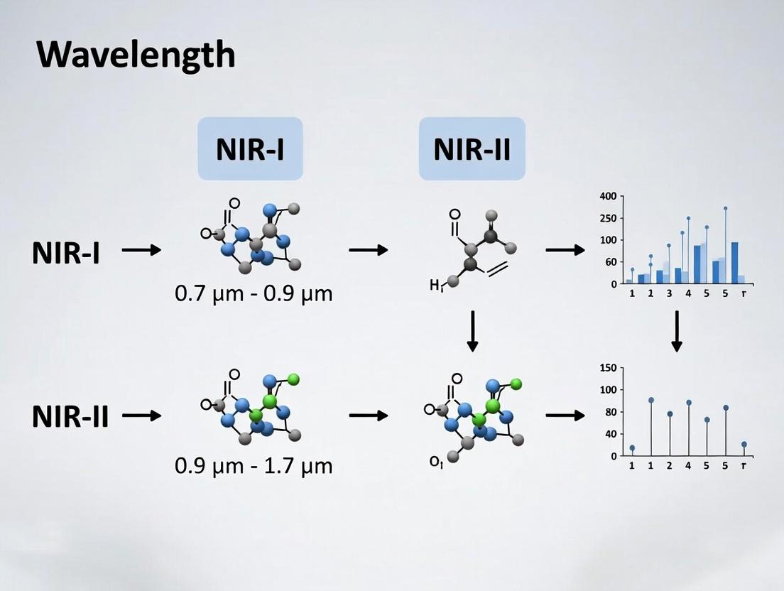
Defining the Spectrum: Core Concepts of NIR-I, NIR-II, and NIR-IIb Wavelengths
Near-infrared (NIR) light occupies a critical region of the electromagnetic spectrum between visible light and mid-infrared radiation. In the context of biomedical research, particularly for in vivo imaging and therapeutic applications, the NIR region is subdivided into distinct windows based on reduced photon scattering and minimal tissue autofluorescence. This whitepaper frames NIR light within the broader electromagnetic spectrum and details the operational definitions for the NIR-I and NIR-II windows as established by current research.
The standard divisions, as per recent consensus in photonics and biomedical optics literature, are as follows:
- NIR-I (First NIR Window): 700 nm – 900 nm
- NIR-II (Second NIR Window): 900 nm – 1700 nm
Regions beyond 1700 nm are often classified as NIR-IIb or short-wavelength infrared (SWIR).
Table 1: The Electromagnetic Spectrum Relevant to Biomedical Optics
| Spectral Band | Abbreviation | Wavelength Range | Primary Interaction with Biological Tissue | Key Applications in Life Sciences |
|---|---|---|---|---|
| Ultraviolet | UV | 10 nm – 400 nm | DNA damage, fluorescence excitation | Microscopy, sterilization |
| Visible | Vis | 400 nm – 700 nm | Absorption by chromophores (hemoglobin, melanin) | Bright-field microscopy, histology |
| Near-Infrared I | NIR-I | 700 nm – 900 nm | Moderate scattering, low autofluorescence | NIR fluorescence imaging (e.g., ICG), optogenetics |
| Near-Infrared II | NIR-II | 900 nm – 1700 nm | Reduced scattering, deep penetration | Deep-tissue vascular imaging, tumor surgery guidance |
| Mid-Infrared | MIR | 3 μm – 50 μm | Molecular vibration absorption | Fourier-transform IR spectroscopy, tissue diagnostics |
| Far-Infrared / Terahertz | FIR/THz | 50 μm – 1 mm | Phonon absorption | Imaging of superficial tissue layers |
Experimental Protocols for NIR Imaging
Protocol: In Vivo NIR-II Fluorescence Imaging of Tumor Vasculature
This protocol details a standard method for visualizing deep-tissue vasculature using NIR-II emitting fluorophores.
Materials:
- Animal model (e.g., nude mouse with subcutaneous xenograft tumor).
- NIR-II fluorophore (e.g., PEGylated single-walled carbon nanotubes [SWCNTs], Ag₂S quantum dots).
- NIR-II imaging system: Typically includes a 808 nm or 980 nm laser for excitation, InGaAs cameras (sensitive from 900-1700 nm), and appropriate long-pass filters (e.g., LP1000 nm, LP1200 nm).
- Anesthesia system (isoflurane).
- Heating pad for animal physiological maintenance.
- Image analysis software (e.g., ImageJ, custom MATLAB/Python scripts).
Procedure:
- Fluorophore Administration: Prepare a sterile solution of the NIR-II fluorophore in phosphate-buffered saline (PBS). Intravenously inject via the tail vein at a dose of ~100-200 μL (e.g., 200 pmol for quantum dots).
- Animal Preparation: Anesthetize the mouse using 2% isoflurane in oxygen. Secure the animal in a prone position on a heated stage. Depilate the imaging region to reduce signal attenuation from hair.
- Imaging System Setup: Power on the NIR-II imaging system and allow the camera to cool to its operating temperature (typically -80°C). Set the excitation laser power to a safe, non-thermal level (e.g., 100 mW/cm²). Configure filter wheels with the appropriate long-pass emission filter.
- Image Acquisition:
- Acquire a pre-injection background image.
- Acquire sequential images post-injection at defined time points (e.g., 1 min, 5 min, 30 min, 1 hr, 24 hrs).
- Maintain consistent exposure times (e.g., 50-200 ms) and laser power across all sessions.
- Data Processing:
- Subtract the background image from all subsequent images.
- Apply flat-field correction if necessary.
- Calculate signal-to-background ratios (SBR) and contrast-to-noise ratios (CNR) in regions of interest (ROI) over the tumor versus adjacent normal tissue.
Protocol: Comparative Penetration Depth Analysis (NIR-I vs. NIR-II)
This protocol quantifies the superior tissue penetration of NIR-II light compared to NIR-I.
Materials:
- Tissue-mimicking phantoms or ex vivo tissue slices (e.g., chicken breast, porcine tissue) of varying thickness (0.5 mm to 10 mm).
- NIR-I fluorophore (e.g., Indocyanine Green, IRDye 800CW).
- NIR-II fluorophore (e.g., IR-1061 dye, PbS quantum dots).
- Dual-channel imaging system capable of detecting both NIR-I (800-900 nm) and NIR-II (1000-1400 nm) emission.
- Caliper for thickness measurement.
Procedure:
- Phantom Preparation: Create a thin layer of fluorophore (either NIR-I or NIR-II) sealed between glass slides. Stack tissue slices or phantom slabs of known, increasing thickness on top of this source.
- Dual-Channel Imaging: For each thickness, acquire images using both the NIR-I and NIR-II detection channels under identical excitation conditions.
- Signal Quantification: Measure the mean fluorescence intensity detected through each tissue thickness for both spectral windows.
- Analysis: Plot detected intensity (log scale) versus tissue thickness. Calculate the attenuation coefficient (μ) for each wavelength range using the Beer-Lambert law (I = I₀ * e^(-μx)). The NIR-II window will demonstrate a lower μ, confirming deeper effective penetration.
Table 2: Comparative Optical Properties of NIR Windows in Biological Tissue
| Property | NIR-I (750-900 nm) | NIR-II (1000-1350 nm) | Measurement Technique |
|---|---|---|---|
| Scattering Coefficient (μs') | High (~1.0 mm⁻¹) | Low (~0.3-0.5 mm⁻¹) | Spatial frequency domain imaging (SFDI) |
| Absorption by Hemoglobin | Moderate | Very Low | Spectrophotometry of whole blood |
| Absorption by Water | Very Low | Low | Spectrophotometry |
| Typical Autofluorescence | Moderate | Negligible | In vivo imaging of wild-type animals |
| Max. Penetration Depth in Tissue | 1-3 mm | 5-10 mm | Measurement of point spread function (PSF) broadening |
The Scientist's Toolkit: NIR Imaging Reagents & Materials
Table 3: Essential Research Reagents for NIR Imaging
| Item | Function & Description | Example Product/Chemical |
|---|---|---|
| NIR-I Fluorescent Dyes | Small molecule probes for labeling, histology, and shallow in vivo imaging. High quantum yield in the 700-900 nm range. | Indocyanine Green (ICG), IRDye 800CW, Cy7 |
| NIR-II Fluorescent Nanomaterials | Inorganic nanoparticles emitting in the NIR-II window for deep-tissue imaging. Often require surface functionalization for biocompatibility. | Ag₂S Quantum Dots, PEGylated SWCNTs, Rare-earth-doped Nanoparticles |
| Targeted Contrast Agents | Fluorophores conjugated to targeting moieties (antibodies, peptides, aptamers) for molecular-specific imaging. | Anti-CD31-Ag₂S QDs (vascular imaging), cRGD-PbS QDs (tumor integrin targeting) |
| Long-Pass Emission Filters | Optical filters that block excitation laser light and shorter wavelengths, allowing only NIR-II emission to reach the detector. Critical for signal purity. | Semrock LP1000 nm, LP1250 nm, LP1500 nm |
| InGaAs Camera | Photodetector array sensitive to wavelengths from 900-1700 nm. Requires thermoelectric or liquid nitrogen cooling to reduce dark noise. | Teledyne Princeton Instruments NIRvana, Hamamatsu C12741-03 |
| Tunable/Single-Wavelength Lasers | Provide precise excitation wavelengths matched to fluorophore absorption peaks (commonly 808 nm, 980 nm, 1064 nm). | Omicron LuxX diode lasers, CNI Lasers |
Visualization of NIR Imaging Workflows and Pathways
This whitepaper serves as a foundational technical guide within a broader research thesis aimed at standardizing the operational definitions and applications of near-infrared (NIR) biological imaging windows. Precise spectral demarcation is critical for advancing imaging technologies, contrast agent development, and quantitative biological analysis. This document delineates the core wavelength ranges—NIR-I, NIR-II, and the NIR-IIb sub-window—based on the interplay between photon-tissue interaction and detector sensitivity, providing a unified framework for researchers and drug development professionals.
Spectral Window Definitions and Rationale
Core Definitions
- NIR-I (First Near-Infrared Window): 700–900 nm. Historically the first window utilized for deep-tissue fluorescence imaging, benefiting from a local minimum in hemoglobin and water absorption.
- NIR-II (Second Near-Infrared Window): 1000–1700 nm. Encompasses a broader region with significantly reduced scattering and autofluorescence, leading to superior imaging depth and resolution.
- NIR-IIb (Second Near-Infrared Sub-window): 1500–1700 nm. A specialized region within NIR-II where tissue scattering is minimized to its lowest, offering the highest fidelity for in vivo imaging.
Quantitative Optical Property Comparison
The following table summarizes key optical properties that define and differentiate these windows.
Table 1: Comparative Optical Properties of NIR Imaging Windows
| Property / Window | NIR-I (700-900 nm) | NIR-II (1000-1350 nm) | NIR-IIb (1500-1700 nm) |
|---|---|---|---|
| Tissue Scattering Coefficient (μs') | ~1.0 mm⁻¹ at 800 nm | ~0.5 mm⁻¹ at 1100 nm | <0.3 mm⁻¹ at 1600 nm |
| Autofluorescence Intensity | High (from biomolecules) | Low | Negligible |
| Water Absorption | Low | Moderate | High (requires consideration) |
| Typical Penetration Depth | 1-3 mm | 3-8 mm | 5-10+ mm (for equivalent contrast) |
| Optimal Resolution (FFP) | ~20-40 μm | ~10-25 μm | ~5-15 μm |
Note: FFP = Fast Fourier Transform; values are approximate and tissue-dependent.
Experimental Protocols for Validation
Protocol: Measuring Tissue Scattering Profiles
Objective: To empirically determine the reduced scattering coefficient (μs') across NIR-I, NIR-II, and NIR-IIb windows. Materials: Tissue-mimicking phantoms (lipids, intralipid), tunable NIR laser source (700-1700 nm), integrating sphere spectrometer, lock-in amplifier. Methodology:
- Prepare phantoms with known, controlled scatterer concentrations.
- For each target wavelength, illuminate the phantom with a collimated beam.
- Use the integrating sphere to collect total diffuse reflectance and transmittance.
- Apply the Inverse Adding-Doubling (IAD) algorithm to the measured data to calculate μs' and absorption coefficient (μa).
- Plot μs' versus wavelength to visualize the sharp decline into the NIR-II and NIR-IIb regions.
Protocol: In Vivo Imaging Contrast-to-Noise Ratio (CNR) Assessment
Objective: To quantify the improvement in CNR when imaging in NIR-IIb versus NIR-I. Materials: NIR-IIb-emitting fluorophore (e.g., PbS/CdS quantum dots, organic dye IR-1061), NIR-I dye (e.g., ICG), mouse model, 2D InGaAs camera (sensitive to 1000-1700 nm), Si CCD camera (for NIR-I), 1500 nm long-pass filter. Methodology:
- Administer the NIR-IIb fluorophore intravenously to an anesthetized mouse.
- Using the InGaAs camera with a 1064 nm laser excitation and a 1500 nm long-pass filter, acquire dynamic video of the cerebral vasculature.
- Switch the filter and excite the same subject with an 808 nm laser, using the Si CCD to capture NIR-I fluorescence.
- Co-register the images. Select a region of interest (ROI) on a vessel and a nearby tissue background region.
- Calculate CNR for each window: CNR = (Signalvessel - Signalbackground) / SD_background.
- Statistically compare the CNR values, demonstrating the superior signal clarity in NIR-IIb.
Diagram: NIR Window Classification & Key Characteristics
Diagram: Experimental CNR Comparison Workflow
The Scientist's Toolkit: Key Research Reagent Solutions
Table 2: Essential Materials for NIR-I/NIR-II Imaging Research
| Item / Reagent | Function & Application | Key Consideration |
|---|---|---|
| Indocyanine Green (ICG) | FDA-approved NIR-I fluorophore (ex/em ~780/820 nm). Used for angiography, lymphography, and liver function testing. | Rapid plasma binding and short half-life limit flexible bioconjugation. |
| PbS/CdS Core/Shell QDs | Nanocrystals emitting in NIR-II (1100-1600 nm). High quantum yield, tunable emission, used for deep-tissue vasculature imaging. | Contains heavy metals (Pb), posing potential long-term toxicity concerns for clinical translation. |
| IR-1061 & Dyes (e.g., CH-4T) | Small-molecule organic dyes emitting beyond 1500 nm (NIR-IIb). Enable high-resolution cerebral and tumor imaging. | Often requires encapsulation in polymer matrices (e.g., PLGA-PEG) for in vivo stability and biocompatibility. |
| Erbium-Doped Nanoparticles | Down-conversion probes excited at ~980 nm, emitting in NIR-IIb (∼1525 nm). Inorganic, photostable labels. | Requires high-power density for excitation; can generate local heating. |
| 1500 nm Long-Pass Filter | Critical optical component placed before the InGaAs detector. Blocks excitation light and NIR-IIa emission, isolating the NIR-IIb signal. | Optical density (OD) >5 at the laser wavelength is required to prevent signal saturation. |
| 2D InGaAs Camera (Cooled) | Primary detector for NIR-II/IIb imaging. Sensitive from 900-1700 nm. Essential for capturing low-flux photons from deep tissue. | Cooling reduces dark noise. Chip format (e.g., 320x256, 640x512) dictates imaging field of view and resolution. |
| Tunable NIR Laser Source | Provides precise excitation from 700 nm to 1700 nm. Allows systematic evaluation of fluorophores and tissue properties across windows. | OPO (Optical Parametric Oscillator) systems offer wide tunability but are costly and complex. |
The study of light-tissue interactions is foundational for advancing biomedical optics, particularly within the Near-Infrared I (NIR-I, 700-900 nm) and Near-Infrared II (NIR-II, 1000-1700 nm) spectral windows. These windows are defined by minimized light absorption by endogenous chromophores like water, hemoglobin, and lipids, allowing for deeper photon penetration. This guide details the core physical principles—scattering, absorption, and the resultant penetration depth—that underpin emerging applications in non-invasive imaging, photothermal therapy, and optically triggered drug delivery.
Core Optical Phenomena in Biological Tissue
Scattering
Scattering is the redirection of photon trajectory due to interactions with microscopic variations in refractive index within tissue (e.g., organelles, membranes, collagen fibers). It is characterized by the scattering coefficient (µ_s, units: cm⁻¹), which denotes the probability of scattering per unit path length. The anisotropy factor (g) describes the directionality of scattering, ranging from isotropic (g=0) to highly forward-directed (g~0.9 for tissue).
Absorption
Absorption is the conversion of photon energy into other forms (heat, fluorescence, chemical energy). It is quantified by the absorption coefficient (µ_a, cm⁻¹), indicating the probability of absorption per unit path length. Primary endogenous absorbers in the NIR windows include:
- Hemoglobin (Hb/HbO₂): Absorption decreases significantly >650 nm.
- Water: Absorption minima around 800-900 nm (NIR-I) and increases beyond 1150 nm, defining the upper limit of the NIR-II window.
- Lipids: Exhibit absorption peaks near 930 nm and 1200 nm.
Attenuation and Penetration Depth
The total attenuation of a light beam in tissue is governed by the combined effects of scattering and absorption, described by the reduced attenuation coefficient: µ'eff = [3µa(µs' + µa)]^(1/2), where µs' = µs(1-g) is the reduced scattering coefficient. The effective penetration depth (δ), defined as the depth at which the fluence rate is reduced to 1/e (~37%) of its surface value, is: δ = 1 / µ'_eff.
Quantitative Comparison: NIR-I vs. NIR-II Windows
Live search data (2023-2024) consolidating values from recent reviews on skin, brain, and breast tissue phantoms reveals key differentials.
Table 1: Typical Optical Properties and Penetration Depth in Biological Tissue
| Parameter / Tissue Type | NIR-I (~800 nm) | NIR-II (~1064 nm) | NIR-II (~1300 nm) | Notes |
|---|---|---|---|---|
| µ_a (cm⁻¹) - Skin | 0.1 - 0.3 | 0.2 - 0.4 | 0.4 - 0.8 | Minima at ~800 nm, increases with longer NIR-II. |
| µ_s' (cm⁻¹) - Skin | 10 - 20 | 6 - 12 | 5 - 10 | Reduced scattering decreases with increasing λ. |
| Penetration Depth δ (mm) - Skin | 2 - 3 | 3 - 5 | 2.5 - 4 | Maximized in 1000-1100 nm range. |
| µ_a (cm⁻¹) - Brain | 0.1 - 0.2 | 0.15 - 0.25 | 0.3 - 0.5 | Water absorption becomes significant >1150 nm. |
| µ_s' (cm⁻¹) - Brain | 8 - 15 | 5 - 9 | 4 - 8 | |
| Penetration Depth δ (mm) - Brain | 3 - 5 | 5 - 8 | 4 - 6 | Enables transcranial optical access. |
| Primary Absorber | Hemoglobin (low), Water (very low) | Water (low), Lipids (moderate) | Water (increasing), Lipids | Chromophore cross-sections define windows. |
Experimental Protocols for Characterization
Protocol: Measuring µa and µs' Using Integrating Sphere with Inverse Adding-Doubling (IAD)
Objective: To determine the optical properties of thin, homogenous tissue samples or phantoms. Materials: See "The Scientist's Toolkit" below. Methodology:
- Sample Preparation: Prepare a tissue phantom (e.g., Intralipid suspension for scattering, India ink for absorption) or a thinly sliced, optically cleared tissue sample.
- Collimated Transmission (T_c) Measurement:
- Place the sample at the entrance port of the integrating sphere.
- Direct a collimated, monochromatic beam (from a tunable laser or filtered source) onto the sample.
- Measure the power of the collimated beam transmitted directly through the sample without scattering (Pc). Calculate Tc = Pc / Pincident.
- Total Transmission (Tt) and Diffuse Reflectance (Rd) Measurement:
- Attach the sample to the sphere's sample port.
- For Tt, illuminate the sample from outside and measure the total power diffusely transmitted into the sphere.
- For Rd, swap source and detector ports to measure total power diffusely reflected from the sample.
- IAD Analysis: Input measured Tc, Tt, and Rd values, along with sample thickness, into an IAD software algorithm (e.g., from Oregon Medical Laser Center). The algorithm iteratively solves the radiative transport equation to output µa and µ_s'.
- Validation: Perform measurements across NIR-I and NIR-II wavelengths using appropriate detectors (Si CCD for NIR-I, InGaAs for NIR-II).
Protocol: In Vivo Measurement of Penetration Depth via Spatial Frequency Domain Imaging (SFDI)
Objective: To map penetration depth and optical properties in vivo over a wide field. Methodology:
- Pattern Projection: Project sinusoidal patterns of light at multiple spatial frequencies (e.g., 0, 0.05, 0.1, 0.2 mm⁻¹) and wavelengths (e.g., 750, 850, 1050, 1300 nm) onto the tissue surface using a DLP projector with NIR capability.
- Image Acquisition: Use a synchronized, calibrated NIR camera (Si or InGaAs) to capture the reflected diffuse light pattern for each frequency/wavelength combination.
- Demodulation: Process images to extract the amplitude of the reflected AC component (AAC) and the average DC component (ADC) at each pixel.
- Model Fitting: For each pixel, fit the measured AAC/ADC vs. spatial frequency data to a Monte Carlo-derived or analytical model of light propagation. This yields maps of µa and µs' across the tissue field.
- Penetration Depth Calculation: Compute the pixel-wise penetration depth map using δ = 1 / µ'eff, where µ'eff = sqrt(3 * µa * (µa + µ_s')).
Visualization: Pathways & Workflows
Diagram Title: Scattering vs. Absorption Pathways in Tissue.
Diagram Title: SFDI Protocol for Mapping Penetration Depth.
The Scientist's Toolkit: Key Research Reagents & Materials
Table 2: Essential Materials for Optical Tissue Property Experiments
| Item | Function | Application Notes |
|---|---|---|
| Intralipid 20% | A standardized lipid emulsion used as a scattering phantom material. Its µ_s' is well-characterized and adjustable via dilution. | Foundation for tissue-simulating phantoms. Must be used with an absorbing agent (e.g., ink) to match µ_a. |
| India Ink / Nigrosin | Absorbing agent with broad-spectrum absorption. Added in minute quantities to phantoms to achieve desired µ_a. | Concentration must be precisely measured. Filtered ink is preferred for uniform particle size. |
| Polydimethylsiloxane (PDMS) | Solid phantom matrix. Allows embedding of scattering and absorbing particles to create stable, reusable solid optical standards. | Curing temperature can affect particle distribution. Optical properties are stable long-term. |
| Tunable NIR Light Source (e.g., Ti:Sapphire Laser, Optical Parametric Oscillator, Supercontinuum Laser with Monochromator) | Provides monochromatic, high-power light across NIR-I and NIR-II for precise spectral measurements. | Essential for spectroscopy. Requires matching detector sensitivity (Si for NIR-I, InGaAs/ HgCdTe for NIR-II). |
| Integrating Sphere (with NIR-optimized coatings) | Collects all diffusely transmitted or reflected light from a sample for accurate total power measurement, minimizing collection loss. | Port size must accommodate sample. Coating (e.g., Spectralon, BaSO₄) must be highly reflective in target wavelength range. |
| Inverse Adding-Doubling (IAD) Software | Algorithmic solver that derives µa and µs' from measured total transmission, collimated transmission, and diffuse reflectance. | Standard, validated tool. Requires accurate input of sample thickness and sphere geometry. |
| Spatial Light Modulator (SLM) / DLP Projector | Generates the sinusoidal patterns required for Spatial Frequency Domain Imaging (SFDI). | Must have sufficient output power in the NIR range (700-1700 nm). |
| NIR-Sensitive Cameras (Si CCD for NIR-I, InGaAs for NIR-II) | Detects diffuse reflected light in imaging-based measurement techniques (e.g., SFDI, diffuse optical tomography). | Cooling is often required to reduce dark noise, especially for InGaAs detectors in NIR-II. |
The concept of the biological optical window describes specific wavelength ranges in the near-infrared (NIR) spectrum where light exhibits maximal penetration depth in living tissue. This phenomenon is primarily due to the reduced scattering and minimal absorption by endogenous chromophores such as water, hemoglobin, lipids, and melanin. This whitepaper, framed within a broader thesis on NIR-I (700-950 nm) and NIR-II (1000-1350 nm, extending to ~1700 nm) definitions, details the biophysical principles, provides comparative quantitative data, and outlines experimental protocols for leveraging these windows in biomedical research and drug development.
Biological tissues are highly scattering and absorbing media for light. The "optical window" is not a single band but a series of spectral regions where the combined effect of absorption and scattering is minimized. The first window (NIR-I) has been utilized for decades in techniques like functional NIRS (fNIRS). More recently, the second (NIR-II) and third (NIR-III, ~1550-1870 nm) windows have garnered significant interest due to even lower scattering coefficients and reduced autofluorescence, enabling superior resolution and penetration depth for in vivo imaging and therapeutic applications.
Biophysical Principles of Light-Tissue Interaction
Light interaction with tissue is governed by absorption ((\mua)) and scattering ((\mus)) coefficients. The reduced scattering coefficient ((\mus')) determines the effective scattering. The penetration depth ((\delta)) is approximately inversely proportional to the effective attenuation coefficient ((\mu{eff} = \sqrt{3\mua(\mua + \mu_s')})).
Key Chromophore Absorption Profiles:
- Hemoglobin (Oxy- and Deoxy-): High absorption in visible range (400-650 nm), drops significantly beyond 650 nm.
- Water: Very low absorption from ~650-900 nm, begins to increase around 970 nm, with strong peaks at ~1200 nm, 1450 nm, and 1900 nm.
- Lipids: Exhibit absorption peaks near 930 nm, 1200 nm, and 1700 nm.
- Melanin: Absorption decreases monotonically with increasing wavelength.
The combined effect creates troughs in total tissue absorption between the peaks of these major chromophores.
Comparative Analysis: NIR-I vs. NIR-II Windows
The following table summarizes key optical properties and performance metrics for the primary optical windows.
Table 1: Quantitative Comparison of Biological Optical Windows
| Parameter | Visible (400-650 nm) | NIR-I Window (700-950 nm) | NIR-II Window (1000-1350 nm) | NIR-III Window (1550-1870 nm) |
|---|---|---|---|---|
| Primary Absorbers | Hb, HbO₂, Melanin | Water (low), Hb/HbO₂ (low) | Water (medium), Lipids | Water (high), Lipids |
| Scattering Coefficient ((\mu_s')) | Very High (~100-200 cm⁻¹) | High (~20-50 cm⁻¹) | Lower (~5-20 cm⁻¹) | Low (~<10 cm⁻¹) |
| Theoretical Penetration Depth | <1 mm | 1-5 mm | 5-20 mm | 3-10 mm* |
| Tissue Autofluorescence | High | Moderate | Very Low | Negligible |
| Typical Resolution (Imaging) | High (surface) | Moderate | Superior (deep tissue) | High (limited by water absorption) |
| Key Applications | Histology, confocal microscopy | fNIRS, indocyanine green imaging | NIR-IIb (1300-1400nm) in vivo imaging, photothermal therapy | Spectral-hole burning microscopy, specialized sensing |
*Penetration in NIR-III is more variable and heavily dependent on water content.
Table 2: Absorption Coefficients of Major Chromophores at Key Wavelengths
| Chromophore | Absorption Coefficient ((\mu_a)) [cm⁻¹] |
|---|---|
| Oxyhemoglobin (HbO₂) at 660 nm | ~2.5 |
| Oxyhemoglobin (HbO₂) at 850 nm | ~0.8 |
| Water at 850 nm | ~0.02 |
| Water at 1064 nm | ~0.12 |
| Water at 1300 nm | ~0.8 |
| Lipid at 1200 nm | ~0.5 |
Experimental Protocols for Characterizing the Optical Window
Protocol 4.1: Measuring Tissue Optical Properties Using Integrating Sphere Spectroscopy
Objective: To quantify the absorption ((\mua)) and reduced scattering ((\mus')) coefficients of ex vivo tissue samples across NIR wavelengths. Materials: Double-integrating sphere setup, broadband NIR light source (e.g., halogen lamp), spectrometer (NIR-sensitive, InGaAs detector for >1000 nm), calibrated reflectance standards, tissue sample (thinly sliced, <2 mm thick). Procedure:
- System Calibration: Measure dark signal, then reflectance and transmittance of calibration standards.
- Sample Measurement: Place tissue sample at the input port of the first sphere. Measure total reflectance (Rₜ) and total transmittance (Tₜ) with the sample in place.
- Inverse Adding-Doubling (IAD): Input Rₜ and Tₜ, along with sample thickness, into an IAD algorithm. This computational model iteratively solves the radiative transport equation to extract (\mua) and (\mus') at each wavelength.
- Validation: Verify results with phantoms of known optical properties.
Protocol 4.2: In Vivo NIR-II Fluorescence Imaging for Vascular Mapping
Objective: To visualize deep tissue vasculature with high spatial resolution using NIR-II emitting fluorophores. Materials:
- Animal Model: Mouse with dorsal skinfold chamber or shaved back.
- Fluorophore: FDA-approved Indocyanine Green (ICG, emits ~900 nm) or NIR-IIb fluorophore (e.g., PbS quantum dots, emits 1300-1500 nm).
- Imaging System: 808 nm or 980 nm laser for excitation, NIR-sensitive InGaAs camera with appropriate long-pass filters (e.g., 1250 nm LP for NIR-IIb imaging), imaging chamber. Procedure:
- Animal Preparation: Anesthetize and position the animal on the heated stage.
- Fluorophore Administration: Inject fluorophore intravenously via tail vein (e.g., ICG: 0.1-0.3 mg/kg).
- Image Acquisition: Turn off room lights. Set laser power to safe limits (<100 mW/cm²). Acquire time-series images at desired frame rate (e.g., 5-10 fps).
- Data Analysis: Use software to calculate signal-to-background ratio (SBR), full-width at half-maximum (FWHM) of vessel profiles, and generate maximum intensity projections (MIPs).
Key Signaling Pathways in NIR Photobiomodulation/Therapy
While NIR imaging exploits passive optical properties, therapeutic applications like photobiomodulation (PBM) involve active biological responses. A primary proposed mechanism involves cytochrome c oxidase (CCO) in the mitochondrial electron transport chain.
Diagram Title: Proposed NIR Photobiomodulation Pathway via Mitochondria
The Scientist's Toolkit: Research Reagent Solutions
Table 3: Essential Materials for NIR Window Research
| Item | Function/Benefit | Example Application |
|---|---|---|
| Indocyanine Green (ICG) | FDA-approved NIR-I fluorophore (Ex/Em ~780/820 nm). Binds plasma proteins, enabling angiography. | Vascular imaging, liver function tests, sentinel lymph node mapping. |
| NIR-II Fluorescent Quantum Dots (QDs) | Semiconductor nanocrystals (e.g., PbS, Ag₂S) with tunable emission in NIR-II. High brightness and photostability. | Deep-tissue, high-resolution vascular and tumor imaging (NIR-IIb). |
| Organic NIR-II Fluorophores | Small molecule dyes (e.g., CH-series) with emission >1000 nm. Potentially improved biocompatibility vs. QDs. | Targeted molecular imaging in the NIR-II window. |
| Intralipid / Lipid Phantoms | Standardized scattering media for calibrating and validating optical systems. Mimics tissue scattering properties. | System calibration, protocol development, Monte Carlo simulation validation. |
| NIR-Transparent Skull Windows | Optical clearing materials (e.g., TiO₂/polymer composites) or thinned skull preparations. | Chronic brain imaging in rodent models, reducing skull scattering. |
| InGaAs Cameras | Photodetectors sensitive from ~900-1700 nm. Essential for capturing NIR-II/NIR-III light. | In vivo NIR-II fluorescence and bioluminescence imaging. |
| Long-Pass Optical Filters (e.g., 1100, 1250, 1500 nm LP) | Block excitation laser light and shorter-wavelength autofluorescence. Isolate NIR-II/NIR-III signal. | Improving signal-to-background ratio in NIR-IIb imaging. |
The biological optical window, particularly in the NIR-II region, represents a powerful tool for minimally invasive biomedical investigation. The minimized scattering and absorption in this range directly translate to enhanced imaging depth, resolution, and data fidelity. As the definitions and understanding of NIR-I, II, and III windows evolve, so too do the opportunities for developing novel diagnostic imaging techniques, targeted phototherapies, and drug delivery monitoring systems. Continued research into advanced fluorophores, optimized instrumentation, and precise protocols is crucial for translating these optical advantages into tangible clinical and pharmaceutical advancements.
Key Historical Milestones in the Development of NIR Biomedical Imaging
The development of Near-Infrared (NIR) biomedical imaging is intrinsically linked to the progressive exploration and utilization of the NIR spectrum, defined as wavelengths from 700 nm to 1700 nm. This technical guide frames its historical analysis within a core thesis on the evolution of NIR-I (700-900 nm) and NIR-II (1000-1700 nm) research, positing that the transition from NIR-I to NIR-II represents a paradigm shift driven by the imperative to overcome fundamental optical tissue scattering and autofluorescence limitations, thereby unlocking unprecedented resolution and penetration depth for in vivo imaging.
Historical Milestones: A Quantitative Timeline
The following table summarizes pivotal breakthroughs, highlighting the shift from NIR-I dyes to NIR-II materials and modalities.
| Year | Milestone Event | Key Wavelength Range | Significance & Quantitative Impact | Primary Researchers/Group |
|---|---|---|---|---|
| 1977 | Discovery of NIR light for tissue oximetry | ~730 nm & ~810 nm (NIR-I) | First non-invasive measurement of tissue oxygenation using differential absorption of hemoglobin. | F. F. Jöbsis |
| 1986 | Introduction of Indocyanine Green (ICG) for angiography | Peak excitation ~780 nm, Emission ~820 nm (NIR-I) | FDA-approved dye enabled first clinical NIR-I fluorescence imaging; penetration depth ~1 cm. | R. K. (Clinical adoption) |
| 1999 | Development of targeted NIR-I fluorescent probes | ~700-900 nm (NIR-I) | Conjugation of NIR-I dyes (e.g., Cy5.5) to antibodies enabled molecular-specific imaging. | R. Weissleder et al. |
| 2003 | First reported semiconductor quantum dots for in vivo imaging | ~700-900 nm (NIR-I) | High-brightness, tunable emission; demonstrated multiplexing but contained toxic metals. | M. Bawendi, S. Weiss et al. |
| 2009 | Carbon Nanotubes demonstrated for NIR-II imaging | 1000-1400 nm (NIR-II) | First demonstration of in vivo imaging in the NIR-II window; showed ~3-4x improved penetration depth vs. NIR-I. | H. Dai et al. |
| 2011 | Inorganic Rare-Earth Nanoparticles (RENPs) for NIR-II | ~1500-1600 nm (NIR-IIb) | Introduced down-converting luminescence in the "NIR-IIb" sub-window, minimizing scattering. | G. Chen et al. |
| 2014 | First small organic molecule dyes for NIR-II imaging | ~1000-1100 nm (NIR-II) | Developed fluorophores like IR-E1050, offering biocompatibility and renal clearance. | H. Dai et al. |
| 2019 | NIR-II imaging for real-time human lymphatic mapping | ~1000-1400 nm (NIR-II) | First clinical translation of NIR-II imaging in humans; achieved resolution of lymphatic vessels <0.5 mm. | H. Dai, J. Wang et al. |
| 2022-2023 | Clinical trials with NIR-II contrast agents | NIR-II / NIR-IIb | Initiation of trials (e.g., based on Ag2S quantum dots) for tumor margin delineation and vascular imaging. | Multiple (e.g., Memorial Sloan Kettering) |
Experimental Protocol: Benchmarking NIR-I vs. NIR-II ImagingIn Vivo
This protocol is central to the thesis, providing a methodology for comparing imaging performance across spectral windows.
Objective: To quantitatively compare spatial resolution, signal-to-background ratio (SBR), and penetration depth of a fluorescent probe imaged in the NIR-I (800-900 nm) and NIR-II (1000-1400 nm) windows.
Materials: (See Scientist's Toolkit below)
- Animal Model: Nude mouse with subcutaneously implanted tumor.
- NIR Fluorescent Probe: An agent with dual NIR-I & NIR-II emission (e.g., PEGylated Ag2S Quantum Dots, emitting at 980 nm and 1200 nm).
- Imaging Systems:
- NIR-I System: 785 nm laser excitation, 830 nm long-pass emission filter, Silicon CCD camera.
- NIR-II System: 808 nm laser excitation, 1000 nm long-pass emission filter, InGaAs camera.
- Analysis Software: ImageJ with custom macros for resolution and SBR calculation.
Procedure:
- Probe Administration: Intravenously inject a bolus of the probe (200 µL, 100 µM concentration) via the tail vein.
- Image Acquisition: Anesthetize the mouse and place it in the imaging chamber.
- Acquire time-series images at both NIR-I and NIR-II channels simultaneously (if using a spectral splitter) or sequentially at predetermined time points (e.g., 0, 1, 5, 10, 30, 60 min post-injection).
- Maintain identical laser power density (e.g., 50 mW/cm²) and integration time (e.g., 100 ms) for both systems where possible.
- Quantitative Analysis:
- Spatial Resolution: Image a resolution target or use the edge-spread function of a sharp anatomical feature (e.g., vessel edge). Calculate the full width at half maximum (FWHM) of the line profile.
- Signal-to-Background Ratio (SBR): Define a region of interest (ROI) over the tumor and a contralateral background ROI. Calculate SBR = (Mean SignalROI - Mean BackgroundROI) / Standard Deviation_Background.
- Penetration Depth Assessment: Image the probe through a tissue phantom of increasing thickness (0-10 mm). Plot normalized intensity vs. thickness and calculate the attenuation length.
Key Signaling Pathways & Workflows in Targeted NIR Imaging
The core logic of molecular-targeted NIR imaging involves specific probe-receptor interaction leading to signal amplification.
Title: Workflow for Targeted NIR Fluorescence Imaging
Title: Molecular Targeting with NIR Probes
The Scientist's Toolkit: Essential Reagents & Materials for NIR Imaging Research
| Item Name | Function & Role in NIR Research | Key Considerations |
|---|---|---|
| Indocyanine Green (ICG) | Benchmark NIR-I (≈820 nm) fluorophore; used for angiography, lymphography, and liver function testing. | FDA-approved, rapid hepatic clearance, prone to aggregation and photo-bleaching. |
| IRDye 800CW | Synthetic small molecule NIR-I dye (≈800 nm); commonly conjugated to antibodies or peptides for targeted imaging. | High chemical stability, commercially available as conjugation-ready succinimidyl ester. |
| Ag2S Quantum Dots | Typical inorganic NIR-II (≈1200 nm) nanoparticle; used for deep-tissue vascular and tumor imaging. | Good biocompatibility, size-tunable emission in NIR-II, requires PEGylation for stability. |
| CH1055-PEG | Organic donor-acceptor-donor (D-A-D) type NIR-II small molecule dye (emission 1055 nm). | Renal clearable, suitable for clinical translation, high molecular brightness. |
| NaYF4:Yb,Er@NaYF4 | Core-shell rare-earth nanoparticle (RENP); emits at 1550 nm (NIR-IIb) under 980 nm excitation. | Extremely low autofluorescence and scattering in NIR-IIb, requires high-power excitation. |
| InGaAs FPA Camera | The standard detector for NIR-II imaging (900-1700 nm). | Critical for NIR-II work; cooled versions reduce dark noise; cost is a major factor. |
| Silicon CCD/CMOS Camera | Standard detector for NIR-I imaging (700-1000 nm). | High quantum efficiency up to ≈1000 nm, low cost compared to InGaAs. |
| 808 nm Laser Diode | Common excitation source for both NIR-I and NIR-II fluorophores. | Must match fluorophore absorption; power density must be within safety limits (ANSI). |
| Long-pass Emission Filters | Optical filters (e.g., 1000 nm LP, 1250 nm LP) to block excitation/ambient light and isolate NIR-II signal. | Cut-on wavelength and optical density (OD) are critical specifications. |
| DSPE-PEG(2000)-Maleimide | A common lipid-PEG derivative for nanoparticle surface functionalization and bioconjugation. | Provides stealth from RES, improves circulation time, and offers a thiol-reactive group for ligand attachment. |
From Theory to Practice: Imaging Techniques and Therapeutic Applications Across NIR Windows
This whitepaper is framed within a broader thesis defining the evolving paradigm of in vivo fluorescence imaging. Historically dominated by the first near-infrared window (NIR-I, 700-900 nm), the field has expanded into the second (NIR-II, 1000-1700 nm) and emerging third (NIR-III, 1500-1900 nm) windows. This thesis posits that each biological window offers distinct trade-offs between photon scattering, tissue autofluorescence, and water absorption, necessitating the parallel development of specialized contrast agents. Optimal imaging depth, resolution, and signal-to-background ratio (SBR) are not achieved by a single universal probe but by matching engineered dyes and nanoparticles to the specific photophysical demands of each spectral window.
Defining the NIR Windows: Optical Properties and Rationale
The segmentation into windows is based on the interplay of light with biological tissue.
- NIR-I (700-900 nm): The conventional window where silicon-based detectors are highly sensitive. Reduced hemoglobin absorption compared to visible light allows ~1-2 mm imaging depth. Significant scattering and residual autofluorescence limit SBR.
- NIR-II (1000-1350 nm): Markedly reduced scattering and negligible autofluorescence lead to superior spatial resolution and SBR, enabling centimeter-deep imaging. The 1000-1350 nm sub-window is optimal, minimizing water absorption.
- NIR-IIa / NIR-III (1500-1900 nm): Further reduced scattering potential exists, but strong water absorption peaks (particularly ~1450 nm, ~1900 nm) limit photon flux, creating a "tissue transparency window" only in specific sub-bands (e.g., 1500-1700 nm). Requires specialized detectors (e.g., InGaAs).
Diagram Title: Light-Tissue Interaction Dictates NIR Window Properties
Contrast Agent Development by Window
NIR-I Agents (700-900 nm)
These are mature technologies, primarily serving as benchmarks and for superficial imaging.
Organic Dyes: Cyanine dyes (e.g., Cy5.5, ICG). ICG is FDA-approved but suffers from aggregation, protein binding, and rapid clearance. Nanoparticles: Quantum dots (QDs, e.g., CdSe/CdS/ZnS core/shell) with bright, tunable emission but concerns over heavy metal toxicity.
NIR-II Agents (1000-1350 nm)
The focus of intense development to leverage the optical advantages of this window.
Organic Dyes: Donor-Acceptor-Donor (D-A-D) structured small molecules (e.g., CH-4T, IR-1061). Key strategies include:
- Molecular Engineering: Extending conjugation and strengthening donor/acceptor units to redshift emission.
- Water-Solubilization: Grafting with polyethylene glycol (PEG) or sulfonate groups.
- Targeting: Conjugation to peptides (e.g., RGD) or antibodies. Protocol: Synthesis of a PEGylated D-A-D Dye (e.g., CH-4T-PEG)
- Core Synthesis: Under inert atmosphere, react a central electron-accepting unit (e.g., thieno[3,4-b]thiophene) with brominated electron-donating arms (e.g., cyclopentadithiophene) via a Pd-catalyzed Stille coupling in degassed toluene/triethylamine.
- Purification: Purify the crude product by silica gel column chromatography (eluent: dichloromethane/hexane gradient).
- PEGylation: React the terminal reactive groups (e.g., -COOH) on the dye with an amine-terminated PEG chain (e.g., mPEG-NH₂, 5 kDa) using EDC/NHS coupling in anhydrous DMF overnight.
- Isolation: Precipitate the product in cold diethyl ether, collect via centrifugation, and further purify by dialysis (MWCO 3.5 kDa) against water for 48h.
- Characterization: Confirm by
¹H NMR,MALDI-TOF, and absorbance/emission spectroscopy in PBS.
Nanoparticles:
- Single-Walled Carbon Nanotubes (SWCNTs): Semiconducting chiralities emit in NIR-II. Functionalized with PEG-phospholipids for biocompatibility.
- Inorganic Nanoparticles: Ag₂S, Ag₂Se, or PbS/CdS core/shell QDs. Offer high quantum yield but require careful surface coating for stability and reduced toxicity.
- Rare-Earth-Doped Nanoparticles (RENPs): NaYF₄ nanoparticles doped with lanthanides (e.g., Nd³⁺, Yb³⁺, Er³⁺). Excited at ~800 nm, emit via cascade processes in NIR-II. Often coated with an inert shell (e.g., NaYF₄).
NIR-III Agents (1500-1900 nm)
Emerging materials designed for the reduced scattering regime, often requiring >1500 nm emission.
Organic Dyes: Heavily modified D-A-D scaffolds pushing emission beyond 1500 nm (e.g., fluorophores based on benzobisthiadiazole). Brightness is often lower due to energy gap law. Nanoparticles: RENPs are prominent.
- Design: Core-shell-shell architectures (e.g., NaYF₄:Nd³⁺@NaYF₄@NaYF₄:Yb³⁺/Er³⁺?).
- Mechanism: 808 nm excitation excites Nd³⁺ (sensitizer). Energy transfers to Yb³⁺, then to Er³⁺, which emits at ~1525 nm. The inert middle shell suppresses surface quenching. Protocol: Synthesis of Nd³⁺-Sensitized RENPs for >1500 nm Emission
- Core Synthesis: Heat a mixture of RE³⁺ chlorides (78% Y, 20% Nd, 2% Gd) with oleic acid and 1-octadecene to 156°C under argon. Add NH₄F and NaOH in methanol, react for 30 min.
- Shell Growth (Inert): Add precursor solution containing Y³⁺ and Gd³⁺ chlorides to the core nanoparticle solution at 310°C, grow for 45 min.
- Shell Growth (Active): Add precursor solution containing Y³⁺, Yb³⁺, and Er³⁺ chlorides to the core-shell nanoparticles at 310°C, grow for 1 hour.
- Purification: Cool, precipitate with ethanol, collect by centrifugation, and disperse in cyclohexane.
- Water Transfer: Ligand exchange with poly(acrylic acid) (PAA) at 70°C for 2h, followed by dialysis against water.
Quantitative Comparison of Representative Agents
Table 1: Comparison of Fluorescent Agents Across NIR Windows
| Agent Class | Example | Peak Emission (nm) | Quantum Yield | Extinction Coefficient (M⁻¹cm⁻¹) | Key Advantages | Key Limitations |
|---|---|---|---|---|---|---|
| NIR-I Dye | Indocyanine Green (ICG) | ~820 | <1% in blood | ~120,000 | FDA-approved, rapid clearance | Poor stability, binds proteins, low QY |
| NIR-II Dye | CH-4T-PEG | ~1050 | ~0.3% (in serum) | ~3.2 x 10⁵ | Good brightness, tailorable chemistry | Moderate QY in aqueous media |
| NIR-II NP | Ag₂S QD (PEG) | ~1200 | ~15% | ~1 x 10⁶ (per NP) | High brightness, photostable | Potential long-term toxicity, size polydispersity |
| NIR-II NP | (6,5) SWCNT-PEG | ~1000 | 0.1-1% | ~1 x 10⁷ (per NT) | Extreme photostability, high aspect ratio | Complex chirality mixture, low QY |
| NIR-III NP | NaYF₄:Nd/Yb/Er@shell | ~1525 | ~0.5% (in water) | ~Nd³⁺: ~1 x 10⁴ | Sharp emissions, low background, long lifetime | Low absorption cross-section, complex synthesis |
The Scientist's Toolkit: Research Reagent Solutions
Table 2: Essential Materials for NIR Fluorophore Development & Evaluation
| Item / Reagent | Function / Application | Example Vendor/Product |
|---|---|---|
| D-A-D Dye Building Blocks | Core acceptor and donor units for organic NIR-II dye synthesis. | Sigma-Aldrich (Thienothiophene, diketopyrrolopyrrole); Luminescence Technology Corp. |
| PEGylation Reagents | Confer water solubility and stealth properties to dyes and nanoparticles. | BroadPharm (mPEG-NH₂, DSPE-PEG); Laysan Bio (PEG-COOH, -NH₂, -SH). |
| EDC / NHS | Carbodiimide crosslinkers for conjugating dyes to targeting ligands or PEG. | Thermo Fisher Scientific (Pierce EDC Sulfo-NHS Crosslinking Kit). |
| Rare Earth Chlorides | High-purity precursors for synthesizing rare-earth-doped nanoparticles. | Stanford Advanced Materials (YCl₃, NdCl₃, YbCl₃, ErCl₃, 99.99%). |
| Oleic Acid / 1-Octadecene | Solvent and coordinating ligands for high-temperature synthesis of NPs. | Sigma-Aldrich (Technical grade, 90%). |
| Dialysis Membranes (MWCO) | Purifying aqueous nanoparticle or dye solutions from small molecule impurities. | Repligen (Spectra/Por Float-A-Lyzer G2). |
| NIR Spectrophotometer | Measuring absorbance of NIR agents (up to ~1600 nm). | Shimadzu (UV-3600i Plus); Agilent (Cary 5000). |
| NIR-II Imaging System | In vitro and in vivo fluorescence imaging in NIR-II/III windows. | NIRVANA (Princeton Instruments); InVivo (INDEC BioSystems). |
| ICG (Reference Std.) | Benchmark NIR-I dye for comparative studies. | Sigma-Aldrich (Diagnostic Green products). |
Diagram Title: Workflow for Developing and Validating NIR Agents
The progression of fluorescence imaging into deeper NIR windows is intrinsically linked to the parallel innovation in contrast agent chemistry. The core thesis is validated: maximizing the benefit of each biological window requires specifically engineered materials—from small organic dyes for molecular targeting in the NIR-II to complex, multi-shell rare-earth nanoparticles for the NIR-III. Future development hinges on improving the aqueous quantum yield and biocompatibility of NIR-II dyes, and on refining the sensitization efficiency and surface chemistry of NIR-III nanoparticles. The ultimate goal is a versatile toolkit of window-optimized probes, enabling researchers to select the ideal balance of resolution, depth, and SBR for their specific biological question.
Photoacoustic imaging (PAI) is a rapidly emerging hybrid modality that combines the high optical contrast of optical imaging with the deep penetration and spatial resolution of ultrasound. This whitepaper frames PAI within the critical research context of near-infrared (NIR) spectral windows, specifically the NIR-I (700-900 nm) and NIR-II (1000-1700 nm) regions. The fundamental principle involves irradiating biological tissue with pulsed laser light. The tissue absorbs the light, undergoes thermoelastic expansion, and generates broadband acoustic waves, which are detected by ultrasound transducers to form images.
The choice between NIR-I and NIR-II illumination is pivotal. While NIR-I offers compatibility with a wide array of endogenous chromophores (e.g., hemoglobin, melanin) and established exogenous contrast agents, NIR-II provides significantly reduced scattering and lower tissue autofluorescence. This results in enhanced penetration depth and improved signal-to-background ratio, making the NIR-II window a frontier for high-resolution deep-tissue imaging in preclinical research and drug development.
Core Physical Principles and Wavelength-Dependent Contrast
The Photoacoustic Effect
The initial pressure rise ( P0 ) generated in an acoustically homogeneous medium is given by the simplified equation: [ P0 = \Gamma \cdot \mua \cdot F ] where ( \Gamma ) is the Gruneisen parameter (dimensionless thermoelastic coefficient), ( \mua ) is the optical absorption coefficient (cm⁻¹), and ( F ) is the local optical fluence (J/cm²).
Optical Properties in NIR-I vs. NIR-II
Tissue optical scattering decreases with increasing wavelength. This reduction is the primary driver for the superior performance of NIR-II. The following table summarizes key quantitative differences.
Table 1: Comparative Optical Properties in NIR-I vs. NIR-II Windows
| Property | NIR-I (e.g., 800 nm) | NIR-II (e.g., 1064 nm) | Impact on PAI |
|---|---|---|---|
| Reduced Scattering Coefficient (µs') | ~8-10 cm⁻¹ (brain) | ~4-6 cm⁻¹ (brain) | Lower scattering in NIR-II enables deeper penetration and less distorted fluence distribution. |
| Absorption of Hemoglobin (Hb) | High (Oxy-Hb ~1.5, Deoxy-Hb ~2.5 cm⁻¹/mM) | Low (~0.1-0.3 cm⁻¹/mM) | NIR-I is optimal for vascular/oxygenation imaging. NIR-II minimizes blood background for agent imaging. |
| Absorption of Water | Very Low | Increases significantly >900 nm | Becomes a dominant absorber >1200 nm, limiting usable window but enabling hydration imaging. |
| Maximum Safe Exposure (ANSI) | ~100 mJ/cm² (700-1050 nm) | ~100 mJ/cm² (1050-1400 nm) | Similar limits allow comparable fluence delivery. |
| Typical Penetration Depth (with contrast) | 2-4 cm | 5-8 cm | NIR-II facilitates imaging of deeper-seated structures. |
| Spatial Resolution at Depth | Degrades faster due to scattering | Better maintained at depth | Enables high-resolution functional imaging in deep tissues. |
Experimental Protocols for Key PAI Applications
Protocol: High-Resolution Vascular Morphology in NIR-II
Objective: To image the deep cerebrovasculature of a mouse skull-intact using an NIR-II absorbing contrast agent.
- Animal Preparation: Anesthetize mouse (e.g., 1.5% isoflurane in O₂). Secure in stereotaxic frame. Maintain body temperature at 37°C.
- Contrast Agent Administration: Intravenously inject 100 µL of IR-1061 dye-loaded nanoparticles (2 mg/mL in PBS) via tail vein.
- System Setup: Use a tunable OPO laser system operating at 1064 nm. Pulse width: 5-10 ns. Pulse repetition rate: 10 Hz. Ultrasound transducer central frequency: 40 MHz.
- Data Acquisition: Position the mouse under the focused transducer. Raster-scan the excitation beam. Acquire raw PA signals (A-lines) at each point. Set laser fluence to 15 mJ/cm² (below ANSI limit).
- Image Reconstruction: Apply time-reversal or back-projection algorithm to reconstruct a 3D image matrix.
- Analysis: Use 3D rendering software to segment and quantify vascular diameter, density, and tortuosity.
Protocol: Multi-Spectral Optoacoustic Tomography (MSOT) for Oxygenation (NIR-I)
Objective: To map tumor hypoxia by quantifying oxygen saturation (sO₂) of hemoglobin.
- Preparation: Implant tumor-bearing mouse in imaging chamber with warm water for acoustic coupling.
- Wavelength Selection: Acquire MSOT images at multiple wavelengths across the NIR-I spectrum (e.g., 750, 800, 850 nm). This spectral unmixing differentiates oxy- and deoxy-hemoglobin.
- Data Acquisition: Perform cross-sectional (tomographic) scans at each wavelength using a 512-element curved array transducer.
- Spectral Unmixing: For each pixel, fit the measured PA amplitude spectrum ( P0(\lambda) ) to the linear model: [ P0(\lambda) = k \cdot [sO2 \cdot \epsilon{HbO2}(\lambda) + (1 - sO2) \cdot \epsilon_{Hb}(\lambda)] ] where ( \epsilon ) are known molar extinction coefficients.
- Calculation: Solve for ( sO_2 ) and ( k ) (total hemoglobin concentration) using a linear least-squares algorithm.
Visualization of Key Concepts
Title: Core Photoacoustic Imaging Signal Pathway
Title: NIR-I vs NIR-II Wavelength Selection Logic
The Scientist's Toolkit: Key Research Reagent Solutions
Table 2: Essential Materials and Reagents for Advanced PAI Research
| Item | Category | Function & Rationale |
|---|---|---|
| Indocyanine Green (ICG) | NIR-I Exogenous Contrast Agent | FDA-approved dye (~800 nm peak). Used for vascular flow imaging, sentinel lymph node mapping, and liver function studies. |
| IRDye 800CW | NIR-I Fluorescent/PA Probe | Conjugatable dye for antibody/peptide targeting. Enables molecular PAI of cell surface markers (e.g., EGFR, HER2). |
| Semiconductor Polymer Nanoparticles (SPNs) | NIR-II Exogenous Contrast Agent | Organic nanoparticles with high photostability and tunable absorption in NIR-II. Ideal for deep-tissue vascular labeling and cell tracking. |
| Single-Walled Carbon Nanotubes (SWCNTs) | NIR-II Exogenous Contrast Agent | Strong, stable NIR-II absorbers. Can be functionalized for targeted molecular imaging and drug delivery monitoring. |
| Bismuth-Based Nanocrystals | Inorganic NIR-II Agent | (e.g., Bi₂Se₃). High atomic number provides strong PA signal. Used for enhanced tumor imaging and therapeutic agent. |
| Multi-Spectral Oxy-Hem/Deoxy-Hem Phantoms | Calibration/Validation | Tissue-mimicking phantoms with known concentrations of hemoglobin derivatives. Essential for validating MSOT sO₂ calculations. |
| Agarose/Gelatin-Based Phantom Materials | System Calibration | Used to create custom vasculature phantoms with defined absorption (India ink) and scattering (lipid) properties for resolution testing. |
| Hematology Analyzer | Supporting Equipment | Quantifies blood hemoglobin concentration in subject, a critical input parameter for accurate spectral unmixing of endogenous signals. |
This whitepaper details established clinical and preclinical applications within the Near-Infrared Window I (NIR-I, 700–900 nm) spectrum. This examination is a foundational component of a broader thesis investigating the comparative advantages, limitations, and definitions of the NIR-I (700–900 nm) and NIR-II (1000–1700 nm) biological windows. While NIR-II imaging is an emerging field promising deeper tissue penetration and reduced scattering, NIR-I technologies have matured into clinically validated tools, particularly in intraoperative guidance and physiological monitoring. This guide provides an in-depth technical analysis of these core NIR-I applications.
Core Principles of NIR-I Tissue Interaction
The utility of NIR-I stems from its specific interaction with biological tissues. Within this window, the absorption of light by endogenous chromophores like hemoglobin, melanin, and water is relatively low but spectrally distinct. This allows photons to penetrate tissue (typically 1-10 mm depth) and enables two primary modalities:
- Contrast-Based Imaging: Using exogenous fluorophores or absorbing agents to highlight specific structures (e.g., tumors, lymph nodes, blood vessels).
- Spectroscopic Oximetry: Leveraging the differential absorption spectra of oxygenated (HbO₂) and deoxygenated hemoglobin (HbR) to calculate tissue oxygen saturation (StO₂).
The primary limitation in NIR-I is scattering, which blurs spatial resolution at depth, a challenge that motivates research into the NIR-II window.
NIR-I in Surgical Guidance
NIR-I fluorescence imaging provides real-time, high-contrast visualization of anatomical and pathological structures during surgery.
Key Clinical and Research Agents
| Agent/Target | Excitation/Emission (nm) | Primary Application | Mechanism |
|---|---|---|---|
| Indocyanine Green (ICG) | ~780/~820 | Angiography, Lymphography, Tumor Delineation | Non-targeted; binds plasma proteins, illuminates vasculature and tissue perfusion. |
| Methylene Blue | ~665/~685 | Parathyroid Identification, Sentinel Lymph Node Mapping | Accumulates in certain tissues; fluorescence identifies parathyroid glands. |
| 5-ALA (Metabolized to PpIX) | ~405/~635 & ~704 | Tumor Resection (Glioblastoma, etc.) | Prodrug metabolized to fluorescent protoporphyrin IX (PpIX) in tumor cells. |
| Targeted Fluorophores (Research) | Varies (~750-850) | Molecular Imaging of Tumor Biomarkers | Antibody or peptide conjugated to NIR-I dye (e.g., IRDye800CW) for specific targeting. |
Quantitative Performance Data
Table 1: Comparison of NIR-I Fluorescence Imaging Systems for Surgical Guidance
| System/Platform (Example) | Typical Sensitivity (nM) | Spatial Resolution | Depth Penetration | Key Clinical Use |
|---|---|---|---|---|
| Open-field camera systems (e.g., FLUOBEAM, PDE) | 1-10 nM (for ICG) | 1-2 mm at surface | ~5-10 mm in tissue | Plastic & reconstructive surgery, bowel perfusion. |
| Laparoscopic/endoscopic systems | 5-20 nM | 2-3 mm | ~3-7 mm in tissue | Minimally invasive oncologic surgery (GI, urology). |
| Microscope-integrated systems (e.g., FLUOROPEN) | ~1 nM | Sub-mm at cortical surface | 1-3 mm in brain tissue | Neurosurgical tumor resection. |
Detailed Experimental Protocol: ICG-Guided Sentinel Lymph Node Biopsy
Objective: To intraoperatively identify the first-draining (sentinel) lymph node(s) from a tumor site using NIR-I fluorescence.
Materials (The Scientist's Toolkit):
- ICG Solution: 0.5-1.25 mg/mL in sterile water. Function: NIR-I fluorophore.
- Human Serum Albumin (HSA) or Patient's Own Blood: Function: To pre-mix with ICG, forming protein-bound ICG for slower lymphatic clearance.
- NIR-I Fluorescence Imaging System: Contains laser/diode for ~780 nm excitation and a filtered CCD camera sensitive to >800 nm emission. Function: To detect and display ICG fluorescence.
- 27-Gauge Intradermal Needle: Function: For precise peritumoral ICG injection.
Methodology:
- Preoperative Preparation: Dilute ICG powder to desired concentration. Mix with a small volume of HSA or draw into a syringe followed by 0.1-0.2 mL of patient's blood. Allow to incubate for several minutes.
- Tracer Injection: In the operating room, inject 0.1-0.5 mL of the ICG mixture intradermally or subdermally around the tumor or biopsy cavity.
- Dynamic Imaging: Immediately activate the NIR-I camera. Observe the real-time video display. Fluorescent lymphatic channels will become visible within minutes, tracing the path to the sentinel node(s).
- Node Identification & Resection: Follow the fluorescent lymphatic tracts through the incision. The sentinel node(s) will fluoresce brightly. Use the imaging system to confirm the node's location before and after dissection.
- Ex Vivo Confirmation: After resection, place the node under the NIR-I camera to confirm fluorescence before sending for pathological analysis.
NIR-I in Oximetry
NIR spectroscopy (NIRS) for oximetry is a non-invasive method for monitoring tissue hemodynamics and oxygenation.
Principle and Modified Beer-Lambert Law
The technique relies on the differential absorption of HbO₂ and HbR in the NIR-I window. Using multiple wavelengths (typically 730-850 nm), the concentration changes can be calculated using the Modified Beer-Lambert Law: ΔA = log(I₀/I) = (ε⋅Δc⋅DPF⋅L) + G Where ΔA is attenuation change, I₀/I is light intensity ratio, ε is extinction coefficient, Δc is concentration change, DPF is differential pathlength factor, L is source-detector distance, and G is scattering loss.
Quantitative Data and Device Specifications
Table 2: Specifications of Typical Continuous-Wave NIRS Oximeters
| Parameter | Clinical Cerebral Oximeter | Research Tissue Oximeter | Wearable Muscle Oximeter |
|---|---|---|---|
| Wavelengths | 2-4 fixed LEDs (~730, 810, 850 nm) | 4-8 lasers (690-850 nm) | 2-3 LEDs (~730, 760, 810 nm) |
| Measurement | Regional Oxygen Saturation (rSO₂) | Tissue Oxygenation Index (TOI) or HbO₂/HbR Concentration | Muscle Oxygen Saturation (SmO₂) |
| Depth Penetration | ~2-3 cm (cortical tissue) | Adjustable via source-detector spacing | ~1-2 cm (muscle) |
| Sampling Rate | 0.5-2 Hz | 1-10 Hz | 1-50 Hz |
| Key Output | rSO₂ (%) | StO₂ (%), Δ[HbO₂], Δ[HbR] (μM) | SmO₂ (%) |
Detailed Experimental Protocol: Measuring Muscle Oxygenation Dynamics During Exercise
Objective: To monitor changes in muscle oxygen saturation (SmO₂) and hemoglobin concentrations during a controlled exercise protocol.
Materials (The Scientist's Toolkit):
- Continuous-Wave NIRS Device: With multiple source-detector separations (e.g., 2.5 cm and 3.5 cm). Function: Emits light and detects attenuation at multiple wavelengths.
- NIRS Sensor Pad: Contains integrated light sources and detectors. Function: To be placed on the skin over the muscle of interest.
- Black Cloth or Opaque Cover: Function: To shield the sensor from ambient light.
- Adhesive Tape or Strap: Function: To secure the sensor and prevent motion artifact.
- Calibration Phantom (Optional): Function: For pre-experiment system validation.
- Ergometer (Treadmill/Bike): Function: To provide standardized exercise workload.
Methodology:
- Sensor Placement: Shave and clean the skin over the target muscle (e.g., vastus lateralis). Adhere the NIRS sensor pad longitudinally along the muscle belly. Cover with black cloth and secure firmly with an elastic strap.
- Baseline Recording: With the subject at rest (seated or supine), record baseline NIRS signals for at least 3-5 minutes. Ensure signal stability.
- Exercise Protocol: Initiate a standardized exercise protocol (e.g., constant workload cycling at 75% VO₂max). Simultaneously trigger the NIRS device to start recording at a high frequency (≥1 Hz).
- Data Acquisition: Record continuously throughout the exercise period (e.g., 10 minutes) and into a recovery period (≥5 minutes post-exercise). Note the exact start/stop times of exercise.
- Data Processing: Use the device's proprietary software or a validated analysis package (e.g., Homer2 in MATLAB) to convert attenuation changes into concentration changes of Δ[HbO₂] and Δ[HbR] using the Modified Beer-Lambert Law. Calculate SmO₂ as [HbO₂]/([HbO₂]+[HbR]) * 100%.
- Analysis: Identify key parameters: baseline SmO₂, magnitude of desaturation during exercise, rate of re-saturation during recovery.
NIR-I techniques in surgical guidance and oximetry represent robust, clinically integrated technologies that exploit the specific optical properties of the 700-900 nm window. Their established protocols, quantitative benchmarks, and reagent solutions form the cornerstone of in vivo optical imaging and monitoring. However, limitations in penetration depth and spatial resolution due to scattering are inherent to NIR-I. This drives the thesis forward into the NIR-II (1000-1700 nm) window, where reduced scattering and autofluorescence promise significant advancements in deep-tissue, high-resolution imaging, building upon the foundational principles and experimental frameworks established by NIR-I research.
The conventional near-infrared window I (NIR-I, 700–900 nm) has been a cornerstone of biomedical optical imaging. However, its utility is constrained by significant photon scattering and autofluorescence in biological tissues. Research into the definitions of extended near-infrared windows has delineated two critical regions: NIR-II (900–1700 nm) and its sub-window, NIR-IIb (1500–1700 nm). This whitepaper, framed within a thesis on NIR wavelength range definitions, elucidates the superior performance of NIR-IIb for deep-tissue, high-resolution vascular and tumor imaging, driven by drastically reduced scattering and minimal autofluorescence.
Quantitative Advantages: NIR-IIb vs. NIR-I & NIR-IIa
The following table summarizes the key optical properties that underpin the superiority of NIR-IIb imaging.
Table 1: Quantitative Comparison of Optical Properties Across NIR Windows
| Property / Metric | NIR-I (700-900 nm) | NIR-IIa (1000-1400 nm) | NIR-IIb (1500-1700 nm) | Measurement/Note |
|---|---|---|---|---|
| Photon Scattering Coefficient (μs') | High (~1.5 mm⁻¹ at 800 nm) | Moderate (~0.7 mm⁻¹ at 1300 nm) | Low (~0.3 mm⁻¹ at 1550 nm) | In brain tissue; decreases with λ⁻ⁿ (n~0.5-1.5) |
| Autofluorescence Background | Very High | Low | Negligible | From tissue biomolecules (e.g., flavins) |
| Tissue Penetration Depth | ~1-3 mm | ~3-6 mm | >5-8 mm | Defined as 1/μeff; highly tissue-dependent |
| Spatial Resolution (In Vivo) | 10-50 μm | 5-20 μm | 3-10 μm | For microscopy; limited by scattering |
| Signal-to-Background Ratio (SBR) | Low (2-5) | High (10-50) | Very High (50-300+) | For vascular imaging with fluorophores |
| Water Absorption Peak | Minimal | Rising | Significant | Limits maximal penetration but reduces scattering |
Core Imaging Principles and Mechanisms
NIR-IIb imaging leverages organic fluorophores, quantum dots, or single-walled carbon nanotubes (SWCNTs) with emission peaks beyond 1500 nm. Upon excitation with ~808 nm or 980 nm lasers, these agents emit long-wavelength photons that experience less scattering (Rayleigh scattering ~λ⁻⁴) and virtually no tissue autofluorescence interference.
Diagram 1: NIR-IIb Imaging Principle and Photon-Tissue Interaction
Experimental Protocols for Key Applications
Protocol 4.1: High-Resolution Cerebral Vascular Imaging in Rodents
Objective: To achieve non-invasive, high-resolution mapping of the cerebral vasculature through the intact skull.
Materials: See "The Scientist's Toolkit" below. Procedure:
- Animal Preparation: Anesthetize mouse (e.g., C57BL/6) with isoflurane. Secure in stereotaxic frame. Maintain body temperature at 37°C.
- Contrast Agent Administration: Intravenously inject 200 µL of IR-E1050 dye (or PEGylated PbS quantum dots) via tail vein at a dose of 10 nmol/g body weight.
- System Setup: Use a 980 nm continuous-wave laser diode for excitation, expanded and homogenized to illuminate the shaved scalp. Use a 1500 nm long-pass filter. Employ a cooled 2D InGaAs camera (640 x 512 pixels) with a 25 mm f/1.4 lens.
- Image Acquisition: Acquire dynamic images at 5-10 frames per second for 5-10 minutes post-injection. Adjust laser power (<100 mW/cm²) and exposure time (20-100 ms) to avoid saturation.
- Data Processing: Calculate Signal-to-Background Ratio (SBR) = (Isignal - Ibackground) / Ibackground. Apply Gaussian filtering and contrast-limited adaptive histogram equalization (CLAHE) for visualization.
Protocol 4.2: NIR-IIb Fluorescence-Guided Tumor Resection
Objective: To delineate tumor margins in real-time during surgical resection.
Procedure:
- Tumor Model: Implant U87MG glioblastoma cells into the flank or brain of a nude mouse.
- Targeted Probe Injection: At 24-48 hours pre-surgery, inject 150 µL of EGFR-targeted Ag2S quantum dots (emission ~1600 nm) intravenously (5 mg/kg).
- Pre-Resection Imaging: Under anesthesia, perform wide-field NIR-IIb imaging (1550 nm LP filter) to locate the primary tumor. Acquire a baseline image.
- Real-Time Guiding: Use a handheld NIR-IIb imaging system during surgical dissection. Continuously monitor the video feed. Regions with SBR > 5:1 above surrounding normal tissue are considered positive.
- Post-Resection Validation: Image the resection cavity and the excised tumor. Fix tissues for ex vivo histology (H&E) to validate margin status.
Diagram 2: NIR-IIb Guided Tumor Resection Workflow
The Scientist's Toolkit: Essential Research Reagents & Materials
Table 2: Key Research Reagent Solutions for NIR-IIb Imaging
| Item | Function & Rationale | Example Products/Composition |
|---|---|---|
| NIR-IIb Fluorophores | Emit light in 1500-1700 nm range; core of imaging. | IR-E1050/1061 dyes, PbS/CdHgTe QDs, Er³⁺-doped nanoparticles, Chirality-sorted (9,4) SWCNTs. |
| Targeting Ligands | Conjugate to fluorophores for specific tumor/vascular targeting. | cRGD, EGFR antibodies, Folic acid, PSMA ligands. |
| Biocompatible Coatings | Render probes water-soluble, stable, and low-toxicity. | PEG chains (SH-PEG-NH₂), DSPE-PEG, poly(maleic anhydride-alt-1-octadecene). |
| Calibration Standards | Quantify fluorescence intensity and system performance. | IR-26 dye in DCE (reference quantum yield), Custom phantoms with Intralipid & India ink. |
| Anesthesia System | Maintain animal viability and immobility during imaging. | Isoflurane vaporizer, nose cone, oxygen supply. |
| NIR-I Laser Source | Excites fluorophores; must match probe absorption. | 808 nm or 980 nm laser diode, fiber-coupled, with collimator. |
| Long-Pass Filters | Block excitation light and NIR-I/IIa emission. | 1500 nm long-pass (LP), 1550 nm LP (Semrock, Thorlabs). |
| Cooled InGaAs Camera | Detect weak NIR-IIb photons with low noise. | Princeton Instruments NIRvana, Teledyne Xenics Xeva, Hamamatsu C12741. |
Data Interpretation and Advanced Analytical Techniques
Table 3: Quantitative Metrics for Image Analysis
| Metric | Formula/Description | Application in NIR-IIb |
|---|---|---|
| Signal-to-Background Ratio (SBR) | (Mean IntensityRegion of Interest - Mean IntensityBackground) / SD_Background | Primary metric for vessel sharpness and tumor detection. Values >100 common. |
| Full Width at Half Max (FWHM) | Measured from cross-sectional intensity profile of a sub-resolution structure. | Quantifies achievable resolution. Can approach 3-5 µm for capillaries. |
| Tumor-to-Background Ratio (TBR) | Mean IntensityTumor / Mean IntensityMuscle or Contralateral Tissue | Critical for oncology. Guides resection when >2-3. |
| Pharmacokinetic Parameters | Derived from time-intensity curves (e.g., AUC, T_max, half-life). | Analyzed from dynamic contrast-enhanced studies for vascular permeability. |
NIR-IIb imaging, as defined within the spectral taxonomy of near-infrared windows, represents a paradigm shift for non-invasive deep-tissue observation. Its defining characteristics—minimal scattering and autofluorescence—enable unprecedented clarity for visualizing vascular architecture and tumor margins. Future research will focus on developing brighter, targeted, clinically translatable fluorophores and integrating this modality with multi-spectral and therapeutic platforms.
This whitepaper details the technical integration of near-infrared (NIR) imaging with photothermal therapy (PTT) and photodynamic therapy (PDT). It is framed within a broader thesis investigating the distinct advantages and applications of the NIR-I (700-900 nm) and NIR-II (1000-1700 nm) biological windows. The primary hypothesis is that NIR-II offers superior performance for deep-tissue theragnostic platforms due to reduced scattering and autofluorescence, leading to higher resolution imaging and more efficient phototherapy. This guide provides the experimental and material foundation for validating this hypothesis.
Wavelength-Dependent Optical Properties: NIR-I vs. NIR-II
The performance of a theragnostic platform is fundamentally governed by the optical properties of biological tissue at the chosen wavelength.
Table 1: Comparative Optical Properties of NIR-I vs. NIR-II Windows
| Optical Property | NIR-I (750-900 nm) | NIR-II (1000-1350 nm) | Impact on Theragnostics |
|---|---|---|---|
| Tissue Scattering | Moderate-High | Significantly Reduced (∝ λ^-α) | NIR-II enables deeper penetration and higher spatial resolution for imaging. |
| Autofluorescence | Present from endogenous fluorophores | Negligible | NIR-II drastically improves signal-to-background ratio (SBR) for imaging. |
| Water Absorption | Low | Local minima at ~1100 nm, rises after ~1150 nm | Optimal window for deep penetration exists between 1000-1350 nm. |
| Maximum Imaging Depth (in vivo) | 2-4 mm | 5-20+ mm | NIR-II facilitates imaging and therapy in deeper-seated lesions. |
| Typical Resolution (FMT) | ~1-2 mm | <1 mm | Improved resolution for precise tumor delineation. |
| Photothermal Conversion Efficiency (PCE) | Can be high in nanomaterials | Often higher due to broader absorption profiles | Enables efficient heat generation with lower laser power densities. |
| Singlet Oxygen Quantum Yield (for PDT) | High in some agents (e.g., ICG) | Challenging; requires specific agent design | NIR-I currently has more clinical PDT agents. NIR-II PDT is emerging. |
Core Theragnostic Platforms: Mechanisms and Agents
Photothermal Therapy (PTT) Platforms
PTT employs light-absorbing agents to generate localized hyperthermia (>42°C), inducing cell death via necrosis, apoptosis, and immunogenic cell death.
Key Agents: Inorganic nanoparticles (Gold nanorods, nanoshells, CuS/Se nanoparticles), carbon-based materials (graphene oxide, carbon nanotubes), and organic polymers (polypyrrole, PEDOT:PSS). NIR-II agents like CuS nanoparticles offer high PCE at 1064 nm.
Photodynamic Therapy (PDT) Platforms
PDT uses photosensitizers (PS) that, upon light activation, convert tissue oxygen to cytotoxic reactive oxygen species (ROS), primarily singlet oxygen (¹O₂).
Key Agents: Traditional porphyrins (limited to visible light), NIR-I agents (Indocyanine Green, Chlorin e6), and emerging NIR-II PS (based on bacteriochlorin, diketopyrrolopyrrole, or coordination complexes).
Experimental Protocols for Platform Validation
Protocol: Synthesis of a Model NIR-II Theragnostic Nanoparticle (CuS@PEG-PS)
This protocol describes creating a core-shell nanoparticle combining PTT (CuS core) and PDT (polymer-shell coated PS).
- Materials: Copper chloride, sodium sulfide, poly(ethylene glycol)-thiol (SH-PEG-COOH), NIR-I/II-active photosensitizer (e.g., IR780 derivative), EDC/NHS coupling agents, dialysis tubing (10 kDa MWCO).
- Synthesis of CuS Core: Under N₂, add 10 mL of 10 mM CuCl₂ to 40 mL water. Rapidly inject 10 mL of 10 mM Na₂S with stirring. Heat at 70°C for 1 hr. Cool to room temperature.
- PEGylation: Add 50 mg of SH-PEG-COOH to the CuS solution. Stir for 24 hrs. Purify via centrifugation (15,000 rpm, 20 min) and resuspend in MES buffer (pH 6.5).
- PS Conjugation: Activate carboxyl groups on PEG with 10 mM EDC/NHS for 30 min. Add amine-functionalized PS (molar ratio PS:CuS = 100:1). React for 12 hrs.
- Purification: Dialyze against DI water for 48 hrs to remove unreacted reagents. Lyophilize and store at 4°C.
- Characterization: Use TEM for size, UV-Vis-NIR spectroscopy for absorption (700-1300 nm), and DLS for hydrodynamic diameter and zeta potential.
Protocol: In Vitro Efficacy and Imaging Assessment
- Cell Culture: Seed cancer cells (e.g., 4T1, U87MG) in 96-well plates (for viability) and glass-bottom dishes (for imaging) at 5x10³ cells/well.
- Nanoparticle Incubation: After 24 hrs, add fresh media containing CuS@PEG-PS at concentrations ranging from 0 to 100 μg/mL. Incubate for 4-6 hrs.
- NIR Imaging:
- Use a NIR-II fluorescence imaging system equipped with a 1064 nm laser for excitation and a 1300 nm long-pass filter for emission collection.
- Image cells to confirm nanoparticle uptake. Quantify intracellular fluorescence intensity.
- Phototherapy:
- PTT Group: Irradiate cells with a 1064 nm laser at 0.5-1.0 W/cm² for 5-10 min. Monitor temperature with an IR thermal camera.
- PDT Group: Irradiate cells with a 660 nm (for PS activation) or 808 nm laser at 0.2-0.5 W/cm² for 10 min.
- Combo Group: Perform sequential irradiation (e.g., 660 nm then 1064 nm).
- Viability Assay: 24 hrs post-irradiation, add CCK-8 reagent and measure absorbance at 450 nm. Calculate % viability relative to untreated control.
Protocol: In Vivo Theragnostic Evaluation in a Murine Model
- Tumor Model: Subcutaneously inject 1x10⁶ cancer cells into the flank of athymic nude mice. Proceed when tumors reach ~100 mm³.
- Administration & Imaging: Intravenously inject 200 μL of CuS@PEG-PS (5 mg/kg). At pre-determined time points (0, 4, 12, 24, 48 h):
- Anesthetize the mouse.
- Acquire NIR-II fluorescence images (1064 nm ex / 1300 nm LP em).
- Acquire photoacoustic images at 1064 nm to confirm deep-tissue localization.
- Phototherapy Treatment: At the time of peak tumor accumulation (e.g., 24 h p.i.):
- Randomize mice into groups: Saline (Control), Saline + Laser, NP only, NP + PTT Laser, NP + PDT Laser, NP + Combo Laser.
- Irradiate tumor with appropriate laser parameters (1064 nm, 1 W/cm², 10 min for PTT; 660 nm, 0.3 W/cm², 15 min for PDT).
- Monitor tumor surface temperature with an IR thermal camera during PTT.
- Monitoring: Measure tumor volume and body weight every 2 days for 14 days. Perform histological analysis (H&E, TUNEL) on excised tumors.
Diagrams of Signaling Pathways and Workflows
Title: Mechanism of NIR-Triggered PTT and PDT Cancer Cell Death
Title: Experimental Workflow for Validating a NIR-II Theragnostic Agent
The Scientist's Toolkit: Key Research Reagent Solutions
Table 2: Essential Materials for NIR Theragnostic Research
| Item Name | Category | Function & Rationale |
|---|---|---|
| Indocyanine Green (ICG) | NIR-I Dye | FDA-approved benchmark for NIR-I imaging and PDT; used for comparative studies against novel NIR-II agents. |
| Gold Nanorods (e.g., 808 nm LSPR) | NIR-I PTT Agent | Tunable, high-PCE standard for photothermal therapy in the NIR-I window. |
| PEG-Thiol (SH-PEG-COOH) | Surface Coating | Provides colloidal stability, reduces non-specific binding, and offers a carboxyl group for bioconjugation of targeting ligands or drugs. |
| IR-1061 or IR-26 Dye | NIR-II Fluorescence Standard | Used to calibrate and benchmark the sensitivity and performance of NIR-II imaging systems. |
| Singlet Oxygen Sensor Green (SOSG) | ROS Detection Reagent | Fluorescent probe specific for ¹O₂, essential for quantifying PDT efficacy of new photosensitizers in vitro. |
| CCK-8 Assay Kit | Cell Viability Assay | A sensitive, one-step colorimetric assay to quantify cell proliferation and cytotoxicity post-phototherapy. |
| Matrigel | Extracellular Matrix | Used for establishing orthotopic or more physiologically relevant tumor models in vivo. |
| IVIS Spectrum CT or Similar | Imaging System | Preclinical in vivo imaging system capable of 2D fluorescence (NIR-I/II) and 3D tomography, critical for biodistribution studies. |
| 1064 nm Diode Laser | NIR-II Light Source | Standard laser for exciting NIR-II imaging agents and activating NIR-II photothermal therapies. |
| InGaAs NIR Camera | NIR-II Detector | A camera sensitive to 900-1700 nm light, required for capturing NIR-II fluorescence signals. |
Overcoming Challenges: Noise Reduction, Probe Design, and System Optimization
Within the broader research on optical imaging in biological tissues, the definition and exploitation of specific near-infrared (NIR) windows are paramount. The conventional NIR-I window (700–900 nm) offers initial advantages over visible light but is limited by persistent tissue autofluorescence and significant photon scattering. The NIR-II window, historically defined as 1000–1700 nm, and often subdivided into NIR-IIa (1300–1400 nm) or NIR-IIb (1500–1700 nm), represents a paradigm shift. This in-depth guide explores the physical principles behind the superior performance of NIR-II imaging, provides quantitative comparisons, details experimental protocols for validation, and outlines essential research tools.
Core Principles: Why the NIR-II Window Excels
The advantages of the NIR-II window stem from fundamental reductions in photon-tissue interactions.
- Mitigated Autofluorescence: Most endogenous fluorophores (e.g., flavins, NADH, collagen elastin) require excitation with higher-energy photons (typically <750 nm) to fluoresce. Their emission spectra decay sharply beyond 900 nm. Excitation and detection within the NIR-II range thus operates in a region of minimal endogenous fluorescence background, drastically improving signal-to-noise ratio (SNR).
- Reduced Scattering: According to Rayleigh or Mie scattering theory, scattering scales inversely with the fourth power (or a lower power for larger structures) of wavelength (~λ^−4). Longer wavelengths in the NIR-II region experience significantly less deflection, enabling deeper tissue penetration and higher spatial resolution due to fewer scattering events.
Quantitative Data Comparison
Table 1: Optical Properties of Biological Tissue Across Spectral Windows
| Property / Metric | Visible (400-700 nm) | NIR-I (700-900 nm) | NIR-II (1000-1700 nm) | Key Implication |
|---|---|---|---|---|
| Tissue Autofluorescence | Very High | Moderate-High | Very Low | NIR-II offers >10-100x lower background. |
| Reduced Scattering Coefficient (μs') | High (~10-50 cm⁻¹) | Moderate (~5-20 cm⁻¹) | Low (~2-10 cm⁻¹) | Deeper penetration, sharper images. |
| Photon Penetration Depth | Shallow (<1 mm) | Moderate (1-3 mm) | Deep (3-10+ mm) | Enables non-invasive whole-body imaging in small animals. |
| Typical Resolution (at depth) | Poor (blurred) | Fair | High (can approach 10-50 μm at several mm depth) | Enables detailed vascular imaging. |
| Optimal In Vivo Imaging Window | Surface only | Superficial organs | Deep tissues, brain, bone | Broadens scope of in vivo studies. |
Table 2: Performance Metrics of Representative NIR-II Fluorophores
| Fluorophore Type | Peak Emission (nm) | Quantum Yield (in vitro) | Key Application | Advantage |
|---|---|---|---|---|
| Single-Walled Carbon Nanotubes (SWCNTs) | 1000-1600 | 0.1-1% | Vascular mapping, tumor targeting | Photostable, multiplexing via chirality. |
| Ag2S/Ag2Se Quantum Dots | 1200-1350 | 5-15% | Sentinel lymph node biopsy, tumor imaging | Bright, tunable emission, potentially lower toxicity. |
| Lanthanide-Doped Nanoparticles | 1500-1600 (Er³⁺) | Varies (~0.1%) | High-contrast deep-tissue imaging | Sharp emission bands, long lifetime for time-gating. |
| Organic Dye (e.g., CH-4T) | ~1060 nm | ~0.3% | Fast pharmacokinetics, renal clearance | Potentially biodegradable, simpler chemistry. |
Experimental Protocols for Validation
Protocol 1: Direct Comparison of Imaging Depth and Resolution in Tissue Phantoms
- Phantom Preparation: Create a 1% (w/v) agarose gel containing a uniform suspension of 1% (v/v) intralipid (scattering agent) and 0.1 μM fluorescein (autofluorescence agent). Allow to set in a rectangular cuvette.
- Capillary Tube Embedding: Fill a glass capillary tube (OD: 0.5 mm) with a high-concentration NIR-I dye (e.g., ICG, 785 nm excitation/820 nm emission) and a NIR-II dye (e.g., Ag2S QDs, 808 nm excitation/1200 nm emission). Embed the tube horizontally at a known depth (e.g., 2 mm, 4 mm, 6 mm) within the phantom.
- Dual-Window Imaging:
- NIR-I Setup: Use an 808 nm laser for excitation. Collect emission through an 830 nm long-pass and 850/40 nm bandpass filter onto a silicon CCD camera.
- NIR-II Setup: Use the same 808 nm laser for excitation. Collect emission through a 1100 nm long-pass filter onto an InGaAs (Indium Gallium Arsenide) camera.
- Analysis: Measure the full-width at half-maximum (FWHM) of the capillary line profile and the signal-to-background ratio (SBR) for each depth and imaging window. NIR-II will show consistently narrower FWHM and higher SBR at increasing depths.
Protocol 2: In Vivo Vascular Imaging for Contrast and Resolution Assessment
- Animal Preparation: Anesthetize a nude mouse (or other appropriate model) and place it on a heated imaging stage. Secure tail vein catheter.
- Fluorophore Administration: Inject a bolus of NIR-II fluorophore (e.g., 200 pmol of PEGylated Ag2S QDs in 100 μL PBS) intravenously.
- Imaging Acquisition: Use a NIR-II imaging system (808 nm or 980 nm laser, InGaAs camera with appropriate long-pass filters). Acquire dynamic video (5-10 fps) immediately post-injection for angiography.
- Post-Processing: Generate maximum intensity projection (MIP) images and select a region of interest (ROI) over a fine vasculature network (e.g., cerebral vasculature through thinned skull). Calculate the contrast ratio between vessel and tissue.
Signaling Pathways and Experimental Workflows
Diagram 1: Comparative Photon Fate in NIR-I vs NIR-II Windows
Diagram 2: Generic NIR-II Bioimaging Workflow
The Scientist's Toolkit: Key Research Reagent Solutions
Table 3: Essential Materials for NIR-II Imaging Research
| Item | Function & Explanation | Example Vendor/Product Type |
|---|---|---|
| NIR-II Fluorophores | The core imaging agent. Emit light in the 1000-1700 nm range upon NIR excitation. | SWCNTs (NanoIntegris), Ag2S Quantum Dots (NN-Labs), Lanthanide Nanoparticles (custom synthesis common). |
| InGaAs (Indium Gallium Arsenide) Camera | Essential detector. Silicon CCDs are insensitive beyond ~1000 nm. InGaAs sensors cover 900-1700 nm. | Princeton Instruments (NIRvana), Hamamatsu (C15550-0205), Teledyne FLIR (OWL). |
| NIR Diode Lasers | Excitation source. Must match fluorophore absorption (commonly 808 nm or 980 nm). High power (>500 mW) needed for deep imaging. | Laserglow, CNI Laser, Oxxius. |
| Long-pass & Band-pass Filters | Block excitation laser light and select specific emission bands. Crucial for eliminating stray light and multiplexing. | Thorlabs, Semrock (for NIR-II), Iridian Spectral Technologies. |
| Dichroic Mirrors | Reflect excitation light to sample and transmit emitted NIR-II light to the camera. | Chroma Technology, Semrock. |
| Tissue Phantoms | Calibration and validation tools. Mimic tissue scattering/absorption (e.g., Intralipid, India Ink in agarose). | Homemade or commercial (e.g., Biotissue Phantoms). |
| Spectral Calibration Source | A known blackbody source for calibrating camera response across NIR-II wavelengths. | Labsphere, Thorlabs. |
The development of contrast agents for in vivo biomedical imaging is intrinsically linked to advancements in near-infrared (NIR) optical windows. The NIR-I (700–900 nm) and NIR-II (1000–1700 nm, sometimes extended to 2100 nm) spectral ranges offer progressively reduced photon scattering, lower tissue autofluorescence, and deeper tissue penetration compared to visible light. The core thesis of modern agent optimization is to engineer materials whose excitation/emission profiles align with these windows, while simultaneously maximizing brightness (quantum yield, extinction coefficient), ensuring physiological stability, and guaranteeing biocompatibility. This guide details the technical parameters and methodologies for achieving this tripartite optimization.
Core Optimization Parameters: A Quantitative Framework
Brightness (Photophysical Performance)
Brightness is the product of molar extinction coefficient (ε, M⁻¹cm⁻¹) and photoluminescence quantum yield (Φ). For NIR-II agents, brightness must be evaluated in vivo, as it is affected by the biological environment.
Table 1: Quantitative Benchmarks for Contrast Agent Classes in NIR Windows
| Agent Class | Typical Core Material | Optimal λ (Ex/Em) | ε (M⁻¹cm⁻¹) | Φ (in vitro) | Φ (in vivo, NIR-II) | Key Advantage | Primary Limitation |
|---|---|---|---|---|---|---|---|
| Organic Dyes (e.g., IRDye, Cy7) | Cyanine derivatives | ~780/820 nm (NIR-I) | 200,000 - 300,000 | 5-15% (NIR-I) | <1% (NIR-II) | Biodegradable, Easily functionalized | Low NIR-II Φ, Poor photostability |
| Single-Walled Carbon Nanotubes (SWCNTs) | (n,m) chiral nanotubes | 785-808/1000-1400 nm | ~10⁷ per particle | 0.1-3% | 0.1-1% | High photostability, Tunable emission | Polydisperse, Complex surface chemistry |
| Quantum Dots (QDs) | PbS, Ag₂S, InAs | 808/1200-1600 nm | ~10⁶ per particle | 10-70% (NIR-II) | 5-20% | Exceptional brightness, Narrow emission | Potential heavy metal toxicity |
| Rare-Earth Doped Nanoparticles (RENPs) | NaYF₄:Yb,Er/Nd | 808/1525 nm (Er) | N/A (absorbance) | N/A | N/A (upconversion) | Anti-Stokes shift, No autofluorescence | Low quantum efficiency (<1%) |
| Polymeric Nanoparticles | D-A-D chromophores | 808/900-1300 nm | ~10⁵ per dye | 10-50% in matrix | 5-25% | High biocompatibility, Tunable | Potential aggregation-caused quenching |
Stability: Chemical, Colloidal, and Photostability
Stability parameters determine the agent's functional lifetime in vivo.
Table 2: Stability Parameters and Target Metrics
| Stability Type | Key Metrics | Target Value for In Vivo Use | Common Test Method |
|---|---|---|---|
| Chemical Stability | % Integrity after 24h in serum (HPLC/MS) | >95% | Incubation in 100% FBS at 37°C |
| Colloidal Stability | Hydrodynamic Diameter (DLS) change in PBS, PDI | ΔD < 10%, PDI < 0.2 | DLS measurement over 7-14 days |
| Photostability | Signal half-life under constant irradiation (mW/cm²) | >30 minutes (NIR-II) | Continuous laser exposure, intensity matched to imaging |
| Thermal Stability | Decomposition temperature (TGA) or structural change | >100°C (for synthesis/storage) | Thermogravimetric Analysis (TGA) |
Biocompatibility: From Cellular to Systemic Levels
Biocompatibility is a multi-faceted requirement encompassing non-toxicity, favorable pharmacokinetics, and eventual clearance.
Table 3: Biocompatibility Assessment Criteria
| Assessment Level | Key Assays/Parameters | Target Outcome | Regulatory Consideration |
|---|---|---|---|
| Cytotoxicity | Cell viability (MTT/CCK-8) after 24-72h exposure | IC50 > 100 µg/mL (or >80% viability at imaging dose) | ISO 10993-5 |
| Hemocompatibility | Hemolysis assay (% hemolysis) | <5% hemolysis at working concentration | ASTM E2524-08 |
| Pharmacokinetics | Blood half-life (α and β phases), AUC | Tuned via size/coating: Renal clearance (<6 nm) or EPR effect (20-150 nm) | FDA guidance on liposomes |
| Clearance & Biodistribution | % Injected Dose per gram (%ID/g) in RES organs vs. target | Low liver/spleen uptake (<20%ID/g at 24h) unless targeting RES | ICH S3 guideline |
| Immunogenicity | Cytokine release (IL-6, TNF-α) assay, complement activation | Minimal cytokine elevation over control | Potential immunotoxicity screening |
Detailed Experimental Protocols for Key Evaluations
Protocol: Absolute Quantum Yield (QY) Measurement for NIR-II Emitters
This protocol uses an integrating sphere to minimize errors from scattering.
- Materials: NIR-II spectrometer with InGaAs detector (900-1700 nm), calibrated integrating sphere, 808 nm or 980 nm laser diode (power calibrated), sample in optically transparent solvent (e.g., D₂O, chloroform), reference black absorber.
- Preparation: Prepare agent at optical density (OD) between 0.05-0.1 at excitation wavelength to minimize inner filter effects.
- Measurement:
a. Place solvent-only cuvette in sphere. Record emission spectrum with laser on (
Emission_Solvent). b. Replace with sample cuvette. Record emission spectrum (Emission_Sample) and reflected excitation peak (Reflected_Excitation). c. Replace with black absorber at sample position. Record background spectrum (Background). - Calculation:
Φ = (Photons emitted by sample) / (Photons absorbed by sample)
= [∫
Emission_Sample- ∫Emission_Solvent] / [∫Reflected_Excitation(Solvent)- ∫Reflected_Excitation(Sample)] Integrate over the full emission band. Correct all spectra by subtractingBackground.
Protocol:In VivoPhotostability Assessment in Mouse Model
- Animal Model: Nude mouse with subcutaneous xenograft tumor.
- Agent Administration: Intravenous injection of contrast agent (e.g., 200 µL of 100 µM dye equivalent).
- Imaging Setup: NIR-II imaging system with 808 nm laser (100 mW/cm², a typical in vivo power), InGaAs camera.
- Procedure: a. At peak tumor uptake time (e.g., 24h post-injection), anesthetize mouse and position in imager. b. Acquire a time-series of images with constant laser illumination and fixed camera settings (e.g., 100 ms exposure, frame every 10 seconds for 10 minutes). c. Draw regions of interest (ROIs) over the tumor and a background tissue area.
- Analysis: Plot mean signal intensity in tumor ROI versus time. Fit to a single-exponential decay: I(t) = I₀ * exp(-t/τ). The photobleaching half-life = τ * ln(2). A robust agent should have τ > 300 seconds under these conditions.
Protocol: ComprehensiveIn VitroBiocompatibility Screening
- Cell Viability (ISO 10993-5 compliant): a. Seed cells (e.g., HepG2, HEK293) in a 96-well plate (10⁴ cells/well). Incubate for 24h. b. Add serial dilutions of contrast agent (0.1-500 µg/mL) in fresh medium. Include medium-only and agent-only controls. c. After 24h and 72h, add CCK-8 reagent (10 µL/well), incubate for 2h, measure absorbance at 450 nm. d. Calculate viability: % = [(Asample - Aagentbackground) / (Amediumcontrol - Ablank)] * 100.
- Hemolysis Assay (ASTM E2524-08): a. Collect fresh human or murine blood in heparin tube. Wash RBCs 3x with PBS. b. Prepare 2% RBC suspension. Mix with agent at final concentrations (10-500 µg/mL). PBS (0% lysis) and 1% Triton X-100 (100% lysis) are controls. c. Incubate at 37°C for 3h with gentle shaking. Centrifuge, measure supernatant absorbance at 540 nm. d. % Hemolysis = [(Asample - APBS) / (ATriton - APBS)] * 100.
- Cytokine Release: a. Seed human peripheral blood mononuclear cells (PBMCs) or THP-1 macrophage line. b. Treat with agent at imaging-relevant dose for 6-24h. c. Collect supernatant, analyze using ELISA kits for IL-1β, IL-6, TNF-α.
Visualizations
Diagram Title: Contrast Agent Development & Optimization Workflow
Diagram Title: In Vivo Pharmacokinetics and Fate Pathways
The Scientist's Toolkit: Essential Research Reagents & Materials
Table 4: Key Research Reagent Solutions for Contrast Agent Development
| Item/Category | Example Product/Type | Primary Function in Optimization | Key Consideration |
|---|---|---|---|
| NIR-I/II Fluorophores | IR-26, IR-1061, CH1055, FD-1080 dye | Benchmarking quantum yield, spectral calibration | Solubility (often in DCE or DMSO), environmental sensitivity. |
| Bioconjugation Kits | Click Chemistry (DBCO-NHS, TCO-Tetrazine), Maleimide-NHS kits | Attaching targeting ligands (antibodies, peptides) for active targeting. | Reaction efficiency in aqueous buffer, impact on agent photophysics. |
| Polymeric Coatings | PEG-SH (varying MW: 2k-10k Da), Polystyrene-b-Polyacrylic acid | Providing stealth properties, colloidal stability, functional groups. | Grafting density, chain length for optimal stealth and circulation time. |
| Characterization Standards | NIST-traceable latex beads, Ludox silica | Calibrating DLS, SEM, for accurate size measurement. | Monodispersity of standard is critical. |
| In Vivo Imaging Matrices | Matrigel (for tumor xenografts), Tissue-mimicking phantoms (Intralipid, India ink) | Ex vivo validation of imaging depth and signal penetration. | Phantom optical properties (µs, µa) must match target tissue. |
| Critical Assay Kits | CCK-8/WST-8 (cytotoxicity), LAL Endotoxin Assay, Complement C3a ELISA | Standardized assessment of biocompatibility and immunogenicity. | Use kits validated for nanoparticles, which can interfere with colorimetric readouts. |
| Chromatography Media | Sephadex G-25/G-100, AKTA FPLC systems with Superdex columns | Purification of nano-agents by size, removal of unreacted small molecules. | Column choice depends on agent size; avoid non-specific adsorption. |
| Anaerobic Solvents/Glovebox | Degassed DMF, Toluene, with 3Å molecular sieves | Synthesis of air-sensitive agents (e.g., PbS QDs, some polymers). | Oxygen and water levels <1 ppm are often required for reproducible synthesis. |
Within the broader thesis on the optical characteristics and biomedical applications of the Near-Infrared (NIR) spectrum, the selection of an appropriate detector for the NIR-II window (1000-1700 nm) is a critical determinant of experimental success. This technical guide provides an in-depth analysis of detector technologies, from standard Indium Gallium Arsenide (InGaAs) to cryogenically-cooled cameras, focusing on their operational principles, performance trade-offs, and optimal use cases in life science and pharmaceutical research.
The NIR spectrum is subdivided into NIR-I (700-1000 nm) and NIR-II (1000-1700 nm). While NIR-I imaging benefits from silicon-based detectors, the NIR-II region offers reduced scattering and autofluorescence, enabling deeper tissue penetration and higher resolution in vivo imaging. Exploiting the NIR-II window necessitates specialized detectors, as silicon becomes transparent beyond ~1000 nm.
Detector Technology Fundamentals
InGaAs Photodiode Arrays (Room Temperature)
The workhorse for NIR-II detection, standard InGaAs arrays are sensitive from 900 nm to 1700 nm. They operate at room temperature or with thermoelectric cooling (TEC) to around -10°C to -40°C, which reduces dark current.
Cryogenically-Cooled InGaAs Cameras
For ultra-low-light applications (e.g., single-molecule fluorescence, deep-tissue dynamic imaging), cryogenic cooling (to -80°C and below) is essential. This drastically reduces dark current by several orders of magnitude, allowing for longer integration times and higher signal-to-noise ratios (SNR).
Emerging Alternatives
- Extended InGaAs: Doped to extend sensitivity to 2200-2500 nm (NIR-IIb).
- HgCdTe (MCT): Offers broad spectral range and high sensitivity but requires deep cryogenic cooling.
- Superconducting Nanowire Single-Photon Detectors (SNSPDs): Provide ultra-high detection efficiency and timing resolution for quantum applications.
Quantitative Performance Comparison
Table 1: Key Performance Parameters of NIR-II Detectors
| Parameter | Standard TEC-Cooled InGaAs | Cryogenically-Cooled InGaAs | MCT (Cryogenic) | Scientific sCMOS (NIR-I) |
|---|---|---|---|---|
| Spectral Range | 900-1700 nm | 900-1700 nm | 400-2500 nm | 400-1000 nm |
| Quantum Efficiency (Peak) | 70-85% @ 1550 nm | 75-90% @ 1550 nm | >80% @ 2000 nm | >90% @ 600 nm |
| Operating Temperature | -10°C to -40°C | -80°C to -150°C | -80°C to -200°C | -20°C to -45°C |
| Dark Current (Typical) | 100-1000 e-/pix/s | 0.01-1 e-/pix/s | <0.1 e-/pix/s | 0.1-1 e-/pix/s |
| Read Noise | 50-200 e- | 10-50 e- | <50 e- | 1-3 e- |
| Frame Rate (Full Frame) | 10-100 Hz | 1-50 Hz | 1-100 Hz | 20-100 Hz |
| Typical Cost | $$ | $$$$ | $$$$$ | $$$ |
Table 2: Suitability for Research Applications
| Application | Recommended Detector | Key Rationale |
|---|---|---|
| NIR-IIb (1500-1700 nm) Imaging | Extended InGaAS or MCT | Required sensitivity beyond 1700 nm. |
| Fast Biodistribution Kinetics | High-Speed Standard InGaAs | High frame rate captures rapid dynamics. |
| Single-Particle Tracking in Deep Tissue | Cryogenic InGaAs or SNSPD | Ultra-low dark current enables single-photon detection. |
| Multiplexed Fluorophore Imaging | Cryogenic InGaAS | High SNR distinguishes spectrally close probes. |
| Standard In Vivo NIR-II Imaging | TEC-Cooled InGaAs | Optimal balance of performance and cost. |
Experimental Protocols for Detector Characterization and Use
Protocol: Measuring System SNR for NIR-II Imaging
Objective: Quantify the Signal-to-Noise Ratio of a complete NIR-II imaging system. Materials: NIR-II point source or fluorescence slide (e.g., IR-26 dye), detector system, calibrated optical power meter, neutral density filters. Methodology:
- Dark Frame Acquisition: Acquire 100 images with the lens cap on, using the intended integration time. Calculate the mean dark signal and its temporal standard deviation per pixel.
- Signal Acquisition: Illuminate the detector uniformly with the NIR-II source at a known, low irradiance. Acquire 100 images.
- Data Analysis: For a defined region of interest (ROI), calculate:
- Mean Signal = (Mean of illuminated frames) - (Mean dark signal).
- Total Noise = Standard deviation of pixel values in illuminated frames.
- SNR = Mean Signal / Total Noise.
- Vary Integration Time & Irradiance: Repeat steps 1-3 to generate SNR curves, identifying the regime where read noise or dark current dominates.
Protocol:In VivoNIR-II Lymphatic Imaging
Objective: Image lymphatic vessel architecture and drainage kinetics. Materials: Animal model (e.g., mouse), NIR-II fluorophore (e.g., ICG, PbS quantum dots), 808 nm or 980 nm laser source, appropriate NIR-II detector (TEC-cooled InGaAs for kinetics, cryogenic for high-resolution), surgical tools. Methodology:
- Anesthesia and Preparation: Anesthetize the animal and position it on a heated stage.
- Fluorophore Injection: Subcutaneously inject 10-20 µL of fluorophore (e.g., 100 µM ICG) into the paw.
- Image Acquisition:
- Use a 808 nm laser for excitation with a 1250 nm long-pass emission filter.
- For kinetics: Use a standard InGaAs camera at 10-30 fps for 10 minutes post-injection.
- For high-resolution vasculature: Use a cryogenically-cooled camera with 500-1000 ms integration time.
- Data Processing: Apply dark frame subtraction, flat-field correction, and use time-color coding to visualize drainage kinetics.
Diagrams
Title: NIR-II Detector Selection Decision Tree
Title: NIR-II Bioimaging Experimental Workflow
The Scientist's Toolkit: Key Research Reagent Solutions
Table 3: Essential Materials for NIR-II Imaging Experiments
| Item | Function & Rationale |
|---|---|
| ICG (Indocyanine Green) | FDA-approved NIR-I/II fluorophore (peak emission ~820 nm, tail into NIR-II). Used for clinical translation and vascular imaging. |
| IR-26 Dye | Standard solid-state reference fluorophore with strong emission at ~1300-1400 nm for system calibration and SNR testing. |
| PbS/CdS Quantum Dots | Tunable, bright NIR-II fluorophores. Enable multiplexed imaging due to narrow emission bands. Require careful biocompatibility assessment. |
| CH1055-PEG Polymer Dye | Organic NIR-II fluorophore with excellent biocompatibility and renal clearance, ideal for long-term in vivo studies. |
| 808 nm & 980 nm Diode Lasers | Common excitation sources matched to fluorophore absorption, minimizing tissue heating. |
| 1250 nm Long-Pass Emission Filter | Critical for blocking laser scatter and NIR-I autofluorescence, isolating the NIR-II signal. |
| NIR-II Calibration Target | Reflectance standard with known IR reflectance for quantitative intensity calibration across the field of view. |
| Anesthesia System (Isoflurane) | Provides stable animal immobilization for longitudinal in vivo imaging without affecting physiology. |
The systematic investigation of biological tissue using near-infrared (NIR) light is fundamentally constrained by the choice of illumination source. This guide examines the core technical considerations for lasers and light-emitting diodes (LEDs) across the NIR-I (700–900 nm) and NIR-II (1000–1700 nm) spectral bands, a critical decision point within broader research into deep-tissue imaging, spectroscopy, and phototherapeutic applications.
Optical Source Characteristics: Lasers vs. LEDs
The selection between laser and LED sources hinges on specific photophysical parameters required for the experimental design.
Table 1: Quantitative Comparison of NIR Illumination Sources
| Parameter | Laser (Diode, Solid-State) | Light-Emitting Diode (LED) |
|---|---|---|
| Spectral Bandwidth | Narrow (0.1 – 5 nm) | Broad (20 – 100 nm) |
| Spatial Coherence | High | Low |
| Beam Divergence | Low (1 – 10 mrad) | High (20° – 120°) |
| Typical Power Output | 10 mW – 10 W (CW) | 1 mW – 500 mW (CW) |
| Power Density | Very High (focusable) | Moderate to Low |
| Modulation Bandwidth | Very High (MHz – GHz) | Moderate (kHz – MHz) |
| Cost (for comparable power) | High | Low to Moderate |
| Lifetime | 10,000 – 50,000 hrs | 25,000 – 100,000 hrs |
Table 2: Source Suitability by NIR Band and Application
| NIR Band | Preferred Laser Types | Preferred LED Types | Key Applications |
|---|---|---|---|
| NIR-I (700-900 nm) | Ti:Sapphire (tunable), GaAs diode lasers | AlGaAs LEDs | Fluorescence imaging (e.g., ICG), oximetry, optogenetics |
| NIR-IIa (1000-1400 nm) | InGaAsP/InP diode lasers, Fiber lasers (Yb-doped) | Custom III-V semiconductor LEDs | Deep-tissue vascular imaging, photon scattering reduction |
| NIR-IIb (1500-1700 nm) | Quantum cascade lasers, GaSb-based diode lasers | Emerging technology | Spectral-hyperspectral imaging, silicon-free detection |
Experimental Protocol: Characterizing Tissue Penetration Depth
A standard protocol for empirically determining effective penetration depth ((\delta_{eff})) for a given source and wavelength.
Objective: To measure the attenuation of NIR light in ex vivo tissue samples and calculate (\delta_{eff}).
Materials: (See "The Scientist's Toolkit" below). Procedure:
- Sample Preparation: Slice homogeneous tissue (e.g., chicken breast, murine liver) into slabs of varying, precisely measured thicknesses (0.5 mm to 10 mm) using a vibratome.
- Source Calibration: Mount the laser or LED on a stable optical bench. Use a power meter to measure and record the incident power ((P_0)) at the sample surface. For LEDs, use a collimating lens.
- Data Acquisition: Place a tissue slab of thickness (d) in the beam path. Place a calibrated NIR photodetector (e.g., InGaAs for >1000 nm, Si for <1000 nm) in direct contact with the rear of the sample to collect transmitted light. Measure transmitted power ((P_t)). Repeat for each thickness. Perform triplicate measurements.
- Analysis: Plot (ln(Pt / P0)) versus thickness (d). Perform a linear fit. The slope of the line is the effective attenuation coefficient (\mu{eff}). Calculate the effective penetration depth as (\delta{eff} = 1 / \mu_{eff}).
Experimental Protocol: In Vivo NIR-II Vascular Imaging
A core methodology for evaluating high-performance illumination in live animal models.
Objective: To visualize deep vasculature using NIR-IIb fluorescence imaging with a 1500 nm laser.
Materials: (See "The Scientist's Toolkit" below). Procedure:
- Animal Model: Anesthetize a mouse (e.g., BALB/c) according to IACUC protocol.
- Dye Administration: Intravenously inject a fluorescent probe (e.g., PbS quantum dots, organic dye CH1055) via the tail vein at a dose of ~100 µL of 100 µM solution.
- Illumination Setup: Use a 1500 nm continuous-wave (CW) diode laser. Pass the beam through a diffuser to create a uniform spot (~2 cm diameter) on the region of interest (e.g., hind limb). Adjust laser power to achieve a safe surface irradiance (<100 mW/cm²).
- Image Acquisition: Use a cooled InGaAs camera (sensitive to 900-1700 nm) equipped with a 1500 nm long-pass emission filter. Acquire time-series images for 10-30 minutes post-injection. Control camera settings (integration time, gain) to avoid saturation.
- Image Processing: Use software (e.g., ImageJ, custom MATLAB) to perform background subtraction, contrast enhancement, and time-intensity curve analysis for specific vascular regions.
The Scientist's Toolkit: Research Reagent Solutions
| Item | Function/Benefit |
|---|---|
| Ti:Sapphire Tunable Laser (680-1080 nm) | Provides wavelength agility across NIR-I/IIa for spectroscopy and multiphoton microscopy. |
| InGaAsP/InP Fixed-Wavelength Laser (e.g., 1310 nm) | Standard source for NIR-IIa imaging, offering optimal tissue scattering reduction. |
| High-Power NIR-I LED Array (850 nm) | Low-cost, low-heat illumination for whole-body optogenetics or photoacoustic tomography. |
| Cooled InGaAs Camera (SWIR Camera) | Essential detector for NIR-II imaging, with high quantum efficiency beyond 1000 nm. |
| NIR Fluorescent Dyes (ICG, IRDye 800CW) | FDA-approved and commercial contrast agents for NIR-I fluorescence imaging. |
| NIR-IIb Organic Dyes (e.g., CH1055) | Small-molecule fluorophores emitting >1000 nm for high-resolution vascular imaging. |
| NIR Long-Pass & Band-Pass Filters | Isolate specific emission bands and reject excitation laser light in fluorescence setups. |
| Integrating Sphere with NIR Ports | For accurate measurement of total radiant flux and spectral power distribution of LEDs. |
| Optical Power Meter with NIR Detectors | Calibrated measurement of source output power (requires appropriate Si or InGaAs sensor head). |
| Collimating & Aspheric Lenses (NIR AR-Coated) | For beam shaping—collimating divergent LED output or focusing laser beams. |
Diagrams
Illumination Source Selection Workflow
NIR Light-Tissue Interaction Pathway
Data Processing Techniques for Enhancing Signal-to-Noise Ratio in Deep Tissue
This technical guide details advanced data processing methodologies for improving signal-to-noise ratio (SNR) in deep-tissue imaging, specifically within the context of Near-Infrared Window I (NIR-I: 750-900 nm) and Window II (NIR-II: 1000-1700 nm) research. Enhanced SNR is critical for extracting meaningful biological data in applications ranging from fundamental physiological research to preclinical drug development.
Deep-tissue optical imaging leverages the biological transparency windows in the NIR spectrum. While NIR-II offers reduced scattering and autofluorescence compared to NIR-I, both regimes generate signals contaminated by various noise sources. Post-acquisition data processing is therefore indispensable for revealing underlying biological information.
The primary challenges include:
- Photonic Noise: Shot noise, dark current from detectors (InGaAs for NIR-II, Si CCD/CMOS for NIR-I).
- Tissue-Induced Noise: Scattered photons, background autofluorescence.
- Instrumental Noise: Readout noise, thermal drift.
- Sample Noise: Non-specific probe binding, heterogeneous tissue absorption.
Data Processing Techniques: A Technical Guide
Temporal Domain Processing
Technique: Time-Gated Fluorescence & Lifetime Imaging Protocol: Use a pulsed laser (e.g., Ti:Sapphire for NIR-I, OPO for NIR-II) and a time-gated detector (ICCD or SPAD array). Acquire signal in discrete time windows post-pulse. Processing: Apply a temporal filter to isolate the early-arriving ballistic photons (signal) from the later-arriving scattered photons (noise). Fit pixel-wise decay curves to a multi-exponential model to separate probe fluorescence lifetime from short-lived autofluorescence.
Table 1: Comparative Efficacy of Temporal Filtering
| Technique | Optimal Wavelength Range | Typical SNR Improvement | Key Hardware Requirement |
|---|---|---|---|
| Time-Domain Deconvolution | NIR-I & NIR-II | 3-5 fold | Ultra-fast PMT/SPAD, <100 ps pulse laser |
| Frequency-Domain Demodulation | Primarily NIR-I | 2-4 fold | RF-modulated laser source & detector |
| Photon-Counting Histogram | NIR-II (low light) | 5-10 fold | Single-photon counting module (SPCM) |
Spectral Domain Processing
Technique: Spectral Unmixing (Linear & Non-linear) Protocol: Acquire hyperspectral image cubes (λ, x, y). For in vivo studies, define reference spectra from control animals/regions for autofluorescence and from ex vivo samples for pure probe emission. Processing: Employ algorithms like Linear Unmixing or Non-negative Matrix Factorization (NMF) to decompose each pixel's spectrum into its constituent contributions from the probe and autofluorescence.
Spectral Unmixing Workflow for SNR Enhancement
Spatial Domain Processing
Technique: Computational Adaptive Optics & Deconvolution Protocol: Acquire a 3D image stack. For adaptive optics, measure the point-spread function (PSF) using a guide star (e.g., injected microsphere or intrinsic feature). Alternatively, use a theoretically modeled PSF. Processing: Apply iterative deconvolution algorithms (e.g., Richardson-Lucy, Bayesian-based) using the measured/estimated PSF to reverse spatial blurring. Deep learning-based networks (e.g., U-Net) trained on paired low/high-SNR images are increasingly used.
Table 2: Spatial Filter Performance Comparison
| Algorithm Type | Principle | Advantage | Computational Load |
|---|---|---|---|
| Wiener Filter | Frequency-domain, statistical | Fast, simple | Low |
| Richardson-Lucy | Iterative, maximum likelihood | Effective for Poisson noise | Medium-High |
| Total Variation | Edge-preserving regularization | Reduces noise while keeping edges | High |
| Deep Learning (U-Net) | Convolutional neural network | Handles complex, non-linear noise | Very High (training) |
Multimodal Fusion & Advanced Analytics
Technique: Co-registration with Anatomical Modalities Protocol: Sequentially or simultaneously image the same subject with NIR fluorescence and an anatomical modality (e.g., MRI, micro-CT, ultrasound). Use fiducial markers for alignment. Processing: Use rigid or non-rigid registration algorithms to align the functional (NIR) data to the high-SNR anatomical map. This constrains fluorescence signal interpretation to correct anatomical locations, effectively enhancing informational SNR.
The Scientist's Toolkit: Essential Research Reagents & Materials
Table 3: Key Research Reagent Solutions for NIR SNR Experiments
| Item | Function & Relevance to SNR |
|---|---|
| NIR-II Fluorescent Probes (e.g., SWCNTs, Ag₂S QDs) | High-quantum-yield emitters in the NIR-II window, minimizing scattering and autofluorescence background at the source. |
| Tissue-Mimicking Phantoms | Calibration standards with known optical properties (μₐ, μₛ') to validate and optimize processing algorithms before in vivo use. |
| Fiducial Markers (NIR-visible/CT-visible) | Essential for accurate spatial co-registration of multimodal data sets, enabling fusion-based SNR enhancement. |
| Enzyme-Linked Assay Kits for Probe Biodistribution | Quantitative validation of processed imaging data via ex vivo tissue analysis, confirming signal specificity. |
| Defined Artificial Serum & Matrigel | For controlled in vitro and ex vivo validation of probe performance and processing techniques in scattering media. |
Integrated Experimental Protocol for Validation
Objective: Validate a spatial deconvolution algorithm for NIR-II bone imaging.
- Sample Preparation: Inject a NIR-II-targeting probe (e.g., bone-targeted Ag₂S QDs) into a mouse model.
- Data Acquisition:
- Acquire in vivo 3D NIR-II fluorescence scan of the hind limb using a 1300 nm long-pass filter.
- Subsequently, acquire a high-resolution micro-CT scan of the same animal.
- PSF Estimation: Image sub-resolution fluorescent beads embedded in a tissue-simulating phantom at multiple depths to create an experimental PSF library.
- Data Processing Pipeline: a. Apply Richardson-Lucy deconvolution to raw fluorescence data using the depth-appropriate PSF. b. Co-register deconvolved fluorescence data to the micro-CT volume using fiducial markers and a rigid-body transformation algorithm. c. Perform spectral unmixing if a reference autofluorescence spectrum was acquired.
- Validation: Quantify SNR as (Mean Signal in Bone ROI) / (Std. Dev. in Adjacent Muscle ROI). Compare pre- and post-processing values. Confirm via ex vivo gamma counting of harvested tissues.
Integrated SNR Enhancement Validation Workflow
Effective SNR enhancement in deep-tissue imaging requires a synergistic combination of probe development, hardware optimization, and sophisticated data processing. Techniques must be selected and tailored based on the specific NIR window (I vs. II), the dominant noise source, and the biological question. The future lies in integrated, intelligent processing pipelines that combine physical models with data-driven machine learning approaches, directly informed by the advancing understanding of NIR tissue optics.
Benchmarking Performance: Quantitative Comparison of NIR-I vs. NIR-II vs. NIR-IIb
This technical guide, framed within a broader thesis on NIR-I (650-950 nm) and NIR-II (1000-1700 nm) biological imaging, provides a direct comparison of the fundamental trade-off between penetration depth and spatial resolution in optical imaging modalities. The inverse relationship between these parameters is paramount for researchers and drug development professionals selecting appropriate techniques for in vivo applications, from superficial cellular imaging to deep-tissue interrogation.
The pursuit of non-invasive, high-fidelity biological imaging is constrained by the physical interaction of light with tissue. Two paramount metrics—penetration depth and spatial resolution—are intrinsically linked and often inversely related. This guide dissects this relationship, with a specific lens on the advantages conferred by the reduced scattering and absorption in the NIR-II window compared to the NIR-I and visible spectra.
Quantitative Comparison of Imaging Modalities
The table below summarizes the core performance characteristics of key optical imaging techniques, highlighting the penetration-resolution paradigm.
Table 1: Penetration Depth vs. Spatial Resolution Across Modalities
| Imaging Modality | Typical Wavelength Range | Max Practical Depth in Tissue | Best Spatial Resolution | Primary Depth-Limiting Factor | Primary Resolution-Limiting Factor |
|---|---|---|---|---|---|
| Confocal Microscopy | Visible - NIR-I | ~200 µm | ~0.2 µm | Scattering, working distance | Diffraction limit, pinhole size |
| Two-Photon Microscopy | NIR-I (~700-1050 nm) | ~1 mm | ~0.5 µm | Scattering, excitation power | Diffraction limit, laser pulse width |
| NIR-II Fluorescence Imaging | NIR-II (1000-1700 nm) | 5-10 mm | ~10-50 µm | Absorption (water), fluorophore brightness | Scattering, detector pixel size |
| Photoacoustic Tomography | Visible - NIR-II | 5-7 cm | ~50-500 µm (depth-dependent) | Attenuation of US, not light | Ultrasound frequency, detector bandwidth |
| Optical Coherence Tomography | NIR-I ( ~1300 nm) | 1-3 mm | ~1-15 µm (axial) | Scattering, coherence length | Source bandwidth, beam focusing |
Physical Principles Governing Depth and Resolution
Penetration Depth: Scattering and Absorption
Light penetration is primarily hindered by scattering and absorption. Scattering events, which deflect photons, increase quadratically with decreasing wavelength ((\lambda^{-2}) to (\lambda^{-4}) depending on scatterer size). Absorption in tissue is dominated by hemoglobin, water, and lipids, with distinct spectral profiles.
Key Insight: The NIR-II window resides in a local minimum for tissue absorption and exhibits significantly reduced scattering compared to NIR-I, enabling deeper photon migration.
Spatial Resolution: The Diffraction Limit and Beyond
Lateral spatial resolution ((\Delta x)) for diffraction-limited optical systems is given by (\Delta x = 0.61 \lambda / NA), where (\lambda) is the wavelength and NA is the numerical aperture. This equation reveals the core conflict: longer wavelengths (NIR-II) for deeper penetration inherently limit the best achievable resolution for a given NA. Super-resolution techniques (STED, SIM) circumvent this but are typically limited to superficial depths due to scattering.
Diagram 1: The Wavelength-Dependent Trade-off (67 chars)
Experimental Protocols for Direct Comparison
Protocol: Quantifying Penetration Depth in Tissue Phantoms
Objective: Measure effective attenuation coefficients ((\mu_{\text{eff}})) for NIR-I vs. NIR-II light.
- Phantom Preparation: Prepare liquid phantoms using Intralipid (scattering agent) and India ink (absorption agent) in PBS to mimic tissue optical properties ((\mus' \approx 1.0) mm⁻¹, (\mua \approx 0.02) mm⁻¹).
- Imaging Setup: Use a tunable laser source (e.g., 808 nm for NIR-I, 1064 nm or 1300 nm for NIR-II) coupled to a collimator. Place a calibrated power meter at increasing distances (0-20 mm) behind a cuvette filled with the phantom.
- Data Acquisition: Record transmitted power (P) at each depth (d). Perform in triplicate.
- Analysis: Fit data to the Beer-Lambert law: (P = P0 \exp(-\mu{\text{eff}} d)). Calculate and compare (\mu_{\text{eff}}) for each wavelength.
Protocol: Measuring Resolution Degradation with Depth
Objective: Image a resolution target through varying tissue thickness.
- Sample Setup: Use a USAF 1951 resolution target. Place freshly excised tissue slices (e.g., mouse brain, thickness: 0, 500 µm, 1 mm, 2 mm) or calibrated tissue phantoms on top.
- Dual-Wavelength Imaging: Acquire reflected light (for superficial) or fluorescence (for emission) images using:
- A NIR-I system: 785 nm excitation, 820 nm emission filter.
- A NIR-II system: 980 nm or 1310 nm excitation, 1500 nm long-pass filter. Use identical objective lenses (e.g., NA=0.3).
- Analysis: Determine the smallest resolvable group element for each wavelength and tissue thickness. Plot resolution (lp/mm) vs. depth.
The NIR-I vs. NIR-II Paradigm: A Data-Driven Analysis
The transition from NIR-I to NIR-II imaging is a strategic response to the depth-resolution trade-off, prioritizing depth and signal-to-background ratio for in vivo applications.
Table 2: NIR-I vs. NIR-II In Vivo Performance Metrics
| Performance Metric | NIR-I (e.g., 800 nm) | NIR-II (e.g., 1300 nm) | Technical Rationale |
|---|---|---|---|
| Scattering Coefficient (µs') | High (~1.5 mm⁻¹) | Low (~0.4 mm⁻¹) | Rayleigh scattering ∝ λ⁻⁴ |
| Autofluorescence | Moderate-High | Very Low | Reduced tissue fluorophore excitation |
| Water Absorption | Low | Moderate (but local min) | Higher at >1400 nm |
| Optimal Depth for Microscopy | ≤ 1 mm | 1-3 mm | Reduced scattering allows clearer focal plane |
| Optimal Depth for Macroscopy | 2-4 mm | 5-10+ mm | Fewer scattered photons reach detector |
| Best-case Lateral Resolution | Superior (shorter λ) | Inferior (longer λ) | Δx ∝ λ (for same NA) |
| Signal-to-Background Ratio (SBR) | Lower | Significantly Higher | Minimal autofluorescence, reduced scattering blur |
Diagram 2: NIR-I vs NIR-II Strategic Choice (61 chars)
The Scientist's Toolkit: Essential Research Reagents & Materials
Table 3: Key Reagent Solutions for Penetration/Resolution Studies
| Item | Function & Relevance to Study | Example Product/Chemical |
|---|---|---|
| NIR-I Fluorophore | Control agent for conventional imaging; high brightness at shorter λ. | ICG (Indocyanine Green), Cy7, Alexa Fluor 790 |
| NIR-II Fluorophore | Emits in NIR-II window for deep, high-SBR imaging. | IR-1061, CH1055, Ag2S quantum dots, single-wall carbon nanotubes (SWCNTs) |
| Tissue Phantom Kit | Provides standardized scattering/absorption properties for controlled depth studies. | Intralipid 20%, India ink, synthetic skin phantoms (e.g., from Biomimic) |
| Attenuation Calibration Standards | Neutral density filters or calibrated absorbers to measure system response. | Schott NG filters, NIST-traceable optical density standards |
| Resolution Test Target | Quantifies spatial resolution and its degradation with depth. | USAF 1951 Target, Siemens Star Target |
| Tissue Clearing Agents | Reduces scattering to improve depth/resolution (an alternative strategy). | CUBIC, CLARITY, ScaleS solutions |
| Matrigel or Tissue Mimic | For 3D cell culture imaging studies of penetration in vitro. | Corning Matrigel, collagen hydrogels |
The choice between NIR-I and NIR-II imaging, or any optical modality, is a direct function of the required balance between penetration depth and spatial resolution for a specific biological question. NIR-II imaging does not "break" the physical trade-off but strategically shifts the operational point towards deeper penetration, accepting a coarser intrinsic resolution for dramatically improved in vivo performance. Advanced computational techniques (adaptive optics, deep learning deconvolution) and hybrid modalities (photoacoustics) are emerging to further mitigate this fundamental constraint, guiding next-generation instrument and reagent development.
Contrast-to-Noise Ratio Analysis in Different Tissue Types and Models
This technical guide, framed within a broader thesis on NIR-I (700-900 nm) and NIR-II (1000-1700 nm) biophotonic windows, provides a detailed analysis of Contrast-to-Noise Ratio (CNR) as a critical performance metric for in vivo imaging. The transition from NIR-I to NIR-II significantly reduces photon scattering and autofluorescence, enabling deeper tissue penetration and superior image contrast. This whitepaper details the theoretical foundations, experimental protocols, and quantitative analyses for CNR evaluation across diverse biological tissues and preclinical models, essential for researchers and drug development professionals optimizing imaging probes and protocols.
Contrast-to-Noise Ratio quantifies the ability to distinguish a signal-generating target (e.g., a tumor labeled with a fluorescent probe) from its background. It is defined as: CNR = |μtarget - μbackground| / σ_background where μ is the mean signal intensity and σ is the standard deviation of the background noise.
In the context of NIR-I/NIR-II imaging, CNR is profoundly influenced by:
- Tissue Optical Properties: Scattering coefficients decrease at longer wavelengths (NIR-II), leading to less photon diffusion.
- Autofluorescence: Endogenous fluorophores (e.g., flavins, porphyrins) exhibit minimal emission in NIR-II, drastically lowering background.
- Probe Performance: Quantum yield, absorption cross-section, and photostability of contrast agents.
Quantitative CNR Data Across Tissue Types & Models
The following tables synthesize recent experimental data from the literature, highlighting the advantage of the NIR-II window.
Table 1: CNR Comparison of Indocyanine Green (ICG) in NIR-I vs. NIR-II Windows
| Tissue Type/Model | NIR-I (800-900 nm) CNR | NIR-II (1000-1300 nm) CNR | Improvement Factor | Reference Year |
|---|---|---|---|---|
| Mouse Brain (Cortical Vessels) | 2.1 ± 0.3 | 8.7 ± 0.9 | ~4.1x | 2023 |
| Human Breast Tissue (Ex Vivo) | 1.5 ± 0.4 | 5.8 ± 1.1 | ~3.9x | 2024 |
| Mouse Hindlimb Tumor (4T1) | 3.4 ± 0.7 | 12.5 ± 2.3 | ~3.7x | 2023 |
| Rat Liver Perfusion | 4.2 ± 0.8 | 15.3 ± 2.5 | ~3.6x | 2024 |
Table 2: CNR of Advanced NIR-II Probes in Different Disease Models
| Probe Type | Model (Target) | Peak Emission (nm) | Reported CNR | Key Advantage |
|---|---|---|---|---|
| Ag2S Quantum Dots | Mouse Glioblastoma | 1200 | 18.2 ± 3.1 | High photostability, deep penetration |
| Lanthanide Doped NPs | Mouse Arthritis (Knee) | 1550 | 22.5 ± 4.0 | No tissue autofluorescence |
| Organic Dye (CH-4T) | Mouse Metastatic Lymph Node | 1060 | 14.8 ± 2.6 | Rapid renal clearance |
| Carbon Nanotubes | Mouse Atherosclerotic Plaque | 1300 | 16.9 ± 3.4 | Multiplexing capability |
Experimental Protocols for CNR Measurement
Protocol 1: Standardized In Vivo CNR Assessment for Tumor Models
- Animal Model Preparation: Implant subcutaneous or orthotopic tumors (e.g., 4T1, U87MG) in nude mice. Allow growth to ~100-200 mm³.
- Probe Administration: Inject NIR-I or NIR-II contrast agent via tail vein at a standardized dose (e.g., 100 µL of 100 µM solution).
- Image Acquisition: Use a calibrated NIR-I/II imaging system (e.g., InGaAs camera for NIR-II). For longitudinal studies, image at t = 0 (pre-injection), 5 min, 1h, 4h, 24h post-injection. Maintain consistent settings: exposure time, laser power, field of view.
- ROI Definition: Using analysis software (e.g., ImageJ, Living Image):
- Target ROI: Draw around the entire tumor boundary.
- Background ROI: Draw on adjacent, healthy tissue of equivalent area.
- Data Calculation: Export mean intensity and standard deviation for each ROI. Compute CNR using the formula above for each time point. Perform statistical analysis across n ≥ 5 animals per group.
Protocol 2: Ex Vivo Tissue Phantom CNR Benchmarking
- Phantom Construction: Prepare 1% agarose slabs. Embed capillary tubes filled with serial dilutions of contrast agent.
- Tissue Overlay: Place uniform slices of different tissues (e.g., skin, muscle, brain, liver) of varying thickness (1-5 mm) over the capillaries.
- Imaging & Analysis: Image through the tissue layer in both spectral windows. Measure signal from the capillary (target) and the surrounding tissue area (background). Plot CNR vs. tissue thickness and wavelength.
Visualizing CNR Determinants in NIR Imaging
Diagram 1: Factors Influencing CNR in NIR-I/II Windows
Diagram 2: Core Workflow for CNR Analysis
The Scientist's Toolkit: Essential Research Reagent Solutions
| Item/Category | Example Product/Description | Function in CNR Research |
|---|---|---|
| NIR-II Fluorescent Dyes | CH-1055, IR-1061, FD-1080 | Organic small molecules emitting >1000 nm; used as baseline probes for CNR benchmarking. |
| Inorganic Nanoparticles | PbS/CdS Quantum Dots, NaYF4:Yb/Er,Nd@NaYF4 Core-Shell NPs | High-quantum-yield, tunable emitters; ideal for deep-tissue, high-CNR imaging studies. |
| Targeted Contrast Agents | cRGD-YSA-CH1055 (for αvβ3 integrin), Anti-EGFR-IRDye 12 | Probe conjugates for molecular targeting; enable CNR measurement of specific biomarkers in disease models. |
| Commercial Imaging Systems | LI-COR Pearl, Siemens INNOVATION, Spectral Instruments Lumina (with NIR-II upgrade kits) | Integrated platforms for standardized, reproducible in vivo CNR quantification. |
| Calibration Phantoms | IR-Thread (Biological Dynamics), NIR-Reflectance Standards (Labsphere) | Provide stable signal and background references for system calibration and cross-study CNR comparison. |
| Image Analysis Software | ImageJ with NIR-II Toolbox, LI-COR Image Studio, MATLAB with Custom Scripts | Essential for precise ROI selection, intensity quantification, and automated CNR calculation. |
| Animal Disease Models | CDX (Cell-Derived Xenograft), PDX (Patient-Derived Xenograft), Genetic (e.g., APPswe/PS1) | Standardized biological contexts for evaluating CNR performance of probes in relevant pathology. |
CNR is the definitive quantitative metric for evaluating the efficacy of optical imaging in biomedical research. The systematic shift from the NIR-I to the NIR-II window provides a fundamental improvement in CNR across all tissue types, due to suppressed scattering and autofluorescence. Rigorous experimental protocols, as outlined, are mandatory for reliable inter-study comparisons. The continued development of bright, bio-compatible NIR-II probes and calibrated imaging systems will further empower researchers and drug developers to visualize biological processes with unprecedented clarity in complex models.
This technical guide, framed within the ongoing research into the distinct advantages of the Near-Infrared I (NIR-I, 700-900 nm) and Near-Infrared II (NIR-II, 1000-1700 nm) biological windows, presents a comparative analysis of imaging applications in oncology, neuroscience, and lymphology. The superior tissue penetration and reduced scattering of NIR-II light, particularly beyond 1500 nm, offer transformative potential for in vivo visualization.
Quantitative Comparison of NIR-I vs. NIR-II Imaging Performance
Table 1: Key Photophysical Properties and Performance Metrics
| Parameter | NIR-I (750-900 nm) | NIR-II (1000-1350 nm) | NIR-IIb (1500-1700 nm) | Primary Impact |
|---|---|---|---|---|
| Tissue Scattering | High (∝ λ^-α) | Reduced (~λ^-1 to λ^-2) | Very Low (~λ^-4) | Spatial Resolution & Penetration |
| Autofluorescence | Moderate-High | Low | Negligible | Signal-to-Background Ratio (SBR) |
| Absorption by Water | Very Low | Low | Increased | Sets upper wavelength limit |
| Typical Penetration Depth | 1-3 mm | 3-8 mm | >8 mm | Imaging Depth |
| Achievable Resolution | ~5-20 μm | ~10-50 μm | ~20-100 μm | Detail Discrimination |
| Optimal SBR | ~10-50 | ~100-500 | Can exceed 1000 | Target Delineation |
Table 2: Case Study Comparison: Agents and Outcomes
| Application | Case Study Focus | Typical Contrast Agent (Class) | Optimal Window | Key Metric Improvement (NIR-II vs NIR-I) |
|---|---|---|---|---|
| Tumor Margins | Intraoperative delineation of mammary carcinoma | ICG derivative (Organic Dye) | NIR-II (1000-1300 nm) | SBR increased from ~2.1 to ~5.2; Residual tumor detection < 1 mm |
| Brain Function | Cortical hemodynamics during pedal stimulation | Single-walled Carbon Nanotubes (Nanomaterials) | NIR-II (1300-1400 nm) | Functional contrast > 12% vs. < 3% in NIR-I; Through-scalp imaging achieved |
| Lymphatic Flow | Sentinel lymph node mapping & lymphatic drainage | IRDye 800CW / CH1055 (Organic Dyes) | NIR-II (1000-1300 nm) | SLN detection depth > 3.5 cm vs. < 1.5 cm; Flow velocity quantification enabled |
Experimental Protocols
Protocol A: Intraoperative NIR-II Imaging for Tumor Margin Assessment
- Animal Model: Orthotopic 4T1 mammary carcinoma in Balb/c mice.
- Contrast Agent: 5 nmol of pegylated CH1055 dye, administered via tail vein 24h prior to surgery.
- Imaging System: NIR-II fluorescence imaging system with 808 nm laser excitation, InGaAs camera (detection range 1000-1700 nm), 150 mW/cm² power density.
- Procedure:
- Primary tumor resection under white light guidance.
- The surgical cavity is imaged in both NIR-I (filter: 845/55 nm) and NIR-II (filter: 1250 LP nm) windows.
- Fluorescence hotspots with SBR > 2 are marked as potential positive margins.
- Resected tumor bed and flagged regions are histologically processed (H&E) for validation.
- Key Analysis: Calculation of SBR = (Signalregion - Background) / (BackgroundStdDev).
Protocol B: NIR-IIb Functional Brain Imaging Through Intact Skull
- Animal Model: C57BL/6 mice.
- Contrast Agent: Erbium-based rare-earth nanoparticles (Emax ~1550 nm), injected intravenously.
- Stimulus: Hindlimb electrical stimulation (2 Hz, 0.3 ms pulses).
- Imaging System: 980 nm laser excitation, cryogenically cooled InGaAs camera with 1500 nm long-pass filter.
- Procedure:
- Anesthetized mouse is placed in stereotactic frame. Scalp is intact or thinned.
- Baseline NIR-IIb fluorescence is recorded for 60s.
- Stimulus is applied for a 30s epoch, followed by a 90s rest. Repeated 5-10 times.
- Data Processing: Time-series analysis, pixel-wise ΔF/F0 calculation, (ΔF/F0 = (F - F0)/F0), and averaging across trials.
- Activation maps are coregistered with a standard mouse brain atlas.
Protocol C: Quantitative Lymphatic Flow Dynamics
- Animal Model: SKH-1 hairless mouse.
- Contrast Agent: 10 µL of 100 µM IRDye 800CW (NIR-I) or IR-12N3 (NIR-II) injected intradermally in the paw.
- Imaging System: Dual-channel NIR-I/II imaging system with simultaneous acquisition.
- Procedure:
- Mouse is placed prone under the imaging system.
- Dye is injected and dynamic imaging initiated at 5 frames per second for 10 minutes.
- Kinetic Analysis:
- Region of Interest (ROI) drawn over the collecting lymphatic vessel.
- Time-intensity curves (TIC) are generated.
- Key parameters extracted: Time-to-arrival, flow velocity (pixel movement/time), and clearance half-life.
Visualizations
Diagram: Tumor Margin Imaging & Validation Workflow
Diagram: NIR-IIb Functional Brain Imaging Pathway
The Scientist's Toolkit: Research Reagent Solutions
Table 3: Essential Materials for NIR-I/II Imaging Research
| Item | Function | Example/Note |
|---|---|---|
| NIR-I Organic Dyes | Targeted or nonspecific contrast for superficial imaging. | ICG, IRDye 800CW; FDA-approved (ICG), but prone to bleaching. |
| NIR-II Organic Dyes | Brighter, more stable dyes for deeper tissue imaging. | CH1055, IR-12N3, FTC; Engineered for high quantum yield in NIR-II. |
| Inorganic Nanomaterials | For NIR-IIb imaging, multiplexing, or theranostics. | Single-walled Carbon Nanotubes (SWCNTs), Ag2S Quantum Dots, Rare-earth nanoparticles. |
| Targeted Conjugates | Molecular imaging of specific biomarkers (e.g., EGFR, PSMA). | Dye/Nanoparticle conjugated to antibodies, affibodies, or peptides. |
| NIR-II Fluorescence Imager | InGaAs camera-based system with LP/SP filters. | Essential for detection >1000 nm; cooling reduces dark noise. |
| Tunable/Specific Wavelength Lasers | Precise excitation source (e.g., 808, 980, 1064 nm). | 808 nm common for dye excitation; 980/1064 nm for nanomaterials. |
| Anatomical Co-registration System | For correlative fluorescence and structural imaging. | Integrated white light camera or multimodal systems (e.g., MRI, CT). |
| Spectral Unmixing Software | To separate overlapping signals from multiple probes or autofluorescence. | Critical for multiplexed imaging and improving SBR. |
Abstract This technical guide, framed within a broader thesis on NIR-I (700-900 nm) and NIR-II (1000-1700 nm) bioimaging research, provides a comparative assessment of these spectral windows. We evaluate technical maturity, cost, and accessibility to inform researchers and drug development professionals in selecting appropriate modalities for in vivo imaging applications.
1. Introduction: Defining the Windows Near-Infrared (NIR) bioimaging leverages the region of relative tissue transparency between visible light and mid-infrared. The NIR-I window (700-900 nm) is historically established, while the NIR-II window (1000-1700 nm, particularly 1000-1350 nm) offers reduced photon scattering and autofluorescence. The choice between them involves a critical trade-off between mature, accessible tools and superior, yet developing, performance.
2. Quantitative Comparison of NIR-I vs. NIR-II Table 1: Core Characteristics of NIR-I and NIR-II Imaging Windows
| Parameter | NIR-I Window (700-900 nm) | NIR-II Window (1000-1700 nm) |
|---|---|---|
| Tissue Penetration Depth | 1-3 mm (typical) | 3-10+ mm (enhanced) |
| Spatial Resolution | 5-20 µm (scattering-limited) | 10-50 µm (improved at depth) |
| Autofluorescence | Moderate-High | Very Low |
| Photon Scattering | High | Significantly Reduced |
| Technical Maturity | Very High (decades of use) | Moderate-High (rapidly advancing) |
| Instrument Cost | $$ (Est. $50k-$150k) | $$$$ (Est. $120k-$300k+) |
| Probe Availability | Extensive (FDA-approved agents) | Growing (primarily research-grade) |
| Accessibility (Labs) | Widespread | Specialized/Increasing |
Table 2: Comparative Analysis of Key Imaging Modalities
| Modality | Optimal Window | Pros | Cons |
|---|---|---|---|
| Indocyanine Green (ICG) | NIR-I (~800 nm) | FDA-approved, low cost, safe. | Broad emission, rapid clearance. |
| Quantum Dots (e.g., PbS/CdSe) | NIR-II (1000-1350 nm) | Bright, tunable, photostable. | Potential toxicity, size, regulatory hurdles. |
| Single-Walled Carbon Nanotubes | NIR-II (1000-1400 nm) | Photostable, multiplexing potential. | Complex functionalization, batch variance. |
| Lanthanide-Doped Nanoparticles | NIR-II (e.g., 1525 nm) | Sharp emissions, long lifetimes. | Lower brightness, synthesis complexity. |
| Organic Dyes/Polymers | NIR-I & NIR-II | Biodegradable, tunable. | NIR-II dyes often lower QY, stability challenges. |
3. Detailed Experimental Protocols
Protocol 1: In Vivo Contrast-to-Noise Ratio (CNR) Comparison Objective: Quantify vessel imaging performance between NIR-I and NIR-II windows using ICG. Materials: ICG, NIR-I camera (Si CCD, 800 nm filter), NIR-II camera (InGaAs, 1000 nm long-pass filter), mouse model, tail vein catheter. Method:
- Anesthetize and secure mouse on heated stage.
- Administer ICG intravenously (2 nmol/g).
- Acquire sequential image stacks over 10 minutes with both systems.
- Analysis: Select a major vessel (V) and adjacent tissue (T). Calculate CNR = (SignalV – SignalT) / Noise_T for each time point and window.
Protocol 2: Biodistribution & Pharmacokinetics of NIR-II Nanoprobes Objective: Assess clearance pathways of a novel NIR-II probe. Materials: NIR-II fluorescent nanoprobe (e.g., Ag₂S QD), NIR-II imaging system, IVIS spectrum or equivalent, major organs post-dissection. Method:
- Administer probe intravenously to cohort (n=5).
- Image whole-body fluorescence at 1, 4, 24, 48 h post-injection.
- Euthanize animals at respective time points, harvest organs (liver, spleen, kidneys, lungs, heart).
- Ex vivo image organs to quantify fluorescence signal.
- Analysis: Plot fluorescence intensity vs. time for each organ to determine clearance half-life and reticuloendothelial system (RES) uptake.
4. Visualizing Key Concepts
Title: NIR-I vs NIR-II Photon Interaction & Imaging Outcomes Workflow
Title: Contextual Relationship of This Analysis to Broader NIR Research
5. The Scientist's Toolkit: Essential Research Reagents & Materials Table 3: Key Research Reagent Solutions for NIR Imaging
| Item | Function | Example/Note |
|---|---|---|
| ICG (Indocyanine Green) | NIR-I clinical standard; vascular/lymphatic imaging. | Sterile powder, reconstitute in water. Light-sensitive. |
| IRDye 800CW | Bright, biocompatible NIR-I dye; conjugation-ready. | Often used for antibody-dye conjugates. |
| CH-4T Dye | Classic organic fluorophore for NIR-II imaging. | Emits ~1064 nm. |
| PbS/CdSe Quantum Dots | Bright, tunable NIR-II emitters (1000-1350 nm). | Require biocompatible coating (e.g., PEG). |
| Ag₂S Quantum Dots | Lower-toxicity alternative for NIR-II imaging. | Emit in 1000-1300 nm range. |
| DSPE-PEG | Amphiphilic polymer for nanoparticle encapsulation. | Provides stealth, improves biocompatibility. |
| Matrigel | Basement membrane matrix for tumor xenograft models. | Provides scaffold for cell growth. |
| IVIS Spectrum CT | Pre-clinical in vivo imaging system. | Enables 2D/3D multi-spectral fluorescence. |
| InGaAs Camera | Essential detector for NIR-II light. | Cooled, 512x640 pixel common. |
| 1500 nm LP Filter | Optical filter for true NIR-II imaging. | Blocks NIR-I and visible light. |
6. Conclusion The NIR-I window remains the accessible, cost-effective choice for many translational applications, bolstered by FDA-approved agents. The NIR-II window offers demonstrably superior physical performance for deep-tissue, high-fidelity imaging but at a higher entry cost and with less mature regulatory pathways. The optimal choice is application-dependent: NIR-I for validated, near-term clinical translation, and NIR-II for pushing the boundaries of preclinical discovery and tackling deeply seated pathologies.
Emerging Standards and Metrics for Validating NIR Imaging Data
The advancement of near-infrared (NIR) bioimaging, spanning the NIR-I (700-900 nm) and NIR-II (1000-1700 nm) windows, has revolutionized in vivo optical imaging. The deeper tissue penetration and reduced autofluorescence offered, particularly by NIR-II, have unlocked unprecedented capabilities for non-invasive monitoring of biological processes. However, the rapid proliferation of novel fluorophores, imaging systems, and applications has highlighted a critical gap: the lack of universally accepted standards and quantitative metrics for data validation. This whitepaper, framed within a broader thesis on NIR wavelength definitions and applications, details the emerging standards and essential validation protocols required to ensure reproducibility, accuracy, and comparability across studies in pharmaceutical and biological research.
Core Challenges in NIR Data Validation
Validation challenges stem from variations in:
- Fluorophore Properties: Quantum yield (QY) dependence on environment, photostability, and batch-to-batch consistency.
- Instrumentation: Disparate laser sources, detector sensitivities (e.g., InGaAs for NIR-II vs. Si CCD for NIR-I), and filter sets.
- Data Processing: Inconsistent background subtraction, normalization methods, and quantification algorithms.
Emerging Standards and Key Validation Metrics
Standardization efforts focus on establishing benchmarks for system performance and reporting requirements.
Table 1: Essential Metrics for NIR Imaging System Characterization
| Metric | Definition | Importance for Validation | Typical Target (NIR-I / NIR-II) |
|---|---|---|---|
| Spatial Resolution | Minimum distance at which two point sources can be distinguished. | Critical for comparative anatomy/angiography studies. Ensures data is not limited by system blur. | < 20 μm (NIR-I); < 40 μm (NIR-II, in vivo) |
| Depth Sensitivity | Maximum depth for detectable signal from a target beneath scattering tissue. | Standardizes penetration claims. Often measured using capillary tubes or targets in tissue phantoms. | Several mm to > 5 mm, dependent on wavelength and tissue type. |
| Temporal Resolution | Minimum time interval between acquired frames. | Validates suitability for dynamic processes (e.g., pharmacokinetics, cardiac imaging). | Milliseconds to seconds, system-dependent. |
| Linearity & Dynamic Range | Detector response linearity to incident photon flux and its operational range. | Ensures quantitative accuracy across signal intensities, preventing saturation. | > 4 orders of magnitude for quantitative tracer studies. |
| System Sensitivity (NEP) | Noise-Equivalent Power; minimum detectable optical power. | Benchmarks ability to detect low-abundance targets or deep-seated signals. | < 1 pW for high-performance NIR-II systems. |
Table 2: Required Reporting Standards for NIR Fluorophore Characterization
| Parameter | Standardized Measurement Protocol | Rationale |
|---|---|---|
| Absorption/Emission Maxima | Measure in relevant biological buffer (e.g., PBS, serum) at defined pH and temperature. | Spectra can shift with environment; buffer data is physiologically relevant. |
| Quantum Yield (QY) | Report using a recognized reference standard (e.g., IR-26 dye for NIR-II) with matched solvent refractive index. | Absolute QY is notoriously difficult; relative measurement to a standard enables cross-lab comparison. |
| Molar Extinction Coefficient (ε) | Provide with confidence intervals, measured via Beer-Lambert law with carefully quantified concentration. | Essential for calculating brightness (ε x QY) and dosing for in vivo studies. |
| Photostability | Report half-life under defined irradiance (mW/cm²) at a specific wavelength. | Allows prediction of signal decay during longitudinal imaging sessions. |
| Stability in Serum | Measure fluorescence intensity over time (e.g., 24h) in 50-100% serum at 37°C. | Predicts in vivo behavior and nanoparticle/complex integrity. |
Detailed Experimental Protocols for Validation
Protocol 1: Spatial Resolution Measurement using a USAF 1951 Target
Objective: To quantitatively determine the in-plane spatial resolution of a NIR fluorescence imaging system. Materials: USAF 1951 resolution test target (chrome on glass), uniform NIR fluorescent slide or solution, imaging system. Methodology:
- Place the fluorescent source directly against the resolution target.
- Image using standard NIR excitation/emission settings. Ensure the target fills the field of view.
- Analyze the image line profile across the smallest resolvable group of bars (Group and Element).
- Calculate resolution using the formula: Resolution (lp/mm) = 2^(Group + (Element-1)/6). Convert line pairs per mm to minimum resolvable distance.
- Report: Laser wavelength, power density, integration time, objective lens, and the calculated resolution in µm.
Protocol 2:In VitroPhantom-Based Depth Sensitivity Assay
Objective: To measure system sensitivity to a fluorescent target at increasing depths within a scattering medium. Materials: Intralipid or lipid suspension (~1-2% to mimic tissue scattering), capillary tube filled with fluorophore, motorized z-stage. Methodology:
- Prepare a scattering phantom with known reduced scattering coefficient (μs') in a transparent container.
- Embed a capillary tube containing a standardized concentration of NIR fluorophore (e.g., 1 µM ICG for NIR-I, IR-26 for NIR-II) horizontally at a known starting depth.
- Acquire fluorescence images, quantifying the signal-to-background ratio (SBR).
- Incrementally cover the capillary with additional layers of scattering medium, repeating imaging.
- Define the maximum imaging depth as the depth where SBR drops below a predefined threshold (e.g., SBR = 2).
- Report: Phantom μs', fluorophore and concentration, excitation power, threshold criterion, and the maximum depth achieved.
Visualization of Key Concepts
NIR Photon Interaction with Tissue
NIR Data Validation Workflow
The Scientist's Toolkit: Key Research Reagent Solutions
Table 3: Essential Materials for NIR Imaging Validation
| Item | Function & Role in Validation |
|---|---|
| NIST-Traceable Wavelength Calibration Source | Provides absolute wavelength calibration for spectrophotometers and imaging systems, ensuring accurate spectral reporting. |
| Standard Reference Fluorophores (e.g., IR-26, IR-1061) | Certified quantum yield standards for NIR-II enable reliable relative QY measurements of novel agents. |
| Tissue-Mimicking Phantoms | Materials with tunable optical properties (μs', μa) to simulate tissue for standardized depth and resolution testing. |
| Radiometric Calibration Kit | A set of known radiance sources to convert camera counts to absolute units of radiance (μW/cm²/sr), enabling quantitative comparison. |
| Stable Control Cell Lines | Engineered to constitutively express NIR fluorescent proteins (e.g., iRFP) for longitudinal instrument performance monitoring. |
| Serum & Plasma from Relevant Species | Used for in vitro stability testing of fluorophores to predict in vivo behavior and pharmacokinetics. |
The establishment and adoption of rigorous standards and metrics are paramount for the maturation of NIR imaging from a promising technology into a reliable, quantitative tool for drug development and biological discovery. By systematically implementing validation protocols for both instrumentation and contrast agents, and by adhering to comprehensive reporting guidelines, the research community can ensure that data generated across NIR-I and NIR-II wavelengths is robust, reproducible, and directly comparable. This foundational work, central to the broader thesis on NIR optical windows, will accelerate the translation of NIR imaging from the bench to the clinic.
Conclusion
The strategic utilization of specific NIR wavelength windows—NIR-I, NIR-II, and NIR-IIb—represents a paradigm shift in non-invasive biomedical observation. While NIR-I offers a mature platform for clinical applications, NIR-II, particularly the NIR-IIb sub-window, provides transformative gains in penetration depth, resolution, and signal clarity, pushing the boundaries of preclinical research. The future lies in the rational design of brighter, targeted NIR-II probes, the development of more accessible and sensitive imaging systems, and the rigorous translational validation needed to move these techniques from bench to bedside. This progression promises to unlock new capabilities in drug development, intraoperative guidance, and the fundamental understanding of dynamic biological processes in vivo, ultimately leading to more precise diagnostics and therapies.