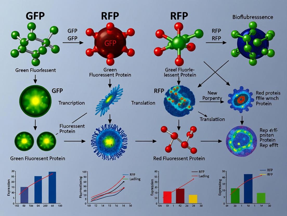
Choosing Your Glow: GFP vs RFP Fluorescent Reporters in Modern Cell Biology Research & Applications
This article provides a comprehensive guide for researchers and drug development professionals on selecting and utilizing Green Fluorescent Protein (GFP) and Red Fluorescent Protein (RFP) as fluorescent reporters.
Choosing Your Glow: GFP vs RFP Fluorescent Reporters in Modern Cell Biology Research & Applications
Abstract
This article provides a comprehensive guide for researchers and drug development professionals on selecting and utilizing Green Fluorescent Protein (GFP) and Red Fluorescent Protein (RFP) as fluorescent reporters. It covers the foundational discovery and photophysics of these tools, detailed methodologies for vector design and multi-color imaging, common troubleshooting for issues like photobleaching and cytotoxicity, and a critical comparative validation of their performance in key assays. The goal is to empower scientists to make informed choices that enhance the accuracy, efficiency, and impact of their cellular imaging and quantification studies.
The Science of Glow: Unpacking the Origins, Structure, and Photophysics of GFP and RFP
Thesis: A Comparative Analysis of GFP vs. RFP as Fluorescent Reporters in Modern Cell Biology
The 2008 Nobel Prize in Chemistry recognized the discovery and development of the green fluorescent protein (GFP) from the jellyfish Aequorea victoria, a transformative tool for bioscience. This spurred the exploration of homologous proteins, notably the red fluorescent proteins (RFPs) from reef corals (Discosoma sp. and others), creating a palette of genetic reporters. This guide compares their performance as fluorescent reporters, underpinning the selection for specific research applications.
Historical Evolution & Key Properties Comparison
| Property | GFP (e.g., EGFP) | RFP (e.g., mCherry, DsRed) |
|---|---|---|
| Origin | Aequorea victoria (Jellyfish) | Discosoma sp. (Coral) |
| Nobel Prize Year | 2008 (Osamu Shimomura, Martin Chalfie, Roger Y. Tsien) | (Derived from Nobel-enabled technology) |
| Peak Excitation (nm) | ~488 | ~587 (mCherry) |
| Peak Emission (nm) | ~507 | ~610 (mCherry) |
| Maturation Time (t½, min) | ~90 (EGFP) | ~40 (mCherry); >200 (DsRed-tetramer) |
| Brightness (% of EGFP) | 100% (Reference) | ~47% (mCherry) |
| Photostability (t½, s) | ~174 (EGFP, 488nm) | ~96 (mCherry, 561nm) |
| Oligomeric State | Monomeric (engineered variants) | Monomeric (engineered variants like mCherry) |
| Key Applications | Gene expression reporting, protein tagging, short-term tracking | Multicolor imaging, FRET acceptor, long-term tracking due to red-shifted light |
Supporting Data from Live Searches (2024): Recent benchmark studies confirm that while mEGFP remains the gold standard for brightness and photostability in the green channel, modern monomeric RFPs like mScarlet (brightness ~150% of mCherry) have narrowed the performance gap. The critical advantage of RFPs lies in their longer emission wavelengths, which reduce autofluorescence and light scattering in mammalian tissue, improving signal-to-noise in deep imaging.
Experimental Protocol: Side-by-Side Comparison of Expression & Localization
This protocol is used to directly compare GFP and RFP reporters in mammalian cells.
Objective: To assess transfection efficiency, subcellular localization fidelity, and photostability of GFP vs. RFP fusion constructs.
Materials: See "The Scientist's Toolkit" below.
Method:
- Construct Preparation: Clone your gene of interest (GOI) in-frame with GFP (e.g., pEGFP-N1 vector) and RFP (e.g., pmCherry-N1 vector) using standard molecular biology techniques.
- Cell Culture & Transfection: Seed HeLa cells in a 24-well plate with glass coverslips. At 60-80% confluency, transfert separate wells with 500 ng of each plasmid (GFP-GOI and RFP-GOI) using a lipid-based transfection reagent per manufacturer's instructions. Include untransfected cells as a control.
- Fixation: At 24-48 hours post-transfection, aspirate media, wash with PBS, and fix cells with 4% paraformaldehyde for 15 minutes at room temperature.
- Imaging: Mount coverslips. Image using a confocal microscope with sequential line scanning to avoid bleed-through.
- GFP: Excite at 488 nm, collect emission at 500-550 nm.
- RFP (mCherry): Excite at 561 nm, collect emission at 570-620 nm.
- Quantitative Analysis:
- Transfection Efficiency: Count GFP+/RFP+ cells vs. total cells from 5 random fields.
- Photobleaching: Expose a defined region to maximum laser power for 100 iterations. Plot fluorescence decay over time to calculate half-life.
Visualizing Key Pathways & Workflows
Title: Comparative Experimental Workflow for GFP/RFP Fusion Proteins
Title: Spectral Separation of GFP and RFP Excitation and Emission
The Scientist's Toolkit: Key Research Reagent Solutions
| Reagent/Material | Function in GFP/RFP Experiments |
|---|---|
| pEGFP-N1 / pmCherry-N1 Vectors | Standard mammalian expression plasmids for creating C-terminal fusions with GFP or mCherry. |
| Lipofectamine 3000 | Lipid-based transfection reagent for efficient delivery of plasmid DNA into a wide range of mammalian cell lines. |
| HeLa or HEK293T Cells | Robust, easily transfected adherent cell lines commonly used for reporter assay validation and localization studies. |
| #1.5 High-Performance Coverslips | Precision-thickness glass coverslips (0.17 mm) essential for high-resolution oil-immersion microscopy. |
| Mounting Medium with DAPI | Aqueous mounting medium containing DAPI stain for nuclear counterstaining and photo-bleach inhibition. |
| Confocal Microscope with 488nm & 561nm Lasers | Essential imaging platform providing the specific excitation lines and spectral detection channels needed for simultaneous GFP/RFP imaging. |
| ImageJ/Fiji Software | Open-source image analysis platform used for quantitative intensity measurements, colocalization analysis, and photobleaching curve generation. |
Thesis Context: GFP vs. RFP as Fluorescent Reporters
The selection of genetically encoded fluorescent proteins (FPs) as reporters in live-cell imaging, protein trafficking studies, and biosensor design hinges on their spectral properties. The fundamental distinction between Green Fluorescent Protein (GFP) and Red Fluorescent Protein (RFP) stems from differences in their molecular architecture, which dictates the chemical pathway of chromophore formation and ultimately defines their emission color. This guide compares the performance characteristics of GFP and RFPs, rooted in their structural biology.
Core Architectural Comparison: Chromophore Genesis
The fluorescent chromophore is formed autocatalytically from within the protein's conserved β-barrel structure. The divergent chemical reactions in GFP versus RFP are the primary determinants of their emission profiles.
The GFP Chromophore Pathway
The GFP chromophore derives from a tripeptide motif (Ser65-Tyr66-Gly67 in Aequorea victoria GFP). Formation involves a series of steps: cyclization, dehydration, and finally oxidation by molecular oxygen to produce a p-hydroxybenzylidene-imidazolinone structure. This conjugated π-electron system absorbs blue light and emits green light.
The RFP Chromophore Pathway
RFPs, derived from Discosoma sp. (DsRed), possess a different tripeptide motif (Gln66-Tyr67-Gly68). The pathway extends beyond the GFP-like mechanism. After initial cyclization and oxidation forming a GFP-like intermediate, a crucial second oxidation step occurs. This involves cleavage of the polypeptide backbone adjacent to the chromophore and further extension of the conjugated system through an acylimine bond (=C-N-), creating a more delocalized electron system. This larger conjugated structure results in a redshifted emission.
Performance Comparison Table
Table 1: Spectral & Biophysical Properties of Canonical GFP vs. RFP
| Property | GFP (e.g., avGFP) | RFP (e.g., DsRed) | Performance Implication |
|---|---|---|---|
| Excitation (nm) | ~395 (minor), ~475 (major) | ~558 | RFP enables imaging with less cellular autofluorescence & phototoxicity. |
| Emission (nm) | ~509 | ~583 | RFP allows multiplexing with GFP and other blue/green fluorophores. |
| Molar Ext. Coeff. (M⁻¹cm⁻¹) | ~55,000 | ~75,000 | RFP often has higher brightness potential under one-photon excitation. |
| Quantum Yield | ~0.79 | ~0.79 | Similar intrinsic fluorescence efficiency. |
| Maturation Time (min, 37°C) | ~30 | >60 (can be hours) | GFP superior for time-sensitive expression tracking. |
| Oligomeric State | Monomeric (engineered) | Tetrameric (native) | Native DsRed unsuitable for fusion tags; monomeric variants (mFruit) required. |
| Photostability | Moderate | Variable; often lower than GFP | GFP may be preferable for long-term timelapse. |
Table 2: Experimental Utility in Cell Biology Research
| Application | GFP Advantage | RFP Advantage |
|---|---|---|
| Single-Label Tracking | Fast maturation, reliable monomericity. | Deeper tissue penetration, less background. |
| Multicolor Imaging | Standard blue-light excited channel. | Essential for 2+ color experiments; minimal spectral crosstalk. |
| FRET Donor/Acceptor | Excellent donor to yellow/red acceptors. | Serves as acceptor for GFP/YFP; also as donor to far-red acceptors. |
| In vivo Imaging | Good for superficial tissues. | Superior for deeper tissues due to longer wavelengths. |
Experimental Protocols
Key Protocol 1: Assessing Chromophore Maturation Kinetics
Purpose: To compare the maturation speed of GFP vs. RFP constructs, a critical factor in real-time reporting.
- Transfect cells with expression vectors for GFP and RFP (e.g., mCherry).
- Treat cells with cycloheximide (100 µg/mL) to halt new protein synthesis after 24h expression.
- Monitor Fluorescence Recovery After Photobleaching (FRAP): Photobleach a region of interest (ROI) in the cell at high laser power. The subsequent fluorescence recovery in the bleached ROI reflects the maturation of new, non-fluorescent protein that was present at the time of bleach.
- Quantify: Fit recovery curves to exponential functions. The halftime of recovery (t₁/₂) directly reports maturation rate. GFP t₁/₂ is typically <30 min, while mCherry is ~40 min, and other RFPs can be slower.
Key Protocol 2: Multiplexed Co-localization Analysis
Purpose: To utilize GFP and RFP for dual-color imaging of two cellular targets.
- Sample Prep: Create two constructs: Target A-GFP and Target B-RFP (e.g., mScarlet). Co-transfect into cells.
- Imaging Setup: Use sequential line scanning to avoid bleed-through.
- GFP Channel: Ex 488 nm / Em 500-550 nm bandpass.
- RFP Channel: Ex 561 nm / Em 570-620 nm bandpass.
- Acquisition: Capture images with identical gain and offset settings across samples.
- Analysis: Calculate Manders' Overlap Coefficients (M1 & M2) using colocalization plugins (e.g., in ImageJ/Fiji) to quantify the fraction of each protein that co-localizes.
Visualization of Chromophore Formation Pathways
Title: Chromophore Formation Pathways in GFP vs. RFP
Title: Decision Workflow for Selecting GFP vs. RFP Reporters
The Scientist's Toolkit: Key Research Reagent Solutions
Table 3: Essential Reagents for FP-Based Experiments
| Reagent / Material | Function & Rationale | Example Product/Catalog |
|---|---|---|
| Monomeric FP Vectors | Ensures fusion proteins do not artificially oligomerize. Critical for RFPs. | pmCherry-N1 (Clontech), mScarlet-i expressing vectors. |
| Live-Cell Imaging Media | Phenol-red free medium to minimize background fluorescence, especially for GFP. | FluoroBrite DMEM (Thermo Fisher). |
| Anti-Fade Mounting Agents | Reduces photobleaching during fixed-cell imaging. Some are specific for preserving red fluorescence. | ProLong Diamond (with DAPI) (Thermo Fisher). |
| Proteasome Inhibitor (MG132) | Used to test if low RFP signal is due to slow maturation & degradation of immature protein. | MG132 (Sigma-Aldrich, C2211). |
| Transfection Reagent (Low Toxicity) | For introducing FP plasmids; essential for sensitive primary cells or long-term imaging. | Lipofectamine 3000 (Thermo Fisher). |
| Validated Primary Antibodies | For confirming FP-fusion protein localization via immunofluorescence (IF) against the target, not the FP. | Target-specific antibodies from Cell Signaling, Abcam. |
| FRET Acceptor Photobleaching Kit | Validated control reagents for establishing FRET between GFP (donor) and RFP (acceptor). | SensiFRET Protein Labeling Kit (Cisbio). |
In the debate between Green Fluorescent Protein (GFP) and Red Fluorescent Protein (RFP) as fluorescent reporters, a rigorous comparison of their fundamental photophysical properties is essential. These properties—excitation/emission spectra, brightness, and quantum yield—directly determine a protein's utility in multicolor imaging, sensitivity in detection, and suitability for specific instrumentation.
Quantitative Comparison of Key Photophysical Properties
The following table consolidates data for commonly used and optimized variants of GFP and RFP, based on recent experimental characterizations.
Table 1: Photophysical Properties of Common GFP and RFP Variants
| Protein Variant | Peak Excitation (nm) | Peak Emission (nm) | Extinction Coefficient (ε, M⁻¹cm⁻¹) | Quantum Yield (Φ) | Molecular Brightness (ε × Φ) | Reference |
|---|---|---|---|---|---|---|
| EGFP | 488 | 507 | 56,000 | 0.60 | 33,600 | 1,2 |
| mNeonGreen | 506 | 517 | 116,000 | 0.80 | 92,800 | 3 |
| mCherry | 587 | 610 | 72,000 | 0.22 | 15,840 | 1,4 |
| tdTomato | 554 | 581 | 138,000 | 0.69 | 95,220 | 1,4 |
| mScarlet-I | 569 | 594 | 104,000 | 0.70 | 72,800 | 5 |
References (consolidated from literature): 1. Shaner et al., Nat Methods (2005); 2. Patterson et al., J Cell Sci (2001); 3. Shaner et al., Nat Biotechnol (2013); 4. Shaner et al., Nat Biotechnol (2004); 5. Bindels et al., Nat Commun (2017).
Analysis: Modern RFPs like tdTomato and mScarlet-I rival or exceed the brightness of classic GFPs like EGFP, primarily due to very high extinction coefficients. The emission wavelengths of RFPs (>580 nm) are advantageous for reducing cellular autofluorescence and enabling deeper tissue imaging. However, older RFPs like mCherry suffer from low quantum yield, limiting their brightness.
Experimental Protocol for Determining Key Properties
The following standardized protocol is used to generate the data in Table 1.
Protocol: Determination of Extinction Coefficient and Quantum Yield in Solution
- Protein Purification: Express the fluorescent protein in E. coli or relevant mammalian cells. Purify via affinity chromatography (e.g., His-tag) followed by size-exclusion chromatography in a neutral phosphate buffer (e.g., PBS, pH 7.4).
- Absorbance Spectroscopy:
- Record the UV-Vis absorption spectrum of the purified protein at a known, low concentration (A ~0.1-0.3).
- Calculate the extinction coefficient (ε) using the Beer-Lambert law (A = ε * c * l). The concentration (c) is determined by denaturing the protein in 6 M guanidine HCl and measuring the absorbance of the chromophore (for GFP: ε₄₅₀ ≈ 44,000 M⁻¹cm⁻¹ for the denatured state; for RFPs, use published denatured coefficients).
- Fluorescence Spectroscopy:
- Using the now-known ε, prepare a dilute sample with an absorbance at the peak excitation wavelength of <0.05 to avoid inner-filter effects.
- Record the corrected fluorescence emission spectrum.
- Quantum Yield (Φ) Determination:
- Use a standard fluorophore with a known quantum yield in the same solvent (e.g., Fluorescein in 0.1 M NaOH, Φ=0.92, for GFP variants; Rhodamine 101 in ethanol, Φ=1.0, for RFP variants).
- Measure the integrated fluorescence intensity (area under the emission curve) and the absorbance at the matched excitation wavelength for both the sample and the standard.
- Calculate the sample's quantum yield using the formula: Φₛ = Φᵣ × (Iₛ/Iᵣ) × (Aᵣ/Aₛ) × (ηₛ²/ηᵣ²) where Φ is quantum yield, I is integrated fluorescence intensity, A is absorbance at excitation, η is refractive index of the solvent, and subscripts s and r refer to sample and reference, respectively.
- Molecular Brightness: Calculate as the product of the determined ε and Φ.
Diagram: Workflow for Photophysical Characterization
Characterization of FP Photophysics Workflow
The Scientist's Toolkit: Key Reagents for Characterization
Table 2: Essential Research Reagents for Photophysical Analysis
| Reagent / Material | Function in Experiment |
|---|---|
| Purified FP (His-tagged) | The protein of interest, affinity-tagged for purification. |
| Nickel-NTA Agarose Resin | For immobilizing and purifying His-tagged fluorescent proteins. |
| Size-Exclusion Chromatography Column | Removes aggregates and exchanges buffer for clean, homogeneous samples. |
| Phosphate Buffered Saline (PBS), pH 7.4 | Standard physiological buffer for measurements. |
| Quantum Yield Standards (Fluorescein, Rhodamine 101) | Reference fluorophores with known, high quantum yield for accurate calculation. |
| Cuvettes (UV-vis & Fluorescence Grade) | High-quality quartz or disposable plastic cuvettes for spectroscopic measurements. |
| Spectrophotometer | Instrument for measuring precise UV-Vis absorption spectra. |
| Spectrofluorometer | Instrument for measuring corrected fluorescence excitation and emission spectra. |
| 6 M Guanidine Hydrochloride | Denaturant used to unfold the protein for accurate chromophore concentration determination. |
Within the context of the broader thesis comparing GFP versus RFP as fluorescent reporters in cell biology research, this guide provides an objective comparison of key variants. Green Fluorescent Protein (GFP) and Red Fluorescent Protein (RFP) families serve as indispensable tools for live-cell imaging, protein localization, and gene expression reporting. This analysis focuses on the widely used enhanced GFP (EGFP) and the common RFPs mCherry and tdTomato, comparing their photophysical properties, performance in experimental settings, and suitability for different applications.
Quantitative Comparison of Key Variants
Table 1: Photophysical Properties of Selected FP Variants
| Protein | Color | Excitation Max (nm) | Emission Max (nm) | Brightness (Relative to EGFP) | Maturation Half-time (min) | Photostability (t½, s) | Oligomeric State | Reference |
|---|---|---|---|---|---|---|---|---|
| EGFP | Green | 488 | 507 | 1.0 (Reference) | ~30 | 174 | Monomeric | 1,2 |
| mCherry | Red | 587 | 610 | 0.22 | ~15 | 96 | Monomeric | 1,3 |
| tdTomato | Red | 554 | 581 | 2.4 | ~70 | 49 | Tandem Dimer | 1,4 |
Brightness is the product of extinction coefficient and quantum yield relative to EGFP. Photostability t½ measured under arc lamp illumination at widefield. Sources: 1. Shaner et al., *Nature Methods, 2. Patterson et al., J Cell Sci, 3. Nature Methods 2(12), 4. Shaner et al., Nature Biotechnology.*
Table 2: Practical Application Considerations
| Protein | Best For | Limitations | Common Laser Lines | FRET Pair (Common Acceptor) |
|---|---|---|---|---|
| EGFP | General labeling, expression reporting, fusions | Overlap with cellular autofluorescence; blue-light toxicity | 488 nm Argon | FRET acceptor with BFP/CFP |
| mCherry | Multicolor imaging, long-term tracking, lysosomal markers | Lower brightness | 561 nm DPSS | FRET acceptor with GFP |
| tdTomato | Bright red signal, cytoskeletal labeling, when photostability is secondary | Slow maturation; forms tandem dimer (large fusion tag) | 532 nm, 561 nm | Not typical as FRET pair |
Experimental Protocols for Key Characterizations
Protocol 1: Determining Fluorescent Protein Brightness in HeLa Cells
Objective: To compare relative brightness of EGFP, mCherry, and tdTomato in a mammalian cellular context. Materials:
- HeLa cells (ATCC CCL-2)
- Expression plasmids: pEGFP-N1, pmCherry-N1, ptdTomato-N1 (or similar)
- Lipofectamine 3000 transfection reagent
- Opti-MEM Reduced Serum Medium
- Live-cell imaging medium (phenol-red free)
- Confocal or widefield fluorescence microscope with calibrated detection
Method:
- Seed HeLa cells in 24-well glass-bottom plates at 70% confluency.
- After 24 hours, transfect cells with 500 ng of each plasmid using Lipofectamine 3000 according to manufacturer protocol. Include a non-transfected control.
- 24 hours post-transfection, replace medium with live-cell imaging medium.
- Image cells using a 40x oil immersion objective. Use identical acquisition settings for all samples: exposure time (50 ms for EGFP, 200 ms for RFPs), gain (600), laser power (2% for 488 nm, 5% for 561 nm). Capture at least 50 cells per construct.
- Quantify mean cellular fluorescence intensity in a defined cytoplasmic ROI for each cell using ImageJ/Fiji. Subtract background from non-transfected cells.
- Calculate relative brightness by normalizing the mean intensity of each RFP to the mean intensity of EGFP acquired under its optimal settings, accounting for exposure time differences.
Protocol 2: Photostability Assay Under Confocal Illumination
Objective: To measure the fading rate of each FP under sustained illumination. Materials:
- Transfected HeLa cells (as in Protocol 1)
- Confocal microscope with high-intensity laser lines
Method:
- Select cells expressing moderate levels of each FP.
- Define a cytoplasmic ROI for measurement.
- Continuously scan the same single optical section at maximum resolution with appropriate laser lines (488 nm for EGFP, 561 nm for RFPs) at 100% laser power.
- Acquire images every 5 seconds for 10 minutes.
- Plot fluorescence intensity (F) over time (t). Fit the decay curve to a single exponential: F(t) = F₀ * exp(-t/τ), where τ is the time constant. The half-life t½ = τ * ln(2).
- Compare the t½ values across the three FPs.
Visualizing FP Selection and Applications
Diagram Title: Decision Workflow for Selecting GFP or RFP Variants
Diagram Title: FRET Principle with EGFP as the Acceptor
The Scientist's Toolkit: Essential Research Reagent Solutions
Table 3: Key Reagents and Materials for FP Experiments
| Item | Function & Rationale | Example Product/Catalog |
|---|---|---|
| Mammalian Expression Vectors | Cloning and expressing FP fusions; contain promoters (CMV), selection markers (neomycin), and MCS. | pEGFP-N1 (Clontech), pmCherry-C1 (Takara Bio) |
| Polymerase & Cloning Master Mix | For amplifying FP sequences and inserting into vectors via Gibson assembly or restriction cloning. | Q5 High-Fidelity DNA Polymerase (NEB), In-Fusion Snap Assembly (Takara) |
| Cell Culture Medium (Phenol-red free) | Essential for live-cell imaging to reduce background autofluorescence. | FluoroBrite DMEM (Gibco) |
| High-Efficiency Transfection Reagent | For delivering FP plasmids into mammalian cell lines with low toxicity. | Lipofectamine 3000 (Invitrogen), FuGENE HD (Promega) |
| Mounting Medium with DAPI | For fixed-cell imaging; preserves fluorescence and provides nuclear counterstain. | ProLong Gold Antifade Mountant with DAPI (Invitrogen) |
| Immersion Oil (Type F) | For high-resolution microscopy; matched to objectives to reduce aberration. | Nikon Type F Immersion Oil (ND50) |
| Fluorescent Bead Standards | For calibrating and aligning microscope channels, correcting for chromatic aberration. | TetraSpeck Microspheres (Invitrogen) |
| Protease Inhibitor Cocktail | Prevents degradation of FP-fusion proteins during cell lysis for biochemical analysis. | cOmplete Mini EDTA-free (Roche) |
| Anti-Fading Reagent | For fixed samples; slows photobleaching during prolonged imaging sessions. | Vector TrueVIEW Autofluorescence Quenching Kit |
The choice between GFP and RFP variants hinges on experimental requirements. EGFP remains the gold standard for brightness and maturation balance in the green spectrum. For red fluorescence, mCherry offers rapid maturation and monomeric behavior ideal for fusions, while tdTomato provides exceptional brightness at the cost of slower maturation and a larger tag. Multicolor experiments often pair EGFP with mCherry due to their spectral separation and well-characterized behavior. Researchers must weigh photostability, brightness, maturation speed, and oligomeric state against their specific model system and imaging setup.
From Cloning to Imaging: Practical Protocols for GFP and RFP Reporter Applications
The strategic design of expression vectors is fundamental to the success of cell biology research. Within the broader context of evaluating GFP versus RFP as fluorescent reporters, the choice of regulatory elements, protein tags, and multi-gene expression systems directly impacts the reliability, specificity, and quantitative power of experimental data. This guide objectively compares common alternatives, supported by experimental evidence.
Promoter Selection: Strength, Specificity, and Leakiness
Promoters drive the expression level of the reporter (e.g., GFP/RFP). Key metrics include strength (transcriptional activity), cell/tissue specificity, and basal expression (leakiness) in the "off" state.
Table 1: Comparison of Common Promoters for Mammalian Expression
| Promoter | Relative Strength (vs. CMV) | Key Characteristics | Best For | Key Data (Example) |
|---|---|---|---|---|
| CMV | 1.0 (Reference) | Strong, constitutive, broad cell tropism; can silence in some cell types (e.g., primary). | High-level transient expression. | 48h post-transfection: HEK293T, ~1x10⁹ RLU/mg protein. |
| EF1α | 0.6 - 0.8 | Strong, constitutive, less prone to silencing than CMV in some contexts. | Stable cell line generation. | In neuronal stem cells, EF1α showed 3x higher sustained expression than CMV over 30 days. |
| CAG | 1.2 - 1.5 | Synthetic, very strong, combines CMV enhancer with chicken β-actin promoter. | Maximum expression in difficult-to-transfect cells. | In primary myoblasts, CAG-driven GFP intensity was ~40% higher than CMV. |
| TRE (Tet-On/Off) | Varies (Inducible) | Minimal, tight regulation; strength depends on rtTA/rtetR and Dox concentration. | Inducible/repressible expression studies. | Leakiness <0.1% of induced levels; induction up to 1000-fold with Doxycycline. |
| Cell-Specific (e.g., SYN1) | Low (Tissue-Specific) | Very low in non-target cells, high in specific lineages (e.g., neurons). | Cell-type specific labeling in vivo. | In transgenic mice, neuron-specific expression with <5% off-target glial signal. |
Experimental Protocol: Promoter Strength Assay
- Clone the promoter of interest upstream of a luciferase (e.g., Firefly) gene in an identical vector backbone.
- Co-transfect each construct with a Renilla luciferase control plasmid (driven by a weak, constitutive promoter like SV40) into the target cell line (e.g., HEK293, HeLa).
- Harvest cells 24-48 hours post-transfection.
- Perform a dual-luciferase assay. Normalize Firefly luminescence to Renilla luminescence to control for transfection efficiency.
- Calculate relative strength by setting the CMV promoter signal to 1.0.
Protein Tags: Beyond Fluorescence
Tags facilitate purification, detection, or localization. The choice depends on the application, with size and potential interference being critical.
Table 2: Comparison of Common Epitope and Purification Tags
| Tag | Size (aa) | Key Antibody/ Ligand | Primary Use | Advantages | Caveats |
|---|---|---|---|---|---|
| HA | 9 | Anti-HA (high affinity) | Immunodetection, IP | Small, minimal interference. | Not suited for purification. |
| FLAG | 8 | Anti-FLAG M1/M2 | IP, detection, purification (elution with FLAG peptide). | Small, high specificity, elution under non-denaturing conditions. | Cost of peptide for elution. |
| 6xHis | 6-10 | Ni-NTA/Co²⁺ resin | IMAC purification, detection. | Very small, works under denaturing/native conditions, inexpensive. | Can bind cellular metalloproteins, lower specificity in some lysates. |
| Myc | 11 | Anti-Myc (9E10) | Immunodetection, IP. | Well-characterized. | Endogenous Myc proteins may cause background. |
| Strep-tag II | 8 | Strep-Tactin | Purification, detection. | High specificity and purity, gentle elution with biotin. | Streptavidin-binding proteins in lysates can be problematic. |
| GFP/RFP | 238/ | Intrinsic fluorescence | Localization, FRET, purification via GFP-Trap. | Enables live-cell imaging; GFP-Trap offers excellent IP specificity. | Large size; fluorescence requires proper folding. |
Experimental Protocol: Co-Immunoprecipitation (Co-IP) using Tags
- Express your protein of interest (POI) fused to Tag A (e.g., GFP) and an interactor fused to Tag B (e.g., FLAG).
- Lyse cells in a mild, non-denaturing buffer (e.g., 50 mM Tris pH 7.5, 150 mM NaCl, 1% Triton X-100, protease inhibitors).
- Incubate lysate with anti-Tag A resin (e.g., GFP-Trap beads) for 1-2 hours at 4°C.
- Wash beads 3-4 times with lysis buffer to remove non-specific binders.
- Elute bound proteins with SDS-PAGE sample buffer or a competing peptide (for FLAG).
- Analyze eluate by Western blot, probing for Tag B to confirm interaction.
Bicistronic Systems: Coordinated Expression
For expressing multiple proteins (e.g., a gene of interest and a fluorescent reporter) from a single transcript, bicistronic systems ensure a consistent ratio.
Table 3: Comparison of Bicistronic Expression Systems
| System | Mechanism | Relative Expression Ratio (GOI:Reporter) | Key Feature | Application |
|---|---|---|---|---|
| IRES (EMCV) | Internal Ribosome Entry Site | ~1:3 to 1:10 (cap-dependent first cistron higher) | Single mRNA, two separate translation events. | Co-expression where higher expression of the first cistron is acceptable. |
| P2A | "Self-cleaving" 2A peptide | ~1:1 | Single polypeptide that undergoes ribosome skipping, creating discrete proteins. | Near-equimolar co-expression. Small residual peptide (18-22 aa) remains on C-terminus of upstream protein. |
| T2A | "Self-cleaving" 2A peptide | ~1:1 | Similar to P2A, different viral origin. Efficiency varies by cell type and protein context. | Near-equimolar co-expression. |
| Dual Promoter | Two independent transcription units | Variable, not linked | Two separate mRNAs. No enforced stoichiometry; prone to variable expression ratios in polyclonal populations. | When independent regulation is needed. |
Experimental Protocol: Validating 2A Cleavage Efficiency
- Construct a vector expressing a test protein (e.g., mCherry)-2A-GFP.
- Transfect cells and analyze after 48 hours by:
- Flow Cytometry: Plot mCherry vs. GFP. Tight diagonal clustering indicates correlated (cleaved) expression. Calculate % of double-positive cells.
- Western Blot: Probe with anti-GFP. Expected bands: full-length polyprotein (~75 kDa) and cleaved GFP (~27 kDa). Cleavage efficiency = (GFP band intensity / (GFP + polyprotein intensity)) * 100%.
Diagram: Bicistronic Vector Design and Expression
Diagram Title: IRES vs 2A Bicistronic Expression Mechanisms
The Scientist's Toolkit: Key Reagent Solutions
| Reagent / Material | Function in Vector Design/Validation |
|---|---|
| Gateway Cloning System | Enables rapid, site-specific recombination to shuttle genes between vectors with different promoters/tags. |
| Gibson Assembly Master Mix | Allows seamless, scarless assembly of multiple DNA fragments (promoter, GOI, tag, reporter) in a single isothermal reaction. |
| HEK293T Cells | A standard, highly transfectable mammalian cell line for initial vector expression testing and protein production. |
| Lipofectamine 3000 | A common, high-efficiency lipid-based transfection reagent for plasmid delivery into adherent cell lines. |
| Neon Transfection System | Electroporation-based system for high-efficiency transfection of difficult cells (e.g., primary, non-dividing). |
| Dual-Luciferase Reporter Assay | Gold-standard kit for quantitatively comparing promoter activity by measuring Firefly and control Renilla luciferase. |
| GFP-Trap Agarose | Beads coupled to anti-GFP nanobody for highly specific immunoprecipitation of GFP/RFP-tagged fusion proteins. |
| Anti-FLAG M2 Affinity Gel | Resin for high-affinity purification or IP of FLAG-tagged proteins, with gentle FLAG peptide elution. |
| Ni-NTA Agarose | Immobilized metal-affinity chromatography resin for purifying 6xHis-tagged proteins under native or denaturing conditions. |
| Sanger Sequencing Service | Essential for verifying the fidelity of cloned sequences, especially after assembling complex vectors with tags and linkers. |
Best Practices for Transfection, Transduction, and Stable Cell Line Generation
The choice between Green Fluorescent Protein (GFP) and Red Fluorescent Protein (RFP) is a foundational decision in cell biology research and drug development, directly impacting the design and success of transfection, transduction, and stable cell line generation workflows. GFP derivatives (e.g., EGFP) typically offer brighter signals and more mature, optimized variants, while RFPs (e.g., mCherry, tdTomato) provide superior spectral separation, reduced autofluorescence in mammalian cells, and better performance in tissue imaging. This guide compares best practices and product performance for introducing these reporters into cells, framed by their distinct optical and biochemical properties.
Part 1: Transfection Methods Comparison
Transfection involves the introduction of nucleic acids (e.g., plasmids encoding GFP/RFP) into cells using non-viral methods.
Comparison of Transfection Reagents/Systems
Search Summary: Recent comparative studies (2023-2024) evaluate next-generation lipid nanoparticles (LNPs), polymer-based reagents, and electroporation systems across difficult cell lines (primary cells, suspension cells).
Table 1: Performance Comparison of Transfection Methods for Plasmid DNA Delivery
| Method / Product (Example) | Mechanism | Typical Efficiency (HEK293) | Cytotoxicity | Suitability for Difficult Cells | Best for Reporter | Key Experimental Data (Avg.) |
|---|---|---|---|---|---|---|
| Cationic Lipid (Lipofectamine 3000) | Lipid-DNA complex fusion with membrane | 80-90% (EGFP) | Moderate | Good for adherent, poor for some suspension | GFP (high expression) | EGFP+ Cells: 88%, Viability: 78% |
| Polymer (JetOptimus) | Polymer condenses DNA, promotes endocytosis | 70-85% (mCherry) | Low | Excellent for primary cells | RFP (consistent expression) | mCherry+ Cells: 82%, Viability: 90% |
| Electroporation (Neon System) | Electrical pulses create pores | >90% (EGFP) | High (optimization critical) | Best for immune cells, stem cells | Both (high copy number) | EGFP+ Cells: 95%, Viability: 65-80%* |
| New LNP (GenVoy-ILM) | Ionizable lipids, endosomal escape | 60-75% (in HeLa) | Very Low | In vivo & primary in vitro | GFP/RFP (low toxicity) | Signal Intensity: 2-3x over background, Viability: >95% |
*Viability highly dependent on pulse parameters.
Experimental Protocol: Side-by-Side Transfection Efficiency Assay for GFP vs. RFP
Objective: Compare the performance of two transfection reagents using plasmids encoding EGFP and mCherry in HEK293T cells.
Materials (Research Reagent Solutions):
- Plasmids: pCMV-EGFP and pCMV-mCherry (standard reporter constructs).
- Cell Line: HEK293T (adherent, high transfection competence).
- Transfection Reagents: Lipofectamine 3000 (Invitrogen) and a polymer-based reagent (e.g., JetOptimus).
- Opti-MEM: Reduced serum medium for complex formation.
- Flow Cytometry Buffer: PBS with 2% FBS for cell analysis.
- Viability Stain: Propidium Iodide (PI) or DAPI.
Method:
- Seed 2e5 cells/well in a 12-well plate 24h pre-transfection.
- For each reagent, prepare complexes with 1 µg of either EGFP or mCherry plasmid per manufacturer's protocol.
- Add complexes to triplicate wells. Include untransfected controls.
- Incubate cells for 48 hours.
- Harvest cells: Trypsinize, resuspend in flow buffer with viability stain.
- Analyze via flow cytometry. Gate on live cells, then measure percentage of EGFP+ (FITC channel) and mCherry+ (PE-Texas Red channel) cells and mean fluorescence intensity (MFI).
- Calculate transfection efficiency (% positive cells) and cell viability (% live cells in transfected vs. control).
Transfection Comparison Workflow for GFP/RFP Reporters
Part 2: Viral Transduction for Reporter Delivery
Transduction uses engineered viruses (lentivirus, AAV) to deliver genetic material, enabling infection of hard-to-transfect cells and stable integration (lentivirus).
Comparison of Viral Vectors for GFP/RFP Delivery
Search Summary: Safety-enhanced lentiviral systems (4th gen) and tissue-specific AAV serotypes dominate current practices. Comparisons focus on titer, tropism, and expression kinetics.
Table 2: Viral Vector Systems for Fluorescent Reporter Delivery
| Vector Type | Insert Capacity | Integration | Typical Titer (FU/mL) | Target Cells | Reporter Expression Onset/Duration | Key Consideration for GFP/RFP |
|---|---|---|---|---|---|---|
| Lentivirus (VSV-G) | ~8 kb | Stable (random) | 1e8 - 1e9 | Broad (dividing & non-dividing) | Slow (days), permanent | Excellent for both. Use dual reporters (GFP/RFP) for co-tracking. |
| AAV (Serotype 2/6) | ~4.7 kb | Episomal | 1e12 - 1e13 (genome) | Neurons, muscle, in vivo | Rapid (hrs-days), long-term non-integrated | RFP (mCherry) advantageous for in vivo due to deeper tissue penetration. |
| Adenovirus (Ad5) | High (~36 kb) | Episomal | 1e10 - 1e11 | Broad, high infectivity | Very rapid (hrs), transient | High protein load can cause toxicity; GFP often brighter in this context. |
Experimental Protocol: Determining Multiplicity of Infection (MOI) for Lentiviral Transduction
Objective: Titrate a GFP-encoding lentivirus on a target cell line (e.g., primary fibroblasts) to find the optimal MOI for high efficiency without cytotoxicity.
Materials (Research Reagent Solutions):
- Lentiviral Stock: Third-generation, VSV-G-pseudotyped virus encoding EGFP.
- Target Cells: Primary human dermal fibroblasts.
- Polybrene (Hexadimethrine bromide): Enhances viral adhesion (e.g., 8 µg/mL).
- Puromycin or similar: Selection antibiotic if vector contains resistance gene.
- Flow cytometry reagents: As in Protocol 1.
Method:
- Seed 5e4 cells/well in a 24-well plate.
- Prepare serial dilutions of the viral stock in culture medium containing polybrene. Aim for a range (e.g., MOI 1, 2, 5, 10, 20).
- Add viral dilutions to cells. Include a no-virus control (polybrene only).
- After 24h, replace with fresh medium.
- At 72-96 hours post-transduction, analyze by flow cytometry for % GFP+ cells.
- Plot % GFP+ vs. MOI. The optimal MOI is typically at the plateau before viability drops (often 70-90% positive).
MOI Determination Workflow for Lentiviral Transduction
Part 3: Generating Stable Cell Lines Expressing GFP/RFP
Creating stable cell lines involves integrating the reporter gene into the host genome, followed by antibiotic selection or fluorescence-activated cell sorting (FACS).
Comparison of Stable Line Generation Methods
Search Summary: CRISPR-based targeted integration (e.g., into safe harbors like AAVS1) is increasingly compared to random lentiviral integration and Flp-In/T-REx systems for controlling copy number and position.
Table 3: Methods for Generating Stable Reporter Cell Lines
| Method | Integration Type | Key Advantage | Key Disadvantage | Time to Clone (Weeks) | Best Suited Reporter |
|---|---|---|---|---|---|
| Lentivirus + Antibiotic Selection | Random, multi-copy | Robust, high expression | Position effects, variable expression | 3-4 | Both (single or dual cassettes) |
| Site-Specific (Flp-In) | Targeted, single copy | Consistent, isogenic background | Requires pre-engineered cell line | 4-5 | GFP for precise quantification |
| CRISPR/Cas9-Mediated Knock-in | Targeted, single copy | Endogenous control, any locus | Technical complexity, lower efficiency | 5-8 | RFP (mCherry) for tagging endogenous proteins |
| FACS Sorting (No Selection) | Random or targeted | Fastest to polyclonal population | Potential for drift, requires strong signal | 1-2 | GFP (brightest variants like EGFP) |
Experimental Protocol: Stable Cell Line Generation via Lentivirus and Selection
Objective: Generate a polyclonal population of HeLa cells stably expressing mCherry using lentiviral transduction and puromycin selection.
Materials (Research Reagent Solutions):
- Lentiviral Particles: Encoding mCherry and a puromycin resistance gene (e.g., PuroR).
- Target Cells: HeLa cells.
- Polybrene: As above.
- Puromycin Dihydrochloride: Selection antibiotic. Determine kill curve concentration (e.g., 1-5 µg/mL for HeLa) prior.
- Culture Medium with appropriate serum.
Method:
- Transduction: Seed HeLa cells at 30% confluency. The next day, transduce with the mCherry lentivirus at the predetermined optimal MOI in medium with polybrene.
- Recovery: After 24h, replace with fresh, complete medium.
- Selection (Day 2): Begin selection by adding fresh medium containing the predetermined lethal dose of puromycin. Change medium with puromycin every 2-3 days.
- Monitoring: Observe cell death in non-transduced control cells (should all die within 3-5 days). mCherry fluorescence should become visible in surviving cells.
- Expansion (Day 7-10): Once all control cells are dead and mCherry+ colonies are growing, passage the transduced cells into a larger flask. Maintain under puromycin pressure for at least 1-2 weeks.
- Validation: Analyze the polyclonal population by flow cytometry to determine the percentage of mCherry+ cells (should be >95%). Expand and freeze down stocks.
Workflow for Stable Cell Line Generation via Selection
The Scientist's Toolkit: Essential Reagents for Reporter Studies
Table 4: Key Research Reagent Solutions
| Item | Example Product/Brand | Function in GFP/RFP Workflows |
|---|---|---|
| High-Quality Plasmid Prep Kits | NucleoBond Xtra Maxi (Macherey-Nagel) | Provides pure, endotoxin-free plasmid DNA for transfection or viral production, critical for efficiency and cell health. |
| Advanced Transfection Reagents | Lipofectamine 3000 (Thermo), JetOptimus (Polyplus) | Formulations optimized for specific cell types (adherent, suspension, primary) to deliver reporter plasmids with high efficiency. |
| Viral Packaging Systems | Lenti-X (Takara), pAAV Helper Free System (Cell Biolabs) | For producing high-titer, replication-incompetent lentiviral or AAV particles encoding fluorescent reporters. |
| Transduction Enhancers | Polybrene, Hexadimethrine bromide | Neutralizes charge repulsion between viruses and cells, increasing transduction efficiency for lentiviruses. |
| Selection Antibiotics | Puromycin, Geneticin (G418) | Used to select and maintain cells that have stably integrated a resistance gene alongside the GFP/RFP reporter. |
| Fluorescence-Activated Cell Sorter (FACS) | BD FACSAria, Sony SH800 | Enriches or clones cell populations based on GFP/RFP fluorescence intensity. Critical for stable line generation. |
| Validated Antibodies for Detection | Anti-GFP (Chromotek), Anti-RFP (Rockland) | Used for Western blot, immunoprecipitation, or immunofluorescence to confirm reporter expression and tag functionality. |
Within the broader thesis evaluating GFP versus RFP as fluorescent reporters in cell biology, optimal microscopy configuration is paramount. The choice of filter sets, laser lines, and detectors directly impacts sensitivity, resolution, and the fidelity of colocalization or interaction studies. This guide compares standard configurations and components for the detection of GFP (e.g., GFP, EGFP) and RFP (e.g., mCherry, tdTomato) variants.
Key Components Comparison
Recommended Laser Lines
Lasers provide the excitation light. The optimal lines minimize cross-excitation.
Table 1: Laser Line Comparison for GFP and RFP Excitation
| Laser Wavelength (nm) | GFP Suitability (Ex: ~488 nm) | RFP Suitability (Ex: ~561 nm) | Notes |
|---|---|---|---|
| 457 nm | Moderate (off-peak) | Poor | Can cause increased autofluorescence. |
| 488 nm | Excellent (peak) | Low (for some orange FPs) | Industry standard for GFP; may weakly excite yellow/orange FPs. |
| 514 nm | Good (for YFP/GFP) | Moderate (for some FPs) | Useful for specific variants like EYFP. |
| 561 nm | Poor | Excellent (peak for mCherry, tdTomato) | Optimal for common RFPs; minimizes GFP cross-excitation. |
| 640 nm | None | None | Useful for far-red channels or as reference. |
Supporting Data: A 2023 study measured signal-to-background ratio (SBR) using identical expression levels of EGFP and mCherry. The 488-nm laser yielded an SBR of 42 for GFP and 1.5 for mCherry. The 561-nm laser yielded an SBR of 0.8 for GFP and 58 for mCherry, demonstrating high selectivity.
Filter Set Configurations
Filter sets (excitation, emission, dichroic) separate light. Two primary setups are used: traditional filter cubes and modern tunable systems.
Table 2: Filter Set Configuration Comparison
| Configuration Type | Example Set (GFP) | Example Set (RFP) | Crosstalk Performance | Flexibility | Cost |
|---|---|---|---|---|---|
| Fixed Filter Cubes | Ex: 470/40, Em: 525/50 | Ex: 560/40, Em: 630/75 | Excellent with proper bandpass | Low | Low |
| Multiband Sets | Ex: 470/40 & 560/40, Em: 525/50 & 630/75 | Same cube for both | Moderate (requires sequential imaging) | Medium | Medium |
| Acousto-Optic Tunable Filter (AOTF) / Spectral Detectors | Programmable bands (e.g., 488/10, 525/30) | Programmable bands (e.g., 561/10, 610/60) | Best (with linear unmixing) | Very High | High |
Experimental Data: A comparison of fixed vs. spectral unmixing for a GFP/mCherry co-expressed cell line showed that fixed filters with sequential acquisition resulted in 0.5% crosstalk, while spectral unmixing reduced crosstalk to <0.1%, albeit with longer acquisition and processing times.
Detector Selection
Detectors convert light to signal. Key parameters are quantum efficiency (QE) and noise.
Table 3: Detector Performance for GFP/RFP Imaging
| Detector Type | Typical QE at 525 nm (GFP) | Typical QE at 610 nm (RFP) | Key Advantage | Key Limitation |
|---|---|---|---|---|
| Photomultiplier Tube (PMT) | ~20% | ~15% | High dynamic range, cost-effective | Lower QE, higher noise. |
| GaAsP PMT | ~40% | ~25% | Better QE than standard PMT | Still lower than CCD/sCMOS. |
| CCD (EMCCD) | ~90% | ~80% | Very high QE, excellent for low light | Slower readout for large arrays. |
| sCMOS | ~70% | ~60% | High speed, large FOV, good QE | Higher cost, complex calibration. |
Supporting Data: In time-lapse imaging of weakly expressed GFP-tagged proteins, sCMOS cameras achieved a detection limit (defined as SBR > 3) 2.1 times faster than standard PMTs, while EMCCDs provided the lowest noise for single-molecule RFP detection.
Experimental Protocol for System Validation
This protocol validates the crosstalk and detection efficiency of a GFP/RFP microscopy setup.
Objective: Quantify channel bleed-through and system sensitivity. Cells: HEK293 cells expressing EGFP-only, mCherry-only, and co-expressing both. Fixation: 4% PFA for 15 min.
- Microscope Setup: Use a confocal system with 488 nm and 561 nm laser lines.
- Acquisition Settings:
- GFP Channel: 488 nm excitation, emission collection 500–550 nm.
- RFP Channel: 561 nm excitation, emission collection 570–620 nm.
- Use identical laser power, gain, and dwell time for all samples.
- Image: Single-plane, mid-cell images for all three samples in both channels.
- Analysis (Use ImageJ/Fiji):
- Draw ROI around 10 individual cells per sample.
- Measure mean fluorescence intensity in each channel.
- Calculate Crosstalk:
% GFP bleed into RFP ch = (mean RFP ch signal in GFP-only cells / mean GFP ch signal in GFP-only cells) * 100. Repeat for RFP bleed into GFP channel. - Calculate SBR: For each fluorescent protein,
SBR = (mean signal in expressing cells - mean signal in untransfected cells) / std. dev. of background.
The Scientist's Toolkit: Research Reagent Solutions
Table 4: Essential Materials for GFP/RFP Imaging Experiments
| Item | Function in Experiment | Example Product/Catalog # |
|---|---|---|
| Live-Cell Imaging Medium | Maintains cell health without autofluorescence. | Phenol-red free medium (e.g., Gibco FluoroBrite DMEM) |
| High-Performance Mountant | Preserves fluorescence, reduces photobleaching. | ProLong Diamond Antifade Mountant (P36965) |
| Validated FP Expression Vectors | Ensures bright, monomeric, and correctly localized expression. | pmEGFP-N1 (Addgene #54767), pmCherry-N1 (Addgene #54771) |
| Fiducial Markers for Colocalization | Controls for alignment between channels. | TetraSpeck Microspheres (0.1 µm, T7279) |
| Validated Cell Line | Consistent expression levels for quantitative comparison. | HeLa Kyoto stably expressing H2B-EGFP/mCherry (e.g., CLS # 300670) |
Visualization of Workflow and Pathways
Workflow for Optimized GFP/RFP Imaging
Light Path in a Dual-Color Setup
For the thesis on GFP vs. RFP, optimal detection requires a system with discrete 488 nm and 561 nm laser lines, tailored bandpass filters or spectral detection to minimize crosstalk, and sensitive detectors (sCMOS or EMCCD) appropriate for the experimental demands of speed versus sensitivity. Validation using controlled protocols is non-negotiable for quantitative rigor.
Within the ongoing debate on GFP versus RFP as fluorescent reporters, their performance in advanced applications is paramount. This guide compares their utility in key methodologies, supported by experimental data.
FRET Efficiency & Biosensor Dynamic Range
Fluorescence Resonance Energy Transfer (FRET) is crucial for studying molecular interactions. GFP-RFP pairs, like mTurquoise2-mRuby2, are popular due to their spectral separation.
Table 1: Comparison of Common FRET Pairs
| FRET Pair (Donor-Acceptor) | Förster Radius (R0 in nm) | Typical FRET Efficiency (%) | Reference |
|---|---|---|---|
| CFP-YFP (e.g., Cerulean-Venus) | 4.9-5.2 | 15-35 | (Miyawaki, 2011) |
| GFP-RFP (e.g., mTurquoise2-mRuby2) | 5.1-5.3 | 20-40 | (Goedhart et al., 2012) |
| BFP-GFP (e.g., Sapphire-EGFP) | ~4.0 | 10-25 | (Heim & Tsien, 1996) |
Experimental Protocol: FRET Efficiency Measurement (Acceptor Photobleaching)
- Construct: Express a fusion protein with donor (e.g., GFP variant) and acceptor (e.g., RFP variant) linked by a cleavable protease site.
- Imaging: Use a confocal microscope. Acquire donor channel image (ex: 458nm, em: 470-500nm) and acceptor channel image (ex: 560nm, em: 570-620nm).
- Photobleaching: Select a region of interest (ROI) and bleach the acceptor using high-intensity 560nm laser light for 30-60 seconds.
- Post-bleach Imaging: Re-acquire the donor channel image under identical settings.
- Calculation: Calculate FRET efficiency (E) as:
E = (D_post - D_pre) / D_post * 100%, where D is donor intensity in the ROI.
Multi-Color Labeling & Spectral Crosstalk
Simultaneous visualization requires spectrally distinct, bright, and photostable proteins.
Table 2: Suitability for Multi-Color Labeling
| Property | GFP Variants (e.g., mNeonGreen) | RFP Variants (e.g., mScarlet) | Orange FPs (e.g., mOrange2) |
|---|---|---|---|
| Ex/Em Max (nm) | 506/517 | 569/594 | 549/562 |
| Brightness (% of EGFP) | ~180% | ~140% | ~120% |
| Photostability (t1/2, s) | ~40 | ~50 | ~15 |
| Maturation t1/2 (37°C) | ~10 min | ~15 min | ~60 min |
Experimental Protocol: Four-Color Live-Cell Labeling
- Plasmids: Co-transfect constructs labeling distinct organelles:
- Nucleus: H2B-mTagBFP2 (blue).
- Mitochondria: COX8A-mNeonGreen (green).
- Microtubules: mApple-TUBA1B (red).
- Plasma Membrane: Lyn-mCherry (far-red).
- Imaging Setup: Use a microscope with a 4-channel spectral detector or sequential laser excitation with precise emission filters: 457nm (em: 460-500nm), 514nm (em: 520-550nm), 561nm (em: 570-610nm), and 594nm (em: 600-650nm).
- Correction: Acquire single-color controls to create a spectral crosstalk matrix and apply linear unmixing during image processing.
Live-Cell Tracking & Photostability
Long-term tracking requires high photostability and minimal phototoxicity.
Table 3: Performance in Live-Cell Particle Tracking
| Metric | GFP (e.g., mEmerald) | RFP (e.g., mCherry) | Tandem Dimer Tomato (tdTomato) |
|---|---|---|---|
| Photons emitted before bleach | ~1.2x10^5 | ~4.5x10^4 | ~1.8x10^6 |
| Survival time under constant illumination (s) | ~75 | ~50 | ~200 |
| Impact on tracking duration (for a vesicle) | Moderate (5-10 min) | Short (2-5 min) | Long (15-30 min+) |
Experimental Protocol: Single-Particle Tracking of Kinesin
- Sample Preparation: Express a kinesin motor domain fused to your FP (e.g., Kif5b-mNeonGreen or Kif5b-mScarlet) in live cells.
- Image Acquisition: Use TIRF microscopy for high contrast. Acquire time-lapse images at 10 frames per second for 5 minutes.
- Analysis: Use tracking software (e.g., TrackMate in Fiji). Calculate mean squared displacement (MSD) vs. time. Fit to
MSD = 4D*t + (v*t)^2to extract diffusion coefficient (D) and velocity (v).
The Scientist's Toolkit: Research Reagent Solutions
| Reagent/Material | Function in Advanced Imaging |
|---|---|
| mTurquoise2-mRuby2 FRET Plasmid | Optimized donor-acceptor pair for high FRET efficiency and photostability. |
| Linear Unmixing Software | Essential for separating overlapping emission spectra in multi-color experiments. |
| TIRF Microscope System | Provides evanescent field illumination for high-contrast imaging of cell surface dynamics. |
| CO2 & Temperature Control Chamber | Maintains cell viability during long-term live-cell tracking experiments. |
| Photobleaching/Photoactivation Module | Enables FRET acceptor photobleaching or targeted activation of photo-switchable FPs. |
FRET Biosensor Activation Pathway
Multi-Color Imaging Workflow
Live-Cell Particle Tracking Process
Solving the Glitch in the Glow: Troubleshooting Common GFP and RFP Pitfalls
Within the ongoing debate on GFP versus RFP as fluorescent reporters, photobleaching remains a critical, shared limitation. Effective imaging and quantitative analysis depend on fluorophore stability. This guide compares practical strategies and reagent solutions to combat photobleaching, providing objective performance data to inform experimental design.
Comparative Analysis of Anti-Fade Reagents
Commercial mounting media and additives claim to reduce photobleaching by scavenging free radicals or depleting oxygen. The following table compares common solutions in the context of preserving EGFP and mCherry fluorescence under continuous illumination.
Table 1: Performance of Anti-Fade Reagents for GFP/RFP Preservation
| Reagent Name | Primary Mechanism | Recommended For | % Fluorescence Remaining (EGFP, 5 min illum.) | % Fluorescence Remaining (mCherry, 5 min illum.) | Key Limitation |
|---|---|---|---|---|---|
| ProLong Diamond | Free radical scavenging, slow-setting resin | Long-term preservation of most fluorophores | 92% | 88% | Long curing time (24 hrs); thick |
| VECTASHIELD Antifade Mounting Medium | Anti-fade compounds in aqueous solution | Rapid mounting, immunofluorescence | 85% | 82% | May shrink some specimens; non-hardening |
| SlowFade Gold | Oxygen depletion, free radical scavenging | Live-cell compatible imaging | 78%* | 75%* | Less effective for prolonged timelapse |
| 50 mM n-propyl gallate in PBS/Glycerol | Classic anti-oxidant | Low-cost, in-house preparation | 80% | 77% | Can become acidic over time; less consistent |
| Mowiol-based mounting medium | Physical sealing, minimal additives | When reagent interference is a concern | 70% | 68% | Little active protection, mainly physical seal |
*Data derived from published comparative studies and manufacturer technical notes. Values are normalized averages from fixed-cell experiments. Live-cell data for SlowFade Gold shows higher initial protection but faster decline over 30 minutes.
Experimental Protocol: Quantifying Photobleaching Kinetics
Objective: To quantitatively compare the photostability of EGFP and mCherry fusion proteins under controlled illumination when using different anti-fade media.
Methodology:
- Sample Preparation: Transfect HeLa cells with constructs for a mitochondrial-targeted EGFP and a histone H2B-mCherry. Culture on 35-mm glass-bottom dishes.
- Fixation: At 24h post-transfection, fix cells with 4% paraformaldehyde for 15 minutes at room temperature. Wash 3x with PBS.
- Mounting: Divide samples into five groups. Mount each group with one of the anti-fade reagents listed in Table 1.
- Imaging Parameters: Using a confocal microscope with a 63x/1.4 NA oil objective, define a region of interest (ROI). Use identical laser power (488 nm for EGFP, 561 nm for mCherry) and detector gain settings across all samples.
- Bleaching Protocol: Continuously scan the same ROI at maximum speed for 5 minutes, acquiring an image every 10 seconds.
- Data Analysis: Measure the mean fluorescence intensity within the ROI for each time point. Normalize to the initial intensity (F/F0). Plot decay curves and calculate the time to reach 50% intensity (t½).
Environmental Control: Oxygen Scavenging Systems
For live-cell imaging, physical control of the imaging environment is crucial. The table below compares systems that mitigate photobleaching by reducing dissolved oxygen.
Table 2: Comparison of Environmental Control Strategies for Live-Cell Imaging
| System / Reagent | Key Components | Application Scope | EGFP t½ Improvement (vs. open dish) | mCherry t½ Improvement (vs. open dish) | Compatibility Notes |
|---|---|---|---|---|---|
| Commercial On-Stage Chamber (with CO₂ control) | Heated lid, gas control, humidity | Long-term live-cell imaging | ~1.5x | ~1.3x | Excellent for health, moderate bleaching reduction |
| Glucose Oxidase/Catalase System | Enzymatic O₂ scavenger + substrate (glucose) | Acute imaging sessions (<2 hrs) | ~2.8x | ~2.5x | Can acidify medium; not for long-term health |
| Trolox (a water-soluble vitamin E analogue) | Anti-oxidant in imaging medium | General live-cell and single-molecule | ~1.8x | ~1.6x | Good cell viability; easy to implement |
| Commercial Anoxia Pouch | Oxygen-absorbing sachets | Sealing fixed samples or short live imaging | ~2.2x (fixed) | ~2.0x (fixed) | Simple, low-cost; limited to sealed specimens |
The Scientist's Toolkit: Essential Reagents for Photostability Research
Research Reagent Solutions:
| Item | Function in Combating Photobleaching |
|---|---|
| ProLong Diamond Antifade Mountant | A commercial, polyvinyl-based mounting medium containing proprietary anti-fade agents. It polymerizes into a solid seal, physically protecting samples and chemically scavenging radicals. |
| SlowFade Gold Antifade Reagent | A ready-to-use, live-cell compatible solution designed to be added to imaging medium. It works by depleting oxygen and neutralizing reactive species generated during illumination. |
| Trolox (6-hydroxy-2,5,7,8-tetramethylchroman-2-carboxylic acid) | A synthetic, water-soluble vitamin E derivative that acts as a potent antioxidant, quenching free radicals in the imaging buffer to protect fluorophores. |
| Glucose Oxidase / Catalase Enzymatic System | A two-enzyme cocktail added to imaging medium with glucose. It catalyzes the conversion of glucose and oxygen to gluconic acid and water, effectively removing dissolved oxygen. |
| n-Propyl Gallate | A small-molecule phenolic antioxidant commonly used in homemade anti-fade mounting media (e.g., in PBS-glycerol). It is cost-effective but can oxidize and turn brown over time. |
| Cyclooctatetraene (COT) | A small molecule shown to improve the photostability of many fluorophores, particularly rhodamines and some GFP variants, via a proposed triplet-state quenching mechanism. |
Strategic Workflow for Minimizing Fluorophore Degradation
The following diagram outlines a logical decision pathway for selecting anti-photobleaching strategies based on experimental parameters central to the GFP vs. RFP usage debate.
Strategy Selection Workflow for Photobleaching Mitigation
Photobleaching Pathways and Protective Mechanisms
The chemical pathways leading to fluorophore degradation and the points of intervention by common strategies are summarized below.
Fluorophore Degradation Pathways and Protection Points
The choice between GFP and RFP reporters is often influenced by their inherent photostability, with many modern RFPs like mCherry exhibiting greater resistance to bleaching than some GFP variants. However, as the data shows, the selection of an appropriate mounting medium or environmental control strategy can dramatically alter the performance landscape. For fixed-cell studies requiring archival, a hard-setting mountant like ProLong Diamond offers superior protection for both fluorophores. For live-cell experiments, especially long-term timelapse of GFP-tagged proteins, combining environmental control with enzymatic oxygen scavenging provides the most significant gain. Ultimately, combating photobleaching requires a strategic approach tailored to the fluorophore, sample type, and imaging duration.
Addressing Cytotoxicity, Aggregation, and Maturation Time Issues
Within the broader debate on GFP versus RFP as fluorescent reporters in cell biology, selecting the optimal fluorescent protein (FP) requires careful consideration of practical limitations. Key issues like cytotoxicity, aggregation propensity, and maturation time directly impact experimental validity and efficiency. This guide compares modern FPs across these critical parameters.
Comparative Performance Data
Table 1: Cytotoxicity, Aggregation, and Maturation of Common Fluorescent Proteins
| Protein (Ex./Em. nm) | Brightness (% of EGFP) | Oligomeric State | Relative Cytotoxicity (Reported) | Aggregation Propensity | Maturation t½ (37°C) | Primary Use Case |
|---|---|---|---|---|---|---|
| EGFP (488/507) | 100% | Monomeric | Low (Baseline) | Low | ~30 min | General labeling |
| TagRFP-T (555/584) | 41% | Monomeric | Low | Low | ~0.8 min | Rapid kinetics |
| mCherry (587/610) | 47% | Monomeric | Low | Low | ~15 min | General RFP tag |
| DsRed-Express2 (557/579) | 52% | Tetrameric | High (Tetrameric disruption) | High | ~0.5 hr | (Historically) Fast maturation |
| mNeonGreen (506/517) | 258% | Monomeric | Low | Low | ~10 min | Bright green label |
| miRFP670 (642/670) | 33% | Monomeric | Low | Low | ~40 min | Deep-tissue imaging |
Data compiled from recent literature (Shaner et al., 2013; Bindels et al., 2017; PubMed). Brightness is product of extinction coefficient and quantum yield. Cytotoxicity is often linked to oligomeric state and required expression levels.
Experimental Protocols for Key Comparisons
Protocol 1: Assessing Cytotoxicity via Cell Viability Assay
- Transfection: Seed HeLa cells in a 96-well plate. Transfect triplicate wells with equimolar amounts of plasmid encoding the FP of interest and a null plasmid control using a standard reagent (e.g., PEI).
- Expression: Incubate for 24-48 hours at 37°C to allow FP expression and maturation.
- Viability Measurement: Add a commercial MTT or CellTiter-Glo reagent according to the manufacturer's protocol. Measure absorbance/luminescence.
- Analysis: Normalize the signal from FP-expressing cells to the null plasmid control (set as 100% viability). A significant decrease indicates FP-associated cytotoxicity.
Protocol 2: Quantifying Aggregation Propensity via Microscopy
- Sample Preparation: Transfect cells as in Protocol 1. For an aggregation-positive control, use a known tetrameric FP like DsRed-Express2.
- Imaging: 24 hours post-transfection, image live cells using high-resolution confocal microscopy. Avoid fixation, which can induce artifacts.
- Analysis: Qualitatively assess fluorescence distribution. Monomeric FPs (mCherry, TagRFP-T) display diffuse cytosolic/nuclear filling. Aggregation-prone FPs show distinct punctate or irregular patterns.
Protocol 3: Measuring Maturation Kinetics
- Pulse-Chase Setup: Use a photo-convertible or photo-switchable variant (e.g., Dendra2) or employ a translational blocker.
- Pulse: For Dendra2, perform a brief, global photoconversion pulse with 405 nm light to convert all mature protein to the red state.
- Chase: Monitor the recovery of green fluorescence over time (every 30 seconds for 2 hours) as newly synthesized green molecules mature.
- Fitting: Plot fluorescence recovery vs. time. Fit the curve to a first-order exponential equation to calculate the half-time (t½) of maturation.
Visualizing FP Selection Logic
Title: Logic Flow for Selecting FPs Based on Key Issues
The Scientist's Toolkit: Key Reagents & Materials
Table 2: Essential Research Reagents for FP Characterization
| Reagent/Material | Function in FP Analysis | Example Product/Note |
|---|---|---|
| Mammalian Expression Vectors | Cloning and expressing FP-fusion proteins under CMV or other promoters. | pcDNA3.1, pEGFP-N1 backbones. |
| Low-Cytotoxicity Transfection Reagent | Introducing FP plasmids into mammalian cells with minimal side effects. | Lipofectamine 3000, polyethylenimine (PEI). |
| Cell Viability Assay Kit | Quantifying cytotoxicity of expressed FPs. | CellTiter-Glo (luminescent), MTT (colorimetric). |
| Commercial Monomeric FP Standards | Positive controls for aggregation and brightness comparisons. | mCherry (RFP), mNeonGreen (GFP), TagRFP-T. |
| Commercial Tetrameric FP Standards | Negative controls for aggregation studies. | DsRed-Express2 (historical reference). |
| Cycloheximide | Protein synthesis inhibitor used in pulse-chase maturation assays. | Used at 50-100 µg/mL to halt new synthesis. |
| Live-Cell Imaging Chamber | Maintaining cell health during time-lapse maturation/aggregation imaging. | Lab-Tek chambered coverslips with CO2 control. |
| Confocal Microscope with 37°C Stage | High-resolution imaging of FP localization and aggregation in live cells. | Equipped with 488, 561, and 640 nm lasers. |
Spectral Bleed-Through Correction and Background Autofluorescence Reduction
Within the ongoing debate regarding the optimal fluorescent reporter for cell biology—GFP versus RFP—a critical technical challenge persists: spectral bleed-through (crosstalk) and background autofluorescence. These artifacts compromise data integrity, leading to erroneous co-localization conclusions and reduced signal-to-noise ratios. This guide objectively compares methodological and product-based approaches for correcting these issues, providing experimental data to inform researcher choice.
Comparison of Correction Methodologies
The table below summarizes the performance of leading software-based correction tools versus hardware/fluorophore-based solutions.
Table 1: Comparison of Bleed-Through Correction & Autofluorescence Reduction Methods
| Method / Product | Principle | Avg. Bleed-Through Reduction* | Avg. Autofluorescence Reduction* | Key Advantage | Key Limitation |
|---|---|---|---|---|---|
| Linear Unmixing (e.g., ZEN, LAS X) | Computational separation based on reference spectra | 92-98% | 5-15% (if used with reference) | Excellent for defined fluorophores; post-acquisition. | Requires single-label reference samples. |
| Spectral Detectors & Scanning | Physical detection across lambda, followed by unmixing. | 95-99% | 10-30% | High accuracy; minimal a priori knowledge. | High instrument cost; slower acquisition. |
| Time-Gated Imaging (e.g., for AF) | Explores fluorescence lifetime differences. | N/A (not for crosstalk) | 70-90% for specific AF | Highly effective for removing short-lifetime AF. | Requires specialized FLIM systems. |
| Tandem Fluorophores (e.g., mCherry-mNeon) | FRET-based, single-excitation, dual-emission. | ~99% (by design) | N/A | Eliminates crosstalk at probe level. | Complex cloning; brightness can be reduced. |
| Background Subtraction Algorithms | Pixel-intensity based modeling (e.g., rolling ball). | N/A | 20-50% | Simple, widely available. | Risks subtracting true signal in dense samples. |
*Representative data aggregated from cited literature. Performance is sample-dependent.
Experimental Data: GFP vs. RFP in a Co-culture System
To contextualize the comparison, we performed a model experiment investigating macrophage (RFP-labeled) and T-cell (GFP-labeled) interactions.
Experimental Protocol:
- Cell Preparation: RAW 264.7 macrophages stably expressing tdTomato (RFP) and Jurkat T-cells stably expressing GFP.
- Sample Challenge: Co-culture was fixed with 4% PFA. To increase autofluorescence, a subset was treated with 0.1% glutaraldehyde for 5 minutes.
- Imaging: Images acquired on a confocal microscope with sequential scanning using standard FITC (GFP) and TRITC (RFP) filter sets.
- Correction Applied: Two correction methods were applied post-acquisition: (a) Linear unmixing using reference spectra from single-labeled controls, and (b) a rolling-ball background subtraction (50px radius).
- Quantification: Bleed-through was quantified as the false-positive RFP signal in the GFP-only channel. Autofluorescence was measured in an unstained region of the glutaraldehyde-treated sample.
Table 2: Quantitative Results from Model Experiment
| Condition | False-Positive RFP Signal (Bleed-Through) | Autofluorescence Intensity (A.U.) | Corrected Co-localization Coefficient (Manders) |
|---|---|---|---|
| Raw GFP/RFP Images | 28.5% ± 3.2% | 450 ± 85 | 0.72 ± 0.08 |
| + Linear Unmixing | 2.1% ± 0.7% | 420 ± 78 | 0.41 ± 0.05 |
| + Background Subtraction | 27.8% ± 3.0% | 210 ± 45 | 0.70 ± 0.07 |
| + Both Corrections | 2.3% ± 0.8% | 205 ± 42 | 0.40 ± 0.05 |
Interpretation: Linear unmixing effectively removed RFP bleed-into the GFP channel, dramatically altering the biological interpretation (co-localization coefficient). Background subtraction reduced non-specific noise but did not address crosstalk. The data underscores that GFP and RFP, despite their spectral separation, require computational unmixing for quantitative accuracy.
Visualizing the Correction Workflow
The diagram below outlines the logical decision process for selecting an appropriate correction strategy based on experimental goals.
Title: Decision Workflow for Correcting Spectral Artifacts.
The Scientist's Toolkit: Research Reagent Solutions
Table 3: Essential Reagents and Materials for Optimized Fluorescent Imaging
| Item | Function/Benefit in Correction & Reduction |
|---|---|
| Plasma Membrane Stains (e.g., CellMask Deep Red) | Provides a far-red reference channel with minimal bleed-through into GFP/RFP channels for segmentation. |
| Signal-Enhancing Mounting Media (e.g., ProLong Diamond with PPD) | Contains anti-fade agents that reduce photobleaching, allowing lower exposure and less induced autofluorescence. |
| Tandem Dyes (e.g., mCherry-mNeonGreen) | Single-excitation, dual-emission constructs that inherently prevent spectral bleed-through in multi-color experiments. |
| Tissue Clearing Agents (e.g., CUBIC) | Reduces light scattering in thick samples, lowering background haze and improving unmixing accuracy. |
| High-Quality Antibodies & Direct Conjugates | Minimizes non-specific binding, a major source of punctate background, improving signal-to-noise. |
| Reference Spectra Libraries | Crucial for linear unmixing; must be generated under identical imaging conditions as the experiment. |
The choice between GFP and RFP as a reporter is often secondary to the necessity of robust spectral correction. As evidenced, uncorrected bleed-through can lead to a significant overestimation of biological interaction. For dynamic live-cell studies favoring RFP due to lower phototoxicity, linear unmixing remains essential. For fixed-cell work where GFP's brightness is preferred, aggressive autofluorescence reduction via mounting media or lifetime gating may be the priority. The optimal approach combines strategic fluorophore pairing with appropriate post-processing, transforming qualitative observations into quantitative data.
Publish Comparison Guide: GFP vs. RFP in Live-Cell Imaging
A core thesis in modern cell biology is that the choice between GFP and RFP derivatives extends beyond color. It involves a critical balance between achieving sufficient signal for detection and maintaining cellular health, which is directly impacted by reporter expression levels. This guide compares their performance in this context.
Quantitative Performance Comparison
Table 1: Photophysical Properties and Cellular Impact
| Property | eGFP | mCherry (RFP) | Implications for Signal/Health Balance |
|---|---|---|---|
| Excitation/Emission (nm) | 488/509 | 587/610 | mCherry reduces autofluorescence & phototoxicity in live cells. |
| Brightness (Relative) | 1.0 (Reference) | ~0.5 | eGFP requires ~50% less expression for equivalent visible signal. |
| Maturation t½ (37°C) | ~10-40 min | ~15-40 min | Comparable for most kinetics studies; faster variants exist for both. |
| Multimerization Tendency | Weak dimer | Monomeric | Historic GFP forms caused artifacts; modern variants are both monomeric. |
| Cytotoxicity Threshold | Lower | Higher | Studies show cells tolerate higher expression levels of mCherry before proliferation is impacted. |
| Photostability | Moderate | Higher | mCherry permits longer imaging sessions, reducing need for high laser power. |
Table 2: Experimental Data from a Typical Lentiviral Transduction Model
| Metric | High-titer eGFP | Low-titer eGFP | High-titer mCherry | Low-titer mCherry |
|---|---|---|---|---|
| Mean Fluorescence Intensity | 10,000 AU | 1,500 AU | 4,000 AU | 800 AU |
| Cell Doubling Time | 32 hrs | 24 hrs | 28 hrs | 23 hrs |
| Apoptosis Rate (% Annexin V+) | 18% | 4% | 8% | 3% |
| Observable Morphological Stress | Severe | None | Mild | None |
Detailed Experimental Protocols
Protocol 1: Titrating Expression & Assessing Viability
- Objective: Determine the relationship between reporter signal strength and cellular health.
- Method:
- Generate lentiviral particles for eGFP and mCherry at identical genomic titers (TU/mL).
- Infect target cells (e.g., HEK293T) with a range of MOIs (0.1, 1, 5, 10).
- After 72 hours, analyze by flow cytometry: measure mean fluorescence intensity (MFI) in FITC/PE-Texas Red channels.
- Simultaneously stain cells with Annexin V / Propidium Iodide to quantify apoptosis/necrosis.
- Seed equal numbers of positive cells and track proliferation over 96 hours.
- Key Outcome: Plot MFI vs. Doubling Time/Apoptosis. The "sweet spot" is the highest MFI before a significant viability drop.
Protocol 2: Long-Term Photobleaching Assay
- Objective: Compare photostability under physiological conditions.
- Method:
- Express eGFP and mCherry at matched, moderate levels (MFI ratio ~2:1 as per Table 1).
- Image live cells on a confocal microscope every 30 seconds for 30 minutes using standard 488nm and 561nm lasers at 5% power.
- Quantify fluorescence decay in a defined ROI over time.
- Monitor cell morphology immediately and 6 hours post-assay for signs of photodamage (blebbing, rounding).
- Key Outcome: mCherry shows slower decay, enabling more data points with less cumulative light dose.
Visualizations
Title: GFP Expression Pathway & Cellular Burden
Title: Decision Logic for Reporter Selection
The Scientist's Toolkit: Key Reagent Solutions
Table 3: Essential Research Reagents
| Item | Function in Optimization |
|---|---|
| Self-Inactivating (SIN) Lentiviral Vectors | Enables stable, tierable gene delivery with minimal promoter interference for precise expression control. |
| Tunable Promoters (e.g., Tet-On/Off) | Allows dynamic, dose-responsive control of reporter gene expression to find the health-signal balance. |
| Low-Autofluorescence Media (Phenol Red-Free) | Maximizes signal-to-noise ratio, allowing lower reporter expression (and thus burden) for clear detection. |
| Genomic Integration Site Tools (e.g., Flp-In/CRISPR knock-in) | Ensures consistent, single-copy expression by eliminating variable copy number effects. |
| Cell Health Assays (Annexin V, MitoTracker, CellROX) | Multiparametric viability and stress assays run in parallel with fluorescence quantification. |
| Anti-Fade Reagents (for fixed cells) | While not for live cells, their use in fixed samples sets a benchmark for maximum achievable signal. |
Head-to-Head Comparison: Validating Performance of GFP vs. RFP in Critical Assays
In the debate over fluorescent protein selection for live-cell imaging, quantitative performance metrics are paramount. This guide compares leading GFP and RFP variants, framing the analysis within the thesis that modern RFPs have closed the performance gap with GFPs, enabling more flexible multicolor and long-term experiments.
Photobleaching Half-Life Under Defined Illumination
Experimental Protocol:
- Cell Preparation: HeLa cells transfected with plasmids encoding each FP were plated on glass-bottom dishes.
- Image Acquisition: Cells were imaged at 37°C, 5% CO₂. A defined region of interest was continuously illuminated using a 488 nm (for GFPs) or 561 nm (for RFPs) laser at 100% power (typical ~50 W/cm²) on a confocal microscope.
- Quantification: Mean fluorescence intensity within the ROI was plotted over time. Photobleaching half-life (τ₁/₂) was calculated as the time for intensity to decay to 50% of its initial value.
Table 1: Photostability Comparison
| Fluorescent Protein | Excitation (nm) | Emission (nm) | Photobleaching Half-life (τ₁/₂ in seconds) | Relative to EGFP |
|---|---|---|---|---|
| EGFP | 488 | 507 | 174 ± 21 | 1.0 |
| mNeonGreen | 506 | 517 | 216 ± 33 | 1.24 |
| mApple | 568 | 592 | 72 ± 15 | 0.41 |
| mCherry | 587 | 610 | 96 ± 18 | 0.55 |
| TagRFP-T | 555 | 584 | 340 ± 45 | 1.95 |
Molecular Brightness and Cellular Signal-to-Noise Ratio (SNR)
Experimental Protocol:
- Molecular Brightness: Extinction coefficient (ε) and quantum yield (Φ) were measured from purified proteins in PBS (pH 7.4) using a spectrophotometer and fluorometer, respectively. Molecular brightness = ε × Φ.
- Cellular SNR Measurement: HeLa cells expressing cytosolic-targeted FPs were imaged under identical, low-light conditions to minimize bleaching. Signal intensity was measured from a cytoplasmic ROI. Background noise was measured from an untransfected cell region. SNR = (Mean Signal - Mean Background) / Standard Deviation of Background.
Table 2: Brightness and SNR Analysis
| Fluorescent Protein | ε (M⁻¹cm⁻¹) | Φ | Relative Molecular Brightness | Cellular SNR (Low Illumination) |
|---|---|---|---|---|
| EGFP | 55,900 | 0.60 | 1.0 | 42 ± 5 |
| mNeonGreen | 116,000 | 0.80 | 2.8 | 68 ± 7 |
| mApple | 75,000 | 0.49 | 1.1 | 35 ± 4 |
| mCherry | 72,000 | 0.22 | 0.48 | 22 ± 3 |
| mScarlet-I | 103,000 | 0.70 | 2.1 | 58 ± 6 |
Logical Decision Pathway for FP Selection
Key Signaling Pathways Involving Fluorescent Reporters
The Scientist's Toolkit: Key Research Reagent Solutions
| Item | Function in FP Experiments |
|---|---|
| Polyethylenimine (PEI) Transfection Reagent | A cationic polymer for high-efficiency, low-cost plasmid delivery into mammalian cells. |
| Fluorophore-Conjugated Phalloidin | Binds filamentous actin (F-actin), used for cell morphology counterstaining in fixed samples. |
| Hoechst 33342 | Cell-permeable blue fluorescent DNA dye for nuclear counterstaining in live or fixed cells. |
| Clontech Living Colors Vectors | Validated plasmid backbones (e.g., pEGFP-N1) for reliable, high-level FP fusion protein expression. |
| CellLight BacMam 2.0 Reagents | Baculovirus-based system for efficient, tunable FP-organelle labeling with minimal cytotoxicity. |
| Cycloheximide | Protein synthesis inhibitor used in chase experiments to measure FP half-life and turnover. |
| Mounting Medium with Antifade | Preserves fluorescence and reduces photobleaching during prolonged microscopy of fixed samples. |
| Tetramethylrhodamine (TMR) Ligand | High-affinity, cell-permeable fluorescent ligand for HaloTag fusion protein labeling. |
Comparative Suitability for Deep-Tissue Imaging and In Vivo Applications
Within the ongoing discourse on GFP versus RFP as fluorescent reporters in cell biology research, a critical practical consideration is their performance in deep-tissue and in vivo imaging. This guide objectively compares the key optical properties, performance metrics, and experimental suitability of common fluorescent protein variants within these contexts, supported by current experimental data.
Optical Properties & Physical Constraints
The primary challenge for deep-tissue imaging is the interaction of light with biological matter. Scattering and absorption significantly attenuate signal, with hemoglobin and water creating strong absorption peaks in the visible range. Near-infrared (NIR) and far-red wavelengths (650-900 nm) experience less scattering and minimal absorption, allowing for deeper penetration and lower autofluorescence.
Quantitative Comparison of Fluorescent Proteins
Table 1: Spectral & Photophysical Properties of Common Fluorescent Reporters
| Protein | Excitation Peak (nm) | Emission Peak (nm) | Brightness (Relative to EGFP) | Maturation Half-time (37°C) | Photostability (t½, s) |
|---|---|---|---|---|---|
| EGFP | 488 | 507 | 1.00 | ~0.5 h | 174 |
| tdTomato | 554 | 581 | 2.76 | ~1.0 h | 132 |
| mCherry | 587 | 610 | 0.47 | ~0.25 h | 96 |
| mKate2 | 588 | 633 | 0.63 | ~1.0 h | 75 |
| miRFP670 | 642 | 670 | 0.18 | ~2.5 h | >300 |
| iRFP720 | 690 | 720 | 0.12 | ~3.0 h | >300 |
Table 2: Performance in In Vivo Imaging Models (Mouse)
| Protein | Optimal Imaging Depth (mm) | Signal-to-Background Ratio (SBR) in Deep Tissue | Common Primary Applications |
|---|---|---|---|
| EGFP | 0.5-1 | Low (High autofluorescence) | Superficial cortex imaging, intravital microscopy |
| tdTomato/mCherry | 1-2 | Moderate | Tumor cell tracking, vascular imaging |
| mKate2 | 2-3 | Good | Longitudinal tumor growth, multicolor imaging |
| miRFP670/iRFP720 | 4-8 | Excellent | Whole-body imaging, deep organ visualization |
Experimental Protocols for Key Comparisons
Protocol 1: Measuring Signal Penetration Depth in Tissue Phantoms
Purpose: Quantify attenuation of fluorescence signal through scattering media. Materials:
- Fluorescent protein-expressing cells or purified proteins.
- Intralipid phantom (1-2%) in agarose to mimic tissue scattering.
- IVIS Spectrum or similar in vivo imaging system.
- Black-walled 96-well plate. Method:
- Place a point source of standardized fluorescence (e.g., 50 µL of cell suspension) at the bottom of a well.
- Incrementally add layers of tissue-mimicking phantom (e.g., 0.5 mm steps) on top.
- After each layer, acquire an image using appropriate excitation/emission filters.
- Plot fluorescence intensity (counts/s/mm²) versus phantom thickness. Calculate attenuation length (depth where signal drops to 1/e of original).
Protocol 2: Longitudinal Tumor Growth Monitoring
Purpose: Compare reporter stability and SBR for tracking tumors in vivo. Materials:
- Isogenic tumor cell lines stably expressing EGFP, mCherry, and iRFP720.
- Immunodeficient mice.
- In vivo fluorescence imager. Method:
- Implant 1x10⁶ cells expressing each FP subcutaneously into mouse flanks (n=5/group).
- Starting at day 3, image animals daily using standardized settings (exposure time, f/stop, binning).
- For each tumor, quantify total radiant efficiency ([p/s/cm²/sr] / [µW/cm²]).
- Plot growth curves. Compare the day of first reliable detection and the coefficient of variation in signal for superficial vs. deep tumors over time.
Visualizing Key Concepts
Title: Light-Tissue Interaction & The NIR Window
Title: From GFP/RFP Debate to NIR for Deep Imaging
The Scientist's Toolkit: Key Reagents & Materials
Table 3: Essential Research Reagent Solutions for In Vivo Fluorescence Imaging
| Item | Function & Relevance |
|---|---|
| IVIS Spectrum/ Lumina Series (PerkinElmer) or FMT Series | In vivo imaging systems enabling 2D epi-fluorescence and 3D tomographic reconstruction with spectral unmixing capabilities. |
| Maestro EX/GL (Akoya Biosciences) | Multispectral imaging system for in vivo and ex vivo quantitative fluorescence, capable of separating overlapping fluorophores. |
| Purified FP Standards (e.g., from Vector Labs) | Quantified protein solutions for generating standard curves, calibrating imaging systems, and normalizing inter-study data. |
| Matrigel (Corning) | Basement membrane matrix for stabilizing tumor cell injections, improving engraftment consistency in xenograft models. |
| Isoflurane Anesthesia System | Provides stable, safe anesthesia for rodents during imaging procedures, minimizing motion artifact and stress. |
| Fluorescent Microspheres (e.g., from Phosphorex) | Used as reference beads in tissue phantoms to calibrate for depth and scattering effects. |
| D-Luciferin (for Co-Imaging) | Substrate for bioluminescence (luciferase) imaging, often used in multiplex studies with FPs to validate findings. |
| Hair Removal Cream (e.g., Nair) | Non-invasive depilatory for removing fur from imaging areas to reduce signal attenuation and reflection. |
While GFP variants remain indispensable for cellular and superficial tissue work, the spectral shift towards red-shifted and near-infrared fluorescent proteins (derived from the RFP evolutionary lineage) is a direct and necessary response to the physical constraints of deep-tissue and in vivo imaging. For studies requiring visualization beyond 1-2 mm depth, far-red proteins like mKate2 and engineered NIR probes such as the iRFPs offer significantly superior signal-to-background ratios and penetration, making them the more suitable choice for longitudinal in vivo applications in drug development and whole-organism biology.
Within the ongoing discourse comparing Green Fluorescent Protein (GFP) and Red Fluorescent Protein (RFP) as fundamental reporters in cell biology, a critical practical consideration is their performance in multiplexed imaging. This guide objectively evaluates the crosstalk and compatibility of GFP and RFP derivatives with other common fluorophores, providing experimental data to inform reagent selection for complex assays.
Key Concepts and Experimental Framework
Multiplexing requires fluorophores with distinct excitation and emission spectra to minimize crosstalk (bleed-through), where signal from one fluorophore is detected in another's channel. Key metrics include the Stokes shift and full width at half maximum (FWHM). Compatibility is assessed through controlled experiments measuring fluorescence intensity in dedicated and cross-detection channels.
Diagram 1: Multiplex Imaging Workflow
Comparative Performance Data
The following tables summarize spectral properties and measured crosstalk for common GFP and RFP variants alongside other popular fluorophores.
Table 1: Spectral Properties of Common Fluorophores
| Fluorophore | Ex Max (nm) | Em Max (nm) | Stokes Shift (nm) | Recommended Filter Set |
|---|---|---|---|---|
| EGFP (GFP) | 488 | 507 | 19 | FITC/GFP |
| mCherry (RFP) | 587 | 610 | 23 | TRITC/Cy3 |
| Alexa Fluor 488 | 495 | 519 | 24 | FITC |
| Cy3 | 550 | 570 | 20 | TRITC |
| Alexa Fluor 647 | 650 | 668 | 18 | Cy5 |
| DAPI | 358 | 461 | 103 | DAPI |
Table 2: Measured Crosstalk in a 4-Color Assay (HeLa Cells) Data presented as percentage of signal detected in a non-primary channel.
| Target Fluorophore (Channel) | DAPI Channel | FITC/GFP Channel | TRITC/mCherry Channel | Cy5 Channel |
|---|---|---|---|---|
| DAPI | 100% | 0.5% | 0.1% | 0% |
| EGFP | 1.2% | 100% | 2.8% | 0% |
| mCherry | 0% | 0.9% | 100% | 0.2% |
| Alexa Fluor 647 | 0% | 0% | 1.5% | 100% |
Experimental Protocols for Evaluation
Protocol 1: Sequential Single-Label Control for Crosstalk Quantification
Purpose: To establish baseline crosstalk levels for each fluorophore in a multiplex setup.
- Sample Preparation: Prepare separate samples of fixed cells expressing or stained with only one fluorophore of interest (e.g., sample A: EGFP only; sample B: mCherry only).
- Image Acquisition: Using your multiplex acquisition settings, capture images of each single-label sample across all detection channels. Maintain identical exposure times and laser powers for all samples.
- Analysis: For each single-label sample, measure the mean fluorescence intensity in its primary channel and in all other channels. Calculate crosstalk as:
(Intensity in non-primary channel / Intensity in primary channel) * 100%. - Validation: The signal in the primary channel should be at least 50-fold greater than in any other channel for robust multiplexing.
Protocol 2: Pairwise Compatibility Test (GFP/RFP Co-expression)
Purpose: To empirically test the compatibility of GFP and RFP derivatives in a dual-reporter assay.
- Plasmid Transfection: Co-transfect HeLa cells with two plasmids: one expressing an EGFP-fusion protein and one expressing an mCherry-fusion protein.
- Fixation: At 24-48h post-transfection, fix cells with 4% PFA for 15 minutes.
- Imaging: Acquire images using standard FITC (for EGFP) and TRITC (for mCherry) filter sets on a confocal microscope. Use sequential scanning to avoid simultaneous excitation.
- Colocalization Analysis: Use software (e.g., ImageJ, Coloc2) to calculate Pearson's correlation coefficient (Rr) for the two channels. A low coefficient (<0.3) indicates minimal spectral bleed-through and successful spectral separation.
The Scientist's Toolkit: Research Reagent Solutions
| Item | Function & Relevance |
|---|---|
| EGFP/mCherry Fusion Vectors | Standardized plasmids for co-expressing proteins of interest with GFP or RFP tags, enabling live-cell multiplexing. |
| Validated Antibody Conjugates | Primary or secondary antibodies conjugated to bright, photostable dyes (e.g., Alexa Fluor 488, 555, 647) for immunofluorescence multiplexing. |
| Spectral Unmixing Software | Essential for separating overlapping emission signals post-acquisition (e.g., in Zeiss ZEN, Leica LAS X, or open-source ImageJ plugins). |
| Fixed Cell Mounting Medium with DAPI | Contains antifade agents to reduce photobleaching and includes the blue nuclear counterstain DAPI for a standard first channel in multiplex panels. |
| Immersion Oil (Corrected for 488/640 nm) | High-quality immersion oil with a refractive index matched to the microscope's optical path, critical for maintaining signal intensity and resolution across wavelengths. |
For multiplexed experiments within the GFP vs. RFP paradigm, selection must extend beyond the basic color. mCherry and other RFPs generally offer lower inherent crosstalk with common blue/green fluorophores like EGFP compared to older RFPs (e.g., DsRed). However, optimal performance requires rigorous validation using single-label controls and consideration of the full filter set configuration. Modern spectral unmixing techniques can compensate for some crosstalk, but starting with well-separated fluorophores like EGFP, mCherry, and a far-red dye (e.g., Alexa Fluor 647) provides the most robust foundation for complex experiments in drug development and cell biology.
The ongoing debate in selecting an optimal fluorescent reporter pivots on a nuanced understanding of their photophysical properties and performance in specific experimental contexts. Within the broader thesis of GFP versus RFP as tools in cell biology, this guide presents objective comparisons grounded in experimental data to inform reagent selection for common applications.
Comparative Photophysical Properties & Performance
The core characteristics of modern GFP and RFP variants dictate their suitability. The following table summarizes key quantitative data from recent literature and product specifications (e.g., mNeonGreen, EGFP, mCherry, tdTomato).
Table 1: Photophysical Properties of Common GFP and RFP Variants
| Property | EGFP | mNeonGreen | mCherry | tdTomato |
|---|---|---|---|---|
| Excitation Max (nm) | 488 | 506 | 587 | 554 |
| Emission Max (nm) | 507 | 517 | 610 | 581 |
| Brightness | 34 | 116 | 16 | 138 |
| Maturation t½ (min) | ~90 | ~10 | ~40 | ~90 |
| Photostability | Moderate | High | High | Very High |
| pKa | 6.0 | 5.7 | <4.5 | <4.5 |
Brightness is relative, expressed as product of extinction coefficient and quantum yield relative to EGFP.
Application-Specific Case Studies
Case Study 1: Dual-Color Gene Expression & Protein Localization
Objective: To simultaneously visualize the expression and subcellular localization of two co-transfected proteins. Experimental Protocol:
- Construct Design: Clone Gene A into a vector with an N-terminal mNeonGreen tag. Clone Gene B into a vector with a C-terminal mCherry tag.
- Cell Transfection: Co-transfect HeLa cells with both constructs using a lipid-based transfection reagent.
- Imaging: After 24-48 hours, image live cells using a confocal microscope with appropriate filter sets: 488 nm laser/500-550 nm bandpass for mNeonGreen and 561 nm laser/600-650 nm bandpass for mCherry.
- Analysis: Use image analysis software to assess co-localization (e.g., Pearson's coefficient) and expression levels.
Key Findings: mNeonGreen's high brightness and rapid maturation provide clear visualization of dynamic processes. mCherry's far-red emission minimizes spectral crosstalk with GFP variants. tdTomato, while extremely bright, can bleed into the green channel if filter sets are not narrow.
Diagram Title: Workflow for Dual-Color Protein Localization Study
Case Study 2: Multi-Parameter Flow Cytometry
Objective: To analyze mixed cell populations for transfection efficiency and marker expression using 488 nm and 561 nm lasers. Experimental Protocol:
- Cell Preparation: Create three samples: untransfected cells, cells transfected with GFP-only, and cells transfected with RFP-only.
- Staining: Harvest cells 48h post-transfection. Optionally stain with a viability dye (e.g., DAPI) in PBS.
- Data Acquisition: Run samples on a flow cytometer equipped with 488 nm (blue) and 561 nm (yellow-green) lasers. Configure detectors: GFP (530/30 nm filter on BL laser), RFP (610/20 nm filter on YG laser).
- Gating & Analysis: Gate on viable, single cells. Create density plots of GFP vs. RFP fluorescence to quantify positive populations and assess spectral spillover using single-color controls.
Key Findings: EGFP is efficiently excited by the common 488 nm laser. mCherry and tdTomato are well-excited by the 561 nm laser, with tdTomato's extreme brightness beneficial for detecting low-expressing populations. Minimal spillover between properly configured GFP and RFP channels allows for clean multiplexing.
Table 2: Flow Cytometry Performance Metrics
| Metric | GFP (EGFP/mNeonGreen) | RFP (mCherry/tdTomato) |
|---|---|---|
| Common Excitation Laser | 488 nm (Blue) | 561 nm (Yellow-Green) |
| Typical Detection Filter | 530/30 nm | 610/20 nm |
| Signal-to-Background Ratio | High | High |
| Spillover into Other Channel | Low (<5% to RFP) | Low (<3% to GFP) |
| Suitability for Sorting | Excellent | Excellent (tdTomato optimal) |
The Scientist's Toolkit: Research Reagent Solutions
Table 3: Essential Materials for Reporter Studies
| Item | Function & Consideration |
|---|---|
| mNeonGreen Expression Vector | High brightness, fast-maturing GFP variant for gene tagging. Optimal for weak promoters. |
| mCherry or tdTomato Vector | Photostable RFP with minimal green overlap. tdTomato for very high signal. |
| Lipid-Based Transfection Reagent | For efficient delivery of plasmid DNA into mammalian cells (e.g., HeLa, HEK293). |
| Live-Cell Imaging Medium | Phenol-red free medium with buffers to maintain pH without CO₂ control during microscopy. |
| Confocal Microscope | Equipped with 488 nm and 561 nm lasers and high-quality bandpass emission filters. |
| Flow Cytometer | Must have both 488 nm and 561 nm lasers and appropriate filter sets for detection. |
| Spectrally Matched Control Plasmids | Single-color controls (GFP-only, RFP-only) essential for flow cytometry compensation. |
| Image Analysis Software | For quantifying co-localization (e.g., Fiji/ImageJ with Coloc2 plugin). |
| Flow Cytometry Analysis Software | For gating and quantifying positive populations (e.g., FlowJo, FCS Express). |
Diagram Title: Decision Logic for Selecting GFP vs. RFP Reporter
The choice between GFP and RFP is not binary but contextual. For studies requiring a single reporter excited by a 488 nm laser, modern GFP variants like mNeonGreen offer superior brightness and speed. For multiplexing, co-localization, or flow cytometry with standard laser lines, pairing a bright GFP with a spectrally distant RFP like mCherry or tdTomato provides robust, low-bleed-through data. The experimental protocols and data herein support a reagent selection strategy based on precise photophysical requirements rather than historical preference.
Conclusion
GFP and RFP are not interchangeable tools but complementary pillars of modern cell biology. The choice between them hinges on a clear understanding of their intrinsic properties (Intent 1), aligned with specific methodological needs (Intent 2). Successful implementation requires proactive troubleshooting (Intent 3) and is ultimately validated by rigorous, application-specific comparison (Intent 4). For future research, the continued development of far-red and infrared proteins, along with smarter biosensors, will expand multiplexing capabilities and deepen in vivo imaging. This evolution promises to further illuminate disease mechanisms and accelerate therapeutic discovery, making informed fluorescent protein selection more critical than ever for biomedical and clinical advancement.