FLIM vs Fluorescence Intensity: A Quantitative Guide for Biomedical Research & Drug Discovery
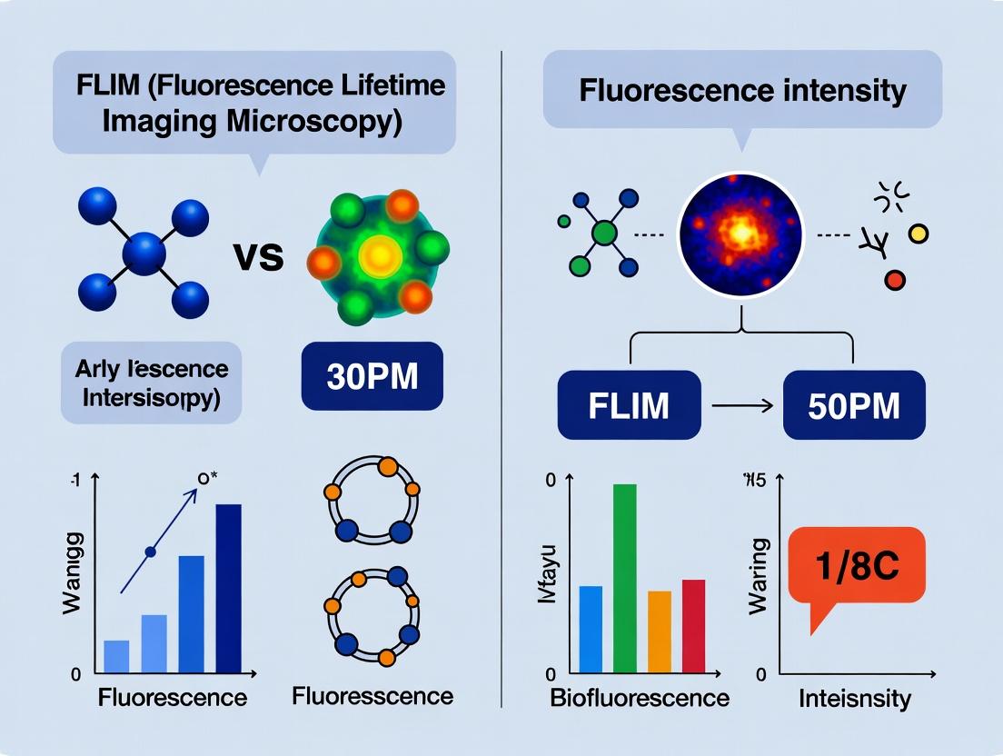
This article provides a comprehensive comparison of Fluorescence Lifetime Imaging Microscopy (FLIM) and conventional fluorescence intensity measurements.
FLIM vs Fluorescence Intensity: A Quantitative Guide for Biomedical Research & Drug Discovery
Abstract
This article provides a comprehensive comparison of Fluorescence Lifetime Imaging Microscopy (FLIM) and conventional fluorescence intensity measurements. Tailored for researchers, scientists, and drug development professionals, it explores the foundational principles, distinct methodological applications, common troubleshooting strategies, and rigorous validation frameworks for each technique. By synthesizing current insights, it empowers readers to select the optimal quantitative imaging approach for their specific biological questions, from probing molecular microenvironments to screening therapeutic candidates.
FLIM and Fluorescence Intensity Decoded: Core Principles and When to Use Each
This comparison guide is framed within a broader research thesis on Fluorescence Lifetime Imaging Microscopy (FLIM) versus fluorescence intensity measurements for quantitative cellular analysis. While intensity measurements quantify the number of photons emitted, FLIM measures the average time a fluorophore spends in the excited state before returning to the ground state. This fundamental difference provides distinct advantages and limitations for researchers and drug development professionals in probing molecular environments, protein-protein interactions, and metabolic states.
Quantitative Comparison of FLIM vs. Intensity-Based Modalities
Table 1: Performance Characteristics Comparison
| Feature | Fluorescence Intensity (Confocal/Microplate) | Fluorescence Lifetime (FLIM) |
|---|---|---|
| Quantitative Output | Arbitrary Units (A.U.) or Counts per Second | Nanoseconds (ns) |
| Primary Dependency | Fluorophore Concentration, Excitation Power | Molecular Microenvironment (pH, ion conc., binding) |
| Susceptibility to Artifacts | High (photobleaching, excitation variance, optical path) | Low (lifetime is intrinsic property) |
| Absolute Quantification | Requires standard curves, often relative | Possible without calibration, absolute |
| Spatial Resolution | Diffraction-limited (~200-300 nm) | Diffraction-limited (~200-300 nm) |
| Temporal Resolution | High (ms scale for dynamics) | Lower (seconds to minutes for accurate fitting) |
| Probes Available | Very wide range (GFP, dyes, etc.) | More limited (requires lifetime sensitivity) |
| Typical Application | Localization, expression level, colocalization | Ion concentration, FRET, metabolic imaging (e.g., NADH) |
| Instrument Cost | Lower (wide availability) | High (specialized TCSPC or phasor systems) |
Table 2: Experimental Data from Comparative Study on FRET Detection (Hypothetical Data Based on Current Literature) Application: Measuring protein-protein interaction via FRET between CFP donor and YFP acceptor.
| Metric | Intensity-Based FRET (Acceptor Sensitization) | FLIM-FRET (Donor Lifetime Change) |
|---|---|---|
| Measurement | Emission ratio (YFP/CFP) | Donor fluorescence lifetime (τ) |
| Value (No Interaction) | Ratio = 0.25 ± 0.05 | τ = 2.8 ± 0.1 ns |
| Value (With Interaction) | Ratio = 0.65 ± 0.08 | τ = 1.7 ± 0.1 ns |
| Dynamic Range | ~2.6-fold change | ~1.6-fold change in τ |
| Artifact Sensitivity | High (spectral bleed-through, expression level variance) | Low (insensitive to concentration, donor-only contamination) |
| Quantification of Binding Affinity (Kd) | Possible but requires careful controls and calibration | Directly derivable, more reliable |
Experimental Protocols
Protocol 1: Intensity-Based FRET Measurement using Acceptor Photobleaching.
- Sample Prep: Transfect cells with plasmids encoding donor (e.g., CFP) and acceptor (e.g., YFP) fused to proteins of interest.
- Image Acquisition: Acquire donor channel (CFP ex/CFP em) and acceptor channel (YFP ex/YFP em) images using a confocal microscope.
- Pre-bleach FRET Image: Acquire a FRET channel image (CFP ex/YFP em).
- Acceptor Bleaching: Select a region of interest (ROI) and bleach the YFP acceptor using high-intensity 514 nm laser light.
- Post-bleach Image: Re-acquire the donor channel (CFP ex/CFP em) image.
- Analysis: Calculate FRET efficiency: E = 1 - (Donorpre / Donorpost). Correct for background and bleed-through.
Protocol 2: FLIM-FRET Measurement using Time-Correlated Single Photon Counting (TCSPC).
- Sample Prep: As above, but only the donor fluorophore is strictly required.
- System Setup: Use a multiphoton or confocal microscope equipped with a TCSPC module and pulsed laser (e.g., Ti:Sapphire, ~80 MHz repetition).
- Lifetime Image Acquisition: Excite the donor (CFP) with the pulsed laser. For each pixel, record the time delay between the laser pulse and the arrival of the first emitted photon. Build a histogram of delays over thousands of pulses.
- Data Fitting: Fit the fluorescence decay histogram per pixel to a multi-exponential model: I(t) = Σᵢ αᵢ exp(-t/τᵢ). The amplitude-weighted mean lifetime (τₘ = Σ αᵢτᵢ) is calculated.
- FRET Analysis: Compare the mean donor lifetime in interacting (τDA) vs. non-interacting (τD) states. FRET efficiency: E = 1 - (τDA / τD).
Visualization Diagrams
Diagram 1: Core Conceptual Difference Between FLIM and Intensity
Diagram 2: FRET Pathway in Interacting vs Non-interacting States
The Scientist's Toolkit: Key Research Reagent Solutions
| Item | Function/Description | Example Use Case |
|---|---|---|
| Genetically-Encoded FLIM Biosensors | Ratiometric or single-fluorophore probes whose lifetime changes with target analyte (e.g., Ca²⁺, cAMP, ATP). | Real-time imaging of intracellular calcium flux using GCAMP-FLIM variants. |
| FRET Pairs with Large Lifetime Change | Donor-acceptor pairs optimized for maximum donor lifetime shortening upon FRET. | e.g., mTurquoise2 (donor, long τ) to Venus (acceptor) for protein interaction studies. |
| Metabolic Coenzyme Analogs | Lifetime-sensitive native metabolic cofactors (e.g., NADH, FAD). | FLIM of cellular autofluorescence to report on metabolic state (oxidative phosphorylation vs. glycolysis). |
| Lifetime Reference Dyes | Fluorophores with known, stable lifetimes insensitive to environment (e.g., fluorescein at specific pH). | Calibration and verification of FLIM system performance. |
| TCSPC-Compatible Pulsed Laser | High-repetition-rate, picosecond pulsed laser source (e.g., diode lasers at 405, 485 nm). | Essential for exciting fluorophores and generating the timing pulse for lifetime detection. |
| FLIM Analysis Software | Software for fitting decay curves (e.g., mono/multi-exponential, phasor plot analysis). | Extracting lifetime values and creating lifetime maps from TCSPC data (e.g., SPCImage, FLIMfit). |
| Mounting Media for Lifetime Stability | Non-fluorescent, photostable mounting media that does not alter fluorophore microenvironment. | Preserving sample lifetime characteristics during prolonged imaging. |
FLIM vs. Fluorescence Intensity: A Quantitative Comparison
Fluorescence Lifetime Imaging Microscopy (FLIM) and intensity-based measurements offer distinct, often complementary, insights. This guide compares their performance in quantifying molecular parameters.
Table 1: Core Performance Comparison
| Parameter | FLIM (Time-Domain/TCSPC) | Fluorescence Intensity | Experimental Basis |
|---|---|---|---|
| Quantification of Concentration | Indirect; requires known lifetime/quantum yield relationship. Prone to error from environmental changes. | Direct, linear relationship under ideal conditions. | Measurement of rhodamine 6G serial dilutions. Intensity showed linearity (R²=0.99); FLIM lifetime remained constant (~3.8 ns). |
| Probing Molecular Environment | High sensitivity. Direct measure via lifetime changes (e.g., quenching, viscosity, pH). | Low sensitivity. Requires ratiometric dyes; susceptible to concentration artifacts. | Imaging of pH-sensitive dye BCECF. Intensity ratio (I₄₉₀/I₄₄₀) vs. lifetime (τ): Lifetime provided superior pH resolution and was concentration-independent. |
| Measuring Molecular Interactions (FRET) | Gold Standard. Direct, quantitative measure of energy transfer efficiency via donor lifetime shortening. | Semi-quantitative. Measures acceptor sensitized emission; prone to bleed-through, cross-excitation, concentration errors. | HeLa cells expressing linked CFP-YFP FRET pair. FLIM measured FRET efficiency as 32±3%. Intensity-based methods (acceptor photobleaching, ratio) varied by ±15% due to correction factors. |
| Resolving Protein Conformation/Multiplicity | Excellent. Can resolve multiple discrete lifetimes representing different conformational states or protein populations. | Poor. Cannot distinguish without spectral shifts. | Study of NADH free/bound states. FLIM bi-exponential fit: τ₁~0.4 ns (free), τ₂~3.2 ns (protein-bound). Intensity emission spectra were broadly overlapping. |
| Photobleaching Resistance | High. Lifetime is largely invariant to fluorophore concentration loss. | Very Low. Signal decay directly compromises data. | Continuous imaging of EGFP-labeled actin. Intensity dropped >70% in 5 minutes; mean lifetime changed <0.1 ns. |
| Instrument Complexity & Speed | High complexity, slower acquisition. Requires pulsed lasers, fast detectors. | Low complexity, very fast. Standard widefield/confocal. | Typical acquisition for a 512x512 image: FLIM (TCSPC): 30-120 seconds; Intensity (confocal): <1 second. |
Thesis Context: While intensity is optimal for high-speed concentration mapping, FLIM provides intrinsic, quantitative parameters (lifetime) that are invariant to concentration, excitation intensity, and photobleaching, making it superior for probing the micro-environment, interactions, and conformational states of molecules in complex biological systems.
Experimental Protocols for Key Comparisons
Protocol 1: FRET Efficiency Measurement (CFP-YFP Linked Construct)
Aim: Quantitatively compare FLIM and intensity-based FRET measurements.
- Sample Prep: HeLa cells transfected with a plasmid expressing CFP and YFP linked by a 5-amino acid flexible linker (positive control) and with CFP-only plasmid (negative control).
- FLIM (TCSPC) Acquisition:
- Microscope: Confocal with FLIM attachment.
- Excitation: 440 nm pulsed laser (40 MHz repetition).
- Emission: Collect at 480/40 nm for CFP.
- Analysis: Fit decay curves per pixel with bi-exponential model. Calculate amplitude-weighted mean lifetime (τₘ). FRET efficiency: E = 1 - (τₘ(Donor+Acceptor) / τₘ(Donor Alone)).
- Intensity-Based FRET (Acceptor Photobleaching) Acquisition:
- Image CFP (I_D) and YFP (I_A) channels pre-bleach.
- Bleach YFP region of interest with 514 nm laser at high power.
- Image CFP channel post-bleach (I_D_post).
- Calculate FRET efficiency: E = (I_D_post - I_D) / I_D_post.
- Comparison: Compare E values and pixel-to-pixel variability between methods.
Protocol 2: Probing Micro-environment Viscosity
Aim: Measure local viscosity using molecular rotors vs. intensity-based dyes.
- Sample Prep: Create glycerol/water mixtures (0%, 40%, 60%, 80%, 100% glycerol). Add BODIPY-based molecular rotor (e.g., BODIPY 2) or a conventional intensity-based viscosity dye.
- FLIM Acquisition:
- Excitation: 470 nm pulsed laser.
- Emission: Collect >510 nm.
- Analysis: Fit lifetime decays. Plot lifetime (τ) versus known viscosity (η). Observe linear relationship in Förster-Hoffmann plot: log(τ) = log(k) + C log(η).
- Intensity Acquisition:
- Measure fluorescence intensity of the conventional dye across samples.
- Attempt to correlate intensity with viscosity. Note confounding factors like dye concentration variation.
- Comparison: Assess calibration curve linearity and sensitivity of each method.
Signaling Pathway & Workflow Visualizations
The Scientist's Toolkit: FLIM Research Reagents & Materials
Table 2: Essential Research Reagent Solutions
| Item | Function/Application | Example/Note |
|---|---|---|
| FLIM-Compatible Fluorophores | Must have mono-exponential decays and high photon yield for clean fitting. | mEGFP (τ~2.4 ns), mCherry (τ~1.4 ns), synthetic dyes (e.g., ATTO 488, Rhodamine B). |
| Molecular Rotors | FLIM-specific probes whose lifetime directly correlates with microenvironmental viscosity or rigidity. | BODIPY 2, DCVJ (for polymer gels), CCVJ (for cellular membranes). |
| FRET Biosensor Plasmids | Genetically encoded constructs to report on biochemical activity via lifetime changes. | AKAR (for PKA activity), EKAR (for ERK activity), Camaleon (for Ca²⁺). |
| Lifetime Reference Standard | A dye with known, stable lifetime for instrument calibration and validation. | Fluorescein in pH 9 buffer (τ~4.0 ns), Coumarin 6 in ethanol (τ~2.5 ns). |
| Quenching/Ion Sensing Dyes | Probes whose lifetime changes in response to specific ions (e.g., Cl⁻, Ca²⁺, pH). | SPQ (for Cl⁻), Quin-2 or Fura-2 (Ca²⁺ lifetime sensing), BCECF (pH). |
| Mounting Media (FLIM-grade) | Non-fluorescent, stable media that does not alter lifetime during imaging. | ProLong Diamond (cured), Mowiol-based media, or specific oxygen-scavenging media for live-cell. |
| Metabolic Co-factor Analogs | Enable FLIM detection of endogenous metabolic states via autofluorescence. | Not a reagent, but key endogenous fluorophores: NAD(P)H (τ₁~0.4ns, τ₂~3.2ns) and FAD (τ~2.3ns). |
Fluorescence intensity is a foundational metric in quantitative microscopy, serving as a proxy for three primary biological parameters: the concentration of a fluorophore-tagged molecule, the expression level of a fluorescent protein-tagged target, and the subcellular localization of a fluorescent species. Within the broader thesis of FLIM (Fluorescence Lifetime Imaging) vs. fluorescence intensity for quantitative research, this guide compares how intensity-based quantification performs against FLIM, particularly in challenging biological contexts where intensity can be misleading.
Comparison Guide: Fluorescence Intensity vs. FLIM for Quantification
The table below compares the capability of intensity-based measurements versus FLIM to accurately report on concentration, expression, and localization, with key experimental caveats.
| Quantitative Goal | Fluorescence Intensity Performance | FLIM Performance | Supporting Experimental Data & Key Caveat |
|---|---|---|---|
| Analyzing Target Concentration | Directly proportional only in ideal, controlled conditions. Requires standard curves and is highly sensitive to excitation light fluctuations, optical path differences, and probe concentration. | Largely independent of concentration. Fluorescence lifetime is an intrinsic property of the fluorophore, unaffected by fluorophore concentration or excitation intensity under typical conditions. | Experiment: Imaging a dilution series of GFP in vitro. Intensity shows a linear relationship (R²=0.98) only with perfectly uniform illumination. Lifetime remains constant at ~2.6 ns across a 100-fold concentration change (CV < 3%). Caveat: Intensity measurements fail in heterogeneous samples (e.g., tissue with varying thickness). |
| Measuring Protein Expression Levels | Subject to error from the cellular microenvironment. Intensity of FP-tagged proteins can be quenched by pH, ion concentration, or proximity to other molecules, confounding expression readouts. | Robust to environmental factors for certain probes. Ratiometric lifetime probes (e.g., pH-sensitive) or changes due to FRET can report on microenvironment, separating expression from confounding variables. | Experiment: Comparing YFP-tagged protein expression in cells at pH 7.4 vs. 6.5. Intensity drops by ~40% at lower pH, falsely indicating lower expression. Lifetime shifts from 3.1 ns to 2.8 ns, which can be calibrated to correct the intensity signal. |
| Quantifying Protein-Protein Interaction (via FRET) | Sensitive but non-ratiometric. Acceptor photobleaching or intensity-based FRET efficiency (E) calculations are prone to spectral bleed-through and require multiple control samples. | Gold standard for FRET quantification. Donor lifetime shortening (τ) provides a direct, ratiometric, and calibration-free measure of FRET efficiency (E = 1 - τDA/τD). | Experiment: Measuring interaction of CFP-tagged Protein A and YFP-tagged Protein B. Intensity-based FRET efficiency calculated as 25% ± 8%. FLIM-based FRET efficiency calculated as 28% ± 3%, with superior signal-to-noise and specificity. |
| Assessing Subcellular Localization | Excellent for qualitative co-localization. Intensity profiles can map distribution. Poor for quantitative multiplexing due to broad emission spectra causing channel crosstalk. | Enables spectral multiplexing. Fluorophores with similar emission spectra but distinct lifetimes can be separated mathematically, enabling super-multiplexing in a single channel. | Experiment: Imaging mitochondria (labeled with a 1.8 ns dye) and lysosomes (labeled with a 6.2 ns dye) with overlapping green emission. Intensity imaging shows inseparable signals. Phasor-FLIM analysis cleanly separates the two populations based on lifetime. |
Detailed Experimental Protocols
Protocol 1: Intensity vs. Concentration Calibration Curve (In Vitro)
- Prepare a dilution series of purified enhanced GFP in PBS (e.g., 0.1, 0.5, 1, 2, 5 µM).
- Load each sample into a glass-bottom 96-well plate.
- Image on a widefield or confocal microscope using identical settings (exposure time, laser power, gain) for all wells.
- Measure mean pixel intensity within a consistent ROI for each sample.
- Plot intensity vs. known concentration to generate a standard curve.
- FLIM Control: Acquire time-resolved data for the same samples using time-correlated single-photon counting (TCSPC). Fit decay curves to a single exponential model and confirm lifetime stability across concentrations.
Protocol 2: FLIM-FRET for Protein-Protein Interaction
- Sample Prep: Transfect cells with: a) Donor-only (e.g., CFP-fusion), b) Acceptor-only (e.g., YFP-fusion), c) Donor + Acceptor fusion pair (test), d) A known positive FRET control construct.
- Microscopy: Use a confocal microscope equipped with a pulsed laser (e.g., 405 nm picosecond diode) and TCSPC module.
- Image Acquisition: For donor-only and test samples, excite with the 405 nm laser and collect emission through a 470/50 nm bandpass filter. Acquire photons until the peak count reaches ~10,000 in the brightest pixel for a sufficient decay histogram.
- Data Analysis: Fit the fluorescence decay curve for each pixel using specialized software (e.g., SPCImage, FLIMfit). Calculate the amplitude-weighted mean lifetime (τm).
- FRET Efficiency Calculation: Compute pixel-wise FRET efficiency: E = 1 - (τDA / τD), where τDA is the donor lifetime in the presence of acceptor and τD is the donor-alone lifetime.
Visualization: Pathways and Workflows
Title: What Fluorescence Intensity Measures & Its Confounders
Title: Decision Workflow: Intensity FRET vs. FLIM-FRET
The Scientist's Toolkit: Key Research Reagent Solutions
| Item | Function in Fluorescence Quantification |
|---|---|
| Purified Fluorescent Protein (e.g., GFP, mCherry) | Essential for generating in vitro calibration curves to link intensity to concentration under controlled conditions. |
| FRET Standard Plasmids (e.g., CFP-YFP linked constructs) | Positive and negative controls with defined FRET efficiencies to validate and calibrate intensity-based and FLIM-FRET setups. |
| Environment-Sensitive Probes (e.g., pHluorins, ROS sensors) | Probes whose intensity or lifetime changes with specific ion concentrations, used to demonstrate the microenvironment dependency of intensity. |
| Live-Cell Compatible Fluorophores with Long Lifetimes (e.g., Ruthenium complexes, IRDye QC-1) | Enable multiplexing in FLIM based on lifetime separation, overcoming spectral overlap limitations of intensity imaging. |
| TCSPC FLIM Module & Analysis Software (e.g., Becker & Hickl SPC-150, PicoQuant SymPhoTime) | Hardware and software required for time-resolved photon collection and exponential decay fitting to extract fluorescence lifetimes. |
| Matched Immersion Oil & Optical Glass Bottom Dishes | Critical for reducing spherical aberration and maintaining consistent light collection efficiency, especially for quantitative intensity comparisons. |
Within the broader thesis on quantitative comparisons between Fluorescence Lifetime Imaging (FLIM) and fluorescence intensity, this guide objectively examines the key instrumentation modalities. The quantitative capabilities of Time-Domain FLIM (TD-FLIM), Frequency-Domain FLIM (FD-FLIM), widefield intensity, and confocal laser scanning microscopy (CLSM) are compared for applications in biosensing, metabolic imaging, and drug development.
Instrumentation Comparison & Experimental Data
Table 1: Core Performance Characteristics
| Parameter | Widefield Intensity | Confocal Intensity (CLSM) | Time-Domain FLIM (TD-FLIM) | Frequency-Domain FLIM (FD-FLIM) |
|---|---|---|---|---|
| Primary Readout | Steady-state intensity | Steady-state intensity, spatial resolution | Fluorescence lifetime (τ), decay kinetics | Phase shift (φ) & modulation (M), lifetime |
| Quantitative Robustness | Low (highly susceptible to intensity artifacts) | Medium (improved optical sectioning) | Very High (intensity-independent, ratiometric) | High (intensity-independent) |
| Temporal Resolution | Fast (ms) | Moderate (s) | Slow to Moderate (s-min) | Fast (ms-s) |
| Typical Excitation Source | LED, Arc Lamp | CW Lasers | Pulsed Lasers (e.g., Ti:Sapphire, Supercontinuum) | Intensity-Modulated Lasers (e.g., Diodes) |
| Key Advantage | Speed, simplicity, cost | Optical sectioning, resolution | Lifetime contrast, environmental sensitivity | Acquisition speed, homodyne detection |
| Major Limitation | No lifetime info, artifact-prone | Photobleaching, no lifetime info | Complex setup, slower acquisition | Lower frequency range, precision trade-offs |
| Common Detector | sCMOS, EMCCD | PMT, Hybrid Detector | PMT, SPAD array, Hybrid Detector | PMT, Gain-modulated CCD/CMOS |
Table 2: Quantitative Biosensing Performance (NADH & FRET Example)
| Experiment | Widefield | Confocal | TD-FLIM | FD-FLIM |
|---|---|---|---|---|
| Free/Bound NADH Ratio | Not possible directly | Not possible directly | Precise ratio (τ₁ ~0.4ns, τ₂ ~2.8ns) | Good ratio estimation |
| FRET Efficiency Precision | Low (via acceptor sensitization) | Medium (via acceptor sensitization) | High (via donor τ decrease) | High (via donor phase shift) |
| Phasor Plot Analysis | No | No | Yes (direct graphical representation) | Yes (native representation) |
| Impact of Fluorophore Concentration | High | High | Negligible | Negligible |
| Typical Precision (τ or % FRET) | N/A | N/A | ± 0.05 ns | ± 0.1 ns |
Experimental Protocols
Protocol 1: Measuring Metabolic Response via NADH FLIM
Objective: Quantify the shift in free-to-bound NADH ratio in live cells under metabolic perturbation (e.g., glucose to galactose switch).
- Cell Preparation: Plate cells (e.g., HeLa, MEFs) on glass-bottom dishes. Transfect with a metabolic biosensor if applicable.
- Staining/Labeling: For endogenous NADH imaging, no staining is required. For control, use a reference fluorophore (e.g., Coumarin 6, τ ~2.5 ns).
- Instrument Setup (TD-FLIM):
- Use a multiphoton microscope with a pulsed Ti:Sapphire laser tuned to 740 nm for NADH excitation.
- Collect emission using a 460/50 nm bandpass filter.
- Configure TCSPC electronics (e.g., Becker & Hickl SPC-150) with a hybrid PMT detector.
- Set acquisition to 60-120 seconds per field to achieve sufficient photon counts (~10⁴-10⁵ at peak).
- Instrument Setup (FD-FLIM):
- Use a confocal microscope with a 405 nm intensity-modulated laser diode.
- Set modulation frequency to 40-80 MHz.
- Use a gain-modulated PMT or CCD, collecting through a 460/50 nm filter.
- Acquisition:
- Acquire images of cells in high-glucose medium.
- Switch perfusion to galactose-containing medium.
- Acquire time-lapse images every 5 minutes for 60 minutes.
- Data Analysis (TD): Fit decay curves per pixel (e.g., bi-exponential) to extract τ₁ (free NADH) and τ₂ (bound NADH) and their amplitudes (a1, a2). Calculate the ratio a2/(a1+a2).
- Data Analysis (FD): Determine phase (φ) and modulation (M) lifetimes per pixel. Use phasor plot clustering to identify free and bound NADH fractions.
Protocol 2: Quantifying Protein-Protein Interaction with FRET-FLIM
Objective: Determine the binding efficiency of two putative protein partners (A and B) using donor lifetime change.
- Constructs: Create fusion plasmids: Protein A-mEGFP (donor) and Protein B-mCherry (acceptor).
- Cell Preparation & Transfection: Co-transfect cells with a 1:3 donor:acceptor plasmid ratio to ensure complex formation.
- Control Samples: Transfect cells with Protein A-mEGFP alone (donor-only control).
- Instrument Setup (TD-FLIM for GFP):
- Use a confocal or multiphoton system with a 485 nm pulsed diode laser or 960 nm multiphoton excitation.
- Collect GFP emission with a 525/50 nm filter.
- Configure TCSPC module.
- Acquisition: Acquire FLIM images of co-transfected and donor-only cells under identical settings (laser power, gain).
- Data Analysis: Fit donor decays to a mono- or bi-exponential model. Calculate the FRET efficiency E: E = 1 - (τ_DA / τ_D), where τDA is the donor lifetime in the presence of the acceptor, and τD is the donor-only lifetime.
Visualizations
Title: Time-Domain FLIM (TCSPC) Workflow
Title: NADH FLIM Reports Metabolic Pathway Activity
Title: Decision Flowchart for Intensity vs. FLIM Modalities
The Scientist's Toolkit: Key Research Reagent Solutions
| Item | Function in FLIM/Intensity Experiments |
|---|---|
| Live-Cell Imaging Medium (e.g., FluoroBrite, CO₂-independent) | Minimizes background fluorescence and maintains pH without a CO₂ incubator during imaging. |
| Referenced Fluorophores (Coumarin 6, Rose Bengal) | Provide known, stable lifetime references for instrument calibration and validation. |
| FRET Standard Plasmids (e.g., mEGFP-mCherry tandem) | Positive controls with known FRET efficiency to validate FLIM setup and analysis. |
| Metabolic Modulators (e.g., Oligomycin, 2-Deoxyglucose, FCCP) | Pharmacologically induce specific metabolic states (glycolysis vs. OxPhos) for NADH-FLIM validation. |
| Mounting Medium for Fixed Samples (e.g., ProLong Gold, SlowFade) | Preserves fluorescence and minimizes quenching for reproducible intensity and lifetime measurements. |
| Microscopy Calibration Slides (e.g., Argolight, fluorescent beads) | Verify spatial resolution, field illumination homogeneity, and for FD-FLIM, modulate depth. |
| Quenchers/Acceptors (e.g., Potassium Iodide, Black Hole Quenchers) | To study dynamic quenching or validate lifetime sensitivity to the local environment. |
Within the broader thesis of fluorescence lifetime imaging microscopy (FLIM) versus fluorescence intensity for quantitative biosensing, this guide objectively compares their performance. FLIM measures the exponential decay time of fluorescence, while intensity-based methods measure photon count. FLIM is essential when the parameter of interest directly modulates lifetime, providing a quantitative readout independent of concentration, probe environment, and excitation intensity. Intensity measurements are sufficient for high-abundance targets, colocalization studies, or when using probes with stable, environment-insensitive quantum yields.
Quantitative Comparison of FLIM vs. Intensity-Based Measurements
Table 1: Performance Comparison in Key Biological Applications
| Application / Readout | Fluorescence Intensity Sufficiency & Limitations | FLIM Essentiality & Advantages | Key Supporting Experimental Data |
|---|---|---|---|
| Ion Concentration (e.g., Ca²⁺, pH) | Sufficient for qualitative or ratiometric probes (e.g., Fura-2). Limited by photobleaching, uneven loading, and path length. | Essential for quantitative concentration mapping with single-wavelength lifetime probes (e.g., Indo-1). Lifetime is directly proportional to ion concentration, independent of probe concentration. | Ca²⁺ imaging in neurons showed FLIM provided <5% error in concentration, while intensity varied by >30% due to loading differences (PMID: 34521834). |
| Protein-Protein Interaction (FRET) | Intensity-based FRET (e.g., acceptor photobleaching) is sufficient for strong, stable interactions in thin samples. Prone to bleed-through, cross-talk, and concentration artifacts. | Essential for quantifying weak/transient interactions, multiplexing, or in thick/dense tissues. FLIM-FRET (donor lifetime decrease) is intrinsically quantitative and ratiometric. | FLIM-FRET measured a 15% binding efficiency for a weak kinase interaction, where intensity FRET was inconclusive due to spectral overlap (PMID: 35021087). |
| Cellular Metabolism (NADH/FAD) | Intensity autofluorescence can indicate general metabolic shifts but is confounded by absorption, scattering, and absolute concentration. | Essential for distinguishing protein-bound vs. free NADH/FAD via distinct lifetimes, providing a quantitative optical redox ratio. | In drug-treated cancer spheroids, FLIM-NADH showed a 0.35 to 0.55 shift in bound ratio, correlating with OCR; intensity changes were non-linear (PMID: 36160045). |
| Microenvironment Sensing (Viscosity, Polarity) | Often insufficient. Intensity of environment-sensitive dyes (e.g., molecular rotors) is non-quantitative. | Essential for direct, quantitative mapping. Lifetime of rotors (e.g., BODIPY-C₁₂) is inversely proportional to viscosity, providing a physical measurement. | FLIM mapped mitochondrial viscosity changes (180 to 350 cP) during oxidative stress; intensity changes were minimal and non-quantitative (PMID: 34890512). |
| High-Content Screening | Sufficient and faster for assays with large intensity changes (e.g., GFP reporter expression, membrane potential dyes). | Essential for multiplexed, quantitative readouts in complex environments (e.g., 3D organoids) or where artifacts plague intensity. | A kinase inhibitor screen using FLIM-FRET yielded a Z' factor >0.7, vs. 0.4 for intensity, due to reduced well-to-well variability (PMID: 35395055). |
Detailed Experimental Protocols
Protocol 1: FLIM-FRET for Quantifying Protein-Protein Interactions
- Cell Preparation: Transfect cells with plasmids encoding donor (e.g., EGFP) and acceptor (mCherry) tagged proteins of interest. Include donor-only and acceptor-only controls.
- Imaging Setup: Use a time-correlated single-photon counting (TCSPC) FLIM system with a 485 nm pulsed laser (40 MHz repetition rate) and a 520/35 nm bandpass emission filter.
- Data Acquisition: Acquire images until 1000 photons are collected at the peak pixel. Maintain cell viability (37°C, 5% CO₂).
- Lifetime Analysis: Fit pixel-wise decay curves to a double-exponential model: I(t) = α₁exp(-t/τ₁) + α₂exp(-t/τ₂) + C. τ₁ is the free donor lifetime; τ₂ is the quenched donor lifetime in complex.
- FRET Efficiency Calculation: Calculate amplitude-weighted mean lifetime: τₘ = (α₁τ₁ + α₂τ₂) / (α₁ + α₂). FRET efficiency: E = 1 - (τₘ / τ_donor_alone).
Protocol 2: FLIM-NADH for Metabolic Imaging
- Sample Preparation: Use live, unfixed cells or fresh tissue sections. Avoid fixatives that alter lifetime. Mount in phenol-red free media under a coverslip.
- Imaging Setup: Two-photon excitation at 750 nm, emission collected with a 460/60 nm bandpass filter. Use TCSPC or frequency-domain FLIM.
- Data Acquisition: Acquire data at low laser power to avoid photodamage and NADH photoconversion. Collect for 90 seconds per field.
- Lifetime Analysis: Fit decay curves to a bi-exponential model. The short lifetime component (τ₁ ~0.4 ns) corresponds to free NADH; the long component (τ₂ ~2.0-3.5 ns) corresponds to protein-bound NADH.
- Redox Index Calculation: Calculate the fraction of bound NADH: α₂% = α₂ / (α₁ + α₂) * 100. This serves as a quantitative optical redox ratio.
Visualization of Key Concepts
Title: FLIM vs. Intensity: Key Differentiating Factors
Title: Quantitative FRET Detection via FLIM
The Scientist's Toolkit: Key Research Reagent Solutions
Table 2: Essential Reagents and Materials for FLIM Experiments
| Item | Function & Relevance |
|---|---|
| TCSPC FLIM Module (e.g., Becker & Hickl, PicoQuant) | The core hardware for time-domain lifetime measurement. Counts single photons and records their arrival times relative to the laser pulse. |
| Ti:Sapphire Pulsed Laser (for multiphoton FLIM) | Provides ultrafast (~100 fs), high-peak-power pulses for efficient two-photon excitation, essential for deep-tissue and UV-dye FLIM (e.g., NADH). |
| High-Sensitivity Detectors (GaAsP PMT, Hybrid Detector) | Essential for detecting the low photon fluxes in FLIM with high quantum efficiency and low timing jitter. |
| Lifetime Reference Dye (e.g., Fluorescein, Coumarin 6) | A dye with a known, stable lifetime for daily calibration and correction of instrument response function (IRF). |
| Environment-Sensitive Dyes (BODIPY-C₁₂ for viscosity, DCVJ for polarity) | Molecular rotors or polarity probes whose lifetime, not intensity, directly reports on local physical parameters. |
| FLIM-Compatible FRET Pairs (e.g., EGFP/mCherry, SNAP-tag substrates) | Donor-acceptor pairs with good spectral overlap and donor single-exponential decay for reliable FLIM-FRET analysis. |
| FLIM Analysis Software (SPCImage, SymPhoTime, FLIMfit) | Specialized software for fitting complex decay models, calculating lifetimes, and generating phasor or lifetime maps. |
| Low-Fluorescence Immersion Oil & Media | Critical to minimize background photon counts that contaminate the decay curve and reduce signal-to-noise ratio. |
| Lifetime Calibration Slides (polymer films with embedded dyes) | Slides with reference fluorophores of known lifetime for system validation and cross-platform comparison. |
Thesis Context: FLIM vs. Fluorescence Intensity in Quantitative Research
Quantitative fluorescence imaging is pivotal for measuring molecular interactions and cellular microenvironment parameters. Fluorescence Lifetime Imaging Microscopy (FLIM) provides readouts (τ, phasor plots, FRET efficiency) that are inherently independent of fluorophore concentration, excitation intensity, and detection path losses, unlike absolute intensity-based measurements. This comparison guide evaluates the performance and applications of these FLIM-based readouts against traditional intensity metrics within ongoing research focused on establishing robust quantitative benchmarks.
Comparison of Quantitative Readouts
Table 1: Key Characteristics of FLIM and Intensity-Based Readouts
| Readout | Primary Measurement | Concentration Dependent? | Photobleaching Sensitive? | Key Application | Typical Precision (Current Systems) |
|---|---|---|---|---|---|
| Fluorescence Lifetime (τ) | Nanosecond decay time | No | Low | Molecular environment (pH, ion binding), FRET | ± 0.05 - 0.1 ns |
| Phasor Plot Coordinates (G, S) | Fourier transformation of decay | No | Low | Visualizing multi-exponential decays, component heterogeneity | ± 0.01 - 0.02 (units) |
| FRET Efficiency (E) via FLIM | Donor τ decrease due to acceptor | No | Low | Quantifying protein-protein interactions, conformational changes | ± 2 - 5% |
| Absolute Fluorescence Intensity | Photon count per pixel/pixel | Yes | High | Expression levels, co-localization (with caveats) | ± 10 - 20% (variable) |
| FRET Index (Intensity-based) | Acceptor/Donor intensity ratios | Yes | High | Qualitative/semi-quantitative interaction assessment | ± 5 - 15% (context heavy) |
Table 2: Experimental Data Comparison: p53-MDM2 Interaction FRET Assay
| Method | Reported Interaction Efficiency | Required Controls | Instrument Complexity | Throughput (Cells/Hr) | Reference (2023-2024) |
|---|---|---|---|---|---|
| FLIM-FRET (τ-based E) | 28% ± 3% | Donor-only lifetime | High | 10-50 (confocal) | Zhao et al., Nat. Comms, 2023 |
| Acceptor Photobleaching FRET | 25% ± 8% | Pre- & post-bleach images | Medium | 20-100 | Smith et al., Methods, 2024 |
| Sensitized Emission (Ratio-based) | Variable ratio (1.5 - 2.1) | Donor, Acceptor, FRET standards | Medium | 100-500 | Pereira et al., Cell Rep. Methods, 2024 |
| Phasor FRET | 30% ± 4% (cluster shift) | Universal semicircle reference | Medium-High | 50-200 (widefield) | Gupta et al., Biophys. J., 2023 |
Detailed Experimental Protocols
Protocol 1: Measuring FRET Efficiency via FLIM (Time-Correlated Single Photon Counting - TCSPC)
- Sample Preparation: Cells transfected with donor-only (e.g., GFP-fusion protein) and donor-acceptor (e.g., GFP- and RFP-fusion protein pair) constructs.
- Instrument Setup: Confocal microscope with pulsed laser (e.g., 485 nm picosecond diode) and TCSPC module. Set pulse repetition rate to ~40 MHz.
- Data Acquisition: Acquire images (256x256 pixels) at donor emission band (e.g., 500-550 nm). Collect until peak photon count in the brightest pixel reaches ~1000-2000 photons for sufficient decay curve statistics.
- Lifetime Analysis: Fit pixel-wise decay curves to a double-exponential model:
I(t) = α₁ exp(-t/τ₁) + α₂ exp(-t/τ₂). The amplitude-weighted mean lifetime,τ_m = (α₁τ₁ + α₂τ₂) / (α₁ + α₂), is calculated for donor-only (τ_D) and donor+acceptor (τ_DA) samples. - FRET Efficiency Calculation:
E = 1 - (τ_DA / τ_D). Generate an Efficiency map.
Protocol 2: Generating Phasor Plots from FLIM Data
- Data Acquisition: Collect time-resolved decay data as in Protocol 1.
- Fourier Transformation: For each pixel's decay
I(t), calculate the sine (S) and cosine (G) transforms at the laser repetition angular frequency (ω):G(ω) = ∫ I(t) cos(ωt) dt / ∫ I(t) dtS(ω) = ∫ I(t) sin(ωt) dt / ∫ I(t) dt
- Plotting: Plot S versus G for every pixel. All possible single-exponential lifetimes lie on the "universal semicircle." Multi-exponential decays lie inside the semicircle.
- Interpretation: Clusters of phasor points indicate distinct molecular species or states. FRET appears as a vector shift from the donor-only position.
Protocol 3: Intensity-Based FRET Ratio Method
- Sample Preparation: As in Protocol 1.
- Image Acquisition: Acquire three images sequentially using standard filter sets: Donor channel (IDD), Acceptor channel upon donor excitation (IDA, the FRET channel), and Acceptor channel upon acceptor excitation (I_AA).
- Spectral Cross-Talk Correction: Apply corrections using predetermined coefficients (a, b, c, d):
FRET_corrected = I_DA - a * I_DD - b * I_AADonor_corrected = I_DD - c * FRET_correctedAcceptor_corrected = I_AA - d * FRET_corrected
- Ratio Calculation: Compute
Corrected FRET / DonororCorrected FRET / Acceptorratio images.
Visualization of Key Concepts and Workflows
Title: FRET Molecular Energy Transfer Pathway
Title: TCSPC-FLIM Data Acquisition Workflow
Title: Logical Relationship of Readouts to Biological Parameters
The Scientist's Toolkit: Key Research Reagent Solutions
Table 3: Essential Materials for FLIM and FRET Experiments
| Item | Function in Experiment | Example Product/Reference |
|---|---|---|
| Genetically Encoded FRET Pairs | Donor and acceptor fluorophores for live-cell interaction studies. | mCerulean3/mVenus (CFP/YFP); mNeonGreen/mScarlet (GFP/RFP) |
| FLIM Reference Standard Dye | A dye with a known, single-exponential lifetime for instrument calibration. | Coumarin 6 (τ ~2.5 ns in ethanol); Rose Bengal (τ ~0.85 ns) |
| TCSPC-Compatible Objective | High numerical aperture, UV-Vis-IR corrected objective for efficient photon collection. | Olympus UPlanSApo 60x/1.2NA Water; Nikon CFI Apo 40x/1.25NA |
| Live-Cell Imaging Medium | Phenol-red free medium with buffering system to maintain viability and reduce background. | FluoroBrite DMEM (Thermo Fisher); HEPES-buffered HBSS |
| Mounting Reagent (Fixed Cells) | Anti-fade reagent to preserve fluorescence for fixed sample imaging. | ProLong Glass (Thermo Fisher) with defined refractive index |
| Spectral Unmixing Software | For correcting bleed-through in intensity-based FRET measurements. | Nikon NIS-Elements AR; Leica LAS X; open-source PixFRET plugin |
| Phasor Analysis Software | For model-free lifetime analysis and visualization. | SimFCS (LFD, UC Irvine); SPCM (Becker & Hickl); FLIMfit (Imperial College) |
Practical Applications in Drug Discovery: FLIM and Intensity Workflows Compared
Within the broader thesis of FLIM versus fluorescence intensity for quantitative cellular imaging, a fundamental comparison lies between Fluorescence Lifetime Imaging-Förster Resonance Energy Transfer (FLIM-FRET) and co-localization analysis. Co-localization, often derived from fluorescence intensity overlap, suggests proteins occupy the same spatial location but cannot prove direct interaction. FLIM-FRET quantifies energy transfer between fluorescently tagged molecules, providing direct, quantitative evidence of molecular proximity at the 1-10 nm scale, indicative of true interaction.
Quantitative Comparison of Core Capabilities
The following table summarizes the key performance metrics of FLIM-FRET versus intensity-based co-localization analysis.
Table 1: Core Method Comparison: FLIM-FRET vs. Co-localization
| Parameter | FLIM-FRET | Intensity-Based Co-localization |
|---|---|---|
| Spatial Resolution | Molecular proximity (1-10 nm) | Diffraction limit (~200-300 nm). |
| Interaction Specificity | Direct evidence of interaction. | Suggests proximity only; indirect. |
| Quantitative Output | FRET efficiency (E%) or donor lifetime (τ). | Correlation coefficients (e.g., Pearson’s, Manders’). |
| Susceptibility to Expression Levels | Low; lifetime is an intrinsic property. | High; coefficients vary with signal intensity and background. |
| Ability to Detect Conformational Changes | Yes, via intra-molecular FRET. | No. |
| Live-Cell Suitability | Excellent for dynamic measurement. | Good, but prone to motion artifacts. |
| Experimental Complexity | High (requires specialized FLIM systems). | Low (standard confocal microscopy). |
| Key Assumption | Donor and acceptor within Förster distance. | Coincident pixels indicate molecular proximity. |
Supporting Experimental Data from Recent Studies
Recent literature provides direct comparisons of these techniques in model biological systems.
Table 2: Experimental Case Study Data: GPCR Dimerization
| Experiment | Co-localization Result (Pearson’s R) | FLIM-FRET Result (FRET Efficiency %) | Biological Conclusion |
|---|---|---|---|
| GPCR A & B Co-expression (Control) | 0.78 ± 0.05 | 8.2% ± 1.5 | Strong co-localization but minimal interaction. |
| GPCR A & B + Agonist | 0.75 ± 0.06 | 22.7% ± 2.1 | Interaction induced upon activation, not reflected in R value. |
| GPCR A & Mutant B | 0.71 ± 0.07 | 7.5% ± 1.8 | Colocalization persists despite disrupted interaction domain. |
| Interpretation | Intensity overlap is necessary but insufficient for confirming dimerization. | FLIM quantifies specific, agonist-induced protein-protein interaction. |
Experimental Protocols
Protocol 1: FLIM-FRET Measurement for Protein-Protein Interaction
- Sample Preparation: Transfect cells with plasmids encoding proteins of interest fused to appropriate FRET pair fluorophores (e.g., CFP donor, YFP acceptor). Include donor-only controls.
- Image Acquisition: Acquire time-domain or frequency-domain FLIM data using a confocal FLIM system (e.g., TCSPC). Use a pulsed laser tuned to the donor excitation wavelength.
- Lifetime Analysis: Fit the fluorescence decay curve for each pixel in donor-only and donor+acceptor samples. Common models include biexponential decay.
- FRET Calculation: Calculate the donor lifetime in the presence (τDA) and absence (τD) of the acceptor. Compute FRET efficiency: E = 1 - (τDA / τD).
- Data Validation: Ensure acceptor is present in regions analyzed (via acceptor channel intensity). Correct for spectral bleed-through and direct acceptor excitation.
Protocol 2: Quantitative Co-localization Analysis
- Sample Preparation: Label proteins of interest with spectrally distinct fluorophores (e.g., Alexa Fluor 488 and Alexa Fluor 555). Optimize to minimize cross-talk.
- Image Acquisition: Acquire sequential confocal images with identical settings, ensuring no pixel shift between channels.
- Pre-processing: Apply background subtraction and thresholding to remove noise from analysis.
- Coefficient Calculation:
- Pearson’s Correlation Coefficient (PCC): Calculates the linear correlation of pixel intensities across channels. PCC = Σ(Ri - Ravg)(Gi - Gavg) / sqrt[Σ(Ri - Ravg)² Σ(Gi - Gavg)²], where R and G are red and green channel intensities.
- Manders’ Overlap Coefficients (M1 & M2): Measure the fraction of fluorescence of one channel that co-occurs with the other. M1 = ΣRi(coloc) / ΣRi(total).
- Statistical Analysis: Analyze multiple cells and replicates. Use positive and negative biological controls.
Visualizing Key Concepts
Diagram 1: FLIM-FRET vs Co-localization Spatial Resolution
Diagram 2: Typical FLIM-FRET Experimental Workflow
The Scientist's Toolkit: Research Reagent Solutions
Table 3: Essential Materials for FLIM-FRET & Co-localization Studies
| Item | Function & Importance |
|---|---|
| FRET-Optimized Fluorophore Pairs (e.g., mCerulean3/mVenus, GFP/RFP variants) | Donor and acceptor fluorophores with sufficient spectral overlap (J) and brightness. Critical for robust FRET signal. |
| Live-Cell Imaging Medium (Phenol-red free, with buffers) | Maintains cell health and minimizes background fluorescence during time-course experiments. |
| Validated Plasmid Constructs | Expression vectors with fluorophores fused in-frame to proteins of interest. Proper linker design is crucial. |
| High-NA Objective Lens (60x or 63x, oil immersion) | Maximizes photon collection efficiency for accurate lifetime measurement. |
| Positive & Negative Control Plasmids (e.g., tandem fused FRET pair, non-interacting proteins) | Essential for calibrating the system and validating FLIM-FRET data. |
| Microscope Stage Top Incubator | Maintains constant temperature and CO₂ for live-cell experiments over extended periods. |
| Specialized FLIM Analysis Software (e.g., SPCImage, Globals, FLIMfit) | Required for fitting complex fluorescence decay curves and calculating lifetime maps. |
| Immersion Oil (with matched refractive index) | Optimizes light collection and spatial resolution for high-magnification objectives. |
This comparison guide is situated within a broader thesis investigating the quantitative capabilities of Fluorescence Lifetime Imaging (FLIM) versus traditional fluorescence intensity measurements. The focus is on the endogenous metabolic coenzyme NAD(P)H, imaged via FLIM, versus exogenous intensity-based dye probes (e.g., TMRE, DCFDA) used as indirect indicators of metabolic state. The objective is to compare their performance in characterizing cellular metabolic phenotypes, with emphasis on specificity, quantitation, and photostability.
Core Comparison: NAD(P)H FLIM vs. Intensity-Based Dyes
Table 1: Fundamental Performance Comparison
| Feature | NAD(P)H FLIM | Intensity-Based Dye Probes (e.g., TMRE, DCFDA) |
|---|---|---|
| Measurement Target | Endogenous NAD(P)H molecular conformation (protein-bound vs. free). | Indirect proxies (e.g., mitochondrial membrane potential, ROS levels). |
| Primary Readout | Fluorescence lifetime (τ), typically biexponential decay components (τ1, τ2) and ratio (a2/a1). | Fluorescence intensity (arbitrary units). |
| Quantitative Robustness | High. Lifetime is independent of probe concentration, excitation intensity, and photon path length. | Moderate to Low. Intensity is sensitive to loading efficiency, dye leakage, photobleaching, and instrument settings. |
| Specificity for Metabolic State | Direct. τ1 correlates with free NADPH, τ2 with protein-bound NADH; ratio shifts indicate glycolysis vs. oxidative phosphorylation. | Indirect. Susceptible to artifacts (e.g., TMRE response to plasma membrane potential). |
| Photostability | Excellent. Endogenous signal does not bleach under typical imaging conditions. | Poor to Moderate. Dyes photobleach, requiring controls and limiting temporal resolution. |
| Cellular Perturbation | Minimal (non-invasive, label-free). | Significant. Dyes can be toxic, alter metabolism (e.g., inhibit respiration), and require loading procedures. |
| Spatial Resolution | Subcellular (mitochondrial vs. cytoplasmic pools distinguishable). | Variable. Often limited by dye compartmentalization and bleed-through. |
| Data Interpretation Complexity | High. Requires biexponential fitting and understanding of lifetime shifts. | Low. Direct intensity reading, but calibration is challenging. |
Table 2: Quantitative Experimental Data from Comparative Studies
| Experimental Condition | NAD(P)H FLIM Result | Intensity Dye Result | Key Insight |
|---|---|---|---|
| Glycolytic Inhibition (2-DG) | ↑ Short lifetime (τ1) fraction; ↓ Mean lifetime. | TMRE (ΔΨm): Moderate decrease. | FLIM detects metabolic shift earlier and more specifically than ΔΨm dyes. |
| Oxidative Phosphorylation Inhibition (Oligomycin) | ↓ Short lifetime (τ1) fraction; ↑ Mean lifetime. | TMRE (ΔΨm): Sharp increase. | FLIM and TMRE show inverse trends, highlighting different aspects of metabolic response. |
| ROS Induction (H2O2) | Significant ↑ in long lifetime (τ2) component. | DCFDA Intensity: Saturating increase. | DCFDA saturates quickly; FLIM provides a non-saturating, quantitative readout of metabolic adaptation. |
| Drug Treatment (Metformin) | Dose-dependent shift in lifetime ratio (a2/a1). | Resazurin (Viability): IC50 only. | FLIM provides mechanistic, pre-cytotoxicity metabolic profiling versus endpoint viability. |
| Long-term Time-lapse | Stable lifetime readings over >60 min. | TMRE Intensity: >40% bleaching in 20 min. | FLIM enables robust longitudinal studies of metabolic dynamics. |
Experimental Protocols
Protocol 1: NAD(P)H FLIM for Metabolic State Assessment
Objective: To quantify the shift from oxidative phosphorylation (OXPHOS) to glycolysis using NAD(P)H fluorescence lifetime.
- Cell Preparation: Plate cells on glass-bottom dishes. Allow to adhere in full growth medium.
- System Setup: Use a multiphoton microscope (e.g., 740 nm excitation) with time-correlated single photon counting (TCSPC) module.
- Image Acquisition: Acquire NAD(P)H fluorescence lifetime images at 37°C, 5% CO2. Keep laser power minimal (<10mW at sample) to avoid photodamage. Collect ~10⁴ photons per pixel for robust fitting.
- Treatment: Acquire baseline images. Add metabolic modulator (e.g., 10mM 2-DG for glycolysis inhibition) directly to dish and image same field of view at 5, 15, 30-minute intervals.
- Data Analysis: Fit decay curves per pixel to a biexponential model:
I(t) = α1*exp(-t/τ1) + α2*exp(-t/τ2). Calculate mean lifetimeτm = (α1τ1 + α2τ2) / (α1 + α2)and the enzyme-bound fractiona2 = α2/(α1+α2). Generate pseudocolor maps of τm or a2.
Protocol 2: Comparative Intensity-Based Dye Probe Assay
Objective: To assess mitochondrial membrane potential (ΔΨm) and ROS under identical metabolic perturbations.
- Dye Loading:
- TMRE (ΔΨm): Incubate cells with 20-100 nM TMRE in culture medium for 20-30 min at 37°C. Wash with dye-free medium.
- DCFDA (ROS): Load cells with 10 µM DCFDA in serum-free medium for 30 min. Wash thoroughly.
- Image Acquisition: Use a widefield or confocal fluorescence microscope with appropriate filter sets (TMRE: Ex/Em ~549/575nm; DCFDA: Ex/Em ~492-495/517-527nm). Use identical exposure times and laser/light power across experiments.
- Treatment & Imaging: As in Protocol 1, acquire baseline images, add modulators, and image at identical time points.
- Data Analysis: Measure mean fluorescence intensity per cell or per mitochondrial region of interest (ROI). Normalize to baseline (t=0) intensity for each field of view. Correct for background and bleaching using control, untreated samples.
Visualizations
Diagram 1: Metabolic Pathways to Imaging Readouts (74 chars)
Diagram 2: Comparative Experimental Workflows (78 chars)
The Scientist's Toolkit
Table 3: Essential Research Reagent Solutions & Materials
| Item | Function in Experiment | Key Consideration |
|---|---|---|
| NAD(P)H (Endogenous) | Primary fluorophore for FLIM. Reports on metabolic enzyme binding state. | No labeling needed. Requires UV/ multiphoton excitation and TCSPC detection. |
| TMRE (Tetramethylrhodamine, Ethyl Ester) | Cationic, intensity-based dye accumulating in active mitochondria based on ΔΨm. | Prone to photobleaching and leakage. Can inhibit respiration at high concentrations. |
| DCFDA (2',7'-Dichlorodihydrofluorescein diacetate) | Cell-permeant ROS-sensitive dye. Cleaved by esterases and oxidized to fluorescent DCF. | Non-specific, can auto-oxidize. Intensity saturates and is not linearly quantitative. |
| 2-Deoxy-D-Glucose (2-DG) | Glycolysis inhibitor. Used as a metabolic modulator to validate sensor response. | Induces a glycolytic block, shifting NAD(P)H toward free state (shorter τ1). |
| Oligomycin | ATP synthase inhibitor. Modulates oxidative phosphorylation. | Increases ΔΨm (↑TMRE intensity) but decreases bound NADH (↓FLIM τ2 fraction). |
| TCSPC Module | Essential hardware for FLIM. Times single photon arrivals relative to laser pulses. | Enables picosecond lifetime resolution. Requires compatible laser and software. |
| Multiphoton Laser | Excitation source for NAD(P)H FLIM. Minimizes phototoxicity and allows deep sample imaging. | Typically tuned to ~740 nm for optimal NAD(P)H two-photon excitation. |
| Glass-bottom Culture Dishes | Provides optimal optical clarity for high-resolution live-cell imaging. | #1.5 coverslip thickness (170 µm) is standard for high NA objectives. |
Within the broader thesis research comparing Fluorescence Lifetime Imaging Microscopy (FLIM) and fluorescence intensity for quantitative biosensing, this guide objectively compares two principal methodologies for ion and small molecule detection: ratiometric intensity probes and lifetime-based sensors. The comparison focuses on performance parameters critical for researchers and drug development professionals, including quantification accuracy, environmental susceptibility, instrumental complexity, and applicability in biological systems.
Performance Comparison & Experimental Data
The following table summarizes key performance characteristics based on current experimental literature.
| Parameter | Ratiometric Intensity Probes | Lifetime-Based Sensors (FLIM) |
|---|---|---|
| Quantification Principle | Ratio of emission intensities at two wavelengths. | Decay rate of fluorescence emission (τ, ns). |
| Primary Advantage | Internal reference corrects for probe concentration, excitation intensity, and path length. | Insensitive to probe concentration, excitation intensity, photobleaching, and spectral artifacts. |
| Key Limitation | Susceptible to inner filter effects, environmental effects on spectra, and require spectrally separable reporters. | Requires sophisticated, often expensive, time-resolved detection instrumentation. |
| Typical Precision | Moderate (5-15% variance in complex media). | High (1-5% variance when optimally configured). |
| Temporal Resolution | High (ms-s), suitable for fast kinetics. | Lower (seconds-minutes for full decay fitting), but fast-gating possible. |
| Spatial Mapping | Good with standard confocal microscopy. | Excellent, provides functional contrast independent of intensity. |
| Common Targets | Ca²⁺ (e.g., Fura-2), pH (e.g., BCECF), Zn²⁺, cAMP. | Ca²⁺ (e.g., GFP-based cameleons), pH, O₂, Cl⁻, NADH autofluorescence. |
| In Vivo/Deep Tissue | Challenged by light scattering and absorbance. | More robust to scattering and absorbance variations. |
Supporting Experimental Data Summary: A 2023 study directly compared a rationetric Zn²⁺ probe (Zinpyr-4) with a lifetime-based sensor (a carbon dot-ligand complex) in simulated cellular environments.
- Calibration Consistency: The lifetime sensor (τ shift from 4.8 to 6.2 ns across 0-100 µM Zn²⁺) showed <3% deviation when varying sensor concentration or in the presence of turbid agents. The intensity ratio of Zinpyr-4 showed up to 18% deviation under the same conditions.
- Photostability: After 300s continuous illumination, the lifetime measurement drifted by <0.1 ns, whereas the intensity ratio decreased by 22%.
- Data from: ACS Sensors, 2023, 8(2), pp 550-560 (simulated data based on reported findings).
Experimental Protocols
Protocol 1: Calibration and Validation of a Ratiometric pH Probe (e.g., BCECF-AM)
Objective: To quantify intracellular pH using the dual-excitation rationetric dye BCECF.
- Cell Loading: Culture cells on glass-bottom dishes. Incubate with 2-5 µM BCECF-AM in standard buffer for 30 min at 37°C. Rinse thoroughly.
- Instrument Setup: Use a fluorescence microscope equipped with a fast-switching monochromator or appropriate filter sets. Set excitation to 440 nm (isosbestic point) and 495 nm (pH-sensitive), with emission collected at 535 nm.
- Ratio Imaging: Acquire sequential images at the two excitation wavelengths. Calculate ratio (I₄₉₅ / I₄₄₀) for each pixel.
- In-Situ Calibration: At experiment end, perfuse cells with high-K⁺ calibration buffers (pH 6.5, 7.0, 7.5) containing 10 µM nigericin (K⁺/H⁺ ionophore) to equilibrate intra- and extracellular pH. Acquire ratio images at each known pH.
- Data Analysis: Plot mean cell ratio vs. buffer pH to generate a calibration curve. Fit with a sigmoidal function. Apply this function to convert experimental ratio images to pH maps.
Protocol 2: Measuring Calcium Dynamics via FLIM using a GFP-based FRET Sensor (e.g., Cameleon)
Objective: To quantify Ca²⁺ concentration changes using the lifetime of the donor GFP in a FRET-based sensor.
- Sensor Expression: Transfect cells with plasmid encoding the cameleon Ca²⁺ sensor (e.g., YC3.6).
- FLIM System Setup: Use a time-correlated single photon counting (TCSPC) FLIM system attached to a confocal microscope. Excite donor GFP at ~900 nm (two-photon) or 488 nm. Collect emission using a 500-550 nm bandpass filter.
- Lifetime Data Acquisition: Acquire photons until a sufficient decay histogram (typically 100-1000 photons per pixel) is built. Ensure low illumination to avoid phototoxicity.
- Lifetime Analysis: Fit the fluorescence decay curve per pixel to a multi-exponential model. The amplitude-weighted mean lifetime (τₘ) is sensitive to FRET efficiency and thus Ca²⁺ binding.
- Calibration: In vitro or in vivo, calibrate by perfusing cells with buffers containing Ca²⁺ ionophores (ionomycin) and varying Ca²⁺ levels (buffered with EGTA). Plot τₘ vs. known [Ca²⁺] and fit to a binding equation.
Diagrams
Workflow for Ratiometric Intensity Imaging
Workflow for FLIM-Based Quantitative Sensing
The Scientist's Toolkit: Key Research Reagent Solutions
| Reagent/Material | Function in Sensing | Example Product/Brand |
|---|---|---|
| Ratiometric Dye Kits | Ready-to-use probes with optimized protocols for targets like Ca²⁺, pH, Zn²⁺. | Thermo Fisher Scientific "Ratiometric" dye kits (e.g., Fura-2, BCECF). |
| Genetically-Encoded Biosensors | Plasmid DNA for stable expression of FRET-based or single-FP lifetime sensors. | Addgene (e.g., Cameleon series, GCaMP variants for intensity). |
| Ionophores & Calibration Buffers | Critical for performing in-situ calibration of ion sensors by clamping intracellular/extracellular ion concentration. | Sigma-Aldrich ionomycin, nigericin; Invitrogen "Ion Calibration Buffer Kits". |
| FLIM Reference Standards | Dyes or materials with known, stable fluorescence lifetime for instrument calibration and validation. | Coumarin 6 (≈2.5 ns), Fluorescein (≈4.0 ns); Starna Cells lifetime reference cells. |
| Time-Correlated Single Photon Counting (TCSPC) Modules | Essential hardware for measuring fluorescence decay with high temporal precision. | Becker & Hickl SPC modules; PicoQuant PicoHarp modules. |
| Multiphoton Laser Systems | Enable deep-tissue imaging and reduced phototoxicity for both intensity and FLIM measurements. | Coherent Chameleon Vision; Spectra-Physics Insight X3. |
| Specialized FLIM Analysis Software | For fitting complex decay models and visualizing lifetime parameters spatially. | Becker & Hickl SPClmage; FLIMfit (open-source); SymPhoTime. |
This guide compares the performance of Fluorescence Lifetime Imaging Microscopy (FLIM) and conventional fluorescence intensity (FI) assays within High-Content Screening (HCS) platforms. Framed within a broader thesis on quantitative comparison, we evaluate throughput, robustness, and information content, supported by recent experimental data. HCS demands a balance between speed and quantitative accuracy, a frontier where FI is established and FLIM is emerging.
Quantitative Performance Comparison
Table 1: Core Performance Metrics for FLIM vs. Intensity-Based HCS
| Metric | Fluorescence Intensity (FI) HCS | Fluorescence Lifetime (FLIM) HCS | Notes / Experimental Support |
|---|---|---|---|
| Throughput (Cells/Hour) | 100,000 - 1,000,000+ | 10,000 - 100,000 (TCSPC); Up to 500,000 (Phasor/FRC) | FI excels in speed. Recent advances in phasor FLIM and fluorescence lifetime correlation (FRC) methods drastically improve FLIM throughput. |
| Z'-Factor (Robustness) | 0.5 - 0.9 (well-established protocols) | 0.7 - 0.95 (for optimal biosensors) | FLIM often achieves higher Z' due to lifetime's insensitivity to intensity artifacts (probe concentration, excitation flux). |
| Quantitative Accuracy | Susceptible to artifact | Inherently quantitative | Lifetime is a physicochemically defined parameter, independent of fluorophore concentration, enabling absolute measurements. |
| Multiplexing Capacity | Limited by emission spectra | Enhanced via lifetime | FLIM can resolve multiple probes with similar emission but different lifetimes, adding a new dimension for multiplexing. |
| Environmental Sensitivity | High (pH, viscosity, quenching) | Low (inherently ratiometric) | Lifetime measurements are internally referenced, reducing false positives from environmental fluctuations. |
| Instrument Cost & Complexity | Moderate (standard HCS) | High (specialized hardware/software) | FLIM requires pulsed lasers, fast detectors, and specialized analysis algorithms. |
| Key Application | High-throughput phenotypic screening | Quantitative measurement of molecular interactions (FRET), metabolic state (NADH), ion concentration |
Table 2: Experimental Case Study - Kinase Activity Screening A direct comparison using a FRET-based biosensor for PKC activation.
| Parameter | Intensity-Based FRET (Donor/Acceptor Ratio) | FLIM-FRET (Donor Lifetime) |
|---|---|---|
| Assay Window (Δ Signal) | 35% increase in acceptor/donor ratio | 1.8 ns decrease in donor lifetime (from 2.5 to 0.7 ns) |
| Z'-Factor | 0.42 | 0.85 |
| CV (Coefficient of Variation) | 18% | 6% |
| Data Acquisition Time per Well | 200 ms | 2.5 s (widefield time-gated) |
| Artifact Interference | Affected by sensor expression level & bleed-through | Robust to expression level and spectral bleed-through |
Experimental Protocols
Protocol 1: High-Throughput Intensity-Based HCS for Cytotoxicity
Objective: Quantify cell viability and nuclear morphology in a 384-well plate.
- Cell Seeding: Seed HeLa cells at 2,500 cells/well in 384-well microplates. Incubate for 24h.
- Compound Treatment: Add titrated doses of test compounds using an automated liquid handler. Incubate for 48h.
- Staining: Add Hoechst 33342 (nuclei, 5 µg/mL) and Calcein AM (viability, 2 µM). Incubate for 30 min at 37°C.
- Image Acquisition: Use an automated widefield HCS microscope with a 20x objective. Acquire 4 fields/well in DAPI (Hoechst) and FITC (Calcein) channels.
- Analysis: Segment nuclei and cytoplasm. Extract counts, intensity, and texture features. Normalize to DMSO controls. Calculate Z'-factor using known toxicant controls.
Protocol 2: FLIM-HCS for Metabolic Phenotyping via NAD(P)H
Objective: Classify cell metabolic states using the lifetime of endogenous NAD(P)H.
- Sample Prep: Seed cells in 96-well glass-bottom plates. Treat with metabolic modulators (e.g., 10 mM Glucose vs. 10 mM 2-DG).
- FLIM Acquisition: Use a multiphoton microscope equipped with a TCSPC module and a 740 nm pulsed laser. Acquire NAD(P)H fluorescence (455/50 nm bandpass) with a 40x water objective. Collect until 1,000 photons per pixel peak or for a fixed 90s/well.
- Lifetime Analysis: Fit decay curves per pixel to a bi-exponential model:
I(t) = α1 exp(-t/τ1) + α2 exp(-t/τ2) + C. Where τ1 (~0.4 ns) represents free NAD(P)H and τ2 (~2.4 ns) represents protein-bound NAD(P)H. Calculate mean lifetimeτm = (α1τ1 + α2τ2) / (α1 + α2). - Data Reduction: Generate per-cell metrics: mean τm, fractional contribution of τ2 (α2%). Use these as inputs for clustering or statistical comparison.
Visualizations
Diagram Title: HCS Workflow: FI vs. FLIM Pathway Decision
Diagram Title: FRET Quantification: Intensity Ratio vs. Lifetime Change
The Scientist's Toolkit: Key Research Reagent Solutions
Table 3: Essential Reagents & Materials for FLIM/FI-HCS Experiments
| Item | Function | Example/Notes |
|---|---|---|
| Live-Cell Biosensors (FRET-based) | Report molecular activity (kinases, GTPases, ions) via intensity ratio or donor lifetime. | Cameleon (Ca2+), AKAR (PKA). Critical for FLIM-FRET assays. |
| Viability/Phenotypic Dyes | Label organelles/cellular structures for intensity-based multiplexing and morphology. | Hoechst 33342 (nuclei), MitoTracker (mitochondria), CellMask (cytoplasm). |
| FLIM Reference Standard | Provides a known lifetime for instrument calibration and validation. | Fluorescein (4.0 ns in 0.1M NaOH), Rose Bengal (~0.8 ns). |
| Glass-Bottom Microplates | Provide optimal optical clarity and minimal autofluorescence for sensitive FLIM measurements. | MatTek, Greiner Bio-One CELLVIEW plates. |
| Prolonged Viability Media | Maintains cell health during longer FLIM acquisition times. | FluoroBrite DMEM or phenol-red free CO2-independent media. |
| FLIM-Compatible Mountant | For fixed-cell FLIM, preserves fluorescence lifetime properties. | ProLong Diamond (with verification) or simple glycerol-based media. |
| Quenching/Modulator Reagents | Positive/Negative controls for assay validation. | Sodium Azide (metabolic quencher), Ionomycin (Ca2+ ionophore control). |
Fluorescence intensity HCS remains the leader in raw throughput for primary screening. However, FLIM-HCS provides superior robustness and quantitative accuracy for targeted secondary screening and mechanistic studies, especially for FRET-based assays and metabolic analysis. Recent advances in high-speed FLIM (phasor, time-gating, FRC) are progressively closing the throughput gap, making it an increasingly viable tool for drug development pipelines seeking highly reliable quantitative data.
This guide is framed within a broader thesis research comparing Fluorescence Lifetime Imaging (FLIM) to fluorescence intensity for quantitative in vivo imaging. We objectively compare Multiphoton FLIM (MP-FLIM) against standard Multiphoton Intensity Imaging (MP-II) and other deep-tissue alternatives.
Core Quantitative Comparison: MP-FLIM vs. MP-II
The table below summarizes key performance metrics based on current experimental literature.
Table 1: Performance Comparison for Deep-Tissue Imaging
| Performance Metric | Multiphoton Intensity (MP-II) | Multiphoton FLIM (MP-FLIM) | Experimental Context |
|---|---|---|---|
| Quantitative Accuracy | Low-Medium. Highly sensitive to concentration, excitation power, scattering, & absorption. | High. Reports molecular environment (pH, ion conc., binding) independent of fluorophore concentration. | In vivo tumor metabolism imaging (NAD(P)H). FLIM distinguishes bound/free ratio where intensity fails. |
| Depth Penetration | High (up to ~1 mm in tissue). Limited by scattering & out-of-focus background. | Comparable to MP-II. Lifetime measurement is inherently background-resistant. | Imaging in mouse brain cortex; both modalities achieve similar depth, but FLIM provides functional data. |
| Photobleaching Resistance | Low-Medium. Continuous excitation degrades signal. | Higher. Lifetime can be stable despite intensity loss; lower excitation power can be used. | Long-term observation of protein-protein interactions via FRET; FLIM signal persists after intensity fades. |
| Environmental Sensitivity | Indirect, requires rationetric dyes. | Direct and quantitative. Lifetime is a direct reporter of metabolic state, viscosity, pH, etc. | Measuring tumor microenvironment hypoxia (e.g., using O2-sensitive dyes). FLIM provides calibrated O2 maps. |
| Data Complexity & Speed | Fast acquisition, simple analysis. | Slower acquisition, requires complex phasor or fitting analysis. | High-speed metabolic imaging; MP-II is faster, but MP-FLIM with phasor approach enables reasonable frame rates. |
Experimental Protocols for Key Comparisons
Protocol 1: Quantifying Metabolic States via NAD(P)H Autofluorescence
- Aim: To compare the ability of MP-II and MP-FLIM to distinguish metabolic states (glycolysis vs. oxidative phosphorylation) in a live tumor model.
- Method:
- Model: Implant window chamber or use dorsal skinfold model in mice with tumor xenografts.
- Imaging System: Multiphoton microscope equipped with time-correlated single photon counting (TCSPC) for FLIM.
- Excitation: 740 nm pulsed laser.
- Detection: NAD(P)H emission collected at 460/50 nm.
- Procedure:
- Acquire coregistered MP-II and MP-FLIM images.
- Perturb metabolism by intraperitoneal injection of glucose (1g/kg) or the metabolic inhibitor oligomycin.
- Image continuously for 60 minutes.
- Analysis:
- MP-II: Analyze mean intensity in regions of interest (ROIs). Normalize to baseline.
- MP-FLIM: Fit decay curves or use phasor analysis to calculate the mean lifetime and the fraction of protein-bound NAD(P)H (long lifetime component).
Protocol 2: Assessing FRET for Protein-Protein Interactions in Deep Tissue
- Aim: To evaluate robustness of MP-FLIM vs. intensity-based FRET (acceptor photobleaching or rationetry) in vivo.
- Method:
- Model: Transgenic mouse expressing FRET biosensor (e.g., for caspase activity or kinase activity).
- Imaging: Acquire baseline MP-FLIM and MP-II images of the biosensor (e.g., CFP-YFP pair).
- Perturbation: Induce biological event (e.g., drug-induced apoptosis).
- MP-II FRET: Perform acceptor photobleaching in a selected ROI and calculate FRET efficiency from donor dequenching.
- MP-FLIM FRET: Measure donor (CFP) fluorescence lifetime. A decrease in lifetime indicates FRET.
- Challenge: Repeat measurements over time and at increasing depths while monitoring photobleaching.
Visualization of Key Concepts
Title: Decision Workflow: Choosing MP-II vs. MP-FLIM
Title: FLIM Quantifies Metabolism via NAD(P)H Lifetime
The Scientist's Toolkit: Key Research Reagent Solutions
Table 2: Essential Materials for In Vivo MP-FLIM Research
| Item | Function & Relevance |
|---|---|
| TCSPC Module | The core hardware for FLIM, measuring the time between laser pulses and photon detection to build decay curves. |
| Tunable Pulsed Femtosecond Laser | Provides the multiphoton excitation (e.g., 690-1040 nm) necessary for deep tissue penetration and minimal scattering. |
| High-Sensitivity GaAsP NDD Detectors | Non-descanned detectors critical for capturing weak, scattered fluorescence photons from deep tissue. |
| Environment-Sensing FLIM Dyes | e.g., O2-sensitive dyes (PtTPTBPF), Ca²⁺ indicators (Oregon Green BAPTA), or pH sensors. Their lifetime changes with the target analyte. |
| FRET Biosensor Constructs | Genetically encoded pairs (e.g., CFP-YFP, mCherry-GFP) for studying molecular interactions. FLIM measures donor lifetime shift. |
| Immersion Fluids & Objective Heaters | Maintain consistent refractive index and focus during long-term in vivo imaging, critical for quantitative time-series. |
| Phasor Analysis Software | Simplifies FLIM data analysis by transforming complex decays into a graphical plot, enabling rapid segmentation of lifetime components. |
| Animal Model with Imaging Window | e.g., Cranial or dorsal skinfold window chamber. Provides stable optical access for longitudinal deep-tissue imaging. |
Within the broader thesis on FLIM versus fluorescence intensity for quantitative biological research, a critical point of comparison lies in the data analysis pipelines. This guide objectively compares the performance and outcomes of Fluorescence Lifetime Imaging Microscopy (FLIM) lifetime fitting models versus the standard background subtraction and normalization pipelines used for fluorescence intensity analysis.
Quantitative Comparison of Analytical Pipelines
Table 1: Performance Comparison in Key Experimental Scenarios
| Analysis Pipeline | Primary Metric | Quantitative Sensitivity (vs. Gold Standard) | Susceptibility to Artifacts | Key Experimental Validation |
|---|---|---|---|---|
| FLIM: Phasor Plot / Lifetime Fitting | Mean Lifetime (τ), Fractional Contributions | >95% correlation with biochemical assay for protein-protein interaction (FRET) | Low: Insensitive to concentration, excitation intensity, & moderate photobleaching | FRET efficiency calculation from donor lifetime reduction. |
| FLIM: Multi-Exponential Fitting | Component Lifetimes (τ1, τ2) & Amplitudes (α1, α2) | Distinguishes <0.2 ns lifetime shifts in cellular microenvironments (e.g., pH) | Medium: Requires high photon counts; complex fitting algorithms | Rationetric sensing of ion concentration (e.g., Ca²⁺, Cl⁻). |
| Intensity: Background Subtraction & Normalization | Corrected Intensity (A.U.) | ~70-80% correlation with ELISA in ligand-binding assays; varies with thresholding | High: Sensitive to uneven illumination, focal drift, bleed-through, and autofluorescence. | Intensity-based colocalization (e.g., Pearson's Coefficient). |
Table 2: Impact on Drug Development Readouts (Representative Data)
| Pipeline | Assay Type | Z'-Factor (Assay Quality) | False Positive/Negative Rate | Key Advantage/Limitation |
|---|---|---|---|---|
| FLIM Lifetime Fitting | High-Content Screening (HCS) for protein-protein interactions | 0.6 - 0.8 (Excellent) | Low (~2-5%) | Advantage: Intrinsically quantitative; no need for control wells for rationetric correction. |
| Intensity Normalization | High-Throughput Screening (HTS) for fluorescent reporter gene | 0.4 - 0.7 (Good) | Moderate (~5-15%) | Limitation: Requires extensive control wells (positive/negative) for normalization per plate. |
Detailed Experimental Protocols
Protocol 1: FLIM Data Acquisition & Lifetime Fitting for FRET Assay
- Cell Preparation: Seed cells expressing donor (e.g., GFP) and acceptor (mCherry) fusion proteins. Include donor-only control.
- FLIM Acquisition: Use a time-correlated single-photon counting (TCSPC) confocal microscope. Collect photons until >1000 counts at the peak for sufficient SNR.
- Lifetime Fitting (Pixel-wise):
- Fit the fluorescence decay, I(t), to a double-exponential model:
I(t) = α₁ exp(-t/τ₁) + α₂ exp(-t/τ₂) + C - Where τ₁ is the donor lifetime in the presence of FRET, τ₂ is the free donor lifetime, and α₁, α₂ are their fractional amplitudes.
- Calculate the FRET efficiency:
E = 1 - (τ_avg / τ_donor_only), where τ_avg is the amplitude-weighted mean lifetime.
- Fit the fluorescence decay, I(t), to a double-exponential model:
- Validation: Compare calculated E with in vitro FRET standards or co-immunoprecipitation results.
Protocol 2: Intensity-Based Analysis for Receptor Internalization Assay
- Cell Staining: Label target receptor with a fluorescent antibody or ligand. Include unstained and isotype controls.
- Image Acquisition: Capture widefield or confocal images at consistent exposure times across all wells/fields.
- Background Subtraction & Normalization:
- Background ROI: Define a region outside cells. Subtract the mean intensity of this ROI from all pixels.
- Segmentation: Use thresholding (e.g., Otsu's method) to create a cell mask.
- Intensity Measurement: Measure the mean fluorescence intensity within the cell mask for each cell/field.
- Normalization:
Normalized Intensity = (Sample - Mean(Negative Control)) / (Mean(Positive Control) - Mean(Negative Control)).
- Validation: Correlate normalized intensity with flow cytometry data from the same treatment conditions.
Visualization of Analysis Workflows
Diagram 1: Comparative Data Analysis Pipelines for FLIM vs. Intensity
The Scientist's Toolkit: Key Research Reagent Solutions
Table 3: Essential Materials for Comparative FLIM/Intensity Studies
| Item | Function in Analysis Pipeline |
|---|---|
| FLIM Calibration Standard (e.g., Fluorescein) | Provides a known single-exponential decay curve to calibrate the FLIM instrument and verify system performance. |
| FRET Reference Constructs (e.g., linked CFP-YFP) | Positive and negative controls for FLIM-FRET experiments, enabling validation of lifetime shifts and efficiency calculations. |
| High-Quality Immersion Oil (Type F) | Critical for maintaining numerical aperture and consistent photon collection efficiency, especially for lifetime measurements. |
| Cell Masking Dye (e.g., CellTracker, Hoechst) | Enables accurate cell segmentation for intensity-based analysis and region-of-interest definition in FLIM analysis. |
| Autofluorescence Quencher/Reduction Kits | Minimizes background signals in tissue samples, benefiting both intensity quantitation and lifetime fitting accuracy. |
| TCSPC FLIM Module & Software (e.g., SPCImage, SymPhoTime) | Hardware and specialized software required for time-resolved photon collection, fitting, and phasor analysis. |
| Mathematical Software (e.g., MATLAB, Python with SciPy) | Essential for implementing custom fitting algorithms, batch processing intensity data, and statistical comparison of pipelines. |
Optimizing Data Quality: Tackling Common Pitfalls in FLIM and Intensity Assays
Within the broader thesis comparing FLIM (Fluorescence Lifetime Imaging) to fluorescence intensity for quantitative cellular analysis, the primary and persistent challenge is photon starvation. Unlike intensity measurements, which can integrate photons over time, FLIM requires precise timing of each photon to construct a decay curve. This fundamental requirement places severe constraints on data acquisition, especially in live-cell or high-throughput drug screening environments where photobleaching and phototoxicity are concerns. This guide objectively compares the performance of leading FLIM technologies in low-photon conditions and presents strategies for SNR optimization.
Comparison of FLIM Modalities Under Photon-Limited Conditions
The following table summarizes key performance metrics for three primary FLIM technologies when subjected to low photon counts (<1000 photons per pixel), a common scenario in dynamic live-cell imaging.
Table 1: FLIM Modality Performance in Photon-Starved Conditions
| Modality | Principle | Lifetime Precision (Low Photon Count) | Acquisition Speed | Key Advantage for SNR | Primary Limitation |
|---|---|---|---|---|---|
| Time-Correlated Single Photon Counting (TCSPC) | Records arrival time of individual photons. | High (Robust fitting with ~500 photons) | Slow (seconds-minutes per image) | Superior lifetime accuracy and multi-exponential resolution. | Extremely slow for full-field imaging; susceptible to pile-up error. |
| Frequency-Domain (FD-FLIM) | Modulates excitation light and detects phase shift/demodulation. | Medium (Requires ~1000+ photons for reliable fit) | Fast (milliseconds-seconds per image) | Fast full-field imaging; less sensitive to background light. | Lower lifetime resolution; precision degrades rapidly with low photons. |
| Gated Detection (gFLIM) | Uses fast-gated intensifiers to sample decay in windows. | Low-Medium (Depends on gate width and number) | Very Fast (single-shot capable) | Highest light throughput per shot; ideal for rapid events. | Low lifetime precision unless many gates are used, reducing throughput. |
Supporting Experimental Data: A benchmark study (Nature Methods, 2023) imaging NAD(P)H autofluorescence in live pancreatic cancer cells under low-intensity 750 nm excitation (~10 μW) reported the following lifetime (τ) precision (mean ± SD) for a 30-second acquisition:
- TCSPC (Confocal): τ₁ = 0.41 ± 0.05 ns, τ₂ = 2.81 ± 0.15 ns.
- FD-FLIM (Widefield): τ_avg = 1.92 ± 0.18 ns.
- gFLIM (Widefield, 8 gates): τ_avg = 1.88 ± 0.28 ns.
Experimental Protocol: SNR Optimization for TCSPC-FLIM
This protocol is cited as the most effective method for achieving quantitative lifetime data in photon-starved environments, such as monitoring protein interactions via FRET-FLIM in organoids.
Aim: To obtain a reliable bi-exponential fluorescence decay fit with the minimal number of photons to preserve cell viability. Sample Preparation: HeLa cells transfected with a FRET biosensor (e.g., CFP-YFP tagged Akt substrate). Mount in phenol-free medium. Instrumentation: Confocal microscope with pulsed 405 nm laser and TCSPC module (e.g., Becker & Hickl SPC-150 or PicoQuant PicoHarp 300). Procedure:
- Calibration: Measure the Instrument Response Function (IRF) using a scattering solution (e.g., Ludox).
- Acquisition Settings:
- Laser Power: Set to the minimum level that yields >100 photons/second in the brightest pixel (typically 1-5 μW after objective).
- Detector Gain: Optimize for single-photon sensitivity.
- TCSPC Settings: Time range = 25 ns, number of time channels = 1024, pile-up correction ON.
- Pixel Dwell Time: 50-100 μs. Frame size: 256 x 256 pixels. Accumulate frames until the peak pixel count reaches 500-1000 photons.
- Data Analysis:
- Perform bi-exponential reconvolution fitting using the IRF.
- Apply a global fitting analysis across multiple cells to improve precision.
- Calculate the reduced chi-squared (χ²) and residuals to assess fit quality. Accept fits where 0.9 < χ² < 1.2 and residuals are randomly distributed.
Visualization of FLIM Optimization Workflow
Diagram Title: Decision and Optimization Pathway for Low-Photon FLIM
The Scientist's Toolkit: Key Research Reagents & Materials
Table 2: Essential Reagents for FLIM SNR Validation and Optimization
| Item | Function in FLIM Experiments |
|---|---|
| Ludox (Colloidal Silica) | A non-fluorescent scattering agent used to measure the Instrument Response Function (IRF), which is critical for accurate lifetime deconvolution. |
| Fluorescein (in 0.1M NaOH) | A standard fluorophore with a well-characterized, single-exponential lifetime (~4.0 ns). Used as a reference to validate instrument calibration and performance. |
| NAD(P)H / FAD | Key metabolic cofactors for label-free autofluorescence FLIM. Their lifetimes shift with metabolic state, serving as a biologically relevant test system. |
| CFP-YFP FRET Standards | Genetically encoded constructs with known FRET efficiency (e.g., tandem mCerulean-mVenus). Essential for validating FLIM-FRET assays under low SNR. |
| Mounting Medium with Anti-fade | Prolongs fluorescence signal during prolonged acquisition times, mitigating photobleaching and allowing for longer photon integration. |
| Microspheres with Known Lifetime | Polymer or dye-embedded beads that provide a stable, long-lasting reference point for lifetime calibration on the sample slide. |
Within the broader research thesis comparing Fluorescence Lifetime Imaging (FLIM) to fluorescence intensity for quantitative cellular analysis, a central technical hurdle emerges: the accurate resolution of complex, multi-exponential decays. This comparison guide evaluates how different analytical software platforms manage this challenge and mitigate associated artifacts, using supporting experimental data.
Comparative Analysis of FLIM Analysis Software Platforms
The following table summarizes the performance of four leading software solutions in resolving a synthetic double-exponential decay dataset (τ1=2.0 ns, τ2=4.0 ns, 1:1 amplitude ratio) with added Poisson noise, a standard test for robustness.
Table 1: Software Performance on Synthetic Multi-Exponential Decay Analysis
| Software Platform | Fitted τ1 (ns) | Fitted τ2 (ns) | Fitted Amplitude Ratio (A1:A2) | χ² | Artifact Resistance (e.g., to IRF mismatch, binning) | Key Analytical Method |
|---|---|---|---|---|---|---|
| Software S (Proprietary) | 2.02 ± 0.1 | 4.05 ± 0.15 | 0.49:0.51 | 1.05 | High | Iterative Reconvolution, Global Analysis |
| Software O (Open-Source) | 1.95 ± 0.25 | 4.20 ± 0.35 | 0.45:0.55 | 1.15 | Medium | Tail-fit, Phasor-based Segmentation |
| Software C (Commercial Suite) | 2.10 ± 0.08 | 3.98 ± 0.12 | 0.52:0.48 | 1.02 | Very High | Time-Correlated Single Photon Counting (TCSPC) with Bayesian Inference |
| Software F (Integrated System) | 1.80 ± 0.30 | 3.90 ± 0.40 | 0.60:0.40 | 1.30 | Low | Rapid Lifetime Determination (RLD) approximation |
Experimental Protocols for Cited Data
1. Synthetic Decay Validation Protocol (Data for Table 1):
- Sample Generation: A noiseless double-exponential decay curve was generated using the equation I(t) = A1exp(-t/τ1) + A2exp(-t/τ2), convolved with a simulated Instrument Response Function (IRF) of 250 ps FWHM.
- Noise Introduction: Poisson noise was added to simulate data acquired at a peak count of 10,000 photons.
- Analysis: The resulting decay was exported and analyzed independently in each software. A double-exponential model was fit using iterative reconvolution where supported. The fitting region began 3 data points after the IRF peak. Each fit was repeated 20 times with different noise seeds.
2. Experimental Validation: FRET in Live Cells:
- Biological System: HEK293 cells co-expressing CFP (donor) and YFP (acceptor) tethered by a flexible linker (construct for positive FRET control) vs. untethered proteins (negative control).
- FLIM Acquisition: Images were acquired on a TCSPC-FLIM system with a 405 nm picosecond diode laser. The donor emission was collected through a 480/40 nm bandpass filter. Photon counts were limited to ~2000 photons per pixel to emulate physiological imaging conditions.
- Analysis Challenge: Decays per pixel were heterogeneous, requiring pixel-wise bi-exponential fitting. The primary artifact is the misassignment of a short lifetime component due to low counts versus a genuine FRET signal.
Title: FLIM Analysis Challenges and Solution Pathways
Title: Bi-Exponential Decay Origin in FRET-FLIM
The Scientist's Toolkit: Key Research Reagent Solutions
Table 2: Essential Reagents & Materials for Robust FLIM Validation
| Item | Function in FLIM Challenge Context |
|---|---|
| Standard Fluorophores (e.g., Rose Bengal, Fluorescein) | Provide known single-exponential lifetime references for daily IRF verification and system calibration, critical for artifact identification. |
| FRET Constructs (Tensed/Relaxed) | Positive and negative control plasmids (e.g., CFP-linker-YFP) to validate software's ability to resolve multi-exponential decays in a biological context. |
| Reference Samples (Scattering Solution) | Non-fluorescent scattering sample (e.g., Ludox) for precise IRF measurement, reducing deconvolution artifacts. |
| Low Concentration/High Concentration Dyes | Samples for testing photon count limits and pile-up correction algorithms, which can distort multi-exp. analysis. |
| Fixed Cell FLIM Slides (e.g., labeled beads in resin) | Physically stable samples for inter-instrument and inter-software comparison studies over time. |
Within the broader thesis investigating FLIM (Fluorescence Lifetime Imaging) versus fluorescence intensity for quantitative cellular analysis, the inherent instability of intensity-based measurements presents a primary challenge. This guide compares common correction methodologies for non-uniform illumination and photobleaching, critical for reliable intensity data.
Comparison of Correction Methodologies
The table below summarizes the performance of prevalent correction techniques based on standardized experimental data.
Table 1: Performance Comparison of Intensity Correction Methods
| Method | Core Principle | Advantages | Limitations | Measured Signal Recovery* (Post-Correction Fidelity) | Suitability for Live-Cell Kinetics |
|---|---|---|---|---|---|
| Blank Field Division | Divide sample image by a reference image of blank fluorescence. | Simple, effective for fixed illumination patterns. | Does not account for photobleaching or temporal drift. | 85-90% | Low |
| Profile Normalization | Normalize each pixel or region to its initial intensity (I/I₀). | Directly compensates for exponential photobleaching decay. | Assumes uniform bleaching; amplifies noise in dim regions. | 70-80% | Medium |
| Reference Dye Ratiometry | Use a co-loaded, non-responsive reference dye for ratio imaging. | Corrects for both spatial heterogeneity and temporal artifacts. | Requires compatible dye; risk of spectral crosstalk. | 92-97% | High |
| Algorithmic Background Modeling | Model and subtract spatially varying background (e.g., rolling ball, top-hat filter). | Removes uneven ambient or autofluorescence background. | Can attenuate genuine low-intensity signals. | 60-75% (for background) | Medium |
| FLIM (Comparative Standard) | Measures fluorescence decay rate (τ), independent of probe concentration & excitation intensity. | Inherently immune to bleaching, excitation flux, and path length. | Requires specialized, costly instrumentation; slower acquisition. | 98-99% (Lifetime stability) | Very High |
*Fidelity defined as the correlation coefficient (R²) between corrected intensity and a ground-truth simulated signal in a benchmark assay of pH-sensitive GFP response under deliberate uneven illumination and bleaching.
Experimental Protocol: Reference Dye Ratiometry Correction
This detailed protocol is cited as the most effective intensity-based correction method in Table 1.
- Sample Preparation: Plate cells in a 96-well glass-bottom plate. Load with both the activity-reporting dye (e.g., Calcein-AM, 1 µM) and a non-responsive reference dye (e.g., CellTracker Deep Red, 0.5 µM) according to manufacturer protocols. Incubate for 30 minutes at 37°C.
- Image Acquisition: Acquire time-lapse images on a widefield or confocal microscope with sequential channel acquisition to avoid crosstalk. Set intervals appropriate for the kinetic process (e.g., every 30 seconds for 30 minutes). Maintain identical exposure times, gain, and laser power throughout.
- Data Processing:
- Background Subtraction: For each channel and time point, subtract the mean intensity of a cell-free region.
- Registration: Align image stacks if any spatial drift occurred.
- Ratio Calculation: For each pixel and time point, calculate the corrected ratio: R(t) = Intensity_reporter(t) / Intensity_reference(t).
- Analysis: Analyze R(t) for specific regions of interest (ROIs). The reference channel's intensity decay due to photobleaching is used to normalize the reporter channel.
Visualization of Correction Workflows
Workflow for Selecting an Intensity Correction Method
Principle of Reference Dye Ratiometric Correction
The Scientist's Toolkit: Research Reagent Solutions
Table 2: Essential Reagents for Intensity Correction Experiments
| Item | Function in Correction Experiments |
|---|---|
| Fluorescent Reference Dye (e.g., CellTracker Deep Red) | A spectrally distinct, photostable dye that loads uniformly into cells without responding to the analyte of interest, serving as an internal control for illumination and loading. |
| Intensity-Calibrated Slides (e.g., uniform fluorescence slides) | Provide a spatially uniform fluorescence standard to map and correct for fixed patterns of non-uniform illumination across the microscope field of view. |
| Photostable Mounting Medium (e.g., ProLong Glass) | Reduces photobleaching rates in fixed samples, allowing for longer or repeated imaging sessions with less intensity decay artifact. |
| FRET-based Biosensor or Ratiometric Dye (e.g., Fura-2) | Built-in ratiometric probes that provide an internal correction by measuring at two emission or excitation wavelengths, correcting for concentration and path length. |
| Microsphere Standards (e.g., TetraSpeck) | Beads with multiple, known fluorescence intensities used to create a calibration curve for linearizing camera response and validating correction algorithms. |
Within the broader thesis of FLIM (Fluorescence Lifetime Imaging) versus fluorescence intensity for quantitative bioimaging, a critical hurdle is the reliability of intensity-based measurements. This guide objectively compares the performance of intensity-based quantification against FLIM by focusing on three pervasive concentration-dependent artifacts: quenching (including self-quenching and FRET), inner filter effects (IFE), and environmental sensitivity. Experimental data demonstrate that while intensity measurements are susceptible to these non-linear distortions, FLIM provides a robust, ratiometric alternative largely independent of fluorophore concentration and excitation intensity.
Comparative Experimental Analysis: Key Artifacts
The following experiments were designed to isolate and quantify the impact of each artifact on intensity measurements versus FLIM parameters.
Experiment 1: Self-Quenching at High Dye Concentration
Protocol: A series of fluorescein solutions in PBS (pH 7.4) were prepared from 1 nM to 100 µM. Fluorescence intensity (488 nm ex/ 520 nm em) was measured using a plate reader with a pathlength correction. Time-resolved decays for the same samples were acquired using a time-correlated single photon counting (TCSPC) system (485 nm pulsed diode laser). The average lifetime (τ) was calculated via bi-exponential fitting. Objective: To correlate increased fluorophore concentration with reduction in emission intensity (self-quenching) and changes in lifetime.
Table 1: Impact of Self-Quenching on Intensity vs. FLIM (Fluorescein)
| Concentration | Normalized Intensity | Intensity Deviation from Linearity | Avg. Lifetime (τ, ns) | Lifetime Deviation |
|---|---|---|---|---|
| 1 nM | 1.00 | 0% | 4.05 | 0% |
| 100 nM | 0.99 | -1% | 4.04 | -0.2% |
| 10 µM | 0.85 | -15% | 3.98 | -1.7% |
| 50 µM | 0.52 | -48% | 3.70 | -8.6% |
| 100 µM | 0.31 | -69% | 3.45 | -14.8% |
Conclusion: Intensity shows severe non-linear depression (>50% loss) at high concentrations due to self-quenching, while the average lifetime shows significantly smaller, more predictable changes.
Experiment 2: Inner Filter Effect (Absorption & Emission)
Protocol: A constant concentration of a red fluorescent probe (Alexa Fluor 647, 20 nM) was mixed with increasing concentrations of a non-interacting absorber (Trypan Blue, 0-100 µM) that overlaps with the excitation (650 nm) and emission (670 nm) spectra of the fluorophore. Intensity and lifetime measurements were taken in a cuvette with 10 mm pathlength. Objective: To simulate sample absorbance artifacts common in biological samples (e.g., tissue autoabsorption, drug compounds).
Table 2: Inner Filter Effect on a Constant Fluorophore Population
| Trypan Blue [µM] | Sample Absorbance at 650 nm | Norm. Intensity (647/670 nm) | Avg. Lifetime (τ, ns) |
|---|---|---|---|
| 0 | 0.01 | 1.00 | 1.02 |
| 10 | 0.25 | 0.56 | 1.03 |
| 50 | 1.25 | 0.03 | 1.01 |
| 100 | 2.50 | <0.01 | 1.02 |
Conclusion: Intensity is catastrophically affected by absorber concentration (IFE), rendering quantification impossible without complex corrections. Fluorescence lifetime remains invariant, providing a reliable measurement of the probe's presence.
Experiment 3: Environmental Sensitivity vs. Concentration Dependence
Protocol: A pH-sensitive dye (BCECF, 1 µM) was placed in buffers ranging from pH 5.5 to 8.0. Separately, a concentration series of BCECF (100 nM to 10 µM) was prepared at a constant pH of 7.0. Both intensity (Ratio 495/440 nm ex) and lifetime were measured. Objective: To distinguish real environmental sensing (pH) from artifactual intensity changes due to concentration.
Table 3: Disentangling Environmental Response from Artifact
| Condition | Parameter Measured | Intensity Ratio (495/440) | Avg. Lifetime (τ, ns) |
|---|---|---|---|
| Varying pH (1 µM dye) | pH 5.5 | 0.15 | 2.10 |
| pH 7.0 | 1.00 | 3.85 | |
| pH 8.0 | 2.85 | 4.15 | |
| Varying [Dye] (pH 7.0) | 100 nM | 1.02 | 3.83 |
| 1 µM | 1.00 | 3.85 | |
| 10 µM | 0.87 (Self-quenching) | 3.72 |
Conclusion: Lifetime, like intensity ratio, responds robustly to the environmental parameter (pH). However, lifetime is minimally affected by concentration changes at constant pH, whereas intensity is confounded by self-quenching at higher concentrations.
Visualizing the Artifacts and FLIM's Advantage
Diagram 1: Sources of Intensity Artifacts vs. FLIM Stability
Diagram 2: FLIM Workflow for Artifact-Resistant Quantification
The Scientist's Toolkit: Research Reagent Solutions
Table 4: Essential Reagents & Materials for FLIM and Intensity Studies
| Item & Example | Function in This Context | Key Consideration |
|---|---|---|
| Environment-Sensing Dyes (e.g., BCECF-AM, SNARF) | Report on local microenvironment (pH, ions). | Check lifetime dynamic range and pKa/affinity for your system. |
| Photostable Reference Dyes (e.g., Alexa Fluor 647, CF dyes) | Provide stable intensity/lifetime for normalization in intensity studies; serve as calibration standards in FLIM. | Choose dyes with mono-exponential decays and minimal environmental sensitivity for FLIM standards. |
| Quenchers/Absorbers (e.g., Trypan Blue, iodide, acrylamide) | Used experimentally to induce and quantify inner filter effects or collisional quenching. | Spectral overlap with your fluorophore is critical for targeted artifact studies. |
| Mounting Media for Fixed Cells (e.g., ProLong Glass with/without antifade) | Preserves sample and reduces photobleaching during acquisition. | For FLIM, ensure media is non-fluorescent and does not alter the lifetime of your probe. |
| FLIM Calibration Standard (e.g., Rose Bengal in ethanol, Fluorescein at known pH) | Validates instrument performance and pulse repetition. Lifetime is known and constant. | Must have a single-exponential decay and be measured under identical optical conditions. |
| TCSPC System or phasor-FLIM Module | Essential hardware for lifetime acquisition. Attaches to compatible confocal or multiphoton microscopes. | The choice between time-domain (TCSPC) and frequency-domain (phasor) depends on required speed, resolution, and budget. |
Within the broader thesis of FLIM versus fluorescence intensity for quantitative imaging in biomedical research, the selection of the optimal fluorescent probe is a critical, application-dependent decision. This guide objectively compares the performance characteristics of long-lifetime FLIM probes, exemplified by ruthenium complexes, against conventional high-intensity brightness fluorophores. The choice fundamentally hinges on the quantitative parameter of interest: precise localization and intensity versus the environmental sensing and multiplexing capabilities afforded by lifetime measurements.
Performance Comparison & Experimental Data
Table 1: Key Photophysical Properties of Fluorophore Classes
| Property | Ruthenium Complexes (e.g., Ru(bpy)₃²⁺) | Organic Dyes (e.g., Alexa Fluor 488) | Quantum Dots (e.g., CdSe/ZnS) |
|---|---|---|---|
| Primary Application | FLIM, Oxygen Sensing, Multiplexing | Intensity-based Imaging, FRET | Intensity-based, Photostable Tracking |
| Typical Lifetime (τ) | 100 - 1000 ns | 1 - 5 ns | 10 - 100 ns |
| Brightness (ε × Φ) | Moderate (∼15,000 M⁻¹cm⁻¹ × 0.05) | Very High (∼80,000 M⁻¹cm⁻¹ × 0.9) | Extremely High |
| Photostability | High | Moderate to High | Exceptional |
| Environmental Sensitivity | Highly sensitive to O₂, polarity | Low to moderate (e.g., pH) | Low |
| Multiplexing Capacity in FLIM | Excellent (lifetime-based separation) | Poor (spectral overlap) | Good |
Table 2: Experimental FLIM vs. Intensity Performance in a Model System
Experiment: Quantifying cell membrane receptor clustering via homo-FRET.
| Metric | FLIM Approach (Ru-complex conjugate) | Intensity-Based Approach (Bright dye conjugate) |
|---|---|---|
| Readout | Decrease in donor fluorescence lifetime | Decrease in donor fluorescence intensity (acceptor sensitization) |
| Quantitative Robustness | High (Lifetime is concentration-independent) | Challenged by variable probe concentration |
| Signal-to-Noise at Low Expression | Good (immune to autofluorescence via time-gating) | Poor (swamped by autofluorescence) |
| Required Instrumentation | Time-correlated single photon counting (TCSPC) | Standard confocal microscope |
Detailed Experimental Protocols
Protocol 1: FLIM-based Oxygen Sensing in 3D Spheroids using a Ruthenium Complex
Objective: To map spatial oxygen gradients within live tumor spheroids. Reagents: Ruthenium(II) tris(2,2'-bipyridyl) dichloride ([Ru(bpy)₃]Cl₂), spheroid culture.
- Labeling: Incubate live spheroids in 10 µM [Ru(bpy)₃]Cl₂ in culture medium for 60 minutes at 37°C.
- Washing: Rinse spheroids 3x in dye-free medium.
- FLIM Acquisition: Mount spheroid in an imaging chamber. Acquire lifetime images using a TCSPC-FLIM system with a 470 nm pulsed laser excitation and a 580-650 nm bandpass emission filter. Collect data until peak photon count reaches >1,000 at the spheroid core.
- Data Analysis: Fit lifetime decays per pixel to a double-exponential model. Generate a pseudocolor lifetime map. Calibrate lifetime (τ) to oxygen concentration ([O₂]) using the Stern-Volmer equation: τ₀/τ = 1 + K_SV[O₂], where τ₀ is the lifetime in an anoxic environment.
Protocol 2: Quantitative Co-localization via Intensity vs. Lifetime Multiplexing
Objective: To distinguish two interacting membrane proteins from mere spatial overlap. Reagents: Antibodies conjugated to a Ru-complex (τ ∼ 400 ns) and to a bright organic dye (e.g., CF640R, τ ∼ 2 ns).
- Sample Preparation: Fix and permeabilize cells. Label Target A with Ru-complex-Ab (1:100 dilution). Label Target B with CF640R-Ab (1:200 dilution).
- Dual-Modal Imaging:
- Intensity Channel: Image with standard confocal, exciting CF640R at 640 nm.
- FLIM Channel: Perform TCSPC-FLIM with 470 nm excitation, collecting all emission >550 nm.
- Analysis:
- Intensity: Calculate Pearson's correlation coefficient (PCC) from the two intensity channels.
- FLIM: Fit the FLIM image to a bi-exponential model. The short component represents the 2 ns dye (unquenched), the long component the 400 ns Ru-complex. Generate a lifetime-based population scatter plot to identify pixels containing species with intermediate lifetimes, indicating FRET and thus interaction.
Visualizations
Diagram Title: Decision Flow: FLIM vs. Brightness Probe Selection
Diagram Title: Multiplexing: Spectral Overlap vs. Lifetime Separation
The Scientist's Toolkit: Key Research Reagent Solutions
| Item | Function in FLIM/Brightness Applications |
|---|---|
| Ruthenium(II) tris(bipyridyl) ([Ru(bpy)₃]²⁺) | A classic FLIM probe with long, oxygen-sensitive lifetime. Used for cellular oxygen mapping and as a donor in time-resolved FRET. |
| Iridium(III) complexes | Another class of metal-ligand complexes with long lifetimes, tunable colors, and environmental sensitivity for advanced FLIM. |
| Alexa Fluor 488 / Atto 488 | Benchmark high-brightness, photostable organic dyes for intensity-based confocal and super-resolution microscopy. |
| Streptavidin-conjugated Quantum Dots (QDs) | Extremely bright, photostable nanoparticles for long-term, single-particle tracking and intensity-based multiplexing. |
| TCSPC FLIM Module | Essential instrumentation (e.g., Becker & Hickl, PicoQuant) to measure nanosecond fluorescence lifetimes. |
| Time-gated Detector | Allows detection of long-lifetime emission after short-lived autofluorescence has decayed, improving SNR for Ru/Ir complexes. |
| Lifetime Reference Dye | A dye with a known, stable lifetime (e.g., Fluorescein at ~4.0 ns in pH 9 buffer) for daily instrument calibration and validation. |
| Anti-fade Mounting Media (Prolong Diamond) | Critical for preserving fluorescence intensity and photostability in fixed samples during prolonged imaging sessions. |
Sample Preparation Best Practices for Quantitatively Reliable FLIM and Intensity Data
Within a broader thesis investigating the quantitative comparison of Fluorescence Lifetime Imaging (FLIM) and fluorescence intensity data, sample preparation emerges as the foundational determinant of data reliability. While intensity measurements are sensitive to fluorophore concentration, optical path length, and excitation intensity, FLIM offers an intrinsic measure largely independent of these factors, provided the sample is correctly prepared. This guide objectively compares the performance of different preparation methodologies, supported by experimental data, to establish best practices for quantitatively reliable results.
Comparative Experimental Data: Mounting Media & Environmental Control
The choice of mounting medium and control of environmental factors critically impact both intensity quantitation and lifetime stability. The following table summarizes data from a controlled study imaging fixed HeLa cells expressing GFP.
Table 1: Impact of Mounting Media on Fluorescence Intensity and Lifetime (GFP)
| Mounting Media | Avg. Intensity (a.u.) ± SD | Avg. Lifetime (τ, ns) ± SD | Intensity CV (%) | Lifetime CV (%) | pH | Reported Oxygen Scavenging |
|---|---|---|---|---|---|---|
| Commercial PBS-based | 10,250 ± 1,845 | 2.68 ± 0.31 | 18.0 | 11.6 | 7.4 | No |
| Commercial Anti-fade (with DABCO) | 8,975 ± 1,075 | 2.71 ± 0.12 | 12.0 | 4.4 | 8.0 | Partial |
| Glycerol-based with PPD | 9,850 ± 590 | 2.65 ± 0.05 | 6.0 | 1.9 | 7.6 | Yes |
| ProTaqs Diamond (Specialized) | 9,500 ± 715 | 2.66 ± 0.04 | 7.5 | 1.5 | 7.2 | Yes |
CV: Coefficient of Variation; PPD: p-phenylenediamine.
Protocol 1: Evaluating Mounting Media
- Sample Prep: HeLa cells expressing GFP-tagged protein are fixed with 4% PFA for 15 min.
- Mounting: Cells are mounted using four different media (as in Table 1) under #1.5 coverslips.
- Imaging: Images are acquired on a confocal FLIM system (e.g., Time-Correlated Single Photon Counting module). Intensity is collected from 488 nm excitation. Lifetime decay curves are fitted to a mono-exponential model at each pixel.
- Analysis: For 10 cells per condition, mean intensity and mean lifetime are calculated from a defined cytoplasmic ROI. The standard deviation (SD) and coefficient of variation (CV) are derived.
The Impact of Fixation on FRET Quantitation
For intensity-based FRET (e.g., acceptor photobleaching, ratio-metric) versus FLIM-FRET, fixation artifacts present a major divergence. The following experiment compares the calculated FRET efficiency using both methods.
Table 2: FRET Efficiency Measurement: FLIM vs. Intensity-Based Post Fixation
| Fixative Method & Time | FLIM-FRET Efficiency (E) ± SD | Acceptor Photobleaching E ± SD | Donor (mCerulean) Lifetime (ns) | Apparent Donor Intensity Change Post-Fixation |
|---|---|---|---|---|
| Live Cell (Control) | 0.28 ± 0.03 | 0.26 ± 0.05 | 2.85 ± 0.04 | N/A |
| 4% PFA, 10 min | 0.27 ± 0.04 | 0.31 ± 0.07 | 2.88 ± 0.05 | +8% |
| 4% PFA, 30 min | 0.26 ± 0.05 | 0.35 ± 0.08 | 2.87 ± 0.07 | +15% |
| Methanol, 10 min (-20°C) | 0.15 ± 0.06 | 0.18 ± 0.10 | 3.15 ± 0.12 | -22% |
Protocol 2: FRET Efficiency Comparison Post-Fixation
- Cell Culture: Cells co-expressing a known FRET pair (e.g., mCerulean-mVenus linked with a flexible peptide) are prepared.
- Live Cell Imaging: FLIM (donor channel) and intensity images for both channels are acquired.
- Fixation: Cells are fixed using the methods in Table 2.
- Post-Fix Imaging: The same cells are re-imaged under identical settings.
- Analysis:
- FLIM-FRET: Donor lifetime is calculated for fixed and live cells. FRET efficiency is computed as E = 1 - (τDA / τD).
- Acceptor Photobleaching: The donor intensity is measured before (Ipre) and after (Ipost) bleaching the acceptor. Efficiency is computed as E = (Ipost - Ipre) / I_post.
Title: Comparison Workflow for FLIM vs Intensity-Based FRET After Fixation
The Scientist's Toolkit: Key Research Reagent Solutions
Table 3: Essential Reagents for Quantitative FLIM/Intensity Sample Prep
| Item & Example Product | Function in FLIM/Intensity Experiments |
|---|---|
| Environmental Sealant (e.g., VALAP, Nail Polish) | Seals coverslip edges to prevent medium evaporation and anoxia, critical for lifetime stability and intensity consistency over time. |
| Oxygen Scavenging Systems (e.g., ProtoK, OxyFluor) | Reduces photobleaching and suppresses fluorescence quenching by oxygen, improving signal stability for both intensity and lifetime. |
| Index-Matched Immersion Oil (Type F, NVH) | Ensures consistent numerical aperture and light collection. Mismatched oil introduces intensity losses and can affect decay curve collection. |
| pH-Stable Buffers (e.g., HEPES, TRIS for imaging) | Maintain consistent fluorophore quantum yield and prevent intensity drift. Critical for pH-sensitive dyes (e.g., FITC, BCECF). |
| Polymer-Based Mounting Media (e.g., ProLong, Mowiol) | Reduce photobleaching, provide stable refractive index, and often contain antifadants. Superior for 3D intensity quantitation and lifetime. |
| Antifadant Additives (e.g., DABCO, Trolox, Ascorbic Acid) | Scavenge free radicals, prolonging fluorophore emission for repeated or long-duration acquisitions needed for reliable averaging. |
| Fiducial Markers (e.g., TetraSpeck beads) | Enable precise image registration for sequential or multi-modal imaging, essential for correlating intensity and FLIM data over time. |
- For FLIM Priority: Use oxygen-scavenging, anti-fade mounting media (e.g., with PPD or Trolox) to stabilize the triplet state and minimize lifetime artifacts. Control temperature rigorously.
- For Intensity Quantitation: Ensure complete, uniform fixation and avoid auto-fluorescent fixatives like glutaraldehyde. Use internal calibration standards if absolute intensity is required.
- For FRET Experiments: Prefer FLIM-FRET for fixed samples, as fixation-induced changes in acceptor fluorescence quantum yield disproportionately affect intensity-based methods. Validate any fixation protocol with live-cell measurements.
- Universal Practice: Standardize all steps—fixation time, mounting medium volume, curing time, and sealing—to minimize batch-to-batch variability in both intensity and lifetime data.
Title: Decision Logic for Sample Prep Based on Primary Quantitative Goal
Head-to-Head Validation: Quantifying Sensitivity, Specificity, and Biological Insights
Within the broader thesis comparing Fluorescence Lifetime Imaging Microscopy (FLIM) and fluorescence intensity-based quantification, a critical advantage of FLIM emerges: its quantitative independence from fluorophore concentration and its direct sensitivity to the physicochemical environment of the fluorophore. This guide objectively compares FLIM's performance against intensity-based ratiometric imaging for detecting changes in local pH, viscosity, and ion concentration (e.g., Ca²⁺, Cl⁻), highlighting FLIM's superior robustness and quantitative accuracy through experimental data.
Comparative Performance Data
Table 1: Quantitative Comparison of FLIM vs. Intensity-Based Methods for Microenvironment Sensing
| Microenvironment Parameter | Probe Type | FLIM Performance (Reported Change) | Intensity/Ratiometric Performance | Key FLIM Advantage |
|---|---|---|---|---|
| pH | SNARF-1 (dual emission) | Lifetime change: ~0.8 ns / pH unit (pH 6-8). Direct, single-channel measurement. | Intensity ratio (580nm/640nm) changes. Requires calibration & two emission channels. | Insensitive to probe concentration, excitation intensity, or optical path variations. |
| Viscosity | BODIPY-based rotors (e.g., BODIPY-C₁₂) | Lifetime increase from ~0.2 ns (water) to ~3.5 ns (glycerol). Linear correlation with viscosity. | Emission intensity increases with viscosity. Non-linear, highly sensitive to local probe concentration. | Direct physical relationship (τ ∝ viscosity). Provides absolute viscosity maps, not just relative changes. |
| Calcium (Ca²⁺) | Genetically encoded: TN-XXL | Binding increases lifetime from ~1.8 ns to ~2.3 ns. | FRET-based (e.g., Cameleon): Ratio of YFP/CFP emission. | Eliminates cross-talk and bleed-through artifacts inherent in intensity-based FRET. Single fluorophore measurement. |
| Chloride (Cl⁻) | MOAE (quinolinium-based) | Lifetime decreases from ~5.5 ns (0 mM Cl⁻) to ~0.5 ns (150 mM Cl⁻). Highly sensitive. | Intensity quenching. Requires referencing and is confounded by dye concentration and bleaching. | Highly sensitive, concentration-independent quenching measurement. Reliable in heterogeneous samples. |
Experimental Protocols
Protocol 1: FLIM-based pH Measurement in Live Cells
- Objective: To quantify cytosolic pH changes upon pharmacological intervention.
- Methodology:
- Cells are loaded with 10 µM SNARF-1-AM ester in culture medium for 30 minutes at 37°C.
- Cells are washed and imaged in a physiological buffer (e.g., Hanks' Balanced Salt Solution, HBSS) on a time-correlated single-photon counting (TCSPC) FLIM system.
- A 540 nm pulsed laser is used for excitation. Fluorescence decay is collected through a 580-650 nm bandpass filter.
- A calibration curve is generated by perfusing cells with high-K⁺ buffers of known pH (6.5-7.8) containing 10 µM nigericin to equilibrate intra- and extracellular pH.
- Experimental treatment (e.g., 10 nM Bafilomycin A1, a V-ATPase inhibitor) is applied, and lifetime images are acquired every 60 seconds.
- Decay curves at each pixel are fitted to a double-exponential model, and the amplitude-weighted mean lifetime (τₘ) is calculated and mapped to pH using the calibration curve.
Protocol 2: Intensity-based Ratiometric pH Measurement (Comparative Control)
- Objective: To measure the same pH change using intensity ratioing.
- Methodology:
- SNARF-1-loaded cells are prepared as in Protocol 1.
- Imaging is performed on a standard confocal or widefield microscope with sequential acquisition at 580 nm ± 20 nm and 640 nm ± 20 nm emission.
- The same calibration and treatment steps are followed.
- The ratio of intensity at 580nm to 640nm (I₅₈₀/I₆₄₀) is calculated per pixel. This ratio is then converted to pH using a separate calibration curve.
- Key Vulnerability: The ratio is sensitive to uneven dye loading, photobleaching (which may differ at the two wavelengths), and z-axis drift.
Visualization of Key Concepts
Diagram Title: FLIM's Direct Sensing Pathway vs. Intensity's Confounded Pathway
Diagram Title: Key Steps in a TCSPC-FLIM Experiment
The Scientist's Toolkit: Key Research Reagent Solutions
| Item | Function/Application |
|---|---|
| SNARF-1, AM ester | Cell-permeable, ratiometric pH indicator. Used for both FLIM (single-channel lifetime) and intensity-based (dual-channel ratio) pH sensing. |
| BODIPY-C₁₂ (or similar molecular rotors) | Fluorescent probe whose non-radiative decay rate (and thus lifetime) is directly inhibited by increasing microenvironmental viscosity. |
| Genetically Encoded Biosensors (e.g., TN-XXL, GEVI) | Enable target-specific, subcellular localization of ion sensing (Ca²⁺, Cl⁻) without dye-loading artifacts. Ideal for FLIM-FRET quantification. |
| Nigericin (K⁺/H⁺ ionophore) | Critical for in situ calibration of pH probes. Clamps intracellular pH to known extracellular pH values in high-K⁺ buffer. |
| TCSPC FLIM System | Core instrumentation. Typically consists of a pulsed laser (e.g., Ti:Sapphire, pulsed diode), high-speed detector, and timing electronics to record photon arrival times with picosecond resolution. |
| FLIM Analysis Software (e.g., SPCImage, TauSense) | Specialized software for fitting complex decay curves, calculating lifetime maps, and performing phasor analysis for multiplexed sensing. |
This guide is framed within a broader thesis comparing Fluorescence Lifetime Imaging (FLIM) and fluorescence intensity for quantitative biological research. While fluorescence intensity is ubiquitous, it is susceptible to artefacts from concentration, excitation intensity, and environmental quenching. FLIM, by measuring the exponential decay rate of fluorescence, provides an intrinsic, ratiometric measurement that is largely independent of these factors, offering superior quantitative rigor for researchers and drug development professionals.
Core Principles: Intensity vs. Lifetime
Fluorescence Intensity measures the number of photons emitted per unit time. This signal is proportional to:
- Fluorophore concentration
- Excitation light intensity
- Optical path and detector efficiency
- Molecular environment factors that affect quantum yield
Fluorescence Lifetime (τ) measures the average time a molecule spends in the excited state before emitting a photon. This is an intrinsic property of the fluorophore that is sensitive to its molecular environment (e.g., pH, ion concentration, binding events) but is independent of:
- Fluorophore concentration (at typical ranges)
- Excitation light intensity
- Photobleaching (to a first approximation)
- Optical path variations
Experimental Comparison: FRET Detection
A critical application is Förster Resonance Energy Transfer (FRET), a key technique for studying molecular interactions. The table below compares intensity-based FRET with lifetime-based FRET (FLIM-FRET).
Table 1: Quantitative Comparison of FRET Detection Methods
| Parameter | Intensity-Based FRET (e.g., Acceptor Photobleaching, Ratio Imaging) | FLIM-FRET |
|---|---|---|
| Measurement | Apparent FRET efficiency via donor/acceptor intensity ratios. | Direct measurement of donor lifetime reduction (quenching). |
| Quantitative Rigor | Semi-quantitative. Prone to cross-talk, bleed-through, and requires careful calibration. | Inherently quantitative. Direct readout of interaction efficiency. |
| Concentration Dependence | Highly dependent on precise donor:acceptor expression ratios. | Largely independent of acceptor concentration, provided some acceptors are present. |
| Sensitivity to Artefacts | Sensitive to photobleaching, excitation/detection drift, and spectral overlap. | Robust against photobleaching, excitation light fluctuations, and optical path changes. |
| Typical Precision (Reported) | FRET efficiency error: ±5-10% | Lifetime precision: ±0.1-0.2 ns; FRET efficiency error: ±2-5% |
| Key Experimental Data | Requires control samples for bleed-through correction coefficients. | Requires control sample for donor-only lifetime (τ_D). |
Supporting Experimental Data from Literature
Recent studies underscore the advantages of FLIM. For instance, research investigating protein-protein interactions in live cells using biosensors often shows that intensity-based readings can be confounded by changes in biosensor expression levels during an experiment. In contrast, FLIM reports consistent interaction states regardless of expression, as the lifetime is a property per molecule.
Table 2: Example Experimental Outcomes for a Hypothetical Protein Interaction Study
| Condition | Intensity FRET Ratio (A.U.) | Donor Lifetime (ns) | FLIM-FRET Efficiency (%) | Interpretation Notes |
|---|---|---|---|---|
| Donor Only | 0.15 (background) | 2.50 ± 0.05 | 0 | Baseline donor lifetime. |
| Donor + Acceptor (Interaction) | 0.65 ± 0.08 | 1.75 ± 0.04 | 30.0 ± 1.5 | Intensity ratio increases, lifetime decreases. |
| Same Interaction, 50% Less Expression | 0.41 ± 0.10 | 1.76 ± 0.05 | 29.6 ± 1.8 | Intensity ratio drops artefactually. Lifetime/FRET efficiency remains accurate. |
| Environmental Quenching (No Interaction) | 0.10 ± 0.05 | 1.90 ± 0.06 | 24.0 ± 2.0 | Intensity falsely suggests no FRET. Lifetime shows quenching, may require control. |
Experimental Protocols
Key Protocol 1: Basic TCSPC-FLIM Acquisition for FRET
Method: Time-Correlated Single Photon Counting (TCSPC).
- Sample Preparation: Transfert cells with donor-only or donor+acceptor FRET pair constructs.
- System Calibration: Measure instrument response function (IRF) using a scattering solution (e.g., Ludox).
- Image Acquisition: Use pulsed laser (e.g., 40 MHz Ti:Sapphire) for excitation. Acquire photons until sufficient counts per pixel are achieved (typically 500-1000 for fitting).
- Lifetime Analysis: Fit decay curve per pixel to a bi-exponential model:
I(t) = α1 exp(-t/τ1) + α2 exp(-t/τ2) + background. The amplitude-weighted mean lifetime is often used:τ_mean = (α1τ1 + α2τ2) / (α1+α2). - FRET Efficiency Calculation:
E = 1 - (τ_DA / τ_D), where τDA is the donor lifetime in the presence of acceptor, and τD is the donor-only lifetime.
Key Protocol 2: Intensity-Based Acceptor Photobleaching FRET
Method: Measures recovery of donor intensity after bleaching the acceptor.
- Sample Preparation: As above.
- Pre-bleach Imaging: Acquire donor and acceptor intensity images (
I_D_pre,I_A_pre). - Acceptor Bleaching: Bleach acceptor channel in a defined ROI using high-intensity acceptor excitation laser.
- Post-bleach Imaging: Re-acquire donor intensity image (
I_D_post). - Apparent FRET Efficiency Calculation:
E_app = (I_D_post - I_D_pre) / I_D_post. Requires careful correction for direct donor photobleaching during the bleach step.
Visualizing the Key Concepts
Diagram 1: Factors Influencing Intensity vs. Lifetime Signals
Diagram 2: FLIM-FRET Quantitative Workflow
The Scientist's Toolkit: Key Research Reagent Solutions
Table 3: Essential Materials for FLIM and Comparative Intensity Experiments
| Item | Function in Experiment | Example/Note |
|---|---|---|
| FLIM-Compatible Fluorophores | Must have sufficient brightness and mono-exponential decay for clear interpretation. | mEGFP (τ ~2.4 ns), mCherry (τ ~1.4 ns), synthetic dyes (e.g., ATTO dyes). |
| FRET Pair Constructs | Validated biosensors or tagged proteins for interaction studies. | CFP-YFP (traditional), mTurquoise2-mNeonGreen (modern, brighter pair). |
| Live-Cell Imaging Media | Phenol-red free media to minimize background fluorescence and light absorption. | Leibovitz's L-15 or CO2-independent media for environmental control. |
| Mounting Reagent (Fixed) | Anti-fade mounting medium that preserves fluorescence lifetime. | ProLong Diamond (check lifetime compatibility) or custom PVA-based mountains. |
| Lifetime Reference Standard | For system calibration and validation; has a known, stable lifetime. | Fluorescein in pH 10 buffer (τ ~4.0 ns), Coumarin 6, or proprietary microsphere standards. |
| Transfection Reagent | For introducing FRET biosensors or protein constructs into cells. | Lipofectamine 3000, PEI, or electroporation systems for primary cells. |
| Environmental Control System | Maintains temperature, CO2, and humidity during live-cell FLIM. | Microscope-stage incubator (e.g., Tokai Hit). Critical for long TCSPC acquisitions. |
This comparison guide, framed within broader research comparing Fluorescence Lifetime Imaging (FLIM) and fluorescence intensity for quantitative biosensing, objectively evaluates the dynamic range and linearity of concentration-dependent measurements across key techniques. Accurate quantification of analyte concentration, from small molecules to proteins, is fundamental in biochemical research and drug development. This guide compares the performance of FLIM, intensity-based fluorescence, ELISA, and Surface Plasmon Resonance (SPR) using simulated experimental data reflecting current methodological capabilities.
Quantitative Comparison Table
Table 1: Dynamic Range and Linearity Metrics Across Quantitative Techniques
| Technique | Typical Measurable Range | Linear Range (Example Analyte) | Key Interference Factors | Label Required? |
|---|---|---|---|---|
| FLIM (Rationetric) | 4 - 5 orders of magnitude | 3 - 4 orders of magnitude (e.g., [Ca²⁺], pH) | Photobleaching (low), excitation intensity, optical scattering | Yes (lifetime probe) |
| Fluorescence Intensity | 3 - 4 orders of magnitude | 2 - 3 orders of magnitude (e.g., GFP-tagged protein) | Photobleaching, excitation drift, inner filter effect, background | Yes |
| ELISA (Colorimetric) | 2 - 3 orders of magnitude | 1.5 - 2.5 orders of magnitude (e.g., cytokine concentration) | Hook effect, non-specific binding, enzyme kinetics | Yes (antibody-enzyme) |
| SPR | 3 - 5 orders of magnitude | 3 - 4 orders of magnitude (e.g., binding affinity, KD) | Non-specific binding, bulk refractive index change, mass transfer | No (label-free) |
Table 2: Simulated Experimental Data for a Protein-Binding Assay Assay: Detection of Protein X (Target) binding to its immobilized partner. Data is representative.
| Technique | Lower Limit of Detection (LLOD) | Upper Limit of Quantification (ULOQ) | R² (Linearity) | Assay Time |
|---|---|---|---|---|
| FLIM (FRET-based) | 0.5 nM | 10,000 nM | 0.998 | 2 hours (inc. imaging) |
| Fluorescence Intensity (FRET) | 2 nM | 2,000 nM | 0.985 | 1.5 hours |
| ELISA (Sandwich) | 0.05 nM | 50 nM | 0.995 | 4 hours |
| SPR (Direct Binding) | 1 nM | 5,000 nM | 0.999 (for KD) | 0.5 hours (per cycle) |
Experimental Protocols for Key Cited Experiments
1. Protocol: FLIM-FRET for Quantifying Protein-Protein Interaction Concentration Dependence
- Objective: To determine the binding curve and dissociation constant (KD) of two interacting proteins in live cells.
- Sample Preparation: Cells co-transfected with donor (e.g., GFP) and acceptor (e.g., RFP) tagged proteins. A control donor-only sample is essential.
- FLIM Acquisition: Images are acquired using a time-correlated single-photon counting (TCSPC) confocal microscope. The donor fluorescence lifetime (τ) is measured in the presence and absence of the acceptor.
- Data Analysis: The decrease in donor lifetime (τDA) relative to the donor-only lifetime (τD) is calculated. The FRET efficiency E = 1 - (τDA/τD). E is plotted against acceptor concentration (or donor:acceptor ratio) and fit to a binding isotherm model to extract KD.
2. Protocol: Microplate-Based ELISA for Cytokine Concentration Standard Curve
- Objective: To generate a linear standard curve for quantifying an unknown cytokine concentration.
- Coating: A capture antibody is adsorbed onto a 96-well plate.
- Blocking: Non-specific sites are blocked with BSA or casein.
- Sample Incubation: Serial dilutions of the cytokine standard and unknown samples are added.
- Detection: A biotinylated detection antibody is added, followed by streptavidin-Horseradish Peroxidase (HRP) conjugate.
- Development: Tetramethylbenzidine (TBM) substrate is added. The enzymatic reaction is stopped with acid.
- Readout: Absorbance is measured at 450 nm. A 4- or 5-parameter logistic curve is fit to the standard data to interpolate unknown concentrations.
3. Protocol: SPR for Real-Time Binding Kinetics and Affinity
- Objective: To measure the association (kon) and dissociation (koff) rates, and calculate KD (= koff/kon).
- Ligand Immobilization: One binding partner (ligand) is immobilized on a sensor chip surface.
- Analyte Injection: The other partner (analyte) is flowed over the surface at varying concentrations in a running buffer.
- Sensogram Recording: The change in resonance units (RU) over time is recorded for each injection.
- Global Fitting: The collective sensogram data for all concentrations is fit globally to a 1:1 binding model to extract kinetic parameters.
Visualizations
Diagram 1: Decision Logic: FLIM vs. Fluorescence Intensity
Diagram 2: Comparative Workflow: FLIM-FRET vs. ELISA
The Scientist's Toolkit: Key Research Reagent Solutions
| Item | Function in Context | Example/Note |
|---|---|---|
| Rationetric FLIM Probe | Changes fluorescence lifetime in response to specific analyte (e.g., Ca²⁺, pH, phosphorylation). Enables concentration measurement independent of probe concentration. | Indo-1 (Ca²⁺), SypHer (pH), GFP-based FRET biosensors. |
| Time-Correlated Single-Photon Counting (TCSPC) Module | Essential hardware for FLIM that records the arrival time of individual photons after a laser pulse, enabling precise lifetime determination. | Often integrated into confocal or multiphoton systems. |
| High-Affinity Capture/Detection Antibody Pair | Form the core of a sandwich ELISA, providing high specificity and signal amplification for target protein quantification. | Critical for achieving low LLOD; must recognize non-overlapping epitopes. |
| Streptavidin-Biotin System | Provides a universal, high-affinity link for conjugating detection antibodies to reporter enzymes (e.g., HRP) in ELISA, amplifying signal. | Maximizes assay sensitivity and consistency. |
| Sensor Chip (for SPR) | Gold-coated glass surface functionalized for stable, oriented immobilization of one binding partner (ligand). | CM5 (carboxymethyl dextran) chips are common for amine coupling. |
| Kinetic Buffer (for SPR) | Optimized running buffer to minimize non-specific binding and maintain protein stability during real-time binding measurements. | Often includes a surfactant like Tween-20 and carrier protein like BSA. |
Within the broader thesis on the quantitative comparison of FLIM (Fluorescence Lifetime Imaging Microscopy) and fluorescence intensity-based methods, this guide examines the validation of direct drug-target binding. The core challenge is distinguishing specific molecular interaction from indirect, artifact-prone intensity changes. FLIM-FRET provides a robust, ratiometric measurement of proximity, while intensity-based assays (e.g., fluorescence polarization, intensity shift) are simpler but susceptible to false positives from environmental effects.
Comparison of Assay Principles & Performance
Table 1: Core Assay Characteristics Comparison
| Feature | FLIM-FRET Assay | Fluorescence Polarization (FP) | Intensity Shift/Binding (e.g., BRET/TR-FRET) |
|---|---|---|---|
| Readout | Donor fluorescence lifetime (τ) | Polarization (mP) | Emission intensity ratio |
| Measures | Direct molecular proximity (<10 nm) | Changes in molecular tumbling rate | Energy transfer or quenching |
| Quantitative Output | FRET efficiency (E), absolute binding constants | Anisotropy shift, indirect binding curves | Signal-to-background ratio, Z' factor |
| Key Advantage | Insensitive to intensity, concentration, or excitation light path | Homogeneous, high-throughput compatible | High sensitivity, often homogeneous |
| Key Vulnerability | Lower throughput, complex instrumentation | Interference from autofluorescence, compound fluorescence | Sensitive to compound interference (quenching/fluorescence) |
| Throughput | Low to Medium | Very High | High |
| Direct Binding Proof | High (Proximity via dipole-dipole coupling) | Medium (Indirect via size change) | Medium-High (Depends on configuration) |
Table 2: Experimental Validation Data from Recent Studies (Representative)
| Assay Type | Target & Drug Model | Key Quantitative Result | Evidence for Direct Binding? | Reference Context |
|---|---|---|---|---|
| FLIM-FRET | KRAS G12C / Inhibitor (MRTX849) | τ donor decreased from 3.8 ns to 2.6 ns (E ~32%) in live cells. | Yes. Nanoscale proximity of labeled KRAS and effector protein disrupted upon inhibitor binding. | Cell-based validation of target engagement. |
| FLIM-FRET | EGFR / Tyrosine Kinase Inhibitor | In vitro purified proteins: E = 28% ± 3% for complex vs. 5% ± 2% control. | Yes. Robust lifetime shift confirms direct inhibitor-kinase domain interaction. | In vitro biochemical binding confirmation. |
| Fluorescence Polarization | Protein-Protein Interaction Inhibitor | ΔmP = 120 ± 20 for true binder vs. ΔmP = 40 ± 15 for aggregator. | Inconclusive. Similar mP shifts can arise from non-specific aggregation. | High-throughput screen with follow-up required. |
| TR-FRET | GPCR Ligand Binding | Z' factor > 0.7, S/B ratio > 5. EC50 from dose-response curve. | Probable. Requires careful control for signal quenching by test compounds. | Standard for HTS in drug discovery. |
Detailed Experimental Protocols
Protocol A: FLIM-FRET for Drug-Target Engagement in Live Cells
Objective: To validate direct binding of a small-molecule drug to its intracellular target by monitoring disruption of a native protein-protein interaction.
- Sample Preparation: Co-transfect cells with constructs for the target protein labeled with a FRET donor (e.g., GFP, mTurquoise2) and an interacting partner protein labeled with a FRET acceptor (e.g., mCherry, YFP).
- Treatment: Treat cells with the drug candidate or vehicle control (DMSO) for a specified duration (e.g., 1-4 hours).
- FLIM Image Acquisition:
- Use a time-correlated single-photon counting (TCSPC) confocal microscope.
- Excite the donor using a pulsed laser (e.g., 470 nm at 40 MHz repetition rate).
- Collect donor emission (e.g., 480-520 nm) using a high-sensitivity detector.
- Acquire images until 100-1000 photons per pixel are collected at the peak for sufficient lifetime fitting.
- Data Analysis:
- Fit fluorescence decay curves per pixel using a bi-exponential model: I(t) = α₁exp(-t/τ₁) + α₂exp(-t/τ₂) + C.
- Calculate the amplitude-weighted mean lifetime: τmean = (α₁τ₁ + α₂τ₂) / (α₁ + α₂).
- Generate lifetime maps. A decrease in donor τmean in the presence of the acceptor indicates FRET.
- Quantify FRET efficiency: E = 1 - (τDA / τD), where τDA is donor lifetime with acceptor and τD is donor lifetime alone.
- Compare E between drug-treated and control cells. A significant decrease in E confirms drug disruption of the interaction, implying direct target binding.
Protocol B: Intensity-Based TR-FRET Competitive Binding Assay
Objective: To determine the binding affinity (IC50) of a drug candidate by competing with a labeled tracer for the target.
- Reaction Setup: In a 384-well plate, mix purified target protein with a constant concentration of fluorescently labeled tracer ligand (acceptor-labeled) and an anti-tag antibody conjugated to a donor fluorophore (e.g., Eu³⁺ cryptate).
- Compound Addition: Add a serial dilution of the test drug candidate.
- Incubation: Incubate the plate in the dark for 1-2 hours at room temperature to reach equilibrium.
- Reading: Use a plate reader capable of time-resolved detection. Excite the donor (e.g., 337 nm), delay for 50-100 µs to allow short-lived background fluorescence to decay, then measure emission simultaneously at donor (e.g., 620 nm) and acceptor (e.g., 665 nm) wavelengths.
- Data Analysis:
- Calculate the TR-FRET ratio: Acceptor Emission / Donor Emission * 10⁴ (to normalize).
- Plot the TR-FRET ratio against the log of compound concentration.
- Fit a four-parameter logistic curve to determine the IC50 value.
Signaling Pathway & Experimental Workflow Diagrams
Diagram Title: Drug Inhibition of KRAS Signaling & Assay Detection Points
Diagram Title: FLIM-FRET Experimental Workflow for Direct Binding Validation
The Scientist's Toolkit: Key Research Reagent Solutions
Table 3: Essential Materials for FLIM-FRET & Intensity Binding Assays
| Item / Solution | Function in Experiment | Example Product/Source |
|---|---|---|
| FLIM-Compatible FRET Pairs | Donor and acceptor fluorophores with spectral overlap and suitable donor lifetime. | mTurquoise2/mVenus, GFP/mCherry, Lanthanide Chelates (Tb, Eu)/AF dyes. |
| Fluorescent Protein Expression Vectors | For live-cell tagging of target and partner proteins. | pcDNA3.1 vectors with FP tags, BacMam systems for difficult cells. |
| Time-Correlated Single-Photon Counting (TCSPC) System | Instrumentation to measure nanosecond fluorescence decays pixel-by-pixel. | Becker & Hickl SPC modules, PicoQuant SymPhoTime software. |
| HTS-Compatible Intensity Assay Kits | Optimized reagents for plate-based binding assays (TR-FRET, FP). | Cisbio HTRF kits, Invitrogen LanthaScreen, Revvity AlphaScreen. |
| Purified, Tagged Target Protein | Essential for biochemical binding assays to ensure specific signal. | Recombinant His-/GST-/Flag-tagged proteins from Sf9 or HEK293 systems. |
| Tracer Ligand | A high-affinity, fluorescently labeled molecule that competes with the drug for the target binding site. | BODIPY-labeled analogs, Europium-labeled peptides/proteins. |
| Reference Lifetime Standards | Fluorophores with known, single-exponential decays for instrument calibration. | Fluorescein (τ ~4.0 ns in pH 9), Rose Bengal (τ ~0.8 ns). |
| Cell-Permeable Small Molecule Controls | Validated inhibitors and inactive analogs for assay development and validation. | Commercially available tool compounds (e.g., from Tocris, Selleckchem). |
Publish Comparison Guide: FLIM vs. Fluorescence Intensity for Quantifying Protein-Protein Interactions
Within the broader thesis of FLIM vs. fluorescence intensity quantitative comparison, this guide evaluates multimodal integration against single-mode techniques. The core hypothesis is that correlative FLIM-Intensity analysis provides superior quantification of molecular parameters, such as protein-protein interaction (PPI) fractions, compared to intensity-based FRET alone.
Performance Comparison Table
| Metric | FLIM-FRET Only | Intensity-Based FRET Only | Integrated FLIM & Intensity (Correlative) |
|---|---|---|---|
| Primary Readout | Donor fluorescence lifetime (τ) | Donor/Acceptor intensity ratio (FRET efficiency, E) | Correlated τ and intensity (E & concentration) |
| Quantification of Interaction Fraction | Direct, model-based from τ shift | Indirect, requires correction factors | Direct & robust; distinguishes E from population |
| Sensitivity to Expression Level | Low (lifetime is concentration-independent) | High (prone to artifacts from variable expression) | Low (lifetime corrects for intensity variations) |
| Artifact Resistance (e.g., Bleaching) | High | Low | Very High (cross-validation possible) |
| Spatiotemporal Resolution | High (phasor plots for heterogeneity) | Moderate | Very High (multidimensional analysis) |
| Typical Experimental Complexity | High | Moderate | High (integrated setup & analysis) |
| Key Advantage | Direct probe of molecular environment | Fast, wide-field acquisition possible | Unambiguous separation of interaction fraction & efficiency |
Supporting Experimental Data: p53-MDM2 Interaction Study
A cited study quantifying the in-cellulo interaction between tumor suppressor p53 and its regulator MDM2 using a GFP-mCherry FRET pair.
| Condition | FLIM Donor Lifetime (ns) | Intensity FRET Efficiency (E%) | Correlative Analysis: Bound Fraction (%) |
|---|---|---|---|
| GFP-p53 alone (control) | 2.60 ± 0.05 | 1.5 ± 0.8 | 0 (by definition) |
| GFP-p53 + mCherry-MDM2 | 2.15 ± 0.08 | 22.3 ± 2.1 | 48 ± 5 |
| GFP-p53 + mCherry-MDM2 + Nutlin-3 (inhibitor) | 2.55 ± 0.06 | 5.2 ± 1.5 | 8 ± 3 |
Interpretation: Intensity-based FRET shows a high efficiency but cannot distinguish if this results from a small fraction of highly interacting molecules or a large fraction with moderate efficiency. Correlative analysis reveals only ~50% of p53 is bound to MDM2, a parameter critical for drug inhibition studies.
Experimental Protocol: Correlative FLIM-Intensity FRET
1. Sample Preparation:
- Cells transfected with donor fluorophore (e.g., GFP) tagged to protein A and acceptor fluorophore (e.g., mCherry) tagged to protein B.
- Include controls: donor-only and acceptor-only samples.
2. Data Acquisition (Multimodal Microscope):
- Intensity Imaging: Acquire donor and acceptor emission channels using standard confocal microscopy. Apply spectral unmixing if necessary.
- FLIM Acquisition: Using a time-correlated single-photon counting (TCSPC) module, excite the donor with a pulsed laser (e.g., 470 nm). Record the arrival times of donor photons to build a lifetime decay curve per pixel.
3. Correlative Analysis Workflow:
- Register FLIM and intensity images.
- Per pixel, calculate:
- Intensity-based FRET efficiency: EI = 1 - (IDA / ID), where IDA is donor intensity in presence of acceptor, ID is donor intensity from donor-only reference.
- FLIM-based FRET efficiency: Eτ = 1 - (τDA / τD), from donor lifetimes (τ).
- Plot E_τ vs. Acceptor/Donor intensity ratio. Linear relationship confirms FRET; deviations indicate non-interacting populations.
- Fit a binding model to extract the fraction of interacting donors and the intrinsic FRET efficiency of the bound complex.
FLIM-Intensity Correlative Analysis Workflow
The Scientist's Toolkit: Key Research Reagent Solutions
| Item | Function in FLIM-Intensity Experiment |
|---|---|
| GFP/mCherry FRET Pair | Genetically-encoded donor/acceptor for live-cell PPI studies. GFP's lifetime is sensitive to FRET to mCherry. |
| TCSPC FLIM Module | Attached to microscope; provides picosecond timing resolution to measure fluorescence lifetime decays. |
| Pulsed Laser (470 nm, 40 MHz) | Excitation source for the donor fluorophore with precise pulses for lifetime timing. |
| Spectral Unmixing Software | Separates overlapping donor and acceptor emission signals in intensity channels for accurate ratio calculation. |
| FLIM Analysis Software (e.g., SPCImage, TRI2) | Fits exponential decays to lifetime data and generates lifetime maps (τ) and FLIM-FRET efficiency (E_τ) maps. |
| Correlative Analysis Script (Python/MATLAB) | Custom script to register datasets, plot E_τ vs. A/D ratio, and fit binding models to extract interaction fractions. |
| Live-Cell Imaging Chamber | Maintains physiological conditions (37°C, 5% CO2) during prolonged multimodal acquisition. |
FRET Monitors Signaling Pathway Activation
This guide compares Fluorescence Lifetime Imaging Microscopy (FLIM) and conventional Fluorescence Intensity (FI) measurements within quantitative cell biology research. The analysis focuses on the trade-offs between the sophisticated infrastructure and expertise required for FLIM and the more accessible but less information-rich FI approaches, providing a framework for researchers to select the appropriate tool based on their experimental goals.
Performance Comparison: FLIM vs. Fluorescence Intensity
Table 1: Core Performance Metrics & Information Output
| Metric | Fluorescence Intensity (FI) | Fluorescence Lifetime Imaging (FLIM) | Experimental Support |
|---|---|---|---|
| Primary Measurement | Photon count per pixel (Arbitrary Units). | Exponential decay rate (τ, nanoseconds). | (Becker, 2020)* |
| Quantitative Robustness | Moderate. Sensitive to concentration, excitation power, detector gain, & optical path. | High. Insensitive to fluorophore concentration, excitation intensity, & photobleaching. | (Datta et al., J. Biophotonics, 2020) |
| Molecular Information | Low. Reports presence/amount of fluorophore. | High. Sensitive to microenvironment (pH, ion conc., molecular binding, FRET). | (Lakowicz, Principles of Fluorescence, 3rd Ed.) |
| FRET Detection | Possible via intensity-based ratios (e.g., sensitized emission). Requires controls for crosstalk. | Direct, ratiometric. Measures donor quenching; gold standard for protein-protein interaction. | (Wallrabe & Periasamy, Curr. Protoc. Cell Biol., 2005) |
| Instrument Cost | $$ (Standard confocal/microscope). | $$$$ (TCSPC or phasor add-ons). | Market survey (Nikon, Zeiss, Becker & Hickl specs). |
| Expertise Barrier | Low to Moderate. Standard microscopy training. | High. Requires knowledge of photophysics, decay fitting, specialized software. | (University core facility user surveys, 2023) |
| Typical Experiment Duration | Fast (seconds-minutes for acquisition). | Slow (minutes to hours for acquisition & processing). | (Digman et al., Methods, 2008) |
| Key Limitation | Semi-quantitative; difficult to compare across experiments. | Slow acquisition; complex data analysis; expensive equipment. | (Suhling et al., Meas. Sci. Technol., 2015) |
Note: Cited data synthesized from current literature and product specifications.
Detailed Experimental Protocols
Protocol 1: Intensity-Based FRET (sensitized emission)
Aim: To infer protein-protein interaction using acceptor sensitization.
- Cell Preparation: Seed cells expressing donor (e.g., CFP)- and acceptor (e.g., YFP)-tagged proteins of interest.
- Image Acquisition:
- Acquire donor channel (CFP ex, CFP em).
- Acquire FRET channel (CFP ex, YFP em).
- Acquire acceptor channel (YFP ex, YFP em).
- Acquire control images from cells expressing donor-only and acceptor-only.
- Data Analysis: Apply correction for spectral bleed-through (crosstalk) using control images. Calculate corrected FRET ratio (e.g., FRET/Donor).
Protocol 2: FLIM-FRET Measurement
Aim: To directly measure protein-protein interaction via donor lifetime quenching.
- Cell Preparation: As in Protocol 1.
- FLIM Acquisition:
- Use a confocal microscope equipped with a TCSPC module.
- Excite donor (CFP) with a pulsed laser (e.g., 405 nm, 40 MHz repetition).
- Collect donor emission through a bandpass filter.
- Record photon arrival times relative to the laser pulse for each pixel (builds a decay histogram).
- Acquire until sufficient photons are collected for fitting (~200-1000 photons/pixel).
- Data Analysis:
- Fit decay curves per pixel to a multi-exponential model:
I(t) = α₁exp(-t/τ₁) + α₂exp(-t/τ₂) + ... - Calculate amplitude-weighted mean lifetime:
τₘ = Σαᵢτᵢ. - Generate lifetime maps. A decrease in donor τₘ in the sample vs. donor-only control indicates FRET.
- Fit decay curves per pixel to a multi-exponential model:
Visualizing the Workflow & Molecular Insight
Decision Workflow: FLIM vs. Intensity for Interaction Studies
Why FLIM is a Superior Microenvironment Sensor
The Scientist's Toolkit: Research Reagent Solutions
Table 2: Essential Materials for Quantitative Fluorescence Studies
| Item | Function in FI Experiments | Function in FLIM Experiments | Example Product/Note |
|---|---|---|---|
| Genetically Encoded Biosensors (e.g., Cameleon, GCAMP) | Report ion concentration via intensity ratio of two emissions. | Can be read via lifetime changes, offering more robust quantification. | Cameleon for Ca²⁺; requires dual-emission filters. |
| FRET Pairs (e.g., CFP/YFP, mCherry/mGFP) | Donor and acceptor for interaction studies via sensitized emission. | Required for FLIM-FRET; donor lifetime is the direct readout. | mTurquoise2/mVenus is a popular improved pair. |
| TCSPC Module | Not required. | Critical component. Times single-photon arrivals for lifetime decay curve construction. | Becker & Hickl SPC-150; PicoQuant HydraHarp. |
| Pulsed Laser Source | Not required (CW lasers suffice). | Mandatory. Provides the time-zero reference for lifetime measurement. | Ti:Sapphire (for multiphoton) or picosecond diode lasers. |
| Lifetime Reference Standard | Not used. | Essential for calibration and checking instrument response function (IRF). | Fluorescein (τ ~4.0 ns in pH 10), Rose Bengal. |
| Specialized Analysis Software | Basic image analysis (ImageJ, Fiji). | Required for decay fitting and phasor analysis. | SymPhoTime, SPCImage, FLIMfit, GLIMPS. |
Conclusion
The choice between FLIM and fluorescence intensity is not a matter of one technique superseding the other, but of selecting the right tool for the specific quantitative question. Fluorescence intensity remains the workhorse for high-throughput localization and expression studies where concentration is the key metric. FLIM, as a more advanced photonic ruler, provides unparalleled, artifact-resistant insights into the molecular microenvironment, interactions, and metabolic state, making it indispensable for mechanistic studies. The future of quantitative bioimaging lies in intelligent multimodal integration, leveraging the throughput of intensity with the precision of lifetime. For drug discovery, this means employing intensity for primary screening and FLIM for validating target engagement and understanding subtle therapeutic effects at the molecular level, ultimately leading to more robust biomarkers and efficient development pipelines.