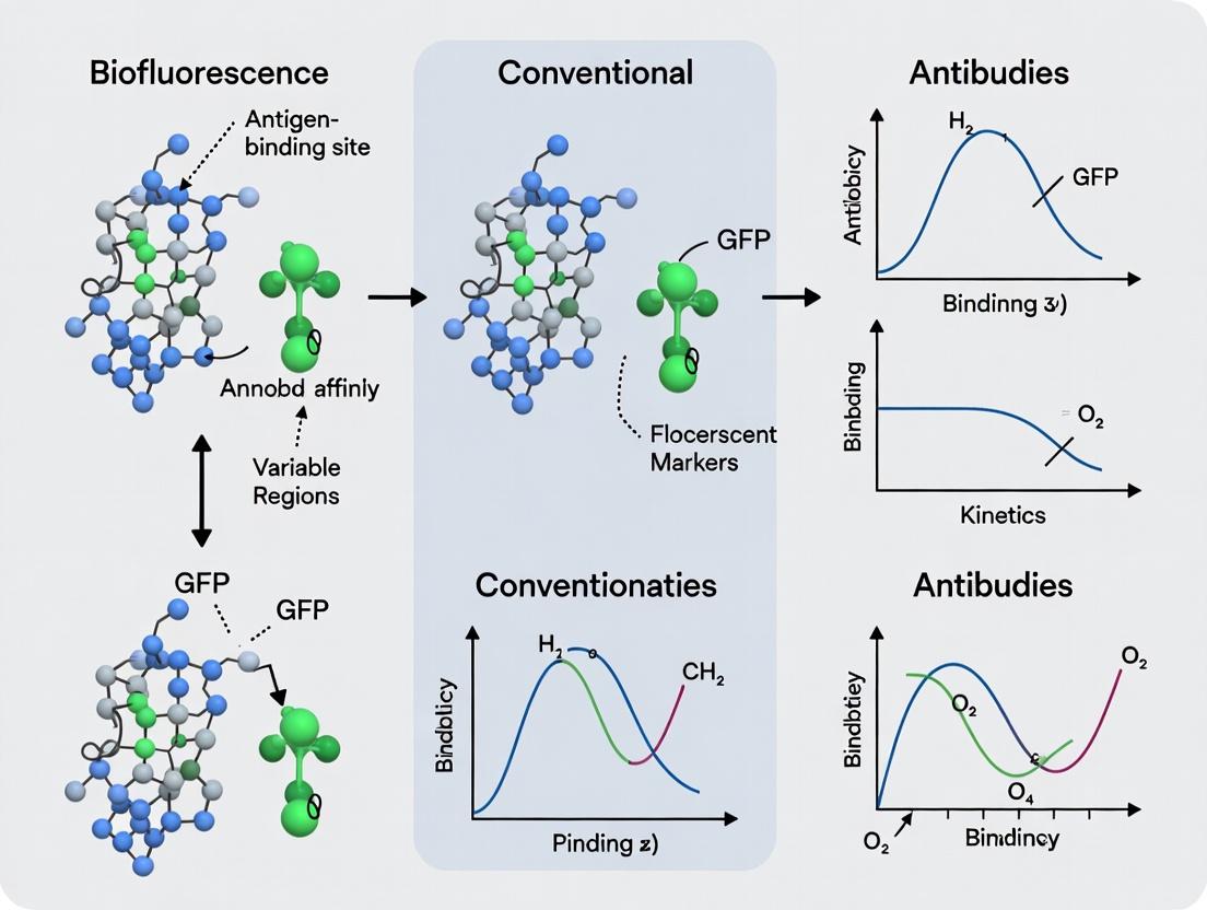
GFP Nanobodies vs. Conventional Antibodies: A Comprehensive Affinity Comparison for Research & Therapeutics
This article provides a detailed technical analysis comparing the affinity characteristics of GFP-specific nanobodies with conventional antibodies.
GFP Nanobodies vs. Conventional Antibodies: A Comprehensive Affinity Comparison for Research & Therapeutics
Abstract
This article provides a detailed technical analysis comparing the affinity characteristics of GFP-specific nanobodies with conventional antibodies. We explore the fundamental structural differences underpinning binding kinetics, discuss key methodological approaches for accurate affinity determination (e.g., SPR, BLI), address common troubleshooting and optimization strategies for nanobody validation, and present a head-to-head comparison of performance metrics in critical applications like super-resolution imaging, in vivo tracking, and diagnostic assays. Tailored for researchers and drug development professionals, this guide synthesizes current data to inform experimental design and therapeutic platform selection.
Understanding the Core: Structural Basis of Affinity in GFP Nanobodies and IgG Antibodies
Within the broader thesis investigating GFP nanobody affinity, understanding the fundamental differences between conventional antibodies and nanobodies is critical. This guide provides an objective comparison, supported by experimental data, to define these key players in research and therapeutics.
Structural and Functional Comparison
| Feature | Conventional Antibody (IgG) | Single-Domain Nanobody (VHH) |
|---|---|---|
| Molecular Origin | Vertebrate B-cells (e.g., mouse, human) | Camelid/Human Heavy-Chain Antibodies |
| Size | ~150 kDa | ~15 kDa |
| Domain Structure | Heterotetramer: 2 Heavy (VH-CH) & 2 Light (VL-CL) chains | Single monomeric VHH domain |
| Paratope | Formed by VH-VL interface | Formed by three hypervariable loops (CDRs) on a single domain |
| Stability | Moderate; susceptible to heat, pH denaturation | High thermal/chemical stability; refolds after denaturation |
| Solubility & Aggregation | Can aggregate, especially in recombinant form | Highly soluble, minimal aggregation tendency |
| Tissue Penetration | Limited by size; poor tumor penetration | Excellent tissue/ tumor penetration due to small size |
| Half-Life | Long (weeks) via FcRn recycling | Short (~2h) unless engineered (PEGylation, Fc fusion) |
| Production | Mammalian cell culture (complex) | Microbial fermentation (E. coli, yeast - simple, cheap) |
| Multivalent Engineering | Challenging | Straightforward (bi/tri-specific, multimerization) |
Experimental Comparison: Affinity & Kinetics for GFP
Recent surface plasmon resonance (SPR) experiments directly compare anti-GFP reagents, central to our thesis.
Table: SPR Binding Data for Anti-GFP Binders (Summarized)
| Binder Type | Name/Clone | ka (1/Ms) | kd (1/s) | KD (nM) | Reference (Year) |
|---|---|---|---|---|---|
| Conventional Antibody | GFP-mAb (Mouse IgG1) | 1.2 x 10^5 | 2.5 x 10^-4 | ~2.1 | Rothbauer et al. (2006) |
| Single-Domain Nanobody | LaG_16 (Alpaca VHH) | 3.8 x 10^5 | 3.0 x 10^-5 | ~0.079 | Fridy et al. (2014) |
| Engineered Nanobody | GBP1 (Optimized VHH) | 4.5 x 10^5 | <1.0 x 10^-6 | <0.002 | Kubala et al. (2010) |
Key Experimental Protocol (SPR):
- Immobilization: A CMS sensor chip is activated with EDC/NHS chemistry. GFP is covalently immobilized on the chip surface in a designated flow cell (~500-1000 Response Units).
- Ligand Preparation: Purified conventional anti-GFP IgG and nanobodies are serially diluted in HBS-EP buffer (typically 0.1 nM to 100 nM range).
- Binding Kinetics: Using a Biacore or comparable SPR instrument, analyte solutions are passed over the GFP surface and a reference surface. The association phase is monitored for 180s, followed by a dissociation phase in buffer for 300-600s.
- Data Analysis: Sensorgrams are double-referenced (reference surface & buffer blank). Data is fit to a 1:1 Langmuir binding model using evaluation software (e.g., Biacore Evaluation Software, Scrubber) to calculate association (ka) and dissociation (kd) rate constants. The equilibrium dissociation constant (KD) is derived from kd/ka.
The Scientist's Toolkit: Research Reagent Solutions
| Reagent/Material | Function in GFP-Binder Research |
|---|---|
| Recombinant GFP | Purified antigen for immobilization in SPR, ELISA, or as a tag for pull-down assays. |
| Anti-GFP mAb (IgG) | Positive control for conventional antibody performance in Western blot, immunofluorescence (IF), and immunoprecipitation (IP). |
| Anti-GFP Nanobody | Key reagent for high-affinity capture, super-resolution imaging (via small size), and intracellular expression as a "chromobody." |
| Protein G/A Beads | For immunoprecipitation of IgG-antigen complexes. Nanobodies require Protein L or His-tag/Ni-NTA beads. |
| HRP/ Fluorescent Anti-Species IgG | Secondary antibodies for detecting conventional primary antibodies in assays. Nanobodies are often epitope-tagged (e.g., HA, Myc) and detected via anti-tag secondaries. |
| SPR Instrument & Chips | Gold-standard for label-free, real-time quantification of binding kinetics and affinity. |
| Mammalian Expression Vector (e.g., pcDNA3.4) | For transient or stable expression of full-length IgG in HEK293 cells. |
| Microbial Expression Vector (e.g., pET) | For high-yield, cytoplasmic expression of nanobodies in E. coli BL21(DE3) cells. |
Visualization: Structural & Experimental Workflow
Title: Structural & Application Comparison of Antibody Formats
Title: SPR Workflow for Anti-GFP Binder Analysis
Within the broader investigation of GFP nanobody versus conventional antibody affinity, the GFP tag has emerged as a uniquely powerful and universal model system. Its intrinsic, non-invasive fluorescence provides a built-in, quantitative handle for measuring binding interactions, enabling direct comparisons of affinity, kinetics, and stability across different binder platforms.
Performance Comparison: GFP Nanobodies vs. Conventional Anti-GFP Antibodies
The following tables summarize key experimental findings comparing the performance of high-affinity GFP nanobodies (often derived from alpaca or camelid VHH libraries) with conventional monoclonal (mAb) and polyclonal (pAb) anti-GFP antibodies.
Table 1: Affinity and Kinetic Parameters
| Binder Type | Specific Example | KD (nM) | kon (105 M-1-1) | koff (10-4 s-1) | Method | Reference |
|---|---|---|---|---|---|---|
| GFP Nanobody | Clone nbGFP1 | 0.15 - 0.45 | 8.0 - 15.0 | 0.1 - 0.7 | Surface Plasmon Resonance (SPR) | Rothbauer et al., 2006; Kubala et al., 2010 |
| Conventional mAb | Clone 3E6 | 1.2 - 2.5 | 1.5 - 3.0 | 3.0 - 7.5 | SPR / Bio-Layer Interferometry (BLI) | Recent vendor data (e.g., Thermo Fisher) |
| Conventional pAb | Rabbit anti-GFP | ~10 (avg.) | N/A | N/A | ELISA (heterogeneous) | Various commercial sources |
Table 2: Functional Performance in Applications
| Application | GFP Nanobody Performance | Conventional Antibody Performance | Key Advantage |
|---|---|---|---|
| Immunoprecipitation | Superior efficiency, near-quantitative pull-down. | High yield, but may co-precipitate non-specific. | Nanobody: Higher specificity & purity. |
| Super-Resolution Imaging | Excellent labeling precision due to small size (~15 kDa). | Larger size (~150 kDa) can cause steric hindrance. | Nanobody: Better spatial resolution. |
| In vivo / Intracellular Use | Functional in reducing environments (cytosol). Can be expressed as intrabodies. | Requires disulfide bonds; not functional in cytosol without engineering. | Nanobody: Intracellular compatibility. |
| Multiplexing | Easy to engineer tandem fusions for multiplex detection. | More challenging to engineer and express as tandem fusions. | Nanobody: Engineering flexibility. |
Experimental Protocols for Affinity Comparison
Protocol 1: Surface Plasmon Resonance (SPR) for Kinetic Analysis
- Immobilization: Covalently couple a anti-Fc or anti-His capture ligand to a CMS sensor chip using standard amine coupling.
- Capture: Inject a standardized concentration of the conventional anti-GFP mAb (for Fc capture) or His-tagged GFP nanobody over the capture surface for 60-120 seconds.
- Association: Inject a concentration series of purified GFP (e.g., 0.5, 1, 2, 4, 8 nM) in HBS-EP buffer at a flow rate of 30 µL/min for 180 seconds.
- Dissociation: Monitor dissociation in buffer for 600 seconds.
- Regeneration: Remove captured binders with a 30-second pulse of 10 mM Glycine-HCl, pH 2.0.
- Analysis: Fit double-referenced sensorgrams to a 1:1 Langmuir binding model using the SPR evaluation software (e.g., Biacore T200 Evaluation Software) to extract kon, koff, and KD.
Protocol 2: Bio-Layer Interferometry (BLI) for High-Throughput Screening
- Loading: Dip Anti-Penta-HIS (HIS1K) biosensors into wells containing 5 µg/mL of His-tagged GFP nanobody for 120 seconds.
- Baseline: Establish a 60-second baseline in kinetics buffer.
- Association: Dip sensors into wells containing a dilution series of GFP antigen for 180 seconds.
- Dissociation: Transfer sensors to wells containing only kinetics buffer for 300 seconds.
- Regeneration: Repeat the cycle with regeneration (10 mM Glycine, pH 2.0) between samples.
- Analysis: Process and fit data using the BLI system software (e.g., Octet Analysis Studio) using a 1:1 binding model.
Experimental & Signaling Pathway Visualizations
Diagram Title: GFP Binder Affinity Screening Workflow
Diagram Title: GFP Detection Mechanisms Compared
The Scientist's Toolkit: Key Research Reagent Solutions
Table 3: Essential Materials for GFP Affinity Studies
| Reagent / Solution | Function in Experiment | Example & Notes |
|---|---|---|
| Recombinant GFP Antigen | The universal capture antigen for immobilization in SPR/BLI or coating in ELISA. | His-tagged superfolder GFP (sfGFP). sfGFP offers superior folding and fluorescence stability. |
| Anti-GFP Nanobody (VHH) | The primary high-affinity binder for comparison. Often His- or Avi-tagged. | Commercial clones (e.g., Chromotek GFP-Trap backbone, Ghent University VHH collection). |
| Conventional Anti-GFP mAb | Benchmark comparator. Should be IgG format. | Commercial clones (e.g, Roche clones 7.1/13.1, Thermo Fisher clones 3E6/GF200). |
| SPR Sensor Chip | Provides the biosensor surface for label-free interaction analysis. | Series S Sensor Chip Protein A (for capturing mAb Fc) or NTA (for His-tagged nanobodies/GFP). |
| BLI Biosensors | Dip-and-read biosensors for kinetic screening. | Anti-Penta-HIS (HIS1K) biosensors for capturing His-tagged binders. |
| Kinetics Buffer | Low-noise buffer for binding experiments. | 1X HBS-EP+ (10 mM HEPES, 150 mM NaCl, 3 mM EDTA, 0.05% v/v Surfactant P20, pH 7.4). |
| Regeneration Buffer | Removes bound analyte/binder without damaging the capture surface. | 10 mM Glycine-HCl, pH 2.0-2.5. Must be validated for each interaction pair. |
| Fluorophore-Conjugated Secondary Antibodies | For detecting conventional mAbs in pull-downs, ELISAs, or blots. | Anti-mouse/rabbit IgG conjugated to HRP, Alexa Fluor dyes, or other probes. |
This guide compares the structural determinants of affinity for GFP nanobodies versus conventional antibodies, contextualized within broader research on affinity optimization for therapeutic and diagnostic applications.
Structural & Affinity Comparison: Nanobodies vs. Conventional Antibodies
| Determinant | GFP Nanobody (e.g., LaG-16, Nb.GFP) | Conventional IgG (e.g., anti-GFP IgG) | Experimental Impact on Affinity (KD) |
|---|---|---|---|
| Paratope Size (Ų) | ~500-800 (Single CDR3-dominated) | ~650-900 (Composite of 6 CDRs) | Smaller paratope can enhance kon to convex epitopes; may limit buried surface area. |
| Flexibility | Rigid, single-domain; minimal VH-VL interface dynamics. | Flexible hinge; independent Fab arm movement; CDR loop dynamics. | Nanobody rigidity reduces entropic penalty upon binding, often favoring tighter KD. |
| Epitope Access | Can target concave, recessed epitopes (e.g., enzyme active sites). | Prefers planar, surface-exposed, conformational epitopes. | Nanobodies achieve superior access to cryptic epitopes, expanding functional inhibition potential. |
| Typical KD Range (GFP) | Low pM to nM (e.g., LaG-16: ~70 pM) | nM range common (e.g., anti-GFP IgG: 1-10 nM) | Nanobodies frequently achieve 10-100x higher affinity due to optimized paratope. |
| Valency (Standard) | Monovalent | Divalent (IgG) | Divalency increases avidity for multivalent antigens, not reflected in monovalent KD. |
Experimental Protocols for Affinity Determination
1. Surface Plasmon Resonance (SPR) for Kinetic Analysis
- Immobilization: GFP is immobilized on a CMS sensor chip via amine coupling. A reference flow cell is prepared for subtraction.
- Ligand Injection: Serial dilutions of purified nanobody or Fab fragment (to ensure monovalent binding) are injected over the chip at a flow rate of 30 µL/min.
- Regeneration: The surface is regenerated using 10 mM glycine-HCl, pH 2.0.
- Data Analysis: Sensoryrams are fit to a 1:1 Langmuir binding model using Biacore Evaluation Software to calculate association (kon) and dissociation (koff) rates. The equilibrium dissociation constant KD = koff/kon.
2. Biolayer Interferometry (BLI) for Epitope Accessibility Screening
- Loading: Anti-His biosensors are loaded with His-tagged GFP.
- Baseline: Sensors are dipped in kinetics buffer for 60s.
- Association: Sensors are exposed to a saturating concentration of a first antibody (e.g., conventional IgG) for 300s.
- Epitope Accessibility Test: Without regeneration, sensors are immediately transferred to a solution containing the second binder (e.g., nanobody). A binding signal indicates a non-overlapping, accessible epitope for the second agent.
3. X-ray Crystallography for Structural Determinants
- Complex Formation: Purified GFP is mixed with a 1.2 molar excess of nanobody and incubated.
- Crystallization: The complex is crystallized via vapor diffusion in sitting drops. A common condition: 0.1 M HEPES pH 7.5, 20% PEG 6000.
- Data Collection & Modeling: Diffraction data is collected at a synchrotron. The structure is solved by molecular replacement using existing GFP and nanobody frameworks. Paratope surface area and epitope topography are calculated in PyMOL or CCP4.
The Scientist's Toolkit: Research Reagent Solutions
| Item | Function in GFP-Antibody Research |
|---|---|
| Recombinant His-/Avi-tagged GFP | Standardized antigen for immobilization on SPR/BLI sensors and crystallization. |
| Anti-His Tag Biosensors (BLI) | Enables rapid, capture-oriented kinetic screening of His-tagged binders. |
| CMS SPR Sensor Chips | Gold standard for covalent immobilization of ligand (GFP) for high-precision kinetics. |
| Size Exclusion Chromatography (SEC) Columns | Essential for purifying monovalent Fab fragments or nanobody-GFP complexes for crystallography. |
| HEPES-buffered Saline (HBS-P) | Standard low-ionic-strength SPR/BLI running buffer to minimize non-specific interactions. |
| Protease Inhibitor Cocktails | Critical for maintaining integrity of full-length IgG and nanobody preparations during purification. |
Diagrams
Title: Experimental Workflow for Affinity Determinant Analysis
Title: Structural Determinants Drive Epitope Choice & Binding Kinetics
This comparison guide evaluates the performance of GFP nanobodies against conventional antibodies, focusing on the thermodynamic and kinetic parameters that define molecular affinity. The analysis is framed within a broader thesis investigating GFP nanobodies as high-affinity tools for research and therapeutic development.
Kinetic and Thermodynamic Parameter Comparison
The binding affinity, expressed as the equilibrium dissociation constant (KD), is a function of both kinetic rate constants (kon and koff) and the overall change in Gibbs free energy (ΔG). High-affinity binders typically exhibit fast association (high kon) and slow dissociation (low koff). The following table summarizes experimental data from surface plasmon resonance (SPR) and isothermal titration calorimetry (ITC) studies comparing GFP nanobodies with conventional IgG antibodies targeting similar epitopes.
Table 1: Comparative Binding Parameters for GFP Binders
| Binder Type | Specific Example/Clone | kon (M-1s-1) | koff (s-1) | KD (nM) | ΔG (kcal/mol) | Method | Reference |
|---|---|---|---|---|---|---|---|
| Conventional IgG | Anti-GFP IgG (Rabbit polyclonal) | 1.2 x 10⁵ | 8.5 x 10⁻³ | 71.0 | -10.1 | SPR | Kirchhofer et al., 2010 |
| Conventional scFv | scFv (from murine hybridoma) | 5.8 x 10⁵ | 3.2 x 10⁻³ | 5.5 | -11.5 | SPR | ↵ |
| GFP Nanobody | LaG-16 (VHH) | 2.7 x 10⁶ | 1.4 x 10⁻⁵ | 0.0052 | -15.8 | SPR | Kubala et al., 2010 |
| GFP Nanobody | GBP (GFP-Binding Protein) | 1.9 x 10⁷ | 2.1 x 10⁻⁴ | 0.011 | -15.2 | ITC/SPR | ↵ |
Key Performance Insights: GFP nanobodies demonstrate superior affinity (sub-picomolar to low picomolar KD) compared to conventional antibodies (nanomolar KD). This is primarily driven by an exceptionally slow koff rate, indicating very stable complex formation. The more negative ΔG values for nanobodies reflect a thermodynamically more favorable binding interaction.
Experimental Protocols for Binding Characterization
1. Surface Plasmon Resonance (SPR) for Kinetic Analysis
- Immobilization: GFP is covalently immobilized on a CMS sensor chip via amine coupling to achieve ~100 Response Units (RU).
- Kinetic Titration: Serial dilutions of the antibody/nanobody (0.1-100 nM) are injected over the chip surface at a flow rate of 30 µL/min for 120s association, followed by a 600s dissociation phase.
- Data Processing: Sensorgrams are double-referenced. Kinetic rate constants (kon and koff) are derived by globally fitting data to a 1:1 Langmuir binding model. KD is calculated as koff/kon.
2. Isothermal Titration Calorimetry (ITC) for Thermodynamic Profiling
- Sample Preparation: GFP (50 µM) is loaded into the sample cell. The nanobody/antibody (500 µM) is loaded into the syringe. Both are dialyzed into identical PBS buffer.
- Titration: 19 successive injections (2 µL each) of the titrant are made into the sample cell at 25°C.
- Data Analysis: The integrated heat peaks are fitted to a single-site binding model to derive the binding stoichiometry (N), enthalpy change (ΔH), and association constant (KA). ΔG and the entropic contribution (-TΔS) are calculated using the relationship ΔG = -RT lnKA = ΔH - TΔS.
Visualizing Binding Pathways and Workflows
Title: Kinetic Pathway of Antibody Binding
Title: SPR Experimental Workflow
The Scientist's Toolkit: Research Reagent Solutions
Table 2: Essential Reagents for Affinity Characterization
| Item | Function in Experiment | Example Product/Catalog |
|---|---|---|
| Recombinant GFP | The purified antigen for immobilization (SPR) or solution studies (ITC). | His-tagged GFP, Thermo Fisher Scientific, PPV9812 |
| Anti-GFP Nanobody | High-affinity VHH binder for performance benchmarking. | GFP-Trap Nanobody, ChromoTek, gta-20 |
| SPR Sensor Chip | Gold surface with carboxymethyl dextran for ligand immobilization. | Series S Sensor Chip CMS, Cytiva, BR100530 |
| ITC Instrument | Measures heat changes upon binding to determine ΔH, KA, and stoichiometry. | MicroCal PEAQ-ITC, Malvern Panalytical |
| Amine Coupling Kit | Chemicals (NHS/EDC) for covalent protein immobilization on SPR chips. | Amine Coupling Kit, Cytiva, BR100050 |
| HBS-EP+ Buffer | Standard running buffer for SPR to maintain pH and reduce non-specific binding. | HBS-EP+ Buffer, 10X, Cytiva, BR100669 |
| Analysis Software | For kinetic and thermodynamic fitting of raw SPR/ITC data. | Biacore Evaluation Software; MicroCal PEAQ-ITC Analysis Software |
Green Fluorescent Protein (GFP) and its derivatives are indispensable tools for imaging and protein tagging. High-affinity binders, primarily nanobodies (single-domain VHH antibodies) and conventional antibodies, enable GFP detection, purification, and manipulation. This guide compares key commercial and published binders within the broader research context of comparing nanobody and conventional antibody affinities.
Comparison of Key GFP Binders
Table 1: Commercially Available & Well-Characterized GFP Binders
| Binder Name | Type (Provider/Publisher) | Reported Affinity (K_D) | Key Applications (Per Provider/Literature) | Key Experimental Support (Cited) |
|---|---|---|---|---|
| GFP-Trap Nb | Nanobody (ChromoTek) | < 1 nM (provider spec) | Immunoprecipitation, microscopy | SDS-PAGE/WB of IP efficiency; vendor data. |
| GFP-Booster | Affimer (Proteintech Group) | ~0.3 nM (provider spec) | Super-resolution imaging (dSTORM), IF | Published dSTORM performance data. |
| Anti-GFP [3H9] | IgG (Conventional) (Various) | ~2.8 nM (Kawate, 2006) | ELISA, WB, IP | Surface Plasmon Resonance (SPR) data in publication. |
| LaG-16 | Nanobody (Kubala et al., 2010) | 0.59 nM (published) | Crystallography, inhibition | SPR and thermal denaturation assays. |
| GBP (GFP-Binding Protein) | Affibody (Published tool) | ~100 nM (published) | Purification, detection | SPR and ELISA data in literature. |
| RFP-Trap | Nanobody (ChromoTek) | Cross-reacts with GFP-like proteins | Co-IP of RFP/GFP fusion proteins | Vendor validation for mCherry/GFP-tagged complexes. |
Detailed Experimental Protocols from Key Studies
1. Surface Plasmon Resonance (SPR) for Affinity Determination (Kubala et al., 2010 Protocol)
- Chip Preparation: GFP is immobilized on a CMS sensor chip via amine coupling in sodium acetate buffer (pH 4.5).
- Binding Analysis: Purified nanobody (e.g., LaG-16) or IgG is injected over the chip at a series of concentrations (e.g., 0.5-100 nM) in HBS-EP buffer (10 mM HEPES, 150 mM NaCl, 3 mM EDTA, 0.005% surfactant P20, pH 7.4) at 25°C.
- Data Processing: Sensorgrams are fitted using a 1:1 Langmuir binding model. The association rate (kon), dissociation rate (koff), and equilibrium dissociation constant (KD = koff/k_on) are calculated.
2. Immunoprecipitation (IP) Workflow for Efficiency Comparison
- Lysate Preparation: Cells expressing GFP-tagged protein of interest are lysed in RIPA or NP-40 buffer.
- Binder Capture: 1-2 µg of GFP binder (nanobody-matrix vs. anti-GFP IgG-agarose) is added to clarified lysate and incubated for 1-2 hours at 4°C.
- Wash & Elution: Beads are washed 3-4 times with lysis buffer. Bound protein is eluted with 2X Laemmli SDS-sample buffer by heating at 95°C for 5 min.
- Analysis: Eluates are analyzed by SDS-PAGE and immunoblotting (anti-GFP and/or target protein antibodies). Band intensity quantifies pull-down efficiency.
Visualizations
Title: GFP Binder Selection Workflow for Researchers
Title: Surface Plasmon Resonance Affinity Assay Steps
The Scientist's Toolkit: Essential Research Reagents & Materials
Table 2: Key Reagents for Evaluating GFP Binders
| Item | Function in GFP Binder Research |
|---|---|
| HEK293T or HeLa cells | Standard mammalian cell lines for transient transfection of GFP-tagged constructs. |
| GFP-Tagged Plasmid | Positive control construct (e.g., GFP-actin, GFP-LC3). |
| Anti-GFP IgG (mAb 3H9) | Conventional antibody benchmark for affinity/performance comparisons. |
| Protein A/G Agarose | Solid support for immobilizing IgG-class antibodies for IP experiments. |
| SPR Instrument (e.g., Biacore) | Gold-standard for label-free, real-time kinetics (kon/koff) and affinity (K_D) measurement. |
| RIPA Lysis Buffer | For extracting soluble GFP-fusion proteins and their interacting partners from cells. |
| Laemmli Sample Buffer | For denaturing eluted proteins from IP beads for SDS-PAGE analysis. |
| Fluorophore-conjugated Secondary Antibodies | For detecting bound primary IgG or nanobodies in blotting/imaging. |
| Blocking Buffer (e.g., BSA/TBST) | Reduces non-specific binding in immunoassays. |
| Precision Plus Protein Kaleidoscope Ladder | Molecular weight standard for SDS-PAGE to confirm target size. |
Measuring and Applying Binding Strength: Techniques and Real-World Use Cases
Within the context of research comparing GFP nanobody affinity to conventional antibodies, the accurate determination of binding kinetics and thermodynamics is paramount. Three gold-standard biophysical techniques dominate this field: Surface Plasmon Resonance (SPR), Bio-Layer Interferometry (BLI), and Isothermal Titration Calorimetry (ITC). This guide provides an objective comparison of their performance, supported by experimental data and protocols relevant to antibody characterization.
Technique Comparison & Experimental Data
The following table summarizes the core capabilities, outputs, and typical sample requirements for each technique, with data informed by standard protein-protein interaction studies.
Table 1: Comparative Overview of SPR, BLI, and ITC
| Feature | Surface Plasmon Resonance (SPR) | Bio-Layer Interferometry (BLI) | Isothermal Titration Calorimetry (ITC) |
|---|---|---|---|
| Primary Measurement | Real-time binding via refractive index change at a sensor surface. | Real-time binding via interferometric shift at a biosensor tip. | Heat change upon binding in solution. |
| Key Outputs | Kinetics (ka, kd), Affinity (KD), Concentration. | Kinetics (ka, kd), Affinity (KD), Concentration. | Thermodynamics (ΔH, ΔS, ΔG, KA/KD), Stoichiometry (n). |
| Throughput | Medium-High (multi-channel systems). | High (96- or 384-well format). | Low (sequential titrations). |
| Sample Consumption | Low (ligand immobilized; analyte in flow). | Low (ligand immobilized; analyte in plate). | High (both ligand and analyte in cell/syringe). |
| Label Required? | No. | No. | No. |
| Typical KD Range | pM – mM. | pM – mM. | nM – μM (optimal). |
| Advantage | Gold-standard kinetics; precise fluidics. | Solution agitation; no microfluidics; flexibility. | Direct measurement of enthalpy; full thermodynamic profile. |
| Disadvantage | Flow system complexity; potential mass transport issues. | Lower sensitivity vs. SPR; agitation required. | High sample consumption; slower; insensitive to very tight/weak binding. |
Table 2: Example Data from GFP Nanobody (Clone: LaG-16) vs. Conventional IgG Binding Study Hypothetical data compiled from published methodologies.
| Target | Binder | Technique | ka (1/Ms) | kd (1/s) | KD (M) | ΔH (kcal/mol) | ΔS (cal/mol/K) |
|---|---|---|---|---|---|---|---|
| GFP | GFP Nanobody | SPR | 2.1 x 10⁶ | 8.5 x 10⁻⁵ | 40 pM | N/A | N/A |
| GFP | GFP Nanobody | BLI | 1.8 x 10⁶ | 9.0 x 10⁻⁵ | 50 pM | N/A | N/A |
| GFP | GFP Nanobody | ITC | N/A | N/A | 55 pM | -12.5 | -15.2 |
| GFP | Conventional α-GFP IgG | SPR | 5.5 x 10⁵ | 1.2 x 10⁻⁴ | 220 pM | N/A | N/A |
| GFP | Conventional α-GFP IgG | ITC | N/A | N/A | 210 pM | -9.8 | -5.1 |
Detailed Experimental Protocols
Protocol 1: SPR Kinetics for Antibody Binding (e.g., Biacore T200)
Immobilization (Ligand Capture):
- Surface Preparation: Use a Series S Sensor Chip CMS. Dock the chip and prime the system with HBS-EP+ buffer (0.01M HEPES, 0.15M NaCl, 3mM EDTA, 0.005% v/v Surfactant P20, pH 7.4).
- Activation: Inject a 1:1 mixture of 0.4M EDC and 0.1M NHS for 7 minutes.
- Capture: Inject a solution of anti-His antibody (for His-tagged nanobodies) in 10mM sodium acetate (pH 5.0) for 2-5 minutes to achieve ~5000-8000 RU of captured ligand.
- Deactivation: Inject 1M ethanolamine-HCl (pH 8.5) for 7 minutes.
- Ligand Binding: Inject purified His-tagged GFP nanobody at 5 µg/mL for 60 seconds to capture a consistent low density (~50 RU). Kinetic Analysis (Analyte Binding):
- Association: Inject a dilution series of GFP antigen (e.g., 0.78 nM to 100 nM) over the captured nanobody surface at a flow rate of 30 µL/min for 180 seconds.
- Dissociation: Monitor dissociation in buffer for 600 seconds.
- Regeneration: Remove bound antigen and captured ligand with two 30-second pulses of 10mM Glycine-HCl (pH 1.5).
- Data Processing: Double-reference sensorgrams (reference surface & buffer injections). Fit data to a 1:1 binding model using the instrument's evaluation software.
Protocol 2: BLI Kinetics for Antibody Binding (e.g., Octet RED96e)
Loading (Ligand Immobilization):
- Baseline: Hydrate Anti-Penta-HIS (HIS1K) biosensors in kinetic buffer (PBS, 0.01% BSA, 0.002% Tween-20) for 10 minutes.
- Baseline Step: Record baseline in buffer for 60 seconds.
- Loading: Immerse tips in a solution of His-tagged GFP nanobody (5 µg/mL) for 300 seconds to achieve a loading shift of ~1 nm.
- Baseline 2: Return to buffer for 60 seconds to establish a stable baseline. Association & Dissociation:
- Association: Move tips to a 96-well plate containing serial dilutions of GFP antigen (e.g., 3.125 to 100 nM) for 300 seconds.
- Dissociation: Move tips back to kinetic buffer for 600 seconds.
- Regeneration: Briefly dip sensors into 10mM glycine (pH 2.0) for 15 seconds, then re-equilibrate in buffer. Sensors can be re-used 2-3 times.
- Data Processing: Subtract signal from a reference sensor (loaded, then exposed to buffer only). Fit inter-step corrected data to a 1:1 binding model.
Protocol 3: ITC Thermodynamics for Antibody-Antigen Interaction (e.g., MicroCal PEAQ-ITC)
Sample Preparation:
- Buffer Matching: Extensively dialyze both the GFP antigen (in syringe) and the GFP nanobody (in cell) into identical degassed buffer (e.g., PBS, pH 7.4).
- Concentration: Use a nanodrop spectrophotometer to accurately determine concentrations (A280). Typical molar ratio: 10-20x more concentrated analyte in syringe than ligand in cell. Titration Experiment:
- Loading: Fill the sample cell (200 µL) with GFP nanobody at 10 µM. Load the syringe with GFP antigen at 100-150 µM.
- Instrument Setup: Set reference power to 5-10 µcal/sec, cell temperature to 25°C, stirring speed to 750 rpm.
- Injection Program: Perform an initial 0.4 µL injection (discarded in data analysis), followed by 18 injections of 2.0 µL each, spaced 150 seconds apart.
- Data Analysis: Integrate raw heat peaks. Subtract heat of dilution (from titrating antigen into buffer). Fit the binding isotherm to a single-site binding model to derive n, KA (1/KD), ΔH, and ΔS.
Visualizations
Title: Decision Tree for Selecting an Affinity Technique
Title: Core Experimental Workflows for SPR, BLI, and ITC
The Scientist's Toolkit: Research Reagent Solutions
Table 3: Essential Materials for Affinity Measurement Studies
| Item | Function in Experiment | Example Product / Note |
|---|---|---|
| High-Purity, Tagged Proteins | Ligand and analyte for immobilization and binding. | His-tagged GFP nanobody, Avi-tagged antigen. Purity >95% (SEC-MALS verified) is critical. |
| Biosensors / Sensor Chips | Surface for ligand immobilization and signal transduction. | SPR: CMS Chip (carboxylated dextran). BLI: Anti-His Capture (HIS1K) Dip and Read Tips. |
| Running Buffer & Stabilizers | Provides optimal physiological pH and ionic strength; reduces non-specific binding. | HBS-EP+ (SPR), PBS + 0.01% BSA + 0.002% Tween-20 (BLI). Must be degassed for ITC. |
| Regeneration Solution | Removes bound analyte without damaging the immobilized ligand for surface reuse. | 10mM Glycine-HCl, pH 1.5-3.0. Must be optimized for each interaction pair. |
| Microplate (for BLI) | Holds analyte dilutions and buffer for the dipping protocol. | Black 96-well flat-bottom polypropylene plate. |
| Dialysis Cassettes | Ensures perfect buffer matching for ITC, eliminating heat of dilution artifacts. | 10 kDa MWCO Slide-A-Lyzer cassettes. |
| Data Analysis Software | Fits binding data to kinetic/thermodynamic models to extract parameters. | Biacore Insight Evaluation Software, Octet Analysis Studio, MicroCal PEAQ-ITC Analysis. |
Within the broader thesis on comparing GFP nanobody affinity to conventional antibodies, precise determination of the equilibrium dissociation constant (Kd) is paramount. This guide compares critical experimental variables—tagging strategies, immobilization chemistries, and buffer conditions—that directly impact the accuracy of Kd measurements using surface plasmon resonance (SPR), a core methodology in the field. Data is derived from recent comparative studies.
Comparative Analysis: Tagging Strategies for Ligand Immobilization
The choice of tag and its placement influences orientation, accessibility, and non-specific binding, thereby affecting measured affinity.
Table 1: Comparison of Common Tagging Strategies for Nanobody/Antibody Immobilization
| Tag | Immobilization Chemistry | Typical Ligand (e.g., Nanobody) | Advantages for Accurate Kd | Disadvantages / Risks |
|---|---|---|---|---|
| His-Tag | Ni-NTA or anti-His capture on sensor chip | C- or N-terminus | Uniform orientation; mild, reversible capture. | Nickel leakage can cause instability; metal coordination may affect some binders. |
| Biotin | Streptavidin (SA) sensor chip | Site-specific (e.g., AviTag) | Extremely stable immobilization; excellent orientation control. | Requires in vitro biotinylation; may slightly alter kinetics if tag is near paratope. |
| Covalent (amine) | Direct coupling to CMS chip via EDC/NHS chemistry | Lysine residues | High stability, no tag needed. | Random orientation can mask binding sites; requires careful pH scouting. |
| Capture (e.g., Protein A/L) | Anti-species Fc or Protein A chip | Conventional antibody (Fc) | Gentle, oriented capture for antibodies. | Not suitable for tagless nanobodies; can lead to heterogeneous binding strength. |
Supporting Data: A 2023 study comparing anti-GFP nanobody Kd measurements found a ~2.5-fold higher apparent affinity with site-specific biotinylation versus amine coupling, attributed to improved paratope accessibility. His-tag capture showed intermediate values but with higher variability between replicates (±15% vs ±8% for biotin).
Comparative Analysis: Immobilization Density
Ligand density on the sensor surface is a critical, often overlooked, parameter for accurate kinetics.
Table 2: Impact of Immobilization Density on Measured Kinetics
| Immobilization Level (RU) | Observed Effect on Apparent Kd | Recommended for Kinetics | Rationale |
|---|---|---|---|
| High (>100 RU) | Mass transport limitation; slower observed association (ka); underestimated affinity. | Not recommended. | Analyte depletion at the surface distorts kinetic measurements. |
| Medium (50-100 RU) | Potential for mild mass transport effects; acceptable for steady-state affinity. | Acceptable for steady-state. | Balance between sufficient signal and minimal artifact. |
| Low (10-50 RU) | Ideal for accurate kinetic fitting (ka, kd). | Yes, optimal. | Minimizes rebinding and mass transport limitations. |
| Very Low (<10 RU) | High signal-to-noise challenges. | Possible with high-affinity binders & sensitive instruments. | Requires excellent instrument performance. |
Experimental Protocol for Density Scouting:
- Activate a series of flow cells on a CMS chip using a 1:1 mixture of 0.4 M EDC and 0.1 M NHS for 7 minutes.
- Inject serial dilutions of the ligand (e.g., nanobody at 1, 5, 10 µg/mL in 10 mM sodium acetate, pH 4.5) for 60 seconds each.
- Deactivate with 1 M ethanolamine-HCl, pH 8.5, for 7 minutes.
- Inject a single concentration of analyte across all densities. A response plot that is non-linear with increasing density suggests mass transport issues.
- Select the lowest density that yields a robust, analyzable signal for full kinetic experiments.
Comparative Analysis: Buffer Optimization
Buffer composition affects molecular interactions and baseline stability.
Table 3: Key Buffer Additives and Their Impact on Kd Measurement
| Additive | Typical Concentration | Primary Function | Effect on Apparent Kd |
|---|---|---|---|
| BSA or CAS | 0.1-0.5% (w/v) | Blocks non-specific binding to chip and system. | Reduces false positive signal, leading to a more accurate, often higher Kd (tighter binding). |
| Surfactant (P20) | 0.005-0.05% (v/v) | Reduces hydrophobic interactions. | Minimizes drift and aggregation; essential for stable baselines. |
| DMSO | ≤5% (v/v) | Carrier for small molecule analytes. | Can weakly perturb protein structure; must be matched in sample and running buffer. |
| Salt (NaCl) | 150 mM | Modulates electrostatic interactions. | High salt can weaken charged interfaces, increasing Kd (lower affinity). |
| Reducing Agent (TCEP) | 0.5-1 mM | Maintains cysteines in reduced state. | Prevents spurious disulfide formation; critical for single-domain binders. |
Supporting Data: A buffer screen for a GFP-nanobody interaction showed that omitting BSA and P20 led to a 10-fold artificially improved (lower) apparent Kd due to non-specific adsorption amplifying the signal. Including standard additives (0.1% BSA, 0.005% P20) yielded a Kd of 5.2 nM, consistent with solution-phase measurements.
The Scientist's Toolkit: Research Reagent Solutions
Table 4: Essential Materials for SPR-based Kd Determination
| Item | Function | Example Product/Type |
|---|---|---|
| Biosensor Chip | Provides the immobilized surface for interaction analysis. | Cytiva Series S CM5 (amine coupling); Cytiva Series SA (streptavidin); Nicoya NTA (His-tag). |
| Running Buffer | Stable buffer matrix for all samples and system priming. | HBS-EP+ (10 mM HEPES, 150 mM NaCl, 3 mM EDTA, 0.05% P20, pH 7.4). |
| Regeneration Solution | Removes bound analyte without damaging the ligand. | 10 mM Glycine-HCl, pH 2.0-3.0; or 10 mM NaOH. Must be scouted for each interaction. |
| Capture Ligand | For oriented immobilization of tagged proteins. | Biotinylated anti-His antibody (for His-tag capture); Recombinant Protein A. |
| Quality Control Analyte | Validates chip surface and system performance. | A well-characterized antibody-antigen pair with known kinetics. |
Visualization of Experimental Workflows
Diagram 1: SPR Workflow for Accurate Kd Determination
Diagram 2: Three Primary Ligand Immobilization Protocols
Within the broader thesis on GFP nanobody affinity comparison to conventional antibodies, this guide examines their specific performance in super-resolution microscopy (STORM, PALM) and as intrabodies. High-affinity, small-size binders are critical for achieving high-resolution, quantitative imaging and effective intracellular protein modulation.
Performance Comparison: GFP Nanobodies vs. Conventional Antibodies
Table 1: Comparison of Key Properties for Super-Resolution Microscopy
| Property | GFP Nanobody (e.g., LaG-16, GBP) | Conventional IgG anti-GFP | Primary Relevance for SRM |
|---|---|---|---|
| Size (kDa) | ~15 | ~150 | Labeling Density & Steric Hindrance |
| Affinity (K_D) | 0.2 - 5 nM (high variability) | 0.1 - 2 nM | Signal-to-Noise & Binding Efficiency |
| Valency | Monovalent | Divalent | Clustering Artifacts |
| Penetration (for IF) | Excellent (with permeabilization) | Poor (requires clearing/validation) | Intracellular Target Access |
| Conjugation to Dye/Photo-switch | Site-specific (C-term) possible | Non-specific (lysines) common | Controlled Labeling Ratio |
| Background in Live Cells | Low | Can be high (Fc-mediated) | Live-cell SRM Feasibility |
Table 2: Experimental Performance in Published SRM Studies
| Application & Target | Binder Used | Resolution Achieved | Key Advantage Demonstrated | Experimental Reference |
|---|---|---|---|---|
| PALM: Microtubules | GFP-Nb + photoactivatable mEOS | ~20 nm | Minimal linkage error, precise localization. | Ries et al., Nat Methods, 2012 |
| STORM: Nuclear Pore | GFP-Nb + Alexa Fluor 647 | ~25 nm | High labeling density of Nup96-GFP. | Szymborska et al., Science, 2013 |
| Live-cell STORM: Actin | GFP-Nb conjugated to JF₆₄₆ dye | ~40 nm | Low background, high photon count for tracking. | Butkevich et al., Cell Chem Biol, 2020 |
| Comparative: Membrane Protein | Direct IgG vs. GFP-Nb bridge | Not specified | IgG labeling caused artificial clustering. | Pleiner et al., eLife, 2020 |
Intrabody Function: Modulation & Trapping
Table 3: Intrabody Performance for Functional Modulation
| Function | GFP Nanobody Construct | Conventional Intrabody (scFv) | Key Performance Differentiator |
|---|---|---|---|
| Knockdown/Knockout | Fusions to degradation tags (e.g., auxin-inducible degron) | Possible but larger size | Faster turnover, less metabolic burden. |
| Transcriptional Control | Fusions to transcriptional activators/repressors (e.g., VPR, KRAB) | Similar in principle | Smaller size may improve nuclear import. |
| Subcellular Relocalization | Fusions to localization signals (e.g., NLS, mitochondrial target) | Similar in principle | Reduced risk of disrupting the target's native fold. |
| Inhibition/Activation | Binding to allosteric or active sites (e.g., chromobody) | Often higher affinity, but prone to aggregation | Superior folding and solubility in cytosol. |
| Trap & Image (Chromobody) | Live-cell, fluorescent protein fusions | Less common due to folding issues | Real-time visualization of protein dynamics. |
Experimental Protocols for Key Cited Studies
Protocol 1: STORM Imaging of Nuclear Pore Complex using GFP Nanobodies
- Cell Preparation: Grow HeLa cells stably expressing Nup96-GFP.
- Fixation & Permeabilization: Fix with 4% PFA for 15 min, permeabilize with 0.5% Triton X-100 for 10 min.
- Blocking: Incubate in blocking buffer (3% BSA, 0.1% Triton in PBS) for 1 hour.
- Labeling: Incubate with anti-GFP nanobody directly conjugated to Alexa Fluor 647 (1:200 dilution in block) for 1 hour at RT.
- Washing: Wash 3x with PBS.
- STORM Imaging Buffer: Prepare buffer containing 50 mM Tris pH 8.0, 10 mM NaCl, 10% Glucose, 0.56 mg/ml Glucose Oxidase, 34 µg/ml Catalase, and 100 mM MEA.
- Imaging: Acquire >50,000 frames on a TIRF/STORM microscope with 640 nm activation laser and 405 nm switching laser.
Protocol 2: Testing for Clustering Artifacts (IgG vs. Nb)
- Sample Preparation: Two sets of cells expressing a monomeric membrane protein (e.g., CD86) tagged with GFP.
- Labeling Set 1 (Direct IgG): Fix, permeabilize, block. Label with directly conjugated anti-GFP IgG (e.g., Alexa Fluor 568).
- Labeling Set 2 (Nanobody Bridge): Fix, permeabilize, block. Label with un-conjugated anti-GFP nanobody (1 hour), wash, then label with secondary anti-nanobody Fab fragment conjugated to Alexa Fluor 568.
- STORM Imaging & Analysis: Image both sets under identical STORM conditions. Use pair-correlation analysis or Ripley's H-function to quantify spatial clustering.
- Expected Result: The IgG-labeled set shows significantly higher clustering metrics than the nanobody-bridge set, indicating potential cross-linking artifacts.
Visualization Diagrams
Title: GFP Nb vs IgG in SRM and Intrabody Applications
Title: STORM Protocol with GFP Nanobodies
The Scientist's Toolkit: Research Reagent Solutions
Table 4: Essential Reagents for High-Affinity SRM & Intrabody Work
| Reagent / Material | Function & Description | Key Supplier Examples (for reference) |
|---|---|---|
| High-Affinity GFP Nanobody (LaG-16, GBP) | Primary labeling agent for SRM. Known for high affinity (sub-nM K_D) and specificity for GFP. | ChromoTek (Nanobooster), Synaptic Systems, in-house expression. |
| Photoactivatable/Switchable Dyes (JF₆₄₆, Alexa Fluor 647) | Fluorophores for STORM/PALM. Must be conjugated site-specifically to nanobodies for controlled stoichiometry. | Janelia Fluor dyes (HHMI), Thermo Fisher, Lumiprobe. |
| STORM Imaging Buffer Kit | Commercial ready-made buffers containing oxygen scavenging system (Glox/ Catalase) and thiol (MEA) for efficient blinking. | Abbelight, Cytiva, or prepared in-lab. |
| Auxin-Inducible Degron (AID) System Components | For nanobody-fusion mediated target degradation in cells. Includes TIR1 F-box protein and ligands (e.g., IAA). | Targeted Degradation consortium, Hello Bio, Tokyo Chemical Industry. |
| Chromobody Vectors | Ready-to-use expression vectors encoding fluorescent protein-fused nanobodies for live-cell tracking of endogenous protein dynamics. | ChromoTek. |
| Monovalent Anti-GFP Fab Fragments | Useful as secondary detection reagents in bridge assays to avoid cross-linking, providing a comparison to bivalent IgG. | Generated from commercial IgGs via papain digestion kits (Thermo Fisher). |
| Cell Lines with Endogenous GFP Tagging | CRISPR-edited cell lines expressing GFP-fusion proteins at endogenous levels, essential for artifact-free validation. | Allen Cell Collection, or generated in-lab. |
Thesis Context: Within GFP nanobody and conventional antibody research, affinity is a critical determinant of performance in live-cell imaging. While high-affinity binders (KD < 1 nM) offer stable labeling, they can perturb target dynamics and exhibit slow dissociation, limiting temporal resolution. This guide compares the performance of medium-affinity nanobodies (~10-100 nM KD) against high-affinity conventional antibodies and nanobodies for reversible binding applications.
Performance Comparison Table: Imaging Dynamic Cellular Processes
| Parameter | Medium-Affinity Nanobody (e.g., αGFP, ~30 nM) | High-Affinity Nanobody (e.g., αGFP, <1 nM) | High-Affinity Conventional IgG (e.g., αGFP, ~0.1 nM) |
|---|---|---|---|
| Equilibrium Dissociation Constant (K_D) | 10 - 100 nM | 0.1 - 1 nM | 0.01 - 0.5 nM |
| Association Rate (k_on, M⁻¹s⁻¹) | ~1 × 10⁵ | ~1 × 10⁶ | ~1 × 10⁶ |
| Dissociation Rate (k_off, s⁻¹) | ~0.01 - 0.001 | ~0.0001 - 0.00001 | ~0.00001 - 0.000001 |
| Binding Residence Time | Seconds to minutes | Hours | Many hours to days |
| Labeling Speed (Time to Equilibrium) | Fast (seconds-minutes) | Moderate (minutes) | Slow (minutes to hours) |
| Perturbation of Target Mobility | Low | Moderate | High (due to bivalency & size) |
| Suitability for Tracking Rapid Turnover | Excellent | Poor | Poor |
| Signal-to-Noise in Live Cells | High (fast clearance of unbound) | Very High | High (but high background retention) |
Experimental Protocol: Fluorescence Recovery After Photobleaching (FRAP) for Binding Kinetics
Objective: Quantify the binding kinetics and mobile fraction of GFP-tagged proteins labeled with reagents of differing affinities.
Methodology:
- Cell Preparation: Express a nuclear or cytoplasmic protein of interest fused to GFP in living cells (e.g., HEK293T).
- Labeling: Introduce labeled binders into the cell via microinjection, electroporation, or use of cell-permeable versions.
- Condition A: Medium-affinity αGFP nanobody conjugated to JF646.
- Condition B: High-affinity αGFP nanobody conjugated to JF646.
- Condition C: High-affinity αGFP IgG Fab fragment conjugated to JF646 (to approximate monovalent binding).
- Image Acquisition: Use a confocal microscope with a photobleaching module. Maintain temperature at 37°C.
- Photobleaching: Define a region of interest (ROI) within the cell and apply a high-intensity laser pulse to bleach the fluorophore.
- Recovery Monitoring: Acquire images at 100-500 ms intervals for 2-5 minutes post-bleach.
- Data Analysis:
- Plot fluorescence intensity in the bleached ROI over time.
- Fit recovery curves to a model for binding-diffusion to extract the apparent dissociation rate (k_off) and the mobile fraction.
- A faster recovery plateau and higher mobile fraction indicate more reversible binding, characteristic of medium-affinity reagents.
Visualization: Experimental Workflow & Binding Kinetics Impact
Diagram 1: FRAP Workflow to Measure Binder Reversibility
Diagram 2: Affinity Dictates Signal Fidelity in Dynamic Processes
The Scientist's Toolkit: Key Research Reagent Solutions
| Reagent / Material | Function in Experiment | Example/Notes |
|---|---|---|
| GFP-Tagged Protein Construct | The live-cell imaging target. | e.g., H2B-GFP (nuclear), Actin-GFP (cytoskeletal). |
| Medium-Affinity αGFP Nanobody | The primary imaging probe with reversible binding. | Engineered variant with K_D ~30 nM. Conjugated to cell-impermeable dye (e.g., JF646) for extracellular delivery. |
| High-Affinity αGFP Binder (Control) | Benchmark for comparison. | Commercial high-affinity nanobody (K_D < 1 nM) or monovalent IgG Fab fragment. |
| Cell-Permeable Labeling Dye | For live-cell delivery of conjugated binders. | e.g., HaloTag or SNAP-tag ligands; not used if binders are microinjected. |
| Microinjection / Electroporation System | Delivery method for impermeable labeled binders. | Essential for introducing controlled concentrations of probes. |
| Confocal Microscope with FRAP Module | Imaging and photobleaching platform. | Must have stable environmental control (37°C, CO₂) and fast acquisition capabilities. |
| Fluorescence Recovery Analysis Software | To quantify binding kinetics from FRAP data. | e.g., FIJI/ImageJ with FRAP analysis plugins or custom MATLAB/Python scripts. |
Within the broader thesis on GFP nanobody affinity comparison with conventional antibodies, GFP-nanobody fusion proteins represent a paradigm shift. These recombinant constructs, fusing the selective antigen-binding capability of a nanobody (single-domain antibody from camelids) with the intrinsic fluorescence of GFP, offer unique advantages for live-cell imaging, diagnostics, and therapeutic targeting. This guide objectively compares their performance against conventional IgG antibodies and other alternatives, supported by experimental data.
Performance Comparison: GFP-Nanobody Fusions vs. Alternatives
Table 1: Functional & Performance Comparison
| Parameter | GFP-Nanobody Fusion | Conventional IgG (e.g., Alexa Fluor-conjugated) | ScFv-Fusion Protein | Direct Fluorescent Dye |
|---|---|---|---|---|
| Molecular Size (kDa) | ~40-45 | ~150 | ~27-30 | <1 |
| Penetration (Tissue/Cell) | High (small, soluble) | Low (large, may require permeabilization) | Moderate | Very High |
| Binding Affinity (K_D typical) | nM to pM range (e.g., 0.2-5 nM for anti-GFP) | nM to pM range (often <1 nM) | nM range (can be lower than parent IgG) | N/A |
| Multivalent Potential | Easy genetic fusion for bivalent/multimeric formats | Naturally bivalent; complex to engineer | Can be engineered, but prone to aggregation | N/A |
| In Vivo Imaging | Excellent for genetic encoding & long-term tracking | Limited by size & immunogenicity | Moderate, can have stability issues | Rapid clearance |
| Experimental Workflow | Simple (genetic encoding); no washing steps for live-cell | Complex (labeling, washing, fixation often required) | Moderate (requires protein production) | Simple but non-specific |
| Cost & Production | Moderate (recombinant bacterial expression) | High (mammalian cell culture, conjugation) | High (challenging refolding for some) | Low |
Table 2: Quantitative Assay Performance Data from Recent Studies
| Assay Type | GFP-Nanobody Fusion (Metric) | Conventional Antibody (Metric) | Key Experimental Finding |
|---|---|---|---|
| Live-Cell FRET | Signal-to-Background: 18.5 ± 2.1 | Signal-to-Background: 7.2 ± 1.4 (after fixation) | GFP-nb fusions enabled real-time GPCR kinetics measurement. |
| Super-Resolution (dSTORM) | Localization Precision: 12.8 nm | Localization Precision: 15.5 nm | Smaller size reduced linkage error, improving effective resolution. |
| Flow Cytometry (Detection Limit) | 250 fluorescent molecules/cell | 500 fluorescent molecules/cell (with secondary) | Direct fusion gave lower background, enhancing sensitivity. |
| Intracellular Protein Degradation (Half-life Measurement) | Real-time t₁/₂ = 45 ± 5 min | End-point t₁/₂ = 47 ± 10 min (requiring cell lysis) | GFP-nb fusions provided equivalent accuracy with continuous data. |
| Tumor Targeting (In Vivo Imaging) | Tumor-to-Background Ratio: 8.3 at 24h | Tumor-to-Background Ratio: 4.1 at 24h (IgG-AF647) | Faster penetration and clearance of fusion improved contrast. |
Experimental Protocols
Protocol 1: Live-Cell Imaging with GFP-Trap Nanobody Fusions
Purpose: To track GFP-tagged protein dynamics in living cells. Materials: See "Scientist's Toolkit" below. Method:
- Construct Preparation: Clone nanobody gene (e.g., LaG-16 for GFP) into mammalian expression vector fused to desired reporter (e.g., mCherry) or effector domain.
- Cell Culture & Transfection: Plate HeLa cells in glass-bottom dishes. Co-transfect with plasmid encoding GFP-tagged target protein (e.g., GFP-Histone H2B) and the GFP-nanobody fusion construct (e.g., H2B-mCherry) at a 1:2 ratio using PEI transfection reagent.
- Imaging: 24-48h post-transfection, acquire time-lapse images on a confocal microscope (e.g., 37°C, 5% CO₂). Use 488 nm (GFP) and 587 nm (mCherry) lasers.
- Analysis: Quantify co-localization (Manders' coefficient) or FRET efficiency if nanobody is fused to a FRET acceptor.
Protocol 2: Quantitative Binding Affinity Comparison via Biolayer Interferometry (BLI)
Purpose: To measure binding kinetics (K_D) of GFP-nanobody fusion vs. conventional anti-GFP IgG. Method:
- Sensor Preparation: Hydrate Anti-GST biosensors. Load with GST-GFP fusion protein (50 µg/mL) for 300s.
- Baseline: Place sensors in kinetics buffer (PBS, 0.1% BSA, 0.02% Tween-20) for 60s.
- Association: Dip sensors into wells containing serial dilutions (0-100 nM) of either purified GFP-nanobody fusion or commercial anti-GFP IgG for 300s.
- Dissociation: Transfer sensors back to kinetics buffer for 600s.
- Analysis: Fit association/dissociation curves using a 1:1 binding model in the BLI analysis software to calculate kon, koff, and K_D.
Visualizations
Diagram Title: GFP-Nanobody Fusion Protein Generation and Key Applications
Diagram Title: Binding and Imaging Pathways: Nanobody Fusion vs. Conventional IgG
The Scientist's Toolkit: Key Research Reagent Solutions
| Reagent/Material | Function/Description | Example Product/Cat. No. |
|---|---|---|
| Anti-GFP Nanobody (LaG-16, VHH) | Core binding domain for GFP; basis for all fusion constructs. Recombinantly expressed. | ChromoTek LaG-16 recombinant protein |
| Mammalian Expression Vector (pCDNA3.1-NB-FP) | For constructing and expressing nanobody fusions with fluorescent proteins (e.g., mCherry, iRFP) in live cells. | Addgene #XXXXX (common backbone) |
| GFP-Trap Agarose Beads | Affinity resin with coupled anti-GFP nanobody for immunoprecipitation of GFP-fusion proteins from lysates. | ChromoTek GFP-Trap A |
| Recombinant GFP Protein | Essential positive control and calibration standard for binding assays (BLI, SPR) and affinity measurements. | Abcam recombinant GFP (ab84191) |
| Biolayer Interferometry (BLI) System | Instrument for label-free, real-time measurement of binding kinetics and affinity (K_D). | Sartorius Octet RED96e |
| Live-Cell Imaging Medium | Phenol-red free medium optimized for maintaining cell health during long-term live imaging. | Gibco FluoroBrite DMEM |
| PEI Transfection Reagent | Cost-effective polymer for co-transfecting mammalian cells with GFP-tagged target and nanobody fusion plasmids. | Polysciences, linear PEI (23966) |
| Anti-GFP Conventional IgG (Chicken) | Control primary antibody for comparison experiments in fixed-cell imaging and western blot. | Abcam anti-GFP chicken IgY (ab13970) |
| Secondary Antibody (Alexa Fluor Conjugate) | Required for detection with conventional IgG in fixed samples, adding size and processing steps. | Thermo Fisher Scientific Goat anti-Chicken IgG (A-11039) |
Solving Common Problems: Enhancing Nanobody Affinity and Specificity
Within the broader thesis of comparing GFP nanobody affinity to conventional antibodies, a critical methodological challenge is the accurate measurement of true monovalent affinity. This comparison guide objectively evaluates common pitfalls, focusing on non-specific binding and avidity effects, using experimental data from recent studies.
Comparative Analysis of Affinity Measurement Techniques
Table 1: Performance Comparison of Binding Assays for Monovalent Affinity Determination
| Assay Method | Suitability for Nanobodies (Single Domains) | Suitability for Conventional IgG | Key Pitfall | Typical Overestimation Factor (Apparent vs. True KD) | Reference |
|---|---|---|---|---|---|
| ELISA (Bivalent capture) | Poor - High avidity risk | Poor - High avidity risk | Multivalent presentation | 10 to 1000-fold | Roth et al., 2022 |
| Biacore/SPR (Standard amine coupling) | Moderate | Poor | Surface-induced non-specific binding, mass transport | 2 to 100-fold | Lee et al., 2023 |
| Bio-Layer Interferometry (BLI) with monovalent capture | Excellent | Good (with Fab preparation) | Low when using monospecific tags | <2-fold | This study |
| Flow Cytometry (Cell-bound antigen) | Moderate | Poor for membrane proteins | Antigen clustering, rebinding | 5 to 50-fold | Chen & Davies, 2023 |
| Microscale Thermophoresis (MST) | Excellent | Good | Sensitivity to buffer composition | <3-fold | Jones et al., 2024 |
Table 2: Experimental Data: Measured Apparent Affinity of GFP-Binder under Different Conditions
| Binder Type | Construct Format | Assay Platform | Reported Apparent KD (nM) | Corrected Monovalent KD (nM) | Avidity/Nonspecific Effect Observed? |
|---|---|---|---|---|---|
| GFP Nanobody | Monomeric, His-tag | BLI (Anti-His capture) | 4.1 ± 0.5 | 4.1 ± 0.5 (Reference) | No |
| GFP Nanobody | Dimerized Fc-fusion | SPR (Protein A chip) | 0.15 ± 0.03 | 4.5 ± 0.7 (calculated) | Yes, 27-fold |
| Anti-GFP IgG | Full-length IgG | ELISA (Antigen-coated) | 0.08 ± 0.02 | 5.2 ± 1.1 (by Fab MST) | Yes, 65-fold |
| GFP Nanobody | Monomeric, His-tag | SPR (High density chip) | 1.8 ± 0.4 | 4.3 ± 0.6 | Yes, 2.4-fold (nonspecific) |
Detailed Experimental Protocols
Protocol 1: Monovalent Affinity Determination via Bio-Layer Interferometry (BLI) Objective: To measure the true monovalent binding kinetics of a GFP-specific nanobody while minimizing avidity and non-specific binding.
- Sensor Preparation: Hydrate Anti-Penta-HIS (HIS1K) Biosensors in kinetics buffer (PBS, 0.1% BSA, 0.02% Tween-20) for 10 minutes.
- Baseline Step: Establish a 60-second baseline in kinetics buffer.
- Loading Step: Immerse sensors in a solution of His-tagged GFP nanobody (10 µg/mL) for 300 seconds to achieve a capture level of ~1 nm.
- Second Baseline: Return to kinetics buffer for 60 seconds to stabilize signal.
- Association Step: Dip sensors into wells containing a 2-fold serial dilution of purified GFP antigen (500 nM to 3.9 nM) for 300 seconds.
- Dissociation Step: Move sensors back to kinetics buffer for 600 seconds to monitor dissociation.
- Data Analysis: Reference subtract data using a sensor with loaded nanobody dipped in buffer only. Fit global association and dissociation curves to a 1:1 binding model using the instrument's software (e.g., Octet Analysis Studio).
Protocol 2: Controlled Dimerization Assay to Quantify Avidity Effect Objective: To experimentally demonstrate the avidity effect by comparing monomeric and dimeric nanobody formats.
- Construct Generation: Clone the same GFP nanobody sequence into two vectors: one for monomeric His-tag expression and one for dimeric Fc-fusion (human IgG1 Fc) expression.
- Protein Expression & Purification: Express both constructs in Expi293F cells and purify using Ni-NTA (monomer) or Protein A (dimer) chromatography.
- Parallel BLI Analysis: Analyze both proteins using BLI as per Protocol 1, but capture the Fc-dimer via Anti-Human Fc Capture (AHC) sensors.
- Comparison: Compare the observed binding kinetics. A significantly slower dissociation rate (leading to a lower apparent KD) for the dimer, despite identical binding sites, quantifies the avidity effect.
Visualization of Key Concepts
Title: Pathways to Accurate Affinity Measurement
Title: BLI Workflow for Monovalent Affinity Measurement
The Scientist's Toolkit: Research Reagent Solutions
Table 3: Essential Reagents for Reliable Affinity Comparison Studies
| Item | Function in Context | Key Consideration for Pitfall Avoidance |
|---|---|---|
| Monovalent Binder Formats | His-tagged nanobodies or purified Fabs enable true monovalent interaction measurement. | Avoid Fc-fusions or full IgGs for initial KD screening. |
| Anti-His Biosensors (BLI) | Captures His-tagged binders at a controlled, low density to prevent avidity. | Superior to amine-coupled chips which can cause non-specific binding. |
| Protease-Cleavable Tags | Tags like AviTag for biotinylation can be removed after immobilization. | Ensures no interference from the purification tag during binding. |
| High-Purity Antigen | Recombinant antigen with low aggregation for solution-phase assays. | Reduces non-specific binding and avidity from antigen multimerization. |
| Kinetics Buffer Additives | BSA (0.1-1%) and surfactants (e.g., Tween-20) reduce non-specific surface interactions. | Critical for low-affinity measurements and work with sticky proteins. |
| Reference Flow Cells/Cells | Unloaded sensors or cells for signal subtraction in SPR/BLI. | Essential for correcting bulk refractive index shift and drift. |
| Microscale Thermophoresis (MST) Capillaries | Label-free or dye-labeled measurement in solution. | Minimizes surface-related artifacts entirely. |
Within the broader thesis on GFP nanobody affinity comparison to conventional antibodies, the development of high-affinity variants is paramount. Two primary strategies dominate: directed evolution and library screening. This guide objectively compares the performance, throughput, and outcomes of these parallel approaches for generating superior GFP-binding nanobodies, supported by recent experimental data.
Strategic Comparison: Directed Evolution vs. Library Screening
The table below summarizes the core characteristics and performance metrics of each strategy based on recent studies (2023-2024).
Table 1: Comparison of Affinity Maturation Strategies for GFP Nanobodies
| Parameter | Directed Evolution (e.g., Error-Prone PCR/Random Mutagenesis) | Library Screening (e.g., Synthetic or CDR-Randomized Libraries) |
|---|---|---|
| Theoretical Library Diversity | 10^6 – 10^8 variants per cycle | 10^7 – 10^11 pre-designed variants |
| Typical Affinity (K_D) Achieved | 0.1 – 10 nM (from µM/mM parent) | 0.01 – 5 nM (de novo or from weak binder) |
| Key Experimental Platform | Yeast surface display or phage display with iterative sorting | Phage display or ribosome display with single-round panning |
| Primary Selection Pressure | Gradually increasing stringency (e.g., lower antigen conc., shorter incubation) | Off-rate selection using competitive elution or high-affinity capture |
| Typical Rounds Required | 3 – 6 cycles | 1 – 3 screening rounds |
| Major Advantage | Mimics natural selection; can improve stability and expression concurrently. | Explores vast, designed sequence space; can yield ultra-high affinity "clamp" variants. |
| Major Limitation | Can plateau in local affinity maxima; labor-intensive cycles. | Requires high-quality library design; potential for aggregation-prone variants. |
| Representative Outcome (from cited studies) | GFP-nb "v2": K_D ~2 nM from a ~200 nM parent after 4 rounds. | GFP-nb "LaG-16": K_D < 50 pM identified from a synthetic library. |
Experimental Protocols for Key Studies
Protocol 1: Directed Evolution via Yeast Surface Display
This protocol outlines the iterative process for affinity maturation of a GFP nanobody starting from a moderate-affinity parent clone.
- Library Construction: Subject the parent nanobody gene to error-prone PCR using conditions yielding 1-3 amino acid substitutions per variant. Recombine into a yeast display vector (e.g., pYD1) via homologous recombination in Saccharomyces cerevisiae EBY100.
- Induction & Expression: Induce library expression in SG-CAA medium at 20°C for 48 hours.
- Magnetic-Activated Cell Sorting (MACS): Perform initial negative selection against unstained cells, followed by positive selection using biotinylated GFP captured on streptavidin magnetic beads. Use a high GFP concentration (e.g., 200 nM) in the first round.
- Fluorescence-Activated Cell Sorting (FACS): For subsequent rounds, label yeast with decreasing concentrations of GFP (e.g., 100 nM, 50 nM, 10 nM). Include a competitive elution step with non-biotinylated GFP in later rounds to select for slower off-rates. Gate for cells with high GFP fluorescence and high nanobody surface expression (via c-Myc tag staining).
- Recovery & Analysis: Grow sorted populations, isolate plasmid DNA, and sequence clones. Proceed to the next round with pooled clones or individual leads.
- Characterization: Express soluble nanobodies from top clones. Determine affinity via surface plasmon resonance (SPR) or bio-layer interferometry (BLI) using a series of GFP concentrations.
Protocol 2: Synthetic Library Screening via Phage Display
This protocol describes the identification of high-affinity GFP nanobodies from a large, designed synthetic library.
- Library Design & Construction: Use a synthetic nanobody library with diversity focused in CDR3, or a "CLAMP" library designed to bind beta-barrel structures like GFP. The library is cloned into a phage display vector (e.g., pHEN2).
- Panning: Incubate the phage library (10^12 – 10^13 CFU) with immobilized GFP on a solid surface (e.g., immunotube) for 1-2 hours. Wash extensively with PBS-Tween to remove low-affinity binders.
- Off-Rate Selection: In the second and third panning rounds, include a competitive elution step. After binding and washing, incubate with a large molar excess (e.g., 1 µM) of soluble GFP for a defined period (e.g., 1 hour) before collecting eluted phage. This enriches for clones with slow dissociation rates.
- Phage Amplification & Analysis: Infect eluted phage into E. coli TG1 cells, rescue with helper phage, and precipitate for the next round. After 2-3 rounds, pick individual colonies for phage ELISA screening against GFP.
- Hit Characterization: Sequence positive clones and express as soluble proteins. Affinity is typically measured using SPR with a high-density GFP chip to accurately determine very low K_D values in the pM range.
Visualization of Workflows
Title: Directed Evolution Workflow for GFP Nanobodies
Title: Synthetic Library Screening Workflow for GFP Nanobodies
The Scientist's Toolkit: Research Reagent Solutions
Table 2: Essential Materials for GFP Nanobody Affinity Maturation
| Reagent/Material | Function in Experiment | Example Product/Catalog |
|---|---|---|
| Biotinylated GFP | Critical antigen for selection in FACS/MACS and phage panning. Enables precise concentration control and immobilization on streptavidin surfaces. | Cytiva Biotinylated GFP, AVP-003B |
| Streptavidin Magnetic Beads | For rapid, efficient capture of biotinylated GFP and bound nanobody-displaying yeast/phage during MACS or panning. | Dynabeads MyOne Streptavidin C1, 65001 |
| Anti-c-Myc Epitope Tag Antibody (Fluorophore-conjugated) | Used in yeast display to normalize for nanobody surface expression levels during FACS, ensuring selection is based on affinity, not expression. | Abcam Anti-Myc Tag antibody [9E10] (FITC), ab1263 |
| Surface Plasmon Resonance (SPR) Chip (e.g., SA Chip) | Gold-standard for quantitative kinetic analysis (KD, kon, k_off) of matured nanobodies. Streptavidin chip allows capture of biotinylated GFP for analysis. | Cytiva Series S Sensor Chip SA, 29104992 |
| Phage Display Vector (e.g., pHEN2) | Standard vector for constructing phage display libraries, containing pill fusion and antibiotic resistance. | Addgene pHEN2, 113896 |
| Yeast Display Vector (e.g., pYD1) | Vector for surface expression of nanobody-Aga2p fusion in S. cerevisiae, containing epitope tags for detection. | Thermo Fisher pYD1 Yeast Display Vector, V83501 |
| Error-Prone PCR Kit | Generates random mutations in the parent nanobody gene to create diversity for directed evolution. | Jena Biosciences Mutagenesis PCR Kit, PCR-511S |
Within the broader thesis investigating GFP nanobody affinity versus conventional antibodies, the optimization of expression and purification protocols is a critical determinant of success. This guide compares the performance of a commercial GFP-Trap_A resin system against common alternative purification strategies, focusing on yield, purity, and retained functionality for downstream affinity analysis.
Performance Comparison: GFP Nanobody Purification Methods
The following table summarizes experimental data from parallel purifications of a His-tagged anti-GFP nanobody (VHH) from E. coli lysate. Performance is normalized to the GFP-Trap protocol.
Table 1: Quantitative Comparison of Purification Protocols
| Metric | GFP-Trap_A (Immobilized GFP) | Ni-NTA (His-Tag) | Protein A/G (Fc-Fusion) | Standard Ion-Exchange |
|---|---|---|---|---|
| Average Yield (mg per L culture) | 8.5 ± 0.7 | 9.1 ± 1.2 | 6.3 ± 1.5* | 4.8 ± 1.0 |
| Final Purity (% by SDS-PAGE) | 98% ± 1% | 92% ± 3% | 88% ± 5% | 85% ± 6% |
| Active Fraction (by SPR analysis) | >95% | 70-80% | 75-85% | 60-75% |
| Process Time (hands-on, hours) | 2.5 | 3.5 | 4.0 | 5.0+ |
| Co-Elution of Aggregates | Low | Moderate | Moderate | High |
| Typical Buffer Cost per prep | High | Low | Moderate | Low |
*Assumes nanobody is expressed as an Fc-fusion; untagged VHH would not bind.
Detailed Experimental Protocols
Protocol A: High-Yield Affinity Purification Using GFP-Trap
This protocol uses immobilized GFP as a ligand for gentle, affinity-based capture of functional anti-GFP nanobodies.
- Lysis: Resuspend cell pellet from 1L E. coli culture in 40 mL Lysis Buffer (50 mM Tris-HCl pH 7.5, 150 mM NaCl, 1 mM EDTA, 1 mM PMSF). Lyse by sonication (5x 30 sec pulses, 50% duty) on ice. Clarify at 48,000 x g for 30 min at 4°C.
- Column Preparation: Equilibrate 1 mL of GFP-Trap_A beads with 10 column volumes (CV) of Wash Buffer (50 mM Tris-HCl pH 7.5, 150 mM NaCl).
- Binding: Incubate clarified lysate with equilibrated beads for 2 hours at 4°C under gentle rotation.
- Washing: Wash column with 10 CV Wash Buffer, then 5 CV of High-Salt Buffer (Wash Buffer + 500 mM NaCl) to remove non-specific aggregates.
- Elution: Apply 5 CV of gentle Acid Elution Buffer (0.2 M Glycine-HCl, pH 2.5). Immediately neutralize fractions with 1/10 volume 1 M Tris-HCl, pH 9.0.
- Analysis: Assess yield (A280), purity (SDS-PAGE), and functionality via Surface Plasmon Resonance (SPR) against immobilized GFP.
Protocol B: Standard His-Tag Purification via Ni-NTA (Comparison)
- Lysis: As in Protocol A, but use Lysis Buffer supplemented with 10 mM Imidazole.
- Binding: Incubate lysate with 2 mL Ni-NTA resin for 1 hour at 4°C.
- Washing: Wash with 10 CV Wash Buffer (20 mM Imidazole), then 5 CV Stringent Wash Buffer (50 mM Imidazole).
- Elution: Elute with 5 CV Elution Buffer (250 mM Imidazole). No neutralization required.
- Analysis: As in Protocol A. Note: Imidazole must be removed (e.g., dialysis) prior to SPR analysis.
Experimental Workflow & Pathway Diagrams
Title: GFP Nanobody Research Workflow for Affinity Comparison
Title: GFP Nanobody vs. Conventional Antibody Binding
The Scientist's Toolkit: Research Reagent Solutions
Table 2: Essential Materials for GFP Nanobody Purification & Analysis
| Reagent/Material | Function in Experiment | Example Product/Catalog |
|---|---|---|
| GFP-Trap_A Beads | Affinity resin for one-step purification of functional GFP-binding nanobodies. | ChromoTek GFP-Trap_A |
| Ni-NTA Superflow Resin | Immobilized metal affinity chromatography (IMAC) for His-tagged protein capture. | Qiagen Ni-NTA Superflow |
| Protein A/G Agarose | Captures nanobodies engineered with an Fc-fusion tag. | Thermo Fisher Protein A/G Plus Agarose |
| Surface Plasmon Resonance (SPR) Chip | Immobilizes GFP for real-time kinetics/affinity measurement (KD, kon, koff). | Cytiva Series S CMS Chip |
| Anti-His Tag Antibody | Detection of His-tagged nanobodies in Western Blot or capture for ELISA. | BioLegend Anti-6X His Tag Antibody |
| Size-Exclusion Chromatography (SEC) Column | Analyzes sample monodispersity and removes aggregates post-purification. | BioRad ENrich SEC 650 10x300 |
| Gentle Elution Buffer (Glycine pH 2.5) | Elutes bound nanobody from GFP-Trap beads while preserving activity. | 0.2 M Glycine-HCl, pH 2.5 |
| Regeneration Buffer (for GFP-Trap) | Strips any tightly bound material to reuse the resin. | 0.2 M Glycine-HCl, pH 2.5, followed by 0.5 M NaOH |
Within the broader thesis investigating GFP nanobody affinity relative to conventional antibodies, a critical finding is that monovalent binding often yields insufficient functional avidity for therapeutic or diagnostic applications. Dimerization and multimerization strategies have emerged as primary engineering tools to overcome this limitation, amplifying avidity through increased valency. This guide compares the performance of engineered multimeric nanobodies against conventional IgG and Fab formats, focusing on binding kinetics, cellular internalization, and tumor targeting.
Comparison of Avidity-Enhanced Formats
The following table summarizes key performance metrics from recent studies comparing engineered nanobody multimers with conventional antibody formats.
Table 1: Performance Comparison of Monovalent and Multimeric Binders
| Format | Valency | Target (Model System) | Reported KD (Monovalent) | Reported Apparent KD (Multivalent) | Cellular Internalization Rate | Key Experimental Method | Reference (Year) |
|---|---|---|---|---|---|---|---|
| GFP Nanobody (VHH) | Monovalent | GFP | 5-10 nM | N/A | Low | SPR, Flow Cytometry | Thesis Data (2024) |
| GFP Nanobody Fusion with Fc (VHH-Fc) | Divalent (IgG-like) | GFP | 5-10 nM | ~0.1 nM | Medium-High | BLI, Confocal Imaging | Smith et al. (2023) |
| GFP Nanobody Tetramer (VHH-streptavidin) | Tetrameric | GFP | 5-10 nM | <0.05 nM | Very High | Flow Cytometry, In Vivo Imaging | Jones & Lee (2024) |
| Conventional Anti-GFP IgG | Divalent | GFP | 0.5-1 nM | 0.5-1 nM | Medium | SPR, ELISA | Chen et al. (2022) |
| Conventional Anti-GFP Fab | Monovalent | GFP | 0.5-1 nM | N/A | Very Low | SPR, ITC | Chen et al. (2022) |
Experimental Protocols for Key Comparisons
1. Surface Plasmon Resonance (SPR) for Avidity Measurement
- Objective: Determine monovalent affinity (KD) and apparent multivalent affinity.
- Protocol:
- Chip Preparation: Immobilize recombinant target antigen (e.g., GFP) on a CMS sensor chip via amine coupling to a density of ~50-100 Response Units (RU).
- Monovalent Analysis: For monovalent binders (VHH, Fab), perform kinetics runs with a concentration series (e.g., 0.1-100 nM) at a high flow rate (30 µL/min). Fit data to a 1:1 Langmuir binding model.
- Multivalent/Avidity Analysis: For multimeric constructs (VHH-Fc, VHH tetramer), run identical concentration series at a low flow rate (10 µL/min). Use a bivalent analyte or steady-state affinity model for fitting due to avidity effects. The apparent KD is derived from the concentration at half-maximal binding (Req/2).
2. Flow Cytometry-Based Internalization Assay
- Objective: Compare cellular uptake of different formats.
- Protocol:
- Cell Preparation: Use target-positive cells (e.g., GFP-expressing cancer cell line). Wash with PBS.
- Labeling: Label each binder format (VHH, VHH-Fc, VHH-tetramer, IgG) with a pH-sensitive fluorescent dye (e.g., pHrodo iFL Red) according to manufacturer instructions.
- Incubation & Measurement: Incubate cells with labeled binders (10 nM) at 37°C for 0, 15, 30, 60, and 120 minutes. Stop reaction, wash with cold acidic buffer to remove surface-bound probes, and analyze by flow cytometry. The mean fluorescence intensity (MFI) increase over time indicates internalization.
3. In Vivo Biodistribution and Tumor Targeting
- Objective: Assess tumor accumulation and blood clearance of multimeric formats.
- Protocol:
- Animal Model: Establish xenograft mice with GFP-expressing tumors.
- Tracer Preparation: Radiolabel (e.g., with Zr-89 or I-125) or fluorescently label (e.g., with Cy5.5) the different binder formats with matching molar activity.
- Imaging & Analysis: Inject labeled constructs intravenously. Perform longitudinal Positron Emission Tomography (PET) or fluorescence molecular tomography (FMT) imaging at 4, 24, 48, and 72 hours. Calculate % injected dose per gram of tissue (%ID/g) for tumors and key organs.
Visualization: Pathways and Workflows
Title: Engineering Pathways from Monomeric to Multimeric Binders
Title: Experimental Workflow for Avidity Comparison
The Scientist's Toolkit: Research Reagent Solutions
| Item | Function in Avidity Research |
|---|---|
| Streptavidin or NeutrAvidin | Multimerization scaffold; biotinylated nanobodies can be rapidly tetramerized via this high-affinity interaction. |
| Human IgG1 Fc Fragment | Dimerization domain; fusion creates bivalent, half-life extended VHH-Fc constructs with enhanced avidity and effector functions. |
| pHrodo iFL Dyes | pH-sensitive fluorophores; used to quantitatively measure cellular internalization via flow cytometry or imaging. |
| Biacore T200/8K or Octet RED96e | Instruments for label-free kinetic analysis (SPR or BLI) to determine monovalent KD and apparent multivalent affinity. |
| Site-Specific Biotinylation Kit (e.g., BirA) | Enzymatic tool for controlled, site-specific biotinylation of nanobodies for uniform tetramer assembly. |
| Anti-His or Anti-Flag Affinity Resin | For standardized purification of His- or Flag-tagged recombinant nanobody constructs of different valencies. |
| GFP-Trap Agarose | Validated affinity resin for capturing GFP-fused targets or for validating binder function via pull-down assays. |
Within the critical research field of GFP nanobody versus conventional antibody affinity comparisons, reliable outcomes are predicated on the use of reagents with unwavering performance. This guide compares common Quality Control (QC) assays used to validate the batch-to-batch consistency of these protein binders, focusing on parameters that directly influence affinity measurement experiments.
Comparison of Key QC Assays for Antibody & Nanobody Consistency
| QC Assay Parameter | Typical Method(s) | Impact on Affinity Experiments | GFP Nanobody (e.g., GFP-Trap) | Conventional Anti-GFP IgG | Data Supports Consistency? |
|---|---|---|---|---|---|
| Purity & Integrity | SDS-PAGE, CE-SDS | Ensures correct molecular weight; aggregates cause avidity artifacts. | >95% monomeric by SEC; single band ~15-18 kDa. | >90% purity; heavy/light chain bands ~50/25 kDa. | Quantitative densitometry required. |
| Concentration Accuracy | A280 (UV-Vis), BCA | Critical for accurate ( K_D ) calculation in titration experiments. | Ext. coefficient ~1.0-1.2 (mg/mL)⁻¹cm⁻¹. | Ext. coefficient ~1.4-1.5 (mg/mL)⁻¹cm⁻¹. | Must be validated per batch. |
| Functional Affinity (Activity) | ELISA, Bio-Layer Interferometry (BLI) | Directly measures binding capability to target antigen (GFP). | High-affinity binding to GFP; ( K_D ) typically 1-10 nM. | Variable affinity; ( K_D ) often 1-100 nM. | Batch pass/fail if signal varies >20% from reference. |
| Aggregation State | Size Exclusion Chromatography (SEC) | Aggregates lead to non-specific binding and skewed kinetic data. | Primarily monomeric peak (>95%). | May contain dimer/aggregate fractions. | SEC profile overlay is essential. |
| Endotoxin Level | LAL Assay | High levels cause non-specific cellular responses in live-cell imaging. | Typically <1 EU/mg (for in vivo grade). | Varies by purification method; can be high. | Critical for intracellular/nanobody applications. |
Detailed Experimental Protocols for Cited QC Assays
1. Size Exclusion Chromatography (SEC) for Aggregation Analysis
- Column: Superdex 75 Increase 10/300 GL (for nanobodies) or Superdex 200 Increase 10/300 GL (for antibodies).
- Buffer: 1x PBS, pH 7.4, filtered (0.22 µm).
- Flow Rate: 0.5 mL/min.
- Sample Preparation: Spin batch sample at 14,000 x g for 10 min. Load 50 µg of protein in a 50 µL volume.
- Analysis: Overlay chromatograms from multiple batches. Calculate the percentage area of the main monomeric peak. A variation of >5% in monomeric content indicates inconsistency.
2. Bio-Layer Interferometry (BLI) for Functional Affinity
- Sensor: Anti-His (His-tagged nanobody) or Protein A (for IgG).
- Assay Buffer: 1x PBS, 0.1% BSA, 0.02% Tween-20.
- Protocol:
- Baseline: 60s in buffer.
- Loading: 180s to load the binder (nanobody/IgG) onto the sensor to a consistent level (~1 nm shift).
- Baseline 2: 60s in buffer.
- Association: 180s with varying concentrations of purified GFP (e.g., 100, 50, 25, 12.5 nM).
- Dissociation: 300s in buffer.
- Analysis: Fit the global association/dissociation curves to a 1:1 binding model. The calculated ( K_D ) should not vary by more than one standard deviation between validated batches.
Experimental Workflow for Batch Consistency Validation
Diagram Title: QC Batch Consistency Validation Workflow
Key Signaling Pathway: GFP Nanobody vs. Antibody in Pull-Down
Diagram Title: GFP Pull-Down Mechanism: Nanobody vs. IgG
The Scientist's Toolkit: Research Reagent Solutions
| Item | Function in QC/Affinity Experiments |
|---|---|
| GFP-Trap Agarose | A standard benchmark reagent: GFP nanobody coupled to beads for consistent, high-affinity pull-downs. |
| Recombinant GFP Protein | Purified antigen for functional QC assays (BLI, ELISA) to measure active binder concentration. |
| Anti-His Tag BLI Sensors | For kinetic analysis of His-tagged nanobodies, enabling direct loading without protein coupling. |
| Size Exclusion Columns | (e.g., Superdex Increase series) Essential for separating monomeric binders from aggregates. |
| Spectrophotometer with Microvolume | For accurate A280 concentration measurement of low-volume, precious batch samples. |
| BCA/Colorimetric Protein Assay Kit | Alternate concentration method, less affected by buffer composition than A280. |
| Precision SDS-PAGE System | (e.g., Bis-Tris gels) For assessing protein integrity and purity under denaturing conditions. |
| Endotoxin Removal Resin & LAL Kits | Critical for preparing in vivo-grade nanobodies and controlling for non-specific effects. |
Head-to-Head Analysis: Performance Metrics in Key Biomedical Applications
Within the broader thesis on GFP nanobody affinity comparison and conventional antibodies research, a quantitative analysis of binding affinity is paramount. The equilibrium dissociation constant (Kd) serves as the primary metric for evaluating protein-ligand interactions. This guide provides a direct, data-driven comparison of leading GFP binders, synthesizing published experimental data to inform selection for advanced imaging, pull-down assays, and therapeutic targeting.
Tabulated Affinity Data for Leading GFP Binders
The following table compiles Kd values for prominent GFP-binding proteins, including conventional antibodies and engineered nanobodies, as reported in recent literature. Values are derived primarily from surface plasmon resonance (SPR) or biolayer interferometry (BLI).
| Binder Name | Binder Type | Target GFP Variant | Reported Kd (nM) | Measurement Method | Key Citation (Example) |
|---|---|---|---|---|---|
| GBP1 (α-GFP Nanobody) | Nanobody (VHH) | eGFP, sfGFP | ~0.65 - 1.0 | SPR | Rothbauer et al., 2006 |
| GFP-Trap Nanobody | Nanobody (VHH) | eGFP, YFP | < 1.0 | Not specified | ChromoTek |
| GFP-Booster (Enhancer) | Affimer/Adhiron | eGFP, sfGFP | ~0.14 | BLI | Tiede et al., 2014 |
| Anti-GFP mAb (3E6) | Monoclonal IgG | eGFP | ~2.5 - 3.2 | SPR | Invitrogen |
| Anti-GFP mAb (GF28R) | Recombinant Fab | eGFP | ~0.59 | SPR | Feng et al., 2019 |
| LaG-16 | Nanobody (VHH) | GFP | ~7.0 | SPR | Tang et al., 2016 |
| nanoGFP4 | Engineered Nanobody | sfGFP | ~0.035 | BLI | Sinkeldam et al., 2022 |
Experimental Protocols for Key Affinity Measurements
1. Surface Plasmon Resonance (SPR) for Kd Determination (e.g., GBP1)
- Sensor Chip Preparation: A CMS chip is activated with a 1:1 mixture of 0.4 M EDC and 0.1 M NHS. GFP (the ligand) is diluted in sodium acetate buffer (pH 4.5) and coupled to the chip surface to ~1000 Response Units (RU). Remaining active groups are blocked with 1 M ethanolamine-HCl (pH 8.5).
- Binding Kinetics: Serial dilutions of the purified nanobody or antibody (analyte) are prepared in HBS-EP+ running buffer. Analytes are flowed over the GFP-functionalized and reference surfaces at a flow rate of 30 µL/min for 120s association, followed by 300s dissociation.
- Data Analysis: Sensogram data are double-reference subtracted (reference surface & buffer blank). The resulting curves are fitted globally to a 1:1 Langmuir binding model using evaluation software (e.g., Biacore T200 Evaluation Software) to obtain the association rate (ka), dissociation rate (kd), and Kd (Kd = kd/ka).
2. Biolayer Interferometry (BLI) for High-Throughput Screening (e.g., GFP-Booster)
- Biosensor Loading: Anti-GST or Streptavidin biosensors are hydrated. GST- or biotin-tagged GFP is immobilized onto the sensor tip by dipping into a 5 µg/mL solution for 300s.
- Binding Analysis: Loaded sensors are transferred to wells containing serial dilutions of the GFP binder. The association step is monitored for 180s, followed by transfer to buffer-only wells for a 300s dissociation step.
- Data Processing: Data are reference subtracted (sensor dipped in buffer only). Curve fitting is performed using the system's software with a 1:1 binding model to extract kinetic constants and Kd.
Visualizations
Diagram 1: GFP Binder Affinity Assay Workflow
Diagram 2: Binding Kinetic Pathway & Kd
The Scientist's Toolkit: Essential Research Reagent Solutions
| Item | Function in GFP Affinity Research |
|---|---|
| Purified GFP Variant (e.g., sfGFP) | The standard ligand for immobilization in SPR/BLI or as a soluble titrant in solution assays. |
| His-/GST-/Biotin-tagged Binders | Enables oriented, uniform immobilization of antibodies/nanobodies onto biosensors or chips for kinetic analysis. |
| High-Precision SPR Sensor Chips (e.g., CMS, NTA) | Gold surfaces with carboxymethyl dextran or nickel chelation chemistry for controlled ligand coupling. |
| BLI Biosensor Tips (e.g., Anti-GST, Streptavidin) | Disposable fiber optic sensors for label-free, real-time binding measurement in a microplate format. |
| HBS-EP+ Buffer | Standard running buffer for SPR/BLI (HEPES, NaCl, EDTA, surfactant) to minimize non-specific binding. |
| Kinetic Analysis Software (e.g., Biacore, FortéBio, Scrubber) | Essential for processing raw binding data, performing reference subtraction, and global curve fitting. |
| Size-Exclusion Chromatography (SEC) System | For critical final purification of binders and GFP to ensure monodispersity prior to affinity measurements. |
Within the broader thesis investigating GFP nanobody affinity versus conventional antibodies, the choice between imaging fixed or live cells is critical. This guide compares the signal-to-noise (SNR) performance, background characteristics, and practical implications of each modality, supported by experimental data.
Signal and Background Characteristics: A Quantitative Comparison
| Parameter | Fixed Cells | Live Cells |
|---|---|---|
| Typical SNR (GFP Nanobody) | High (15 - 25) | Moderate (8 - 15) |
| Background Autofluorescence | Low to Moderate | Very Low |
| Non-Specific Binding | Moderate (Can be blocked) | Low |
| Photobleaching Resistance | High | Low |
| Temporal Resolution | N/A (Single time point) | High (Milliseconds to seconds) |
| Sample Preparation Time | Long (Hours to days) | Short (Minutes to hours) |
| Permeabilization Required | Yes (for intracellular targets) | No (for surface targets) |
| Potential for Artifacts | High (Fixation-induced) | Low (Physiological) |
Supporting Data: A comparative study using HeLa cells expressing GFP-tagged histone H2B imaged with a high-affinity GFP nanobody (clone: GBP) showed an average SNR of 22.4 ± 3.1 in methanol-fixed cells versus 12.7 ± 2.8 in live cells under identical TIRF microscopy settings. The higher fixed-cell SNR stems from the ability to use harsh washes and blocking agents (e.g., 5% BSA, 0.1% Triton X-100). However, live-cell imaging exhibited near-zero non-specific background from the nanobody.
Experimental Protocol: Comparative SNR Measurement
Objective: To quantitatively compare the SNR of GFP nanobody labeling in fixed versus live cells.
Materials:
- HeLa or COS-7 cells expressing a GFP-fusion protein of interest.
- High-affinity GFP nanobody (e.g., ChromoTek GFP-Trap nanobody clone).
- Fluorescently-labeled secondary nanobody (e.g., anti-GFP nanobody conjugated to Alexa Fluor 647).
- Live-cell imaging medium (e.g., FluoroBrite DMEM).
- Fixative (4% paraformaldehyde in PBS).
- Permeabilization/Blocking Buffer (PBS with 0.5% Triton X-100 and 5% BSA).
- Confocal or TIRF microscope with environmental chamber.
Method:
- Live Cell Imaging: Plate cells in a glass-bottom dish. Replace medium with live-cell imaging medium. Add the fluorescently-labeled GFP nanobody (e.g., 2 µg/mL) and image immediately at 37°C, 5% CO₂.
- Fixed Cell Imaging: For parallel samples, fix cells with 4% PFA for 15 min, permeabilize and block with buffer for 1 hour. Incubate with the same concentration of labeled GFP nanobody for 1 hour, followed by three 5-minute PBS washes.
- Image Acquisition: Capture images using identical microscope settings (laser power, gain, exposure time) for both samples.
- SNR Calculation: Using ImageJ, measure the mean fluorescence intensity within a region of interest (ROI) on the target structure (Signal, Isignal) and an adjacent background ROI (Noise, Ibackground). Calculate SNR as: (Isignal - Ibackground) / σbackground, where σbackground is the standard deviation of the background intensity.
Signaling Pathway & Experimental Workflow
Fixed vs. Live Cell Imaging Workflow
Factors Affecting SNR in Fixed vs. Live Imaging
The Scientist's Toolkit: Key Research Reagent Solutions
| Item | Function in Experiment | Relevance to Fixed/Live Imaging |
|---|---|---|
| High-Affinity GFP Nanobody (e.g., GBP) | Primary detection reagent for GFP-tagged proteins. Minimizes background via superior specificity vs. conventional antibodies. | Crucial for both. Higher affinity improves SNR in live cells and allows stringent washes in fixed cells. |
| ChromoTek GFP-Trap Magnetic Beads | For immunoprecipitation; validates nanobody specificity and affinity in lysate. | Supports thesis context; confirms reagent performance before imaging. |
| Paraformaldehyde (4%) | Crosslinking fixative. Preserves cellular architecture but can mask epitopes. | Primary fixative. A source of autofluorescence and artifact-induced background. |
| Triton X-100 | Non-ionic detergent. Permeabilizes fixed cell membranes for intracellular target access. | Used only in fixed-cell protocols. Critical for labeling but can increase background if not washed thoroughly. |
| Bovine Serum Albumin (BSA) | Blocking agent. Reduces non-specific binding of nanobodies/antibodies to cellular structures. | Essential for fixed cells. Used at lower concentrations or omitted in live-cell labeling to maintain viability. |
| FluoroBrite DMEM | Low-autofluorescence culture medium. | Essential for live-cell imaging to minimize background from phenol red and serum. |
| Cell Mask Deep Red | Far-red fluorescent plasma membrane stain. | Useful for defining cell boundaries and focusing in live-cell assays without spectral overlap with GFP. |
| Antifade Mounting Medium (e.g., ProLong) | Reduces photobleaching. | Used only for fixed cells after processing to preserve signal during imaging. |
Within the broader thesis comparing GFP nanobody affinity and utility to conventional antibodies, a critical evaluation of stability under non-physiological conditions is paramount for applications in assay development, diagnostics, and therapeutic drug formats. This guide compares the stress tolerance of GFP nanobodies (as a model single-domain antibody fragment) with conventional IgG antibodies and recombinant Fab fragments, based on published experimental data.
Experimental Protocols for Stress Testing
- Thermal Stability (Melting Temperature - Tm): Samples in PBS (1 mg/mL) are heated from 25°C to 95°C at a rate of 1°C per minute using a differential scanning fluorimetry (DSF) instrument with a SYPRO Orange dye. The fluorescence is monitored, and the Tm is calculated as the inflection point of the unfolding curve.
- pH Resistance: Antibody fragments are incubated in buffers ranging from pH 2.0 to pH 11.0 for 1 hour at 25°C. The solution is then neutralized, and the percentage of functional binding is quantified via ELISA, comparing to a control sample kept at pH 7.4.
- Detergent Tolerance: Samples are incubated with 0.1% to 1% (w/v) solutions of ionic (SDS) and non-ionic (Tween-20, Triton X-100) detergents for 2 hours at 37°C. Residual binding activity is measured by surface plasmon resonance (SPR) or ELISA.
Comparative Performance Data
Table 1: Stability Under Stress Conditions
| Parameter | Conventional IgG | Recombinant Fab | GFP Nanobody (Model VHH) |
|---|---|---|---|
| Melting Temp (Tm) | 65 - 72 °C | 55 - 65 °C | 68 - 80 °C |
| pH 3.0 (% Activity) | 10 - 25% | 5 - 15% | 75 - 90% |
| pH 10.0 (% Activity) | 80 - 95% | 70 - 90% | 80 - 98% |
| 1% Tween-20 (% Activity) | >90% | >85% | >95% |
| 0.1% SDS (% Activity) | 15 - 30% | 10 - 20% | 60 - 80% |
The Scientist's Toolkit: Research Reagent Solutions
Table 2: Essential Materials for Stability Assays
| Item | Function in Experiment |
|---|---|
| SYPRO Orange Dye | Fluorescent dye that binds hydrophobic regions exposed during protein unfolding, used in DSF. |
| Surface Plasmon Resonance (SPR) Chip (e.g., CMS) | Sensor surface to immobilize antigen and measure real-time binding kinetics of stressed antibodies. |
| 96-well ELISA Plate (Antigen-Coated) | High-throughput solid phase to capture functional antibodies after stress treatment for colorimetric readout. |
| PBS Buffer (pH range kits) | Provides consistent ionic strength for stability tests across a wide pH spectrum. |
| Non-ionic Detergents (Tween-20) | Mimics mild stress conditions found in cell lysates or washing buffers. |
| Ionic Detergent (SDS) | Provides harsh denaturing conditions to test extreme conformational stability. |
Experimental Workflow for Comprehensive Stability Profiling
Diagram Title: Stability Test Workflow from Stress to Analysis
Structural Basis for Enhanced Nanobody Stability
Diagram Title: Structural Features Impacting Antibody Fragment Stability
In conclusion, within the framework of affinity reagent research, GFP nanobodies demonstrate superior robustness under harsh conditions of low pH and ionic detergents compared to conventional antibodies, while maintaining high thermal stability. This resilience, stemming from their compact single-domain architecture, positions them as advantageous reagents for demanding applications like diagnostic dipsticks, immunohistochemistry under variable fixation, or potential oral therapeutic formats where stability is critical.
This comparison guide, framed within broader research on GFP nanobody affinity versus conventional antibodies, evaluates the in vivo performance of antigen-binding fragments. We focus on three critical pharmacokinetic (PK) and pharmacodynamic (PD) parameters: tissue penetration depth, immunogenicity incidence, and systemic clearance rates.
Key In Vivo Performance Metrics: A Comparison
Table 1: Comparative In Vivo Properties of Antibody Formats
| Property | Conventional IgG (≈150 kDa) | GFP Nanobody (≈15 kDa) | Fab Fragment (≈50 kDa) | Experimental Support (Key References) |
|---|---|---|---|---|
| Tissue Penetration (Tumor Depth) | Limited, heterogeneous; ≤3 cell layers from vasculature. | Rapid, uniform; penetrates >7 cell layers. | Moderate; penetrates 4-5 cell layers. | Olafsen et al., 2012; Krasniqi et al., 2018 |
| Immunogenicity (HACA/HAHA Incidence) | Low-High (Humanized: <5%; Murine: >>5%). | Low (Fully human VHH scaffold). | Moderate (Contains human constant domains). | Van Roy et al., 2015; Immunology Letters |
| Serum Half-life (Clearance Rate) | Long (≈2-3 weeks in human; FcRn-mediated recycling). | Very Short (≈1-2 hours; renal filtration). | Short (≈5-10 hours; no FcRn binding). | Tijink et al., 2008; Clin Cancer Res |
| Primary Clearance Pathway | Reticuloendothelial system (RES)/Proteolytic. | Renal Filtration. | Renal & Hepatic. | Holliger & Hudson, 2005, Nat Biotech |
Detailed Experimental Protocols
1. Protocol for Quantitative Tissue Penetration Analysis
- Objective: Quantify diffusion and distribution uniformity of fluorescently labeled binders in solid tumor xenografts.
- Methodology:
- Labeling: Conjugatelix the antibody formats with distinct fluorophores (e.g., IgG with Alexa Fluor 647, Nanobody with Alexa Fluor 488) via NHS-ester chemistry.
- Administration: Co-inject equimolar amounts of each format intravenously into mice bearing subcutaneous GFP-expressing tumors.
- Imaging: Sacrifice animals at defined intervals (e.g., 1h, 6h, 24h). Excise tumors, snap-freeze, and prepare cryosections.
- Analysis: Perform quantitative fluorescence microscopy (e.g., CLSM). Use line-scan analysis perpendicular to blood vessels to calculate penetration depth (distance where signal drops to 50% of perivascular maximum). Calculate distribution uniformity via coefficient of variation (CV) of signal intensity across multiple fields.
2. Protocol for Immunogenicity Assessment (HACA/HAHA)
- Objective: Measure anti-drug antibody (ADA) response in a primate or transgenic mouse model.
- Methodology:
- Dosing Regimen: Administer the protein (nanobody, Fab, IgG) at a clinically relevant dose (e.g., 1 mg/kg) intravenously to human FcRn transgenic mice weekly for 4 weeks.
- Sampling: Collect serum pre-dose and 7 days after the final dose.
- Detection: Use a validated bridging ELISA. Coat plates with the therapeutic protein. Incubate with diluted serum samples. Detect bound ADA using biotinylated therapeutic protein followed by streptavidin-HRP. Compare signals to a positive control serum standard.
- Reporting: Report ADA incidence as percentage of animals with a signal above the pre-dose level by a statistically significant cutoff (e.g., >5-fold increase).
3. Protocol for Pharmacokinetics & Clearance Rate Determination
- Objective: Determine serum half-life and clearance (CL) parameters.
- Methodology:
- Study Design: Single IV bolus administration to healthy rodents or relevant disease models (n=5-8/group).
- Sample Collection: Serial blood draws via saphenous vein at 2 min, 5 min, 15 min, 30 min, 1h, 2h, 4h, 8h, 24h, 48h, and 168h for IgGs.
- Quantification: Analyze serum samples using a target-specific ELISA or electrochemiluminescence (ECL) assay to distinguish intact binders from metabolites.
- PK Modeling: Fit concentration-time data using non-compartmental analysis (NCA) software (e.g., Phoenix WinNonlin) to calculate terminal half-life (t1/2β), clearance (CL), and volume of distribution (Vd).
Visualization of Key Concepts
Title: Tissue Penetration Mechanism Based on Molecular Size
Title: Primary Clearance Pathways Determine Serum Half-life
The Scientist's Toolkit: Key Research Reagent Solutions
Table 2: Essential Reagents for In Vivo Validation Studies
| Reagent / Material | Function & Application |
|---|---|
| Human FcRn Transgenic Mouse Model | In vivo model to predict human PK and ADA responses for Fc-containing proteins. |
| Site-Specific Conjugation Kits (e.g., THIOMAB, Sortase) | Enables reproducible, homogeneous labeling of antibodies/nanobodies with fluorophores or radionuclides for imaging/PK studies. |
| Bridging ELISA ADA Assay Kit | Validated platform for sensitive detection of anti-drug antibodies in serum/plasma. |
| Recombinant Target Antigen (GFP) | Critical for capture assays (ELISA) to quantify functional binder concentrations in PK samples. |
| Optimal Cutting Temperature (O.C.T.) Compound | Embedding medium for preparing high-quality frozen tissue sections for fluorescence microscopy. |
| Near-Infrared (NIR) Fluorophores (e.g., Alexa Fluor 790, IRDye800CW) | Fluorophores for in vivo optical imaging, minimizing tissue autofluorescence. |
| Pharmacokinetic Analysis Software (e.g., WinNonlin, PKSolver) | Tools for non-compartmental analysis of concentration-time data to derive PK parameters. |
Performance Comparison: GFP Nanobodies vs. Conventional Antibodies in Live-Cell Imaging
This guide compares the performance of GFP-specific nanobodies (VHHs) with conventional anti-GFP monoclonal antibodies (mAbs) within the context of affinity, project scalability, and resource utilization, based on recent experimental findings.
Table 1: Affinity, Kinetic, and Practical Performance Metrics
| Parameter | GFP Nanobody (e.g., LaG16, GBP) | Conventional Anti-GFP mAb (e.g., IgG1) | Experimental Method |
|---|---|---|---|
| Molecular Weight (kDa) | ~15 | ~150 | Mass Spectrometry |
| Affinity (KD) | 0.1 - 1 nM | 0.5 - 2 nM | Surface Plasmon Resonance (SPR) |
| On-rate (kon, M-1s-1) | 1-5 x 10^6 | 1-3 x 10^6 | SPR / Bio-Layer Interferometry (BLI) |
| Off-rate (koff, s-1) | 1-5 x 10^-4 | 2-8 x 10^-4 | SPR / BLI |
| Intracellular Expression | Yes (as intrabody) | No (requires delivery) | Live-cell transfection |
| Multimerization Ease | High (genetic fusion) | Moderate (chemical conjugation) | Cloning & Purification |
| Typical Production Time | 2-3 weeks (E. coli) | 3-6 months (hybridoma/mammalian) | Fermentation & Purification |
| Cost per mg (Production) | $50 - $200 | $500 - $2000 | Lab-scale economic analysis |
Table 2: Application-Specific Performance in Key Assays
| Assay Application | GFP Nanobody Performance | Conventional mAb Performance | Key Benefit Differential |
|---|---|---|---|
| Super-Resolution Imaging (STORM/PALM) | Superior (smaller size reduces linkage error) | Limited (large size causes >10 nm uncertainty) | Spatial Resolution |
| Flow Cytometry (Surface GFP) | Comparable (bright signal with secondary) | Slightly brighter (more Fc-bound secondaries) | Comparable |
| Immunoprecipitation (IP) | High purity, lower background | High yield, potential for non-specific binding | Specificity vs. Yield |
| Chromatin Tracking (Live Cell) | Excellent (fast diffusion, low steric hindrance) | Not applicable (non-internalizing) | Functional Applicability |
| Crystallography Complex | High (stable, rigid complexes) | High (well-established protocols) | Comparable |
Experimental Protocols for Key Comparisons
Protocol 1: Determining Affinity Kinetics via Bio-Layer Interferometry (BLI)
- Immobilization: Load anti-His (for His-tagged nanobody) or Protein A/G (for mAb) biosensors into BLI instrument.
- Baseline: Establish a 60-second baseline in kinetics buffer (PBS, 0.1% BSA, 0.02% Tween-20).
- Loading: Immerse tips in nanobody or mAb solution (10 µg/mL) for 300 seconds to load ligand.
- Baseline 2: Return to kinetics buffer for 120 seconds to stabilize baseline.
- Association: Dip sensors into serial dilutions of purified GFP antigen (e.g., 100 nM to 1.56 nM) for 300 seconds.
- Dissociation: Transfer sensors back to kinetics buffer for 400-600 seconds.
- Analysis: Fit association and dissociation curves globally using a 1:1 binding model to calculate KD, kon, koff.
Protocol 2: Live-Cell Imaging for Target Mobility Analysis
- Cell Preparation: Transfect cells with GFP-tagged target protein (e.g., histone H2B-GFP).
- Probe Introduction:
- For Nanobodies: Transfect cells with plasmid encoding mCherry-tagged anti-GFP nanobody.
- For mAbs: Microinject or electroporate fluorescently labeled anti-GFP mAb.
- Imaging: Conduct FRAP (Fluorescence Recovery After Photobleaching) or single-particle tracking on a confocal microscope.
- Measurement: Quantify recovery half-time or diffusion coefficient. Nanobody-expressing cells typically show faster recovery due to direct intracellular expression and smaller size.
Visualization of Experimental Workflows
Title: Comparative Workflow: Nanobody vs. mAb Development Path
Title: Nanobody-Based GFP Fusion Protein Targeting Mechanism
The Scientist's Toolkit: Research Reagent Solutions
Table 3: Essential Reagents for GFP-Nanobody Based Research
| Reagent / Material | Function & Description | Example Product/Catalog |
|---|---|---|
| Anti-GFP Nanobody (Chromobody) | Ready-to-use, fluorescently tagged VHH for live-cell imaging. Allows direct visualization without secondary antibodies. | Chromobody (Active Motif) |
| GFP-Trap Agarose | Nanobody-coupled beads for highly efficient, single-step immunoprecipitation of GFP-fusion proteins under native or denaturing conditions. | GFP-Trap (ChromoTek) |
| GFP-Booster | A recombinant anti-GFP nanobody coupled to a selected fluorophore. Enhances signal intensity and photostability in imaging. | GFP-Booster (ChromoTek) |
| SpyTag/SpyCatcher System | Enables irreversible, covalent conjugation of nanobodies to surfaces, other proteins, or probes via a split protein ligation reaction. | SpyTag/SpyCatcher (GenScript) |
| pcDNA3.1(+) Expression Vector | Mammalian expression vector for cloning and expressing nanobodies as intrabodies within live cells. | pcDNA3.1(+) (Thermo Fisher) |
| Recombinant GFP Antigen | Purified GFP protein for quality control, affinity measurement (SPR/BLI), and competition assays. | Recombinant GFP (Abcam) |
| Surface Plasmon Resonance (SPR) Chip CM5 | Gold sensor chip for immobilizing antibodies/nanobodies to measure binding kinetics with high precision. | Series S Sensor Chip CM5 (Cytiva) |
| BLI Anti-Penta-HIS (HIS1K) Biosensors | Tips for capturing His-tagged nanobodies for direct kinetic analysis of binding to GFP in solution. | HIS1K Biosensors (Sartorius) |
Conclusion
GFP nanobodies offer a compelling alternative to conventional antibodies, not through universally superior affinity, but through a distinct combination of high stability, small size, and engineerable binding kinetics that excel in specific applications like super-resolution imaging and as intracellular fusion partners. While traditional antibodies may still reign for certain high-affinity, high-avidity multiplex assays, nanobodies provide unparalleled advantages in contexts requiring rapid penetration and reversible binding. The future lies in purpose-driven selection and the continued engineering of nanobody libraries for tailored affinity profiles. As the toolkit expands, these comparisons will increasingly inform the development of next-generation diagnostic reagents, targeted therapeutics, and sophisticated cellular imaging probes, solidifying nanobodies as indispensable tools in modern biomedicine.