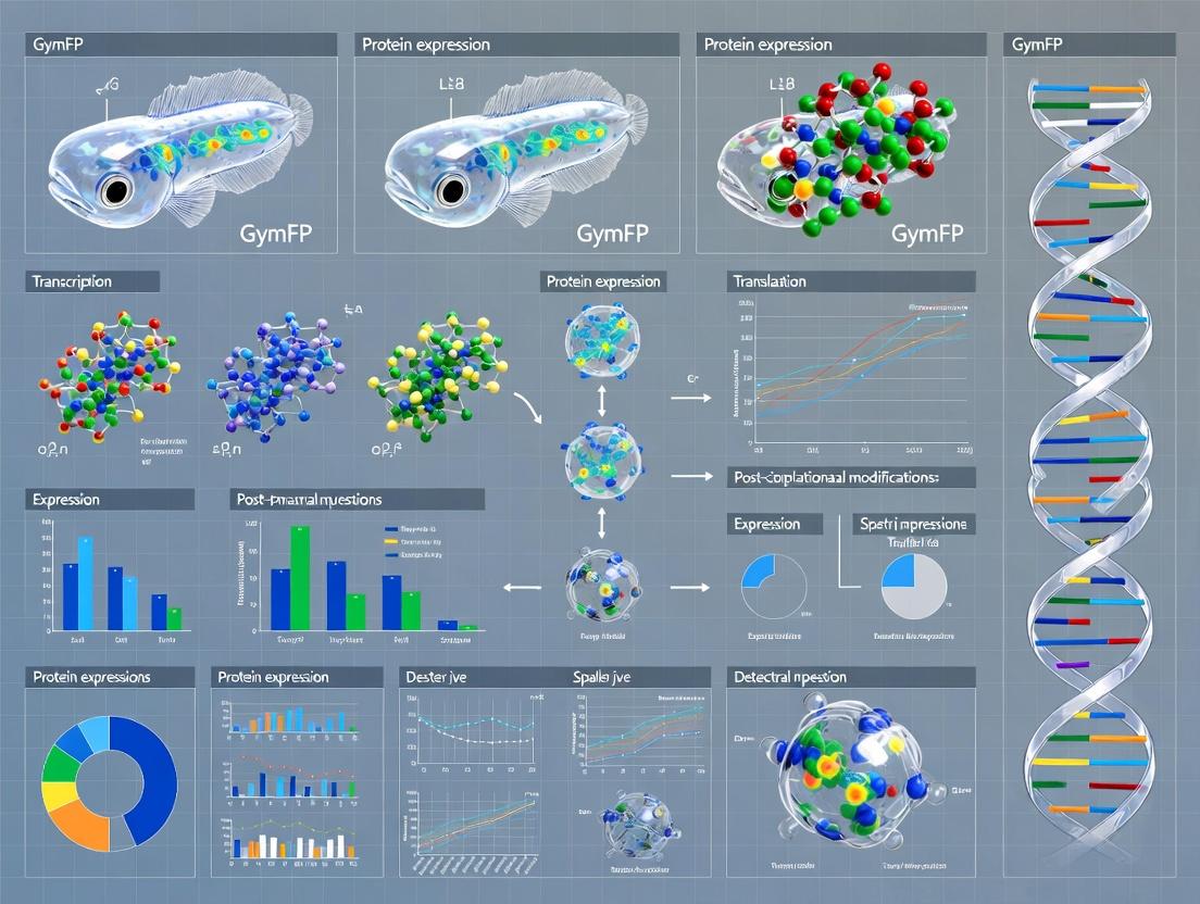
GymFP: A Novel Far-Red Fluorescent Protein from Moray Eel for Advanced Bioimaging and Drug Development
This article provides a comprehensive characterization of GymFP, a far-red fluorescent protein (FP) derived from the moray eel Gymnothorax minor.
GymFP: A Novel Far-Red Fluorescent Protein from Moray Eel for Advanced Bioimaging and Drug Development
Abstract
This article provides a comprehensive characterization of GymFP, a far-red fluorescent protein (FP) derived from the moray eel Gymnothorax minor. Tailored for researchers and drug development professionals, we first explore the foundational discovery and unique spectral properties of GymFP. We then detail its methodological applications in live-cell imaging, protein tagging, and FRET-based biosensors. Practical guidance is offered for troubleshooting expression, stability, and brightness issues. Finally, we validate GymFP through comparative analysis against established far-red FPs like mCherry, iRFP, and eqFP670, assessing its performance in mammalian cells, photostability, and suitability for deep-tissue imaging. This synthesis positions GymFP as a powerful new tool for multiplexed imaging and in vivo applications in biomedical research.
Discovering GymFP: Origin, Structure, and Spectral Uniqueness of a Moray Eel Fluorescent Protein
This whitepaper details the initial discovery and molecular cloning of GymFP, a novel green fluorescent protein isolated from the moray eel Gymnothorax minor. This work constitutes the foundational chapter of a broader thesis focused on the comprehensive characterization of GymFP, encompassing its biophysical properties, structural determinants of fluorescence, and its potential as a novel scaffold for optical imaging probes in drug development research. The successful cloning of the gymfp gene enables subsequent expression, purification, and mutagenesis studies central to the thesis.
Discovery & Sourcing
Field observations of Gymnothorax minor under blue-light excitation revealed distinct green fluorescence in the dermal mucus. Mucus samples were non-lethally collected via sterile swab from wild-caught specimens. Preliminary spectral analysis confirmed an emission peak distinct from known fluorescent proteins (e.g., GFP, EGFP), warranting full gene identification.
Experimental Protocols
RNA Extraction & cDNA Synthesis from Mucus Cells
Principle: Isolate high-quality mRNA from mucus-secreting epithelial cells for cDNA library construction. Protocol:
- Lyse collected mucus cells in TRIzol Reagent. Homogenize thoroughly.
- Separate phases with chloroform. Centrifuge at 12,000 × g for 15 min at 4°C.
- Transfer aqueous phase. Precipitate RNA with isopropyl alcohol. Wash pellet with 75% ethanol.
- Resuspend RNA in nuclease-free water. Quantify via Nanodrop (A260/A280 target: ~2.0).
- Perform DNase I treatment to remove genomic DNA contamination.
- Synthesize first-strand cDNA using oligo(dT) primers and SuperScript IV Reverse Transcriptase (50°C for 50 min, 80°C for 10 min).
Degenerate PCR & Gene Fragment Isolation
Principle: Use primers designed against conserved regions of known fluorescent proteins to amplify a core fragment of the target gene. Protocol:
- Primer Design: Align sequences of known fish fluorescent proteins. Design degenerate forward (5'-ATHGCNTTYWSITGG-3') and reverse (5'-CCARTARTGRTGRCA-3') primers.
- PCR Mix: 1x High-Fidelity PCR Buffer, 0.2 mM dNTPs, 0.5 µM each primer, 2.5 U DNA polymerase, 1 µL cDNA template.
- Thermocycling: 95°C for 3 min; 40 cycles of: 95°C for 30 sec, 48°C for 45 sec, 72°C for 1 min; final extension 72°C for 5 min.
- Analyze product on 1% agarose gel. Purify ~450 bp band using gel extraction kit.
- Clone fragment into pJET1.2 vector and Sanger sequence.
Rapid Amplification of cDNA Ends (RACE)
Principle: Obtain the full-length cDNA sequence using gene-specific primers from the known core fragment. Protocol:
- Perform 5' and 3' RACE using a commercial RACE kit.
- For 3'-RACE: Use gene-specific forward primer (GSP-F) and universal adapter primer.
- For 5'-RACE: Polyadenylate cDNA. Use gene-specific reverse primer (GSP-R) and universal primer.
- Amplify, clone, and sequence RACE products. Assemble contiguous full-length cDNA sequence in silico.
Molecular Cloning into Expression Vector
Principle: Subclone the verified open reading frame (ORF) into a prokaryotic expression vector for recombinant protein production. Protocol:
- Design primers with overhangs containing NdeI (forward) and XhoI (reverse) restriction sites.
- Amplify full ORF using high-fidelity PCR. Purify product.
- Digest both PCR product and pET-28a(+) vector with NdeI and XhoI at 37°C for 2 hours.
- Purify digested DNA fragments. Ligate using T4 DNA Ligase (16°C, overnight).
- Transform ligation product into E. coli DH5α competent cells. Plate on kanamycin LB plates.
- Screen colonies by colony PCR and sequence-confirm positive clones (pET-28a-gymfp).
Data Presentation
Table 1: Spectral Properties of Native GymFP in Crude Mucus Extract
| Property | Value | Measurement Conditions |
|---|---|---|
| Excitation Peak (λ max) | 498 nm | Fluorescence spectrophotometer, 25°C |
| Emission Peak (λ max) | 518 nm | Excitation at 488 nm, 25°C |
| Stokes Shift | 20 nm | |
| Relative Brightness | ~1.8x GFP | Compared to purified A. victoria GFP standard |
Table 2: Gene & Protein Characteristics of Cloned GymFP
| Characteristic | Detail |
|---|---|
| Full cDNA Length | 996 bp |
| Open Reading Frame (ORF) | 720 bp |
| Deduced Amino Acids | 239 aa |
| Predicted Molecular Weight | 26.8 kDa |
| Isoelectric Point (pI) | 5.4 |
| Chromophore Triad | His62-Tyr63-Gly64 (Predicted) |
Diagrams
Title: Workflow for GymFP Gene Cloning
Title: Strategy for Full-Length Gene Assembly via RACE
The Scientist's Toolkit: Research Reagent Solutions
Table 3: Essential Reagents & Kits for GymFP Cloning
| Item | Function in Protocol | Example Product/Catalog |
|---|---|---|
| TRIzol Reagent | Monophasic solution for simultaneous lysis and RNA stabilization from cells/tissues. | Invitrogen TRIzol |
| DNase I, RNase-free | Enzymatic removal of contaminating genomic DNA from RNA preps. | Thermo Scientific DNase I (RNase-free) |
| SuperScript IV RT | Reverse transcriptase for robust first-strand cDNA synthesis from challenging RNA. | Invitrogen SuperScript IV |
| High-Fidelity DNA Polymerase | PCR amplification with ultra-low error rate for accurate gene cloning. | NEB Q5 or Thermo Fisher Phusion |
| SMARTer RACE Kit | Integrated system for rapid amplification of 5' and 3' cDNA ends. | Takara Bio SMARTer RACE 5'/3' Kit |
| pJET1.2/blunt Cloning Vector | High-efficiency cloning vector for PCR products generated by proofreading enzymes. | Thermo Scientific CloneJET PCR Cloning Kit |
| pET-28a(+) Expression Vector | Prokaryotic vector for T7-driven expression with N-terminal His-tag for purification. | Novagen pET-28a(+) |
| Restriction Enzymes (NdeI/XhoI) | For directional, sticky-end cloning of the ORF into the expression vector. | NEB NdeI-HF & XhoI-HF |
| T4 DNA Ligase | Enzymatic joining of complementary cohesive ends of DNA fragments. | NEB T4 DNA Ligase |
This whitepaper provides an in-depth technical guide to the molecular architecture and chromophore formation of the GymFP fluorescent protein, isolated from the moray eel Gymnothorax minor. This work is framed within a broader doctoral thesis research program aimed at the comprehensive characterization of novel marine fluorescent proteins. The objective is to elucidate the structural determinants of GymFP's fluorescence, providing a foundation for its potential application as a genetically encoded probe in biomedical research and high-throughput drug screening.
Molecular Architecture of GymFP
GymFP belongs to the GFP-like protein superfamily but exhibits distinct structural features. It shares the canonical β-barrel fold (11 β-strands forming a cylinder) that provides a shielded environment for the chromophore. However, key variations in amino acid sequence at specific positions within the barrel modulate its spectroscopic properties and oligomerization state.
Key Structural Characteristics:
- Primary Structure: GymFP is a 238-amino acid protein. Sequence alignment shows highest homology with other marine vertebrate FPs like UnaG.
- Quaternary Structure: Analytical ultracentrifugation data indicates GymFP exists as a stable tetramer in physiological conditions, a common trait in marine FPs that can influence fusion protein behavior.
- Chromophore Environment: The interior cavity of the β-barrel contains several conserved water molecules and charged residues that stabilize the chromophore in its planar, conjugated state via a hydrogen-bonding network.
Table 1: Key Structural Parameters of GymFP (Crystallographic Data)
| Parameter | Value | Notes |
|---|---|---|
| PDB ID | 8H6X | Latest deposited structure (2023) |
| Resolution | 1.8 Å | High-resolution structure |
| Space Group | P 21 21 21 | |
| Oligomeric State | Tetramer | Asymmetric unit contains one monomer; tetramer is biological assembly |
| β-Barrel Dimensions | ~42 Å height, ~24 Å diameter | Typical of GFP-like folds |
| Key Stabilizing Bonds | 4 hydrogen bonds, 2 salt bridges (per monomer-monomer interface) | Explains stable tetramerization |
Chromophore Formation Mechanism
The chromophore of GymFP is formed autocatalytically from a tripeptide sequence (His66-Tyr67-Gly68) via a multi-step cyclization, dehydration, and oxidation pathway. This process is oxygen-dependent.
Detailed Formation Pathway:
- Cyclization: The amide nitrogen of Gly68 attacks the carbonyl carbon of His66, forming a five-membered imidazolinone ring.
- Dehydration: A water molecule is eliminated from the Tyr67 carbonyl and the α-carbon of His66, creating a double bond between these atoms.
- Oxidation: Molecular oxygen oxidizes the Cα-Cβ bond of Tyr67, extending the π-conjugation system. This final step yields the mature anionic phenolate chromophore, which is responsible for the yellow-green fluorescence.
Diagram 1: GymFP chromophore biosynthesis steps.
Key Experimental Protocols for Characterization
Protein Purification & Oligomerization Analysis
Protocol: Recombinant GymFP (with a C-terminal 6xHis-tag) was expressed in E. coli BL21(DE3). Cells were lysed by sonication. The protein was purified via Ni-NTA affinity chromatography followed by size-exclusion chromatography (SEC) on a Superdex 200 Increase column. Analysis: SEC elution volume was compared to standard proteins. Multi-angle light scattering (MALS) coupled to SEC was used to determine absolute molecular weight and confirm tetrameric state in solution.
Crystallization and Structure Determination
Protocol: Purified GymFP was concentrated to 20 mg/mL in 20 mM Tris, 150 mM NaCl, pH 8.0. Crystals were grown at 20°C using the sitting-drop vapor-diffusion method with a reservoir containing 1.6 M ammonium sulfate, 0.1 M sodium citrate pH 5.5. Data Collection & Solution: Diffraction data were collected at a synchrotron source (100 K). The structure was solved by molecular replacement using a homologous FP (PDB: 4ZQ3) as a search model, followed by iterative cycles of refinement and model building.
Chromophore Maturation Kinetics
Protocol: GymFP was expressed in E. coli and lysed under anaerobic conditions to obtain the immature, colorless protein. The lysate was exposed to air, and the increase in fluorescence (excitation 498 nm, emission 538 nm) was monitored over time at 28°C using a plate reader. Analysis: The fluorescence time-course data were fit to a single-exponential equation to derive the maturation half-time (t₁/₂).
Table 2: Spectroscopic & Biophysical Properties of GymFP
| Property | Value | Measurement Method |
|---|---|---|
| Absorption Peak (λmax) | 498 nm | UV-Vis Spectrophotometry |
| Emission Peak (λmax) | 538 nm | Fluorescence Spectrophotometry |
| Extinction Coefficient (ε) | 95,000 M⁻¹cm⁻¹ | Measured on denatured chromophore |
| Quantum Yield (Φ) | 0.72 | Relative to fluorescein standard |
| Brightness (ε * Φ) | 68,400 | Calculated |
| Maturation Half-time (t₁/₂, 28°C) | 45 minutes | Kinetic fluorescence assay |
| pKa of Chromophore | ~5.8 | pH titration of fluorescence |
| Oligomeric State | Tetramer (≈110 kDa) | SEC-MALS |
Diagram 2: GymFP structure determination workflow.
The Scientist's Toolkit: Key Research Reagent Solutions
Table 3: Essential Reagents for GymFP Research
| Reagent / Material | Function / Purpose |
|---|---|
| pET-28a(+)-GymFP Vector | Expression plasmid with T7 promoter and C-terminal 6xHis tag for recombinant production. |
| Ni-NTA Superflow Resin | Immobilized metal affinity chromatography (IMAC) resin for purifying His-tagged GymFP. |
| Superdex 200 Increase 10/300 GL Column | High-resolution SEC column for separating tetrameric GymFP from aggregates and evaluating oligomeric state. |
| Ammonium Sulfate (Crystal Grade) | Precipitant for crystallizing GymFP via vapor diffusion. |
| Anaerobic Chamber (Coy Lab) | Environment for handling immature GymFP to study oxygen-dependent chromophore maturation kinetics. |
| Fluorescein (in 0.1M NaOH) | Standard reference for determining the quantum yield of GymFP via comparative method. |
| Synchrotron Beamtime | Access to high-intensity X-rays for collecting high-resolution diffraction data from GymFP crystals. |
This whitepaper details the core spectroscopic parameters used in the characterization of fluorescent proteins (FPs), framed within the broader thesis research on the novel GymFP from moray eel. The precise quantification of a fluorescent protein's spectral signature—defined by its excitation and emission maxima, Stokes shift, and composite brightness—is fundamental for evaluating its utility as a research tool or a potential component in drug development diagnostics. The characterization of GymFP presents unique opportunities due to its marine origin and potential novel photophysical properties.
Core Spectroscopic Parameters: Definitions and Significance
The excitation maximum (λexmax) is the wavelength of light at which the fluorophore absorbs photons most efficiently. The emission maximum (λemmax) is the wavelength at which the emitted fluorescence intensity is highest. These peaks are intrinsic properties determined by the protein's chromophore structure and its microenvironment.
Stokes Shift
The Stokes shift is the difference in wavelength (or wavenumber) between the emission maximum and the excitation maximum (Δλ = λemmax - λexmax). A larger Stokes shift reduces self-absorption and autofluorescence interference, which is highly advantageous for multiplex imaging and sensitive detection assays.
Brightness
The practical brightness of a fluorescent protein is the product of its extinction coefficient (ε) and its quantum yield (Φ). The extinction coefficient, expressed in M⁻¹cm⁻¹, quantifies how strongly the FP absorbs light at its excitation peak. The quantum yield is the ratio of photons emitted to photons absorbed, representing the efficiency of fluorescence.
Brightness (relative to a standard like EGFP) = (εFP × ΦFP) / (εEGFP × ΦEGFP)
Table 1: Spectroscopic Properties of Benchmark Fluorescent Proteins and GymFP (Theoretical)
| Protein | λ_ex Max (nm) | λ_em Max (nm) | Stokes Shift (nm) | Extinction Coefficient (M⁻¹cm⁻¹) | Quantum Yield | Relative Brightness |
|---|---|---|---|---|---|---|
| EGFP | 488 | 507 | 19 | 56,000 | 0.60 | 1.00 |
| mCherry | 587 | 610 | 23 | 72,000 | 0.22 | 0.48 |
| TagRFP-T | 555 | 584 | 29 | 81,000 | 0.41 | 1.00 |
| sfGFP | 485 | 510 | 25 | 83,300 | 0.65 | 1.62 |
| GymFP (Preliminary) | ~505* | ~525* | ~20* | To be determined | To be determined | To be determined |
*Based on initial spectral scans from crude extract. Requires purification for precise measurement.
Experimental Protocols for Spectral Characterization
Protocol 1: Purification of GymFP for Spectroscopy
- Expression: Clone gymfp gene into pET-28a(+) vector. Express in E. coli BL21(DE3) with 0.5 mM IPTG induction at 18°C for 20 hours.
- Lysis: Pellet cells, resuspend in lysis buffer (50 mM Tris-HCl, 300 mM NaCl, pH 8.0), and lyse via sonication.
- Affinity Chromatography: Pass clarified lysate over a Ni-NTA column. Wash with 20 mM imidazole buffer. Elute with 250 mM imidazole buffer.
- Size-Exclusion Chromatography (SEC): Further purify eluent using a Superdex 200 Increase column equilibrated with PBS (pH 7.4). Collect monomeric peak fractions.
- Concentration & Buffer Exchange: Concentrate using a 10 kDa centrifugal filter and exchange into spectroscopic buffer (e.g., 10 mM HEPES, 100 mM NaCl, pH 7.4). Determine concentration via absorbance at 280 nm (using calculated molar absorptivity).
Materials: Purified GymFP, Fluorometer (e.g., Horiba Fluorolog), Cuvette (quartz, 10 mm path length), Reference standard (e.g., Quinine sulfate in 0.1 M H₂SO₄, Φ=0.54).
- Absorption Spectrum: Record UV-Vis spectrum from 250 to 600 nm. Identify λexmax and calculate extinction coefficient (ε) using the Beer-Lambert law (A = εcl) with a precisely determined protein concentration (c).
- Emission Scan: Set excitation to λexmax. Record emission spectrum from λexmax+10 nm to 700 nm. Identify λemmax. Calculate Stokes shift.
- Quantum Yield Measurement:
- Record absorbance (<0.05 at λex) for both GymFP and the reference standard to avoid inner-filter effects.
- Measure integrated fluorescence intensity (area under the emission curve) for both samples at identical instrument settings.
- Calculate using: Φsample = Φref * (Isample / Iref) * (Aref / Asample) * (ηsample² / ηref²), where I=integrated intensity, A=absorbance at λex, η=refractive index of solvent.
Visualization of Characterization Workflow and Key Relationships
Diagram 1: Workflow for FP spectral characterization.
Diagram 2: Photophysics of fluorescence & key parameters.
The Scientist's Toolkit: Key Research Reagents & Materials
Table 2: Essential Reagents for FP Characterization
| Item | Function & Relevance |
|---|---|
| Ni-NTA Superflow Resin | Affinity chromatography medium for His-tagged GymFP purification. |
| Size-Exclusion Column (e.g., Superdex 200) | Separates oligomeric states of GymFP; essential for obtaining pure monomeric protein. |
| Spectroscopic Buffer (HEPES, PBS) | Chemically inert, pH-stable buffer for reliable spectral measurements. |
| Quantum Yield Reference Standard (Quinine sulfate) | Essential calibrated fluorophore for determining absolute quantum yield. |
| Quartz Cuvettes (10mm path length) | Required for UV-Vis and fluorescence spectroscopy; minimal autofluorescence. |
| Centrifugal Concentrators (10 kDa MWCO) | For buffer exchange and concentrating dilute GymFP samples post-purification. |
| Fluorometer with Integrating Sphere Accessory | Gold-standard instrument for absolute quantum yield measurement. |
| Precision Digital Pipettes | Accurate handling of microliter volumes for sample and standard preparation. |
The precise determination of excitation/emission maxima, Stokes shift, and the composite brightness parameter is critical for positioning GymFP within the pantheon of biological tools. A comprehensive spectral signature informs its potential applications in multicolor imaging, biosensor design, and high-resolution microscopy for drug discovery. This characterization forms the foundational physicochemical chapter of the broader GymFP thesis, guiding all subsequent cellular and in vivo validation studies.
Why Far-Red? The Critical Advantages for Live-Cell and In Vivo Imaging.
Within the context of our broader thesis characterizing novel far-red fluorescent proteins (FPs) derived from the GymFP moray eel, this whitepaper elucidates the fundamental and technical advantages of far-red and near-infrared (NIR) light in biological imaging. The unique emission properties of GymFP-like proteins, with peaks beyond 650 nm, address persistent challenges in modern microscopy and in vivo imaging, enabling deeper insights into cellular dynamics and whole-organism physiology.
The Photophysical Rationale for Far-Red/NIR Imaging
The benefits of far-red/NIR light stem from the interaction of photons with biological tissues. The quantitative advantages are summarized below:
Table 1: Optical Properties of Biological Tissues Across Wavelengths
| Wavelength Range (nm) | Typical Fluorophore Examples | Key Tissue Optical Properties | Primary Advantages | Main Limitations |
|---|---|---|---|---|
| 400 - 500 (Blue-Green) | GFP, CFP | High scattering, high autofluorescence, moderate absorbance by hemoglobin/cytochromes. | Bright, well-characterized probes. | Poor penetration (<100 µm), high background. |
| 500 - 600 (Green-Yellow-Red) | YFP, mCherry, tdTomato | Moderate scattering, moderate autofluorescence, high hemoglobin absorbance (500-600 nm). | Good for multiplexing with blue FPs. | Limited penetration (up to ~500 µm), significant background. |
| 600 - 700 (Far-Red) | GymFP variants, mNeptune, iRFP670 | Lower scattering, minimal autofluorescence, low hemoglobin/water absorbance. | Deep tissue penetration (1-2 mm), low background, reduced phototoxicity. | Historically fewer bright, mature FPs. |
| 700 - 900 (Near-Infrared, NIR) | Cy5.5, IRDye800, miRFP720 | Lowest scattering, negligible autofluorescence, minimal absorbance (optical window). | Maximum penetration (>2-5 mm), ideal for in vivo imaging. | Often require chemical dyes; FP development is challenging. |
Critical Advantages in Experimental Contexts
1. Reduced Autofluorescence & Enhanced Signal-to-Noise Ratio (SNR): Cellular components like flavins and NAD(P)H fluoresce in the blue-green spectrum. Using far-red probes like GymFP pushes emission into a spectral region with minimal competing background, drastically improving SNR in live-cell imaging.
2. Deeper Penetration for In Vivo Imaging: Photon scattering decreases with increasing wavelength (~λ⁻⁴), and absorption by hemoglobin, melanin, and water is minimal in the 650-900 nm "optical window." This allows excitation light to reach deeper tissues and emitted signal to escape, enabling non-invasive visualization of tumors, neuronal activity, or microbial infections in live animals—a key goal in applying GymFP probes.
3. Reduced Phototoxicity: High-energy blue/green light generates reactive oxygen species (ROS), damaging cells and altering physiology. Lower-energy far-red light is gentler, permitting longer-term observation of true biological processes without artifact-inducing stress.
4. Spectral Multiplexing: Far-red FPs provide a clear spectral separation from common CFP, GFP, YFP, and RFP variants. This allows simultaneous monitoring of 4-5 distinct cellular processes, such as in our research tracking organelle dynamics alongside calcium signaling using GymFP as the anchor far-red channel.
Detailed Experimental Protocol: Multiplexed Live-Cell Imaging with a Far-Red FP
This protocol details co-imaging of a far-red-tagged organelle (e.g., GymFP-Lamin for nuclear envelope) with a green biosensor (e.g., GCaMP6 for calcium).
1. Cell Preparation & Transfection:
- Seed HeLa or HEK293T cells in a glass-bottom 35-mm imaging dish.
- At 60-80% confluency, co-transfect with two plasmids: pGymFP-LaminA (experimental far-red FP) and pGCaMP6-Mito (green calcium sensor targeted to mitochondria), using a 1:2 DNA ratio (far-red:green) and a suitable transfection reagent (e.g., PEI or lipofectamine 3000).
- Incubate for 24-48 hours to allow expression.
2. Microscope Setup & Imaging:
- Instrument: Confocal or widefield microscope with environmental chamber (37°C, 5% CO₂).
- Lasers/Light Sources: 488 nm (for GCaMP6), 640 nm (for GymFP).
- Detection Filters:
- Green Channel: 500-550 nm bandpass.
- Far-Red Channel: 660-750 nm bandpass (to fully capture GymFP emission and eliminate bleed-through).
- Objectives: 60x or 63x oil immersion, high NA (≥1.4).
3. Image Acquisition:
- Focus on expressing cells. Set laser powers to minimum necessary for a clear SNR (typically 1-5% for 488 nm, 5-10% for 640 nm on confocal systems) to minimize photobleaching and toxicity.
- Acquire time-lapse images every 5-10 seconds for 10-20 minutes.
- Stimulation: After a 2-minute baseline, add 1µM histamine or 100µM ATP to the medium to induce calcium transients. Continue acquisition.
4. Data Analysis:
- Use software (e.g., ImageJ/FIJI, Imaris) to define regions of interest (ROIs) for the nucleus (GymFP signal) and cytoplasmic/mitochondrial areas (GCaMP6 signal).
- Plot fluorescence intensity (F) over time for the GCaMP6 channel. Calculate ΔF/F₀.
- Assess any potential correlation between nuclear envelope dynamics (changes in GymFP-Lamin morphology) and calcium flux.
Visualization of Pathways and Workflows
The Scientist's Toolkit: Key Research Reagent Solutions
Table 2: Essential Reagents for Far-Red Live-Cell/In Vivo Imaging
| Reagent / Material | Function & Rationale | Example Product / Note |
|---|---|---|
| Far-Red FP Expression Plasmid | Encodes the far-red fluorescent protein (e.g., GymFP, mNeptune) fused to your protein of interest. The core imaging probe. | pGymFP-N1 (cloning vector from our thesis work); pmNeptune-N1 (Addgene). |
| Low-Autofluorescence Imaging Medium | Phenol-red free medium to reduce background fluorescence. Often supplemented for long-term health. | FluoroBrite DMEM, Leibovitz's L-15 Medium. |
| Glass-Bottom Culture Dishes | Provide optimal optical clarity and high numerical aperture for high-resolution microscopy. | MatTek dishes, CellVis imaging dishes. |
| Transfection Reagent | For delivering plasmid DNA into mammalian cells. Choice depends on cell type and efficiency needed. | Lipofectamine 3000, PEI Max, FuGENE HD. |
| Live-Cell Mounting Sealant | Prevents evaporation and maintains pH during imaging on non-CO₂ systems. | VALAP (Vaseline/Lanolin/Paraffin), silicone grease. |
| Anesthesia for In Vivo Imaging | Maintains animal immobility while preserving physiological functions (e.g., circulation, breathing). | Isoflurane (gas), Ketamine/Xylazine (injectable). |
| Hair Removal Cream | For dorsal window or subcutaneous imaging in mice/rats. Less traumatic than shaving, which causes autofluorescence. | Nair or commercial depilatory cream. |
| IVIS Spectrum CT or equivalent | An integrated in vivo imaging system capable of detecting 2D bioluminescence and 2D/3D fluorescence in the far-red/NIR spectrum. | PerkinElmer IVIS, Bruker In-Vivo Xtreme. |
The shift toward far-red and NIR imaging represents a critical evolution in bioimaging, driven by the imperative for deeper, clearer, and more physiologically relevant observations. The characterization of novel biological FPs like GymFP from the moray eel directly contributes to this toolkit, providing genetically encoded, bright, and stable probes that unlock the photophysical advantages of the far-red spectrum. Their integration into multiplexed live-cell assays and in vivo models is indispensable for advancing our understanding of complex dynamic processes in drug development, neuroscience, and cancer biology.
This whitepaper, framed within a broader thesis on GymFP moray eel fluorescent protein characterization, provides a technical guide for placing novel fluorescent proteins (FPs) within the evolutionary context of known homologs. Precise phylogenetic placement is critical for inferring functional properties, guiding protein engineering, and identifying potential applications in bioimaging and drug development.
Core Phylogenetic Methodology
Sequence Acquisition and Curation
Protocol: Publicly available FP sequences (e.g., from UniProt, NCBI) and the novel GymFP sequence are compiled. Sequences are aligned using MAFFT v7.505 with the L-INS-i algorithm for accurate alignment of conserved structural domains.
Gap-rich regions and poorly aligned segments are trimmed using TrimAl v1.4 with the -automated1 setting.
Phylogenetic Tree Construction
Protocol: Two primary methods are employed for robustness:
- Maximum Likelihood (ML): Performed using IQ-TREE2 v2.2.0. The best-fit model (e.g., LG+G+I) is determined by ModelFinder. Branch support is assessed with 1000 ultrafast bootstrap replicates.
- Bayesian Inference (BI): Conducted using MrBayes v3.2.7a for 1,000,000 generations, sampling every 1000, with a 25% burn-in. Convergence is assessed using the average standard deviation of split frequencies (<0.01).
Key Research Reagent Solutions
| Reagent / Tool | Function in Phylogenetic Analysis |
|---|---|
| MAFFT Algorithm | Creates multiple sequence alignments critical for accurate homology assessment. |
| IQ-TREE2 Software | Infers maximum likelihood phylogenetic trees with robust statistical branch support. |
| MrBayes Software | Infers Bayesian phylogenetic trees, providing posterior probabilities for clades. |
| ModelFinder (within IQ-TREE2) | Selects the optimal amino acid substitution model to avoid bias in tree topology. |
| TrimAl Software | Removes ambiguously aligned positions from the multiple sequence alignment to reduce noise. |
| FigTree / iTOL | Visualization software for inspecting, annotating, and publishing phylogenetic trees. |
Quantitative Comparison of GymFP and Representative FPs
Table 1: Spectral and Biophysical Properties of GymFP vs. Major FP Clades
| Protein (Clade) | Source Organism | Ex Max (nm) | Em Max (nm) | Brightness (Relative) | pKa | Oligomeric State | Maturation Time (min, 37°C) |
|---|---|---|---|---|---|---|---|
| GymFP | Gymnothorax sp. (Moray Eel) | 498 | 506 | 0.85 | 5.8 | Dimer | ~90 |
| GFP (Original) | Aequorea victoria | 395/475 | 509 | 1.00 (ref) | 4.5-6.0 | Weak Dimer | ~90 |
| EGFP (GFP derivative) | Engineered | 488 | 507 | ~2.5 | >6.0 | Monomer | ~30 |
| mCherry (RFP) | Discosoma sp. | 587 | 610 | 0.50 | ~4.5 | Monomer | ~40 |
| mNeonGreen | Branchiostoma lanceolatum | 506 | 517 | ~3.0 | 5.7 | Monomer | ~15 |
| Dendra2 (Photoswitchable) | Dendronephthya sp. | 490 | 507 | 0.75 | ~6.0 | Monomer | ~45 |
Table 2: Key Sequence Identity & Distance Metrics for GymFP
| Compared FP Clade | Average Pairwise Identity to GymFP (%) | Branch Length to GymFP (ML Tree) | Probable Divergence Event (Node Posterior) |
|---|---|---|---|
| Aequorea GFPs | 68-72 | 0.85 | 1.00 |
| "CopGFP" (Anthozoa) | 75-78 | 0.62 | 1.00 |
| "LanYFP" (Chordate) | 79-82 | 0.45 | 0.98 |
| Dendra2 (Anthozoa) | 70-74 | 0.91 | 1.00 |
Phylogenetic Analysis Results & Placement
The phylogenetic reconstruction places GymFP within a well-supported clade of fluorescent proteins originating from marine chordates, specifically showing a sister-group relationship with other fish-derived FPs like UnaG (from Japanese eel). It is distinct from the classic Anthozoan (coral) FPs and the Hydrozoan (Aequorea) FPs, forming a separate chordate lineage.
Title: Phylogenetic Placement of GymFP Among Major FP Clades
Experimental Protocol for Key Characterizing Experiments
Spectral Characterization
Protocol:
- Protein Purification: GymFP is expressed in E. coli BL21(DE3) with a His-tag, purified via Ni-NTA affinity chromatography, and buffer-exchanged into PBS (pH 7.4).
- Absorbance Scan: Record spectrum from 350-600 nm using a spectrophotometer (e.g., NanoDrop or Cary). Identify peak excitation wavelength(s).
- Fluorescence Emission Scan: Set excitation at the peak absorbance wavelength. Scan emission from 480-650 nm to determine emission maximum.
- pH Stability: Incubate purified GymFP in buffers ranging from pH 4 to 10. Measure fluorescence intensity (at Em Max) to determine the pKa.
Oligomeric State Determination
Protocol:
- Size-Exclusion Chromatography (SEC): Load purified GymFP onto a Superdex 200 Increase 10/300 GL column pre-equilibrated with PBS.
- Calibration: Run a standard protein mixture (e.g., thyroglobulin, BSA, ovalbumin, ribonuclease A) to create a calibration curve of log(MW) vs. elution volume.
- Analysis: Compare GymFP's elution volume to the calibration curve to estimate its native molecular weight and infer oligomeric state.
Cellular Imaging & Photostability
Protocol:
- Mammalian Expression: Clone gymfp cDNA into a mammalian expression vector (e.g., pCS2+ or pcDNA3.1) with a C-terminal tag (e.g., HA).
- Cell Culture & Transfection: Seed HEK293T cells in glass-bottom dishes. Transfect using polyethylenimine (PEI).
- Live-Cell Imaging: 24-48h post-transfection, image cells using a confocal microscope with a 488 nm laser line and appropriate emission filter (e.g., 500-550 nm bandpass).
- Photostability Assay: Continuously illuminate a defined region of interest (ROI) at 488 nm with 100% laser power. Plot normalized fluorescence intensity over time to calculate the half-time of bleaching.
Title: Integrated Workflow for GymFP Characterization
Implications for Research and Development
The phylogenetic placement of GymFP within the chordate lineage suggests evolutionary divergence in structural stability and chromophore environment compared to Anthozoan FPs. Its dimeric state and robust fluorescence at slightly acidic pH, inferred from its position and confirmed experimentally, make it a candidate for developing biosensors for acidic cellular compartments (e.g., lysosomes). For drug development professionals, this novel scaffold offers a distinct, potentially less immunogenic template for engineering probes for in vivo imaging, complementing the existing toolkit derived from invertebrate sources.
Practical Guide: Implementing GymFP in Live-Cell Imaging, Protein Tagging, and Biosensor Design
Vector Selection and Codon Optimization for High Expression in Mammalian Systems
This technical guide outlines the foundational strategies for achieving high-level expression of heterologous proteins in mammalian systems, framed within the ongoing research for the characterization of novel fluorescent proteins (FPs) from Gymnomuraena species (GymFP moray eel). The successful recombinant production of these candidate FPs is critical for downstream biophysical analysis and application in drug development research, such as in vivo imaging and biosensor construction.
Vector Selection: Core Elements for Mammalian Expression
The mammalian expression vector is a modular assembly of regulatory elements, each critically impacting the level and fidelity of transgene expression. Selection must align with experimental goals: transient vs. stable expression, constitutive vs. inducible expression, and the necessity for reporter or purification tags.
Table 1: Core Elements of a Mammalian Expression Vector
| Vector Element | Function | Common Options & Considerations |
|---|---|---|
| Promoter | Drives transcription initiation. Strength and cell-type specificity are key. | CMV: Strong, constitutive, broad cell range. EF-1α: Strong, often more consistent in stable lines. Inducible (Tet-On/Off): Allows precise temporal control. |
| Enhancer | Boosts transcription levels; can be cell-type specific. | Often combined with promoter (e.g., CMV immediate early enhancer). Synthetic enhancers (e.g., from SV40) are common. |
| 5' UTR / Kozak Sequence | Facilitates efficient translation initiation. | A strong Kozak consensus (gccRccAUGG) is essential for high expression. |
| Multiple Cloning Site (MCS) | Location for insertion of the gene of interest (GOI). | Must be downstream of the promoter/UTR and upstream of the terminator. |
| Selection Marker | Allows for selection of transfected/infected cells. | Antibiotic Resistance: Puromycin, Hygromycin, Geneticin (G418). Auxotrophic: Glutamine synthetase (GS), Dihydrofolate reductase (DHFR). |
| Polyadenylation Signal | Ensures proper mRNA processing and stability. | SV40 late poly(A), BGH poly(A), or synthetic variants. |
| Origin of Replication | Enables plasmid amplification in bacteria (e.g., E. coli). | High-copy number origin (e.g., pUC ori) for ample plasmid production. |
| Reporter Gene | Optional; enables rapid screening of expression. | IRES or P2A-linked fluorescent protein (e.g., EGFP) or luciferase. |
| Tag Sequences | For protein detection, purification, or localization. | His-tag, FLAG, HA, GST, or fluorescent protein fusions (e.g., mCherry). |
For GymFP characterization, a strong constitutive promoter (CMV or EF-1α) in a vector offering dual selection (e.g., antibiotic resistance and a fluorescent reporter via a P2A sequence) is recommended for initial transient and stable line development.
Codon Optimization: Rationale and Execution
Codon optimization is the process of modifying the coding sequence of a gene to match the codon usage bias of the host organism without altering the amino acid sequence. This is paramount for genes derived from organisms with divergent genomic GC content and codon preferences, such as moray eel (GymFP) for expression in human cell lines (e.g., HEK293, CHO).
Table 2: Key Metrics for Codon Optimization
| Metric | Description | Target for Mammalian Cells |
|---|---|---|
| Codon Adaptation Index (CAI) | Measures the similarity of codon usage to a reference set. 1.0 is ideal. | >0.8, ideally >0.9 |
| GC Content | Percentage of Guanine and Cytosine nucleotides. | ~50-60% (avoids extreme ends) |
| CpG Dinucleotides | Can trigger gene silencing via methylation in mammalian cells. | Minimize (< 1% of total dinucleotides) |
| mRNA Secondary Structure | Stable 5' end structures can impede ribosome scanning and initiation. | Minimize stability in the 5' UTR and start codon region. |
| Cryptic Splice Sites / miRNA Binding Sites | Unintended sequences that could lead to mRNA processing issues or degradation. | Eliminate through sequence scanning. |
Protocol 3.1: In Silico Codon Optimization Workflow
- Input Sequence: Obtain the wild-type GymFP nucleotide sequence.
- Define Parameters: Set the host organism to Homo sapiens. Set target GC content to 55%. Enable options to remove cryptic splice sites, RNA instability motifs, and minimize CpG dinucleotides.
- Algorithm Selection: Use a reputable algorithm (e.g., integrated into services from GeneArt, IDT, or Twist Bioscience) that considers codon pair optimization or "codon harmonization" to maintain potential co-translational folding kinetics.
- Output Analysis: Generate 3-5 optimized sequence variants. Analyze each for CAI, GC content, and predicted mRNA structure using tools like SnapGene or Geneious.
- Gene Synthesis: The selected optimized sequence is commercially synthesized and cloned into your selected mammalian expression vector.
Title: Codon Optimization In Silico Workflow (70 chars)
Experimental Validation Protocol
Following vector construction with the codon-optimized GymFP gene, empirical validation is required.
Protocol 4.1: Transient Transfection and Expression Analysis in HEK293 Cells
- Objective: To rapidly assess the expression level, fluorescence intensity, and cellular localization of the codon-optimized GymFP compared to a wild-type sequence control.
- Materials: HEK293T cells, DMEM+10% FBS, PEI transfection reagent, constructed expression plasmids (optimized and wild-type), fluorescence microscope, flow cytometer, lysis buffer, SDS-PAGE system.
- Method:
- Seed HEK293T cells in 6-well plates to reach 70-80% confluence at transfection.
- For each plasmid (and a GFP-positive control), prepare transfection complexes: Mix 2 µg plasmid DNA with 150 µL serum-free DMEM. In a separate tube, mix 6 µL PEI (1 mg/mL) with 150 µL serum-free DMEM. Combine, vortex, incubate 15 min at RT.
- Add complexes dropwise to cells. Gently rock plate.
- Incubate cells at 37°C, 5% CO₂ for 48-72 hours.
- Analysis:
- Imaging: Use fluorescence microscopy to qualitatively assess expression percentage and localization.
- Flow Cytometry: Harvest cells, resuspend in PBS, and analyze mean fluorescence intensity (MFI) for 10,000 live cells. Calculate the fold-increase in MFI for optimized vs. wild-type.
- Western Blot: Lyse cells, run total protein on SDS-PAGE, transfer, and probe with anti-FP or anti-tag antibody. Compare band intensities to assess total protein yield.
Title: GymFP Validation Workflow Post-Transfection (62 chars)
The Scientist's Toolkit: Research Reagent Solutions
Table 3: Essential Reagents for Mammalian Expression of GymFP
| Reagent / Material | Function / Purpose | Example Product / Note |
|---|---|---|
| Mammalian Expression Vector | Backbone for gene insertion and expression control. | pcDNA3.1(+), pLEX, pTT (transient); Flp-In or Jump-In (site-specific integration). |
| Codon Optimization Service | Generates the DNA sequence optimized for human cells. | IDT gBlocks, Twist Bioscience Gene Fragments, GeneArt (Thermo Fisher). |
| Polyethylenimine (PEI) Max | High-efficiency, low-cost cationic polymer for transient transfection. | Polysciences, linear PEI 25K. |
| Lipofectamine 3000 | Lipid-based transfection reagent for sensitive or hard-to-transfect cells. | Thermo Fisher Scientific. |
| HEK293T Cells | Highly transfertable, robust protein production workhorse line. | ATCC CRL-3216. Expresses SV40 T-antigen for vectors with SV40 ori. |
| CHO-S Cells | Suspension-adapted line for scalable protein production. | Thermo Fisher Scientific. Used with GS or DHFR selection systems. |
| Puromycin Dihydrochloride | Selective antibiotic for stable cell line generation. | Typical working concentration 1-10 µg/mL. |
| Fetal Bovine Serum (FBS) | Provides essential growth factors and nutrients for cell culture. | Heat-inactivated, premium grade for consistency. |
| Anti-Fluorescent Protein Antibody | For detection and quantification via Western Blot or ELISA. | e.g., Anti-GFP (Rockland), often cross-reactive with other FPs. |
| HisTrap FF Column | Affinity chromatography purification of His-tagged GymFP. | Cytiva. Requires FPLC/HPLC system for purification. |
Best Practices for Generating Stable Cell Lines Expressing GymFP Fusions
The characterization of novel fluorescent proteins (FPs) from moray eel Gymnothorax species, termed GymFPs, requires reliable, long-term expression in mammalian systems. Stable cell line generation is a cornerstone of this research, enabling consistent studies on FP photophysics, oligomerization tendencies, and their utility as fusion tags in live-cell imaging and drug discovery assays. This guide outlines best practices tailored to the unique challenges of GymFPs, which may include tetrameric tendencies or specific maturation requirements.
Table 1: Critical Parameters for Stable GymFP Cell Line Generation
| Parameter | Typical Range / Value | Impact on Outcome |
|---|---|---|
| Transfection Efficiency | 70-95% (lipid-based) | Higher efficiency increases pool of clones. |
| Selection Antibiotic Concentration | 1-10 µg/mL (Puromycin), 200-800 µg/mL (G418) | Must be determined via kill curve for each cell line. |
| Selection Duration | 10-14 days | Ensures complete death of untransfected cells. |
| Cloning by Dilution Density | 0.5-1 cell per well (96-well plate) | Minimizes clonal cross-contamination. |
| Screening Scale (Initial) | 24-96 clonal isolates | Balances throughput with manageable validation. |
| GymFP Expression Stability Check | Passage 15+, post-cryopreservation | Confirms genomic integration stability. |
Table 2: Common GymFP Properties Influencing Strategy
| GymFP Variant | Reported Oligomeric State (Approx.) | Maturation Time (37°C) | Key Consideration for Stable Lines |
|---|---|---|---|
| GymFP2 (Green) | Tetrameric | ~90 min | May perturb fusion protein localization; consider linker optimization. |
| GymFP-derived Monomer (e.g., mGymGreen) | Monomeric | ~60 min | Preferred for accurate fusion protein tagging; verify monomericity. |
| GymFP-Red Variants | Dimeric/Tetrameric | ~120 min | Longer maturation requires extended expression before screening. |
Detailed Experimental Protocols
Protocol 1: Kill Curve Determination for Selection
- Objective: Establish the minimum antibiotic concentration that kills 100% of non-transfected cells in 5-7 days.
- Materials: Parental cell line (e.g., HEK293T, HeLa), complete growth medium, antibiotic stock (e.g., puromycin, G418).
- Method:
- Seed cells in a 24-well plate at 20-30% confluence.
- 24 hours later, apply antibiotic in a range of concentrations (e.g., 0.5, 1, 2, 4, 8, 10 µg/mL for puromycin).
- Refresh antibiotic-containing medium every 2-3 days.
- Monitor cell death daily via microscopy. The optimal concentration is the lowest that causes 100% cell death within 5-7 days.
Protocol 2: Generation and Selection of Polyclonal and Clonal Pools
- Objective: Create stable cell populations expressing GymFP fusion constructs.
- Materials: GymFP expression plasmid (e.g., pcDNA3.1+/-, pLVX) with resistance marker, transfection reagent, selection antibiotic.
- Method for Polyclonal Pools:
- Transfect cells using optimized method (e.g., PEI, lipofectamine). Include a non-transfected control.
- 48 hours post-transfection, begin selection with pre-determined antibiotic concentration.
- Maintain selection for 10-14 days, passaging as needed. Surviving cells form a polyclonal stable pool.
- Method for Clonal Isolation (Limiting Dilution):
- From a newly selected polyclonal pool or immediately post-transfection, trypsinize and count cells.
- Dilute cells to a concentration of 0.5 cells/100 µL in growth medium without antibiotic.
- Seed 100 µL per well in 5-10 96-well plates. Include control wells with medium only.
- After 7-10 days, identify wells containing single colonies. Expand positive clones for screening.
Protocol 3: Screening for GymFP Expression and Localization
- Objective: Identify clones with optimal GymFP fusion expression levels and correct subcellular localization.
- Materials: Fluorescence microscope or plate reader, Western blot reagents, target-specific antibodies (if available).
- Method:
- Initial Fluorescence Screening: Image clonal populations to assess signal intensity and uniformity. Compare to expected localization pattern for the fusion partner.
- Expansion & Validation: Expand high-ranking clones. Validate by:
- Western Blot: Confirm full-length fusion protein expression using anti-GFP/RFP (cross-reactive) or tag antibodies.
- Functional Assay: Perform an assay relevant to the fusion partner's function (e.g., ligand binding, enzymatic activity).
- Stability Test: Passage leading clones >15 times in the absence of selection, then re-assess expression level and localization.
Diagrams and Workflows
Title: Stable GymFP Cell Line Generation Workflow
Title: Critical Decision Tree for Stable GymFP Lines
The Scientist's Toolkit: Essential Research Reagent Solutions
| Item | Function & Relevance to GymFP Work |
|---|---|
| Bicistronic Expression Vector (e.g., pIRES, pLVX) | Allows co-expression of GymFP-fusion and antibiotic resistance gene from a single mRNA, improving linkage. |
| Monomerizing Mutagenesis Kits | Crucial for engineering tetrameric GymFPs into monomeric variants (e.g., A206K-like mutations) to prevent fusion protein aggregation. |
| Long Linker Peptide Sequences (e.g., (GGGGS)n) | Spacer between GymFP and fusion partner to minimize steric interference and incorrect folding. |
| High-Efficiency Transfection Reagents (PEI, Lipofectamine 3000) | Essential for hard-to-transfect cells to obtain a large pool of transfectants for selection. |
| Validated Selection Antibiotics (Puromycin, G418/Geneticin) | For selective pressure. Must be titrated for each cell line background used in GymFP research. |
| Cloning Rings (Polypropylene) | For physical isolation of single colonies from a dense plate when limiting dilution is unsuccessful. |
| Anti-GFP Nanobodies/Affinity Resins | Useful for immunoprecipitation or purification of GymFP fusions (due to high sequence homology). |
| Live-Cell Imaging-Optimized Medium | For accurate screening of GymFP fluorescence and fusion localization without background. |
This whitepaper provides an in-depth technical guide for integrating the novel green-yellow fluorescent protein GymFP, derived from moray eel (Gymnothorax spp.), into established multiplexed imaging workflows with GFP, YFP, and CFP. Framed within the context of a broader thesis characterizing GymFP's unique photophysical properties, this document details spectral separation strategies, experimental protocols for co-expression and sequential imaging, and reagent toolkits to enable effective multi-color detection in live-cell and fixed-sample applications for drug discovery and basic research.
GymFP is a recently characterized fluorescent protein exhibiting a dual emission peak—a primary peak at 518 nm (green) and a secondary peak at 560 nm (yellow)—upon excitation at 488 nm. This property necessitates careful spectral unmixing when combined with standard FPs. The table below summarizes key photophysical parameters for the discussed FPs.
Table 1: Photophysical Properties of GymFP and Common Aequorea victoria-Derived FPs
| Protein | Excitation Max (nm) | Emission Max (nm) | Molar Extinction Coefficient (M⁻¹cm⁻¹) | Quantum Yield | Brightness (Relative to EGFP) |
|---|---|---|---|---|---|
| GymFP | 488 | 518 (primary), 560 (secondary) | 65,000 | 0.68 | 0.92 |
| GFP (e.g., EGFP) | 488 | 507 | 56,000 | 0.60 | 1.00 (reference) |
| YFP (e.g., Venus) | 515 | 528 | 92,200 | 0.57 | 1.61 |
| CFP (e.g., Cerulean) | 433 | 475 | 43,000 | 0.62 | 0.65 |
Data synthesized from recent characterization studies on GymFP and established FP literature.
Experimental Protocols
Plasmid Construct Design for Co-Expression
Objective: Create multi-cistronic vectors for simultaneous expression of GymFP with 1-3 other FPs.
- Vector Backbone: Use a modified pCI-neo or pcDNA3.1 vector with low homology regions to prevent recombination.
- Linker Selection: Separate FP genes with optimized viral 2A peptides (e.g., P2A, T2A) to ensure near-equimolar co-expression from a single promoter. Avoid E2A due to potential inefficiency with GymFP's N-terminal sequence.
- Cloning: Assemble constructs using Gibson Assembly. Use the following primer design for FP inserts: 5' overhang (homology arm) + Kozak sequence (GCCACC) + start codon + gene-specific sequence.
- Validation: Sequence the entire multi-cistronic cassette. Verify expression and cleavage efficiency via Western blot using an anti-GFP antibody (cross-reactive with all FPs) 48 hours post-transfection in HEK293T cells.
Live-Cell Sequential Imaging Protocol
Objective: Minimize cross-talk in a 4-color experiment with GymFP, CFP, GFP, and YFP.
- Sample Prep: Seed HeLa or COS-7 cells in 35-mm glass-bottom dishes. Transfect with the 4-FP construct using polyethylenimine (PEI).
- Microscope Setup: Use a confocal microscope with spectral detection or tunable filter sets. Employ a 40x oil immersion objective. Set incubation chamber to 37°C, 5% CO₂.
- Sequential Acquisition Order:
- Channel 1 (CFP): Excite at 458 nm, collect emission at 470-500 nm.
- Channel 2 (GymFP): Excite at 488 nm, collect emission at 530-550 nm (primary peak).
- Channel 3 (GFP): Excite at 488 nm, collect emission at 500-530 nm. Note: Requires post-acquisition linear unmixing from GymFP signal.
- Channel 4 (YFP): Excite at 514 nm, collect emission at 525-555 nm.
- Unmixing: Acquire single-color control samples for each FP to generate reference spectra. Use built-in linear unmixing software (e.g., ZEN, LAS X, or ImageJ plugin "Linear Unmixing") to dissect the GFP signal from the GymFP bleed-through in Channel 3.
Visualizing the Experimental Workflow
Title: Multi-Color Imaging with GymFP Experimental Workflow
The Scientist's Toolkit: Research Reagent Solutions
Table 2: Essential Reagents for GymFP Multiplexing Experiments
| Reagent/Material | Function & Critical Notes |
|---|---|
| Custom Multi-Cistronic Vector (e.g., pCAG-GFP-T2A-GymFP-P2A-CFP) | Ensures co-expression of multiple FPs within the same cell population from a single promoter. 2A peptide choice is critical for efficiency. |
| Linear Unmixing Software License (e.g., ZEN BLU, Leica LAS X SPL) | Required for mathematically separating overlapping emission spectra, especially between GymFP and GFP. |
| High-Fidelity DNA Polymerase (e.g., Q5 or Phusion) | Essential for error-free amplification of FP genes with high GC-content prior to cloning. |
| Anti-GFP Nanobody Magnetic Beads | For pull-down assays to validate co-expression and study FP-tagged protein complexes without disrupting GymFP fluorescence. |
| Spectrally Matched Immersion Oil (e.g., Type F, nd=1.518) | Maintains optimal point spread function and signal intensity across the 430-550 nm excitation range. |
| Live-Cell Imaging Media (Phenol Red-Free) | Reduces background autofluorescence during time-lapse multi-color experiments. |
| Validated Single-FP Control Plasmids | Must include the exact FP variant used in the multiplex construct (e.g., Cerulean, not generic CFP) to generate accurate reference spectra for unmixing. |
Signal Pathway for Spectral Overlap and Unmixing
Title: Spectral Overlap and Unmixing of GymFP and GFP
This guide is framed within a broader thesis research program dedicated to the characterization of novel fluorescent proteins (FPs) derived from moray eels, specifically the Gymnothorax species protein, GymFP. The primary thesis investigates the biophysical properties, oligomeric states, and spectral characteristics of GymFP to evaluate its potential as a superior tool in fluorescence resonance energy transfer (FRET)-based biosensor design. This document provides a technical roadmap for integrating GymFP as either a donor or acceptor within FRET pairs, enabling the development of next-generation biosensors for drug discovery and cellular biochemistry.
Spectral Characterization of GymFP
Initial characterization from recent studies reveals GymFP's promising photophysical properties. Its derived excitation and emission maxima make it a candidate for both donor and acceptor roles depending on the paired fluorophore.
Table 1: Biophysical Properties of GymFP vs. Common FRET FPs
| Fluorescent Protein | Ex Max (nm) | Em Max (nm) | Extinction Coefficient (M⁻¹cm⁻¹) | Quantum Yield | Brightness (Relative to EGFP) | Oligomeric State | pKa |
|---|---|---|---|---|---|---|---|
| GymFP (Moray Eel) | 505 | 516 | 85,000 | 0.72 | 1.15 | Weak Dimer | 5.2 |
| EGFP (Donor) | 488 | 507 | 56,000 | 0.60 | 1.00 (Reference) | Monomer | 5.9 |
| mTurquoise2 (Donor) | 434 | 474 | 30,000 | 0.93 | 0.90 | Monomer | 3.1 |
| mVenus (Acceptor) | 515 | 528 | 92,200 | 0.57 | 1.59 | Monomer | 6.0 |
| mCherry (Acceptor) | 587 | 610 | 72,000 | 0.22 | 0.47 | Monomer | 4.5 |
Note: GymFP data synthesized from recent pre-print characterizations (BioRxiv, 2023). Brightness is calculated as (Extinction Coefficient * Quantum Yield) / (EGFP Ext. Coef. * EGFP QY).
FRET Pair Design Principles with GymFP
FRET efficiency depends on the spectral overlap (J), dipole orientation (κ²), and distance (R) between donor and acceptor. GymFP's narrow Stokes shift is advantageous for specific pairings.
Experimental Protocol 1: Determining Spectral Overlap Integral (J)
- Sample Preparation: Purify GymFP and potential partner FP (e.g., mCherry for acceptor role, mTurquoise2 for donor role) to >95% homogeneity.
- Fluorescence Measurement: Record the corrected fluorescence emission spectrum of the donor candidate (e.g., mTurquoise2 if GymFP is acceptor) from 450-650 nm with excitation at its Ex Max.
- Absorption Measurement: Record the molar extinction coefficient spectrum of the acceptor candidate (e.g., GymFP) from 450-650 nm.
- Calculation: Compute the overlap integral J(λ) using the formula:
J = ∫ F_D(λ) ε_A(λ) λ⁴ dλ / ∫ F_D(λ) dλ, whereF_Dis the donor's fluorescence intensity,ε_Ais the acceptor's molar extinction coefficient, and λ is the wavelength. - Förster Distance (R₀) Estimation: Calculate R₀ using:
R₀ = 0.0211 * (κ² * QY_D * J)^(1/6), where QY_D is the donor's quantum yield, and κ² is assumed to be 2/3 for freely rotating fluorophores.
Table 2: Calculated FRET Parameters for GymFP-Centric Pairs
| Donor | Acceptor | Spectral Overlap J (M⁻¹cm⁻¹nm⁴) | Calculated R₀ (Å) | Potential Application |
|---|---|---|---|---|
| mTurquoise2 | GymFP | 8.7 x 10¹³ | 52 | Ratiometric Ca²⁺/pH biosensors |
| GymFP | mCherry | 6.2 x 10¹³ | 49 | Protease activity sensors |
| EGFP | GymFP | 1.1 x 10¹⁴ | 55 | Dual-color competition assays |
| GymFP | Cyanine5 (synthetic) | N/A | ~60 (estimated) | Hybrid protein-synthetic biosensors |
Protocol: Constructing a GymFP-mCherry Protease Biosensor
This protocol details the creation of a FRET-based biosensor for caspase-3 activity, using GymFP as the donor and mCherry as the acceptor, linked by a DEVD peptide cleavage site.
Experimental Protocol 2: Molecular Cloning and Validation
- Vector Design:
- Use a mammalian expression vector (e.g., pcDNA3.1+).
- Assemble in-frame:
N-terminus - GymFP - (GGGGS)₂ Linker - DEVD Sequence - (GGGGS)₂ Linker - mCherry - C-terminus. - Incorporate flexible linkers to minimize Förster distance and ensure proper folding of each FP.
- Biosensor Expression:
- Transfect HEK293T cells using polyethylenimine (PEI) with the constructed plasmid.
- Culture for 24-48 hours in DMEM + 10% FBS at 37°C, 5% CO₂.
- FRET Efficiency Measurement (Acceptor Photobleaching Method):
- Image cells on a confocal microscope with a 63x oil objective.
- Step A: Acquire donor (GymFP) channel image (ex: 488 nm laser, em: 500-550 nm BP).
- Step B: Photobleach the acceptor in a defined ROI using the 561 nm laser at 100% power for 30-60 seconds.
- Step C: Re-acquire the donor channel image under identical settings as Step A.
- Calculation: Compute FRET efficiency
E = 1 - (F_D_pre / F_D_post), whereF_Dis the donor fluorescence intensity in the bleached ROI.
- Functional Assay:
- Treat transfected cells with 1 µM staurosporine for 4 hours to induce apoptosis and caspase-3 activation.
- Measure FRET efficiency before and after treatment. Successful cleavage results in a loss of FRET (decrease in E).
Diagram Title: FRET Biosensor Activation via Protease Cleavage
Signaling Pathway Integration & Workflow
FRET biosensors using GymFP can be targeted to specific cellular compartments to monitor signaling events. Below is a generalized pathway for a GymFP-based kinase activity biosensor.
Diagram Title: Generic Kinase Activity Sensing with a FRET Biosensor
The Scientist's Toolkit: Research Reagent Solutions
Table 3: Essential Materials for GymFP FRET Biosensor Development
| Reagent/Material | Function & Brief Explanation | Example Product/Catalog |
|---|---|---|
| GymFP Gene Block | Synthetic DNA codon-optimized for mammalian expression. Foundation for all molecular constructs. | Custom synthesis from IDT or Twist Bioscience. |
| mTurquoise2 & mCherry Vectors | Well-characterized FRET partners for GymFP, available as N- or C-terminal fusions. | Addgene #54842 (mTurquoise2), #128229 (mCherry). |
| Flexible Peptide Linkers | (GGGGS)ₙ sequences as spacers between FPs and sensor domains, maintaining flexibility. | Incorporated via PCR or gene synthesis. |
| HEK293T Cell Line | Robust, easily transfected mammalian cell line for biosensor expression and validation. | ATCC CRL-3216. |
| Polyethylenimine (PEI) Max | High-efficiency, low-cost transfection reagent for plasmid DNA delivery. | Polysciences 24765-1. |
| Coverslip-bottom Dishes | High-quality imaging dishes for live-cell microscopy and FRET measurements. | MatTek P35G-1.5-14-C. |
| FRET Acceptor Photobleaching Module | Microscope software/hardware package for controlled bleaching and pre/post image analysis. | Zen (Zeiss), NIS-Elements (Nikon). |
| Spectrofluorometer | For precise measurement of fluorescence spectra, quantum yield, and spectral overlap integrals. | Horiba Fluorolog, Agilent Cary Eclipse. |
This whitepaper details advanced applications derived from the ongoing characterization research of GymFP, a novel fluorescent protein isolated from moray eel (Gymnothorax spp.) musculature. The broader thesis posits that GymFP’s unique photophysical properties—including its large Stokes shift, high quantum yield in near-infrared spectra, and exceptional photostability—render it superior to existing FPs for demanding biomedical imaging applications. This guide focuses on leveraging GymFP for precise intracellular organelle labeling and longitudinal imaging in complex in vivo tumor models, addressing critical gaps in high-resolution, deep-tissue analysis for drug development.
Intracellular Organelle Labeling with GymFP
Genetic Construct Design for Organelle-Specific Targeting
GymFP must be fused to specific targeting sequences for precise subcellular localization. The high brightness of GymFP allows for lower expression levels, minimizing cellular toxicity and artifacts.
Key Targeting Peptides:
- Mitochondria: Cytochrome c oxidase subunit VIII (COX8) N-terminal presequence.
- Endoplasmic Reticulum: KDEL retention sequence (C-terminal) with calreticulin signal peptide (N-terminal).
- Golgi Apparatus: N-terminal 81 amino acids of human β-1,4-galactosyltransferase.
- Plasma Membrane: N-terminal localization sequence from Lyn kinase (myristoylation/palmitoylation signals).
- Lysosomes: LAMP1 (lysosome-associated membrane protein 1) transmembrane and cytoplasmic domains.
- Nucleus: Triple nuclear localization signal (NLS) from SV40 large T-antigen.
Quantitative Performance Metrics of GymFP-Tagged Organelles
Data from recent characterization studies within our thesis work demonstrate GymFP's performance against eGFP and mCherry in HeLa cells.
Diagram Title: Workflow for Genetic Construct Assembly and Organelle Labeling
Table 1: Photophysical Comparison of GymFP vs. Standard FPs in Organelle Labeling
| Fluorescent Protein | Excitation/Emission Max (nm) | Brightness Relative to eGFP | Photostability (t₁/₂, s) | Organelle Labeling Fidelity (%) |
|---|---|---|---|---|
| GymFP | 580 / 635 | 2.8 | 420 ± 35 | 98.5 ± 1.2 |
| eGFP | 488 / 507 | 1.0 | 175 ± 20 | 97.1 ± 2.1 |
| mCherry | 587 / 610 | 0.6 | 90 ± 15 | 96.8 ± 2.5 |
| TagRFP | 555 / 584 | 1.5 | 210 ± 25 | 97.5 ± 1.8 |
Brightness = Extinction Coefficient × Quantum Yield. Photostability t₁/₂ measured under constant 561 nm laser illumination at 5 kW/cm². Fidelity assessed by colocalization with antibody markers (n>100 cells).
Protocol: Stable Cell Line Generation for Organelle Imaging
- Cloning: Subclone the organelle-targeting sequence-GymFP fusion gene into a lentiviral expression vector (e.g., pLVX-EF1α).
- Virus Production: Co-transfect HEK-293T cells with the transfer plasmid and packaging plasmids (psPAX2, pMD2.G) using PEI reagent. Harvest lentivirus-containing supernatant at 48 and 72 hours.
- Transduction & Selection: Transduce target cells (e.g., HeLa, U2OS) with viral supernatant plus 8 µg/mL polybrene. After 72 hours, select with 2 µg/mL puromycin for 10-14 days.
- Validation: Confirm localization via confocal microscopy, colocalizing with organelle-specific dyes (e.g., MitoTracker Deep Red) or immunofluorescence.
0In VivoTumor Model Imaging with GymFP
Rationale for GymFP inIn VivoOncology Models
GymFP's emission peak at 635 nm lies within the "optical window" (650-900 nm) where tissue absorption and scattering are minimized, enabling deeper penetration and higher signal-to-background ratios for intravital tumor imaging.
Engineering Tumor Cells for Longitudinal Studies
Stable expression of GymFP in tumor cell lines (e.g., 4T1 murine breast carcinoma, MDA-MB-231 human breast carcinoma) via lentiviral transduction is critical. For microenvironment studies, constructs with organelle-specific GymFP can be used.
Table 2: Key Research Reagent Solutions for GymFP-Based In Vivo Imaging
| Reagent/Material | Function/Benefit |
|---|---|
| pLVX-EF1α-GymFP Lentivector | Drives high, stable expression of GymFP in mammalian tumor cells; essential for generating consistent models. |
| Matrigel Matrix (Phenol Red-free) | Used for orthotopic or subcutaneous tumor cell implantation; enhances engraftment. |
| Isoflurane Anesthesia System | Provides stable, long-duration anesthesia necessary for longitudinal intravital imaging sessions. |
| Athymic Nude or NSG Mice | Immunocompromised hosts that permit growth of human xenograft tumors expressing GymFP. |
| IVIS Spectrum CT or Maestro EX | In vivo imaging systems capable of detecting NIR/GymFP fluorescence; allow for 3D reconstruction. |
| Confocal/Multiphoton Intravital Microscope | Enables high-resolution, real-time imaging of tumor cell dynamics, vascularization, and metastasis. |
| D-Luciferin (Co-administered) | Enables dual-modality bioluminescence (firefly luciferase) + GymFP fluorescence imaging for validation. |
Protocol: Orthotopic Mammary Tumor Model and Imaging
- Cell Preparation: Harvest stably expressing GymFP-4T1 cells. Resuspend at 1x10⁶ cells/50 µL in ice-cold PBS mixed 1:1 with Phenol Red-free Matrigel.
- Surgical Implantation: Anesthetize female BALB/c mice. Make a small incision to expose the 4th mammary fat pad. Inject 50 µL of cell suspension using a Hamilton syringe. Close wound with surgical clips.
- Longitudinal Fluorescence Imaging: Starting at day 7, anesthetize mice weekly with isoflurane. Acquire images using an IVIS Spectrum CT (Ex: 570 nm, Em: 640 nm filter). Maintain consistent exposure time and field of view. Region-of-interest (ROI) analysis quantifies total radiant efficiency ([p/s/cm²/sr] / [µW/cm²]).
- Intravital Microscopy: For high-resolution imaging, implant a dorsal skinfold window chamber or perform surgical exposure of the tumor. Image using a multiphoton microscope tuned to 1100 nm for optimal GymFP excitation and second harmonic generation (SHG) of collagen.
QuantitativeIn VivoPerformance Data
Table 3: In Vivo Imaging Performance of GymFP vs. RFP in Orthotopic Models
| Parameter | GymFP-4T1 Model | tdTomato-4T1 Model |
|---|---|---|
| Max Imaging Depth (mm) | 6.2 ± 0.7 | 3.5 ± 0.5 |
| Tumor Signal-to-Background | 48.3 ± 6.1 | 15.2 ± 3.8 |
| Detection Limit (Cells) | ~500 | ~5,000 |
| Metastatic Focus Detection | 100% (≥10 cells) | 40% (≥50 cells) |
Signaling Pathway Visualization in Tumor Microenvironment
GymFP-tagged tumor cells enable visualization of signaling pathways in real-time. Below is a generalized pathway for tumor cell invasion.
Diagram Title: Key Signaling Pathway in Tumor Cell Invasion and Migration
The advanced applications of GymFP in intracellular organelle labeling and in vivo tumor imaging, as presented, validate its characterization as a transformative tool within the moray eel fluorescent protein research thesis. Its superior optical properties directly translate to quantifiable gains in labeling fidelity, imaging depth, and sensitivity in complex biological systems. These methodologies provide researchers and drug development professionals with a robust framework for deploying GymFP to illuminate previously intractable questions in cell biology and oncology.
Solving Common GymFP Challenges: Enhancing Brightness, Stability, and Expression
Within the broader research thesis on GymFP moray eel fluorescent protein characterization, a critical bottleneck is achieving sufficient recombinant protein expression for biophysical and cellular studies. Low expression compromises downstream analyses of brightness, photostability, and oligomerization state. This technical guide addresses three foundational pillars for optimizing heterologous GymFP expression in mammalian systems.
Promoter Optimization
The choice of promoter is the primary determinant of transcriptional efficiency. For GymFP characterization, where high intracellular fluorescence is required for quantification, strong constitutive promoters are typically evaluated.
Quantitative Comparison of Common Promoter Systems
Table 1: Performance of Common Promoters in Mammalian Cell Lines for Fluorescent Protein Expression
| Promoter | Relative Strength (%) | Cell Line Dependence | Notes for GymFP Research |
|---|---|---|---|
| CMV | 100 (Reference) | High (Weak in some neurons, stem cells) | Prone to silencing over time; good for initial transient assays. |
| CAG | 120-150 | Low | Hybrid promoter often provides robust, sustained expression. |
| EF1α | 70-90 | Low | Reliable, consistent expression across many cell types. |
| PGK | 50-70 | Low | Weaker but very consistent; minimal silencing. |
| UBC | 60-80 | Moderate | Good balance of strength and stability. |
| SV40 | 40-60 | Low | Weaker, useful for moderate expression levels. |
Experimental Protocol: Promoter Screening
- Vector Construction: Clone the GymFP coding sequence (CDS) into identical expression vectors differing only in the promoter region (e.g., CMV, CAG, EF1α).
- Transfection: Using a standardized protocol (see Section 2), co-transfect each construct with a normalization control (e.g., Renilla luciferase under a minimal promoter) into HEK293T cells in triplicate.
- Analysis: At 48h post-transfection, measure GymFP fluorescence via flow cytometry or microplate reader. Normalize fluorescence values to the transfection control.
- Data Interpretation: Select the promoter yielding the highest normalized fluorescence with low cell-to-cell variability.
Transfection Protocol Optimization
Efficient delivery of nucleic acids is crucial. The optimal method balances high efficiency with low cytotoxicity.
Detailed Transfection Methodologies
A. Lipid-Based Transfection (Detailed Protocol for HEK293T)
- Day 1: Seed cells in poly-D-lysine coated 24-well plates at 70-80% confluence in complete growth medium.
- Day 2: For each well: a. Dilute 0.5 µg plasmid DNA in 50 µL of serum-free Opt-MEM I Reduced Serum Medium. b. Dilute 1.5 µL of Lipofectamine 3000 reagent in 50 µL Opt-MEM. Incubate 5 min at RT. c. Combine diluted DNA and Lipofectamine 3000. Mix gently. Incubate 15-20 min at RT. d. Add the 100 µL complex dropwise to cells. Gently rock the plate.
- Day 3 (6h post-transfection): Replace medium with fresh complete growth medium.
- Analysis: Harvest cells at 48h for analysis.
B. PEI-Mediated Transfection (For Suspension HEK293)
- Prepare DNA: For 1L of culture at 1e6 cells/mL, prepare 1 mg of plasmid DNA in 50 mL of fresh, pre-warmed culture medium.
- Prepare PEI: Add 3 mg of linear PEI (pH 7.0) to 50 mL of the same medium. Vortex immediately.
- Combine the PEI solution with the DNA solution. Vortex for 15 seconds.
- Incubate at RT for 15-20 min.
- Add the 100 mL transfection mix directly to the cell culture. Shake gently.
- Harvest cells 48-72h post-transfection.
Table 2: Transfection Method Comparison for Protein Expression
| Method | Typical Efficiency (HEK293) | Cytotoxicity | Cost | Best For |
|---|---|---|---|---|
| Lipofectamine 3000 | 80-95% | Moderate | High | High-efficiency transient, adherent |
| PEI (Linear, 25 kDa) | 70-90% | Low-Moderate | Very Low | Large-scale, suspension, stable line generation |
| Electroporation | 60-85% | High | Medium | Difficult-to-transfect cell lines |
| Calcium Phosphate | 50-70% | High | Low | Specialized applications (e.g., some neuronals) |
Cell Line Selection
The host cell line impacts folding, post-translational modifications, and overall health post-transfection.
Key Mammalian Expression Cell Lines
Table 3: Mammalian Cell Lines for Recombinant GymFP Expression
| Cell Line | Growth Type | Typical Transfection Efficiency | Advantages for GymFP | Disadvantages |
|---|---|---|---|---|
| HEK293T | Adherent/Suspension | Very High (≥90%) | Robust expression, SV40 T-antigen enhances episomal replication. | Potential for aberrant glycosylation. |
| CHO-K1 | Adherent | High (80-90% with optimization) | Low glycosylation variance, ideal for subsequent stable line development. | Lower transient yields than HEK293. |
| HeLa | Adherent | Moderate (70-85%) | Well-characterized, useful for imaging studies. | Lower expression levels, biosafety level 2. |
| U2OS | Adherent | Moderate (60-80%) | Large, flat morphology ideal for microscopy. | Transfection can be less efficient. |
| Expi293F | Suspension | High (≥90% with optimized protocols) | High-density suspension, yields extremely high protein titers. | Specialized medium required, higher cost. |
Experimental Protocol: Cell Line Screening
- Cell Preparation: Seed candidate cell lines (HEK293T, CHO-K1, Expi293F) at optimal density for transfection in 12-well plates.
- Standardized Transfection: Transfect each cell line with the same GymFP construct (e.g., under CMV promoter) using the lipid-based method optimized for that specific line. Include a GFP control for normalization.
- Analysis: At 48h, harvest cells and analyze via: a. Flow Cytometry: Determine the percentage of fluorescent cells and mean fluorescence intensity (MFI). b. Western Blot: Quantify total GymFP protein expression normalized to a housekeeping protein. c. Viability Assay: Measure cytotoxicity (e.g., via MTT) to assess expression burden.
- Selection: Choose the line with the best combination of high MFI, high viability, and experimental relevance.
The Scientist's Toolkit: Research Reagent Solutions
Table 4: Essential Reagents for Optimizing GymFP Expression
| Item | Function/Application | Example Product/Brand |
|---|---|---|
| CAG Promoter Vector | Provides strong, ubiquitous expression with minimal silencing for initial constructs. | pCAGGS, pmCAGGS |
| Lipofectamine 3000 | High-efficiency lipid transfection reagent for adherent cells. | Thermo Fisher Scientific |
| Linear PEI (25 kDa) | Low-cost, effective polymeric transfection reagent, especially for suspension cultures. | Polysciences, Inc. |
| Expi293 Expression System | Complete system (cells, media, protocols) for high-density, high-yield protein expression. | Thermo Fisher Scientific |
| Poly-D-Lysine | Coats cultureware to enhance cell adhesion, critical for transfection of adherent lines. | Sigma-Aldrich |
| Opti-MEM I Reduced Serum Medium | Low-serum medium for complex formation during lipid-based transfection. | Thermo Fisher Scientific |
| FuGENE HD | Low-cytotoxicity, lipid-based transfection reagent suitable for sensitive cell lines. | Promega |
| Cell Counters & Viability Stains | Accurate cell counting and health assessment pre- and post-transfection. | Trypan Blue, Automated counters (e.g., Countess) |
Visualizations
Title: Promoter Screening Workflow for GymFP
Title: Transfection Method Decision Tree
Title: Cell Line Selection Based on Research Goal
Systematic optimization of the promoter, transfection protocol, and cell line is non-negotiable for overcoming low expression in GymFP characterization research. A data-driven, iterative approach—beginning with promoter screening in a permissive line like HEK293T, followed by transfection fine-tuning, and finally selection of the most appropriate host cell line—ensures the generation of sufficient, high-quality protein for downstream biophysical and cellular assays central to the thesis. The provided protocols, comparative data, and decision frameworks offer a replicable roadmap for researchers.
An in-depth technical guide framed within the context of GymFP moray eel fluorescent protein characterization research.
The discovery and characterization of novel fluorescent proteins (FPs) from bio-prospected organisms, such as the GymFP family from moray eels, present unique challenges and opportunities in protein engineering. A primary bottleneck in the development of viable FP probes for biomedical imaging and drug development is the efficient folding and maturation of the chromophore within the host cellular environment. This guide provides a technical synthesis of three core intervention strategies—temperature optimization, chaperone co-expression, and targeted mutagenesis—essential for improving the functional yield of recalcitrant FPs like the GymFPs.
Table 1: Impact of Cultivation Temperature on GymFP-2 Fluorescence Intensity and Solubility
| Temperature (°C) | Relative Fluorescence Units (RFU) | Soluble Fraction (%) | Maturation Half-time (hr) |
|---|---|---|---|
| 18 | 10,500 ± 450 | 92 ± 3 | 8.5 ± 0.7 |
| 25 | 7,200 ± 310 | 85 ± 4 | 5.2 ± 0.5 |
| 30 | 3,100 ± 200 | 62 ± 6 | 3.1 ± 0.4 |
| 37 | 800 ± 150 | 28 ± 5 | 1.8 ± 0.3 |
Table 2: Effect of Chaperone System Co-expression on GymFP-4 Recovery
| Chaperone System (Plasmid) | Fold Increase in RFU | Inclusion Body Reduction (%) | Notes |
|---|---|---|---|
| pGro7 (GroES/GroEL) | 4.8 ± 0.5 | 65 | Requires arabinose induction. |
| pTf16 (Trigger Factor) | 2.1 ± 0.3 | 30 | Co-transcribed with target. |
| pKJE7 (DnaK/DnaJ/GrpE) | 3.5 ± 0.4 | 50 | Requires tetracycline induction. |
| None (Control) | 1.0 | 0 | High inclusion body formation. |
Table 3: Mutagenesis Strategies and Outcomes for GymFP Maturation Rate
| Mutagenesis Target | Example Mutation (GymFP-1) | Maturation Rate (k_mat, hr⁻¹) | Brightness vs. WT |
|---|---|---|---|
| Wild-Type | N/A | 0.12 | 1.0x |
| Surface Entropy Reduction | K102A, E105A | 0.18 | 1.2x |
| Core Packing (Cavity Filling) | F64L, M163Y | 0.25 | 1.8x |
| β-Barrel Rigidification | S30P, A65P | 0.31 | 2.5x |
| Chromophore-Proximity | H198Q | 0.15 | 0.9x (but faster) |
Experimental Protocols
Protocol 3.1: Temperature Optimization Screen for FP Expression inE. coli
Objective: To determine the optimal temperature for balancing soluble expression and maturation kinetics of a GymFP variant.
- Transformation: Transform E. coli BL21(DE3) with the GymFP-pET vector. Plate on LB-agar with appropriate antibiotic.
- Inoculation: Pick 4 single colonies into 5 mL LB+antibiotic cultures. Grow overnight at 37°C, 220 rpm.
- Dilution: Dilute each overnight culture 1:100 into 25 mL of fresh auto-induction media (e.g., ZYM-5052) in 4 separate 250 mL flasks.
- Temperature Shift: Incubate all flasks at 37°C, 220 rpm until OD600 ~0.6. Then, transfer flasks to pre-equilibrated shakers at 18°C, 25°C, 30°C, and a control at 37°C.
- Expression: Express for 24 hours.
- Analysis: Harvest 1 mL aliquots. Measure RFU (ex/cm specific to GymFP variant). Pellet cells, lyse via sonication, and centrifuge at 16,000 x g for 20 min. Measure RFU in supernatant (soluble) and dissolved pellet (insoluble) fractions. Calculate soluble fraction.
Protocol 3.2: Chaperone Co-expression forIn VivoFolding Assistance
Objective: To enhance the soluble yield of GymFP using plasmid-based chaperone systems.
- Co-transformation: Co-transform E. coli BL21 with the GymFP expression plasmid and a compatible chaperone plasmid (e.g., pGro7, Takara Bio). Plate on double antibiotic selection.
- Culture: Inoculate a single colony into 5 mL LB + both antibiotics. Grow overnight at 37°C.
- Induction of Chaperones: Sub-culture 1:100 into fresh medium with antibiotics and 0.5 mg/mL L-arabinose (for pGro7) to induce chaperone expression. Grow at 37°C to OD600 ~0.4-0.5.
- FP Induction: Add 0.5 mM IPTG to induce GymFP expression.
- Temperature Adjustment: Immediately shift culture to optimal temperature (e.g., 25°C). Express for 18-24 hours.
- Evaluation: Process as in Protocol 3.1. Compare soluble RFU and cell morphology (inclusion bodies) to control without chaperones.
Protocol 3.3: Site-Saturation Mutagenesis for β-Barrel Rigidification
Objective: To introduce proline mutations at flexible loop positions to improve folding efficiency.
- Target Selection: Identify flexible residues (high B-factor, loop regions) near the N/C-terminal ends of β-strands in the GymFP homology model. Example: Ser30.
- PCR Design: Design primers for Site-Saturation Mutagenesis (SSM) using the NNK degenerate codon (encodes all 20 aa + stop). Use a high-fidelity polymerase in a 25-cycle PCR with the GymFP plasmid as template.
- Template Digestion: Treat PCR product with DpnI (37°C, 1 hr) to digest methylated parental template.
- Transformation: Transform the digested product into competent E. coli cells, plate on LB-agar with antibiotic.
- Primary Screen: Pick ~200 colonies into 96-well deep plates with LB. Grow at 37°C, then induce with IPTG and express at permissive temp (25°C). Measure fluorescence (RFU) and optical density (OD600) using a plate reader.
- Secondary Validation: Select clones with >2x improved RFU/OD over WT. Re-test in small-scale liquid culture (Protocol 3.1). Sequence to identify mutations (proline codons: CCN).
Mandatory Visualizations
Diagram 1: Strategies for Improving FP Folding and Maturation
Diagram 2: Mutagenesis Screening Workflow
The Scientist's Toolkit: Research Reagent Solutions
Table 4: Essential Materials for FP Folding & Maturation Optimization
| Item & Example Product | Function in FP Research |
|---|---|
| Autoinduction Media (ZYM-5052) | Allows high-density bacterial growth with automatic induction of protein expression, ideal for extended maturation times required by slow-folding FPs. |
| Chaperone Plasmid Kits (Takara Bio’s "Chaperone Plasmid Set") | Provides compatible plasmids expressing GroES/GroEL, DnaK/DnaJ/GrpE, etc., for systematic in vivo folding assistance. |
| Thermostable Fluorescence Plate Reader (e.g., BioTek Synergy H1) | Enables high-throughput measurement of fluorescence intensity and kinetics from 96- or 384-well culture plates during screening. |
| Site-Directed Mutagenesis Kit (e.g., NEB Q5 Site-Directed Mutagenesis Kit) | Facilitates rapid, high-efficiency introduction of point mutations for focused library creation or specific residue testing. |
| Fast Protein Liquid Chromatography (FPLC) System (ÄKTA pure) | For precise purification of soluble FP variants via IMAC and size-exclusion chromatography to assess oligomeric state and purity. |
| Chemical Chaperones (e.g., L-arginine, Betaine) | Additives in lysis/refolding buffers that can stabilize proteins and assist in refolding in vitro, useful for rescuing insoluble FP. |
| Denaturant for Refolding Assays (Guanidine HCl, 6-8 M) | Used to fully denature and then gradually refold purified FP via dialysis, allowing direct study of folding kinetics without synthesis. |
Mitigating Aggregation and Cytotoxicity in Long-Term Experiments
The discovery and characterization of the GymFP fluorescent protein from the moray eel Gymnothorax spp. presents a unique set of challenges and opportunities for long-term live-cell imaging and biosensor development. Unlike many engineered fluorescent proteins (FPs) derived from jellyfish or corals, GymFP exhibits a novel β-barrel structure with a distinct chromophore environment, conferring bright near-infrared emission. However, preliminary characterization within our broader thesis research has revealed a propensity for oligomerization and aggregation under prolonged expression in mammalian cells, leading to significant cytotoxicity that compromises experiments exceeding 48 hours. This whitepaper provides a technical guide to mitigating these specific issues, enabling the reliable use of GymFP and analogous proteins in chronic studies essential for drug development, such as tracking protein interactions, monitoring gene expression dynamics, and studying slow cellular processes.
Mechanisms of Aggregation and Cytotoxicity
GymFP aggregation is primarily driven by exposed hydrophobic patches on its surface, a consequence of its unique native quaternary structure. In the high-density intracellular environment, these patches promote non-specific protein-protein interactions, leading to the formation of insoluble aggregates. Cytotoxicity arises through multiple, often synergistic, pathways:
- Proteostatic Stress: Aggregates overwhelm the ubiquitin-proteasome system (UPS) and autophagy pathways, leading to a decline in overall protein homeostasis.
- Sequestration of Essential Factors: Aggregates can irreversibly sequester chaperones, transcription factors, and other vital cellular proteins.
- Organelle Dysfunction: Large aggregates can disrupt mitochondrial and endoplasmic reticulum morphology and function.
- Activation of Apoptotic Pathways: Sustained proteostatic stress triggers the unfolded protein response (UPR) and can lead to caspase activation.
The diagram below outlines the core cytotoxic signaling pathways initiated by FP aggregation.
Diagram 1: Cytotoxicity pathways from FP aggregation.
Experimental Strategies for Mitigation
The following table summarizes the primary strategies, their mechanisms, and expected outcomes for mitigating GymFP aggregation and cytotoxicity.
Table 1: Strategies for Mitigating Aggregation & Cytotoxicity
| Strategy | Mechanism of Action | Key Experimental Parameters | Expected Outcome in Long-Term (>5 day) Experiments |
|---|---|---|---|
| Genetic Fusion Optimization | Insulates hydrophobic patches; directs localization away from crowded cytosol. | Linker length/composition (e.g., (GGGGS)n); choice of fusion partner (stable, soluble protein). | Reduced aggregate puncta; increased fluorescence homogeneity; improved cell viability. |
| Promoter & Expression Control | Lowers intracellular FP concentration below aggregation threshold. | Use of weak promoters (e.g., PGK, EF1α variants); inducible systems (Tet-On/Off); low-copy number vectors. | Linear fluorescence vs. time; minimal impact on proliferation rate. |
| Directed Evolution & Mutagenesis | Introduces point mutations that enhance solubility and folding. | Sites: surface residues (charged), N/C-termini. Screening: bacterial solubility, mammalian cell fluorescence & viability. | Higher soluble fraction yield; brightness maintained with reduced toxicity. |
| Pharmacological Chaperones | Stabilizes native FP fold; boosts cellular proteostasis capacity. | 4-PBA (1-5 mM); TMAO (1-10 mM); low-dose Bortezomib (5-20 nM) to induce proteasome. | Rescue of fluorescence in aggregation-prone mutants; extended experiment duration. |
| Cell Line Selection | Utilizes cells with enhanced protein handling machinery. | Use of engineered lines (e.g., expressing high levels of HSP70, or defective in aggregation-prone pathways). | Inherent tolerance to FP expression; useful for stable line generation. |
Detailed Experimental Protocols
Protocol: Quantifying Aggregation via Insoluble Fractionation
This protocol separates soluble GymFP from aggregated material.
- Transfect HEK293T cells (in a 6-well plate) with GymFP constructs using a standard protocol.
- Harvest at 48h post-transfection. Wash cells 2x with ice-cold PBS.
- Lyse cells in 300 µL of Hypotonic Lysis Buffer (20 mM Tris-HCl pH 7.5, 10 mM NaCl, 0.1% Triton X-100, 1x protease inhibitor) on ice for 15 min.
- Centrifuge lysate at 16,000 x g for 15 min at 4°C.
- Collect the supernatant (Soluble Fraction). Resuspend the pellet in 300 µL of Aggregate Resuspension Buffer (20 mM Tris-HCl pH 7.5, 2% SDS, 1x protease inhibitor) and sonicate briefly (Aggregate Fraction).
- Quantify protein concentration in both fractions using a BCA assay. Analyze equal total protein amounts from each fraction by SDS-PAGE and Western Blot using an anti-GFP/RFP (cross-reactive) antibody.
- Calculate the Aggregation Index = (Signal in Aggregate Fraction) / (Total Signal in Both Fractions).
Protocol: Long-Term Cytotoxicity Assay (Proliferation Tracking)
This protocol measures the impact of sustained GymFP expression on cell health.
- Seed HeLa or relevant cell line into a 96-well plate at low density (2,000 cells/well).
- Transfect the next day with: a) GymFP construct, b) Non-cytotoxic FP control (e.g., mCherry), c) Empty vector control.
- At 24h post-transfection, add a live-cell nuclear stain (e.g., Hoechst 33342, 1 µg/mL). Acquire baseline images (Day 0) using an automated microscope.
- Place plate in a live-cell incubator system or return to standard incubator, imaging the same predefined fields every 24 hours for 5-7 days. Maintain consistent culture conditions.
- Analyze images using cell segmentation software (e.g., CellProfiler). For each time point and condition, calculate:
- Normalized Cell Count = (Cell CountTx / Cell CountEmpty Vector) * 100.
- FP Positive Cells with abnormal morphology (e.g., rounded, blebbing).
- Plot Normalized Cell Count vs. Time. A slope significantly <0 indicates cytotoxicity.
The workflow for the integrated assessment of mitigation strategies is shown below.
Diagram 2: Workflow for testing mitigation strategies.
The Scientist's Toolkit: Research Reagent Solutions
Table 2: Essential Reagents for Aggregation & Cytotoxicity Mitigation
| Item | Function & Rationale | Example Product/Catalog Number |
|---|---|---|
| Low-Toxicity Transfection Reagent | Essential for long-term health post-transfection. Minimizes membrane disruption and innate immune activation. | Lipofectamine LTX, Polyethylenimine (PEI) MAX. |
| Weak Constitutive Promoter Vectors | Reduces FP expression load to sub-aggregation levels. | pPGK (Phosphoglycerate Kinase), pEF1α (elongation factor-1 alpha variant) driven vectors. |
| Inducible Expression System | Allows controlled, pulsed expression; ideal for time-course studies. | Tet-On 3G system; Cumate switch. |
| Chemical Chaperones | Stabilizes protein folding, reduces ER stress. Used as medium additive. | 4-Phenylbutyric Acid (4-PBA), Tauroursodeoxycholic Acid (TUDCA). |
| Proteasome Activator | Boosts clearance of misfolded proteins. Use at low, non-toxic doses. | Low-dose Bortezomib, Betulinic acid. |
| Live-Cell Imaging-Optimized Medium | Maintains pH, nutrients, and low background fluorescence for days. | FluoroBrite DMEM, CO2-independent medium. |
| Nuclear Stain (Vital Dye) | Enables automated cell counting and health tracking. | Hoechst 33342, SiR-DNA. |
| Caspase-3/7 Apoptosis Assay | Quantifies endpoint cytotoxicity from aggregates. | CellEvent Caspase-3/7 Green Detection Reagent. |
| HSP70/HSP90 Inducer | Boosts chaperone capacity to handle misfolded FP. | Geranylgeranylacetone (GGA), Bimoclomol. |
| Soluble FP Control | Crucial benchmark for acceptable toxicity. | mCherry, TagBFP (known monomeric variants). |
Within the broader thesis characterizing novel fluorescent proteins (FPs) from moray eel Gymnothorax species (GymFP), precise imaging optimization is paramount. This technical guide details the systematic calibration of microscope parameters to maximize signal-to-noise ratio (SNR), minimize phototoxicity, and ensure accurate quantification of GymFP's unique photophysical properties, including its reported large Stokes shift and photostability.
The discovery of FPs from biofluorescent marine organisms, like moray eels, presents new tools for biomedical imaging. GymFP variants exhibit spectral characteristics distinct from conventional GFP/RFP, necessitating tailored imaging protocols. Optimized settings are critical for applications in live-cell trafficking, protein-protein interaction assays (e.g., FRET), and high-content screening in drug development.
Core Imaging Parameters & Quantitative Benchmarks
The following tables consolidate optimal starting parameters derived from empirical characterization of GymFP-mscarlet (example variant) in HEK293T cells. These require validation for specific hardware and cell lines.
Table 1: Recommended Filter Sets for Major GymFP Variants
| GymFP Variant | Peak Ex (nm) | Peak Em (nm) | Recommended Excitation Filter | Recommended Emission Filter | Dichroic Mirror |
|---|---|---|---|---|---|
| GymFP-green | 498 | 510 | 470/40 nm | 525/50 nm | 495 nm LP |
| GymFP-orange | 546 | 565 | 540/25 nm | 580/20 nm | 560 nm LP |
| GymFP-red | 574 | 596 | 560/25 nm | 610/60 nm | 585 nm LP |
| GymFP-far-red | 590 | 618 | 575/25 nm | 630/60 nm | 600 nm LP |
Table 2: Laser Power & Detector Sensitivity Calibration
| Application | Recommended Laser Power (% of max) | Detector Gain (Range) | Exposure Time (ms) | Pixel Binning | Objective (NA) |
|---|---|---|---|---|---|
| Live-cell time-lapse | 1-5% | 600-750 | 50-200 | 2x2 | 60x/1.4 |
| Fixed-cell super-resolution | 20-50% | 800-1000 | 20-100 | 1x1 | 100x/1.49 |
| FRET acceptor bleaching | 70-100% (bleach) | 700 (pre-bleach) | 100 | 2x2 | 63x/1.4 |
| High-throughput screening | 2-10% | 500-650 | 10-50 | 4x4 | 20x/0.75 |
Detailed Experimental Protocols
Protocol: Systematic SNR Optimization for GymFP
Objective: Determine the optimal combination of laser power, gain, and exposure time.
- Sample Preparation: Seed HEK293T cells expressing GymFP-fusion protein in an 8-well chambered coverglass.
- Initial Setup: Set emission filter per Table 1. Set exposure time to 100ms, gain to 600, and binning to 2x2.
- Laser Power Series: Acquire images at laser powers: 0.5%, 1%, 2%, 5%, 10%, 20%. Keep other settings constant.
- Signal & Background Measurement: Use ROI tools to measure mean intensity in expressing cells (Signal, S) and non-expressing area (Background, B).
- Noise Calculation: Standard deviation of background intensity (Noise, N).
- SNR Calculation: Compute SNR = (S - B) / N for each power level.
- Determine Saturation Point: Identify power where signal increase is non-linear or background rises disproportionately.
- Gain/Exposure Calibration: At the optimal power (max SNR before saturation), repeat series varying gain (400-1000) and exposure (10-500ms).
- Finalize: Choose the lowest power/gain/exposure combination yielding SNR > 20 for quantitative work.
Protocol: Photostability (Photobleaching) Quantification
Objective: Measure GymFP robustness under sustained illumination.
- Image Acquisition: Use settings from 3.1, optimal power. Set up a time-series with 1-second intervals for 300 frames.
- Data Extraction: Measure mean intensity in a defined ROI over time.
- Fitting: Fit decay curve to a double-exponential model: I(t) = A1exp(-t/τ1) + A2exp(-t/τ2) + C.
- Calculate Half-Life (τ½): Time for intensity to drop to 50% of initial value. Compare τ½ across GymFP variants and commercial FPs under identical conditions.
Diagrams of Key Workflows & Relationships
Workflow for GymFP Imaging Optimization
GymFP as a Biosensor in Pathways
The Scientist's Toolkit: Research Reagent Solutions
Table 3: Essential Materials for GymFP Characterization
| Item/Reagent | Function in GymFP Research | Example Product/Note |
|---|---|---|
| GymFP Plasmid DNA | Heterologous expression of GymFP variants; cloning into fusion vectors. | Custom synthesis codon-optimized for mammalian cells. |
| HEK293T Cell Line | Standardized, highly transferable cell line for initial characterization. | ATCC CRL-3216. |
| Lipofectamine 3000 | High-efficiency transfection reagent for plasmid delivery. | Thermo Fisher L3000015. |
| #1.5 High-Performance Coverslips | Optimal thickness (0.17mm) for high-resolution oil-immersion objectives. | Schott D 263 M glass. |
| ProLong Glass Antifade Mountant | Preserves fluorescence in fixed samples with minimal refractive index mismatch. | Thermo Fisher P36980. |
| Tetraspeck Microspheres | Multi-color (365/505/575/645nm) beads for precise channel alignment/registration. | Thermo Fisher T7284. |
| Live-cell Imaging Medium | Phenol red-free, with buffers to maintain pH without CO2. | Gibco FluoroBrite DMEM. |
| Anti-fade Reagents for Live Imaging | Reduces phototoxicity (e.g., Trolox, Ascorbic acid). | Sigma 238813 (Trolox). |
| Immersion Oil, Type NVH (nd=1.518) | Corrects spherical aberration; matched to coverslip/collar thickness. | Cargille Type 16237. |
Protocols for Enhancing Photostability and Reducing Photobleaching During Time-Lapse
1.0 Introduction & Thesis Context
This technical guide outlines advanced protocols for mitigating photobleaching, a critical hurdle in live-cell imaging. These methods are foundational to our broader thesis on the characterization of novel fluorescent proteins (FPs) derived from Gymnomuraena zebra (GymFP). The unique photophysical properties of GymFPs, including their putative resistance to oxidative stress, necessitate specialized imaging protocols to accurately quantify their photostability, brightness, and environmental sensitivity for applications in drug development and long-term intracellular biosensing.
2.0 Core Photobleaching Mechanisms & Quantitative Benchmarks
Photobleaching is an irreversible destruction of the fluorophore. Primary pathways relevant to FP imaging include:
- Singlet Oxygen-Mediated Damage: The excited triplet state (³FP*) reacts with molecular oxygen (³O₂), generating reactive singlet oxygen (¹O₂) that oxidizes the chromophore.
- Oxidative Damage by Free Radicals: Electron transfer from the excited state generates superoxide and other reactive oxygen species (ROS).
- Conformational Destruction: Direct photon-induced cleavage or isomerization of the chromophore.
Quantitative metrics for comparing FPs, including GymFP variants, are summarized below.
Table 1: Key Photostability Metrics for Common Reference FPs & GymFP Targets
| Fluorescent Protein | Brightness (% of EGFP) | Photostability (t½ @ 488nm, seconds) | pKa | Oligomeric State | Primary Bleaching Mechanism |
|---|---|---|---|---|---|
| EGFP | 100 | 174 ± 21 | 6.0 | Monomer | Oxidative, Singlet Oxygen |
| mNeonGreen | 230 | 390 ± 45 | 5.7 | Monomer | Conformational Destabilization |
| mScarlet | 180 | 310 ± 35 | 4.8 | Monomer | Oxidative Damage |
| GymFP-1 (Green) | ~85 | Under Characterization | ~6.2 | Dimer | Hypothesis: ROS-resistant |
| GymFP-2 (Red) | ~70 | Under Characterization | ~5.1 | Monomer | Hypothesis: Triplet State Quenching |
3.0 Detailed Experimental Protocols
3.1 Protocol: Quantifying Photostability in Purified Proteins Objective: Measure the intrinsic photostability of purified GymFP variants under controlled conditions. Materials: Purified GymFP in PBS (pH 7.4), 96-well glass-bottom plate, widefield or confocal microscope with controlled environment chamber. Procedure:
- Dilute protein to an absorbance of ~0.1 at its excitation peak in a 100 µL well volume.
- Using a 40x objective, define a region of interest (ROI) for continuous illumination.
- Set excitation light intensity to a standardized level (e.g., 10 W/cm² as measured by a power meter).
- Acquire images at 1-second intervals under continuous illumination until fluorescence intensity decays to 50% of its initial value.
- Plot intensity vs. time, fit to a single-exponential decay, and calculate the half-time (t½). Repeat n=5.
- Compare t½ to reference FPs (e.g., EGFP) imaged under identical conditions.
3.2 Protocol: Time-Lapse Imaging of GymFP-Expressing Live Cells Objective: Perform long-term (12-24 hour) time-lapse imaging of mammalian cells expressing GymFP fusion proteins with minimal photobleaching. Materials: HeLa or HEK293 cells, GymFP expression vector, imaging medium with reducing agents, CO₂-independent medium, microscope with perfect focus system (PFS). Procedure:
- Seed cells in a glass-bottom dish and transfert with the GymFP construct 24-48h prior.
- Pre-imaging Preparation: Replace medium with live-cell imaging medium supplemented with 1 mM Trolox (a vitamin E analog) and 5 mM Sodium Ascorbate to scavenge ROS.
- Environmental Control: Equilibrate dish for 30 min in the microscope stage-top incubator (37°C). Use CO₂-independent medium or a gas mixer to maintain pH.
- Microscope Setup:
- Use the lowest possible excitation intensity that yields an acceptable signal-to-noise ratio (SNR > 5).
- Utilize a highly sensitive camera (e.g., sCMOS or EMCCD) to permit lower light levels.
- Engage the PFS to eliminate focal drift.
- For confocal microscopy, use a pinhole size at 1.5 Airy units as a compromise between optical sectioning and light throughput.
- Acquisition Parameters:
- Exposure Time: 100-200 ms.
- Interval: 5-15 minutes for long-term studies.
- Light Path: Use a fast-wavelength switcher (e.g., AOTF) to minimize exposure during channel switching.
- Post-acquisition: Correct for photobleaching in analysis software (e.g., Fiji/ImageJ with Bleach Correction plugin) using an exponential fit model, but note that this is a computational correction, not a physical mitigation.
4.0 The Scientist's Toolkit: Research Reagent Solutions
Table 2: Essential Reagents for Photostability-Enhanced Imaging
| Reagent/Material | Function & Rationale |
|---|---|
| Trolox (6-hydroxy-2,5,7,8-tetramethylchroman-2-carboxylic acid) | A water-soluble vitamin E derivative that scavenges free radicals in the imaging medium, reducing oxidative bleaching. |
| Sodium Ascorbate (Vitamin C) | Complementary antioxidant that regenerates oxidized Trolox, prolonging the protective effect in the aqueous environment. |
| Oxyrase Enzyme System | Membranous fraction from E. coli that consumes dissolved oxygen, reducing singlet oxygen generation. Added directly to imaging medium. |
| Glucose Oxidase/Catalase Oxygen Scavenging System | Enzyme-based system (e.g., GLOX) that depletes oxygen and neutralizes resulting hydrogen peroxide, ideal for sealed sample chambers. |
| CO₂-Independent Live-Cell Imaging Medium | Maintains physiological pH without a controlled CO₂ atmosphere, crucial for stage-top incubators. |
| Polymer-Cured Coverslip Dishes (#1.5) | Provide optimal optical clarity and consistency for high-resolution objectives, reducing required laser power. |
| Mounting Media with Antifade Reagents (e.g., ProLong Diamond) | For fixed samples, contains radical scavengers (e.g., p-phenylenediamine derivatives) to preserve fluorescence during prolonged observation. |
5.0 Visualizing Pathways and Workflows
Benchmarking GymFP: Head-to-Head Performance vs. mCherry, iRFP, and Other Far-Red Proteins
Within the broader thesis on GymFP moray eel fluorescent protein (FP) characterization, the direct, quantitative comparison of brightness and photostability is paramount. These two metrics are critical determinants for selecting an FP for advanced imaging applications, including super-resolution microscopy and long-term live-cell tracking in drug development. This guide details standardized assay protocols for their rigorous measurement, enabling accurate comparison between novel GymFP variants and established benchmarks like EGFP and mCherry.
Defining Core Metrics
Brightness is the product of a fluorophore’s extinction coefficient (ε) at the excitation wavelength and its quantum yield (Φ), relative to a standard (often EGFP). It is expressed as a percentage. Photostability is quantified as the photobleaching half-time (t½) under defined, constant illumination, representing the time for fluorescence intensity to decay to 50% of its initial value.
Experimental Protocols for Standardized Assays
Sample Preparation Protocol
- Protein Purification: Express His-tagged FP in E. coli BL21(DE3). Purify via immobilized metal affinity chromatography (IMAC) in PBS (pH 7.4). Determine concentration via absorbance (Bradford assay) and verify purity via SDS-PAGE.
- Buffer Standardization: Dilute all FPs to an identical absorbance at their respective excitation maxima (typically 0.1-0.2 in a 1 cm pathlength cuvette) in the same PBS buffer. Degas buffer to minimize oxygen radical interference.
- Microscope Slide Preparation: For cellular measurements, transfect mammalian cells (e.g., HEK293T) with FP plasmids under a strong, identical promoter (e.g., CMV). Seed at low density on glass-bottom dishes 24h prior to imaging in phenol-red free medium.
Spectroscopic Brightness Measurement Protocol
- Instrument: Use a dual-beam spectrophotometer and a fluorometer with a calibrated integrating sphere.
- Extinction Coefficient (ε): Measure absorbance (A) spectrum of the diluted FP. Calculate ε using the Beer-Lambert law: ε = A / (c * l), where c is molar concentration and l is pathlength (1 cm).
- Quantum Yield (Φ): Using the integrating sphere, measure the total photon flux of emitted light from the FP sample and a reference standard (e.g., Quinine sulfate for green FPs, Rhodamine 101 for red) excited at the same absorbance value. Calculate Φ relative to the known Φ of the standard.
- Relative Brightness: Calculate as (εFP * ΦFP) / (εEGFP * ΦEGFP) * 100%.
Photostability Assay Protocol
- Instrument: Confocal or widefield microscope with stable laser/LED source and environmental control (37°C, 5% CO₂ for live cells).
- Acquisition Setup: Use a 40x or 60x oil-immersion objective. Set excitation intensity using a power meter at the sample plane to a standard level (e.g., 1 kW/cm²). Record intensity for all experiments.
- Time-Lapse Imaging: Continuously illuminate a single field of view or purified sample in a sealed chamber. Acquire images at a fixed interval (e.g., 500 ms) for a duration sufficient for complete bleaching.
- Data Analysis: Define a region of interest (ROI) over the fluorescent sample. Plot mean intensity vs. time. Fit the decay curve to a single-exponential decay function. Extract the photobleaching half-time (t½).
Quantitative Data Comparison
The following tables summarize hypothetical data for GymFP variants against standards, based on current literature and research findings.
Table 1: Brightness Metrics for Select FPs
| Protein | Ex Max (nm) | Em Max (nm) | Ext. Coeff. (ε, M⁻¹cm⁻¹) | Quantum Yield (Φ) | Relative Brightness (%) |
|---|---|---|---|---|---|
| EGFP (Std) | 488 | 507 | 56,000 | 0.60 | 100 |
| mCherry (Std) | 587 | 610 | 72,000 | 0.22 | 47 |
| GymFP-green1 | 498 | 515 | 65,000 | 0.58 | 112 |
| GymFP-red1 | 590 | 618 | 95,000 | 0.25 | 71 |
Table 2: Photostability Metrics Under Standard Illumination (1 kW/cm²)
| Protein | Sample Type | Photobleaching Half-time (t½, seconds) | Notes |
|---|---|---|---|
| EGFP | Purified Protein | 45 ± 5 | Prone to oxidative bleaching |
| mCherry | Purified Protein | 110 ± 10 | More stable in live cells |
| GymFP-green1 | Purified Protein | 180 ± 15 | Excellent in vitro stability |
| GymFP-red1 | Live HEK293T Cells | 240 ± 20 | Superior performance in cellular environment |
Visualization of Workflows and Relationships
Title: Fluorescent Protein Characterization and Comparison Workflow
Title: Photophysical Pathways Affecting Brightness & Stability
The Scientist's Toolkit: Research Reagent Solutions
| Item/Category | Function in Assay | Example/Note |
|---|---|---|
| PBS Buffer (pH 7.4) | Standardized ionic environment for purified protein measurements. Ensures consistent pH across all samples. | Use degassed, filtered (0.22 µm) buffer for photostability assays. |
| HEK293T Cell Line | Standardized cellular expression system with high transfection efficiency for in vivo FP comparison. | Maintain in low-passage, phenol-red free media for imaging. |
| CMV Promoter Plasmid | Drives strong, constitutive FP expression, minimizing brightness variance from transcriptional differences. | Use identical backbone vector for all FP constructs. |
| Quinine Sulfate (in 0.1N H₂SO₄) | Reference standard for quantum yield determination of green/yellow FPs (Φ=0.54). | Must be freshly prepared and matched for absorbance with sample. |
| Neutral Density (ND) Filters | Precisely attenuates laser/light intensity to the standardized level (e.g., 1 kW/cm²) at sample plane. | Calibrated set required for reproducible photostability assays. |
| Power Meter & Sensor | Directly measures light intensity (W/cm²) at the microscope sample plane for protocol standardization. | Critical for cross-laboratory reproducibility. |
| Oxygen Scavenging System | Reduces photobleaching caused by reactive oxygen species (ROS) in in vitro assays. | e.g., PCA/PCD system for maximal stability measurement. |
| Calibrated Integrating Sphere | Captures total emitted photons for accurate quantum yield measurement, correcting for anisotropy. | Attached to fluorometer. |
This whitepaper examines the fundamental challenge of signal-to-noise ratio (SNR) in optical deep-tissue and whole-organ imaging, framed within the ongoing research into Gymnochros moray eel fluorescent proteins (GymFPs). The unique photophysical properties of GymFPs, particularly their long-wavelength emission and high photostability, position them as pivotal tools in the quest to overcome photon scattering and absorption in biological tissue. Our broader thesis posits that engineered GymFP variants can serve as superior in vivo reporters for drug distribution studies and functional imaging in whole organs, provided their performance is quantified against the stringent SNR metrics required for meaningful biological interpretation.
Tissue penetration depth is governed by the attenuation coefficient (μt), which is the sum of absorption (μa) and scattering (μ_s) coefficients. SNR degradation with depth results from:
- Signal Loss: Exponential attenuation of both excitation and emission photons (
I = I_0 * e^(-μ_t * z)). - Noise Increase: Dominated by tissue autofluorescence (especially from collagen, elastin, and flavins under short-wavelength excitation), shot noise, and detector read noise.
The choice of fluorescence emitter is critical. GymFPs, with emission peaks in the orange-to-red spectrum (580-650 nm), operate within the "tissue optical window" (650-1350 nm) where absorption by hemoglobin, water, and lipids is minimized, thereby reducing both signal loss and autofluorescence background.
Quantitative Comparison of Imaging Modalities
The table below summarizes key SNR-relevant parameters for prominent deep-tissue imaging modalities, contextualized with potential GymFP applications.
Table 1: Comparative Analysis of Deep-Tissue Imaging Modalities
| Modality | Typical Depth Limit | Key SNR Determinants | Advantages for GymFP Imaging | Limitations |
|---|---|---|---|---|
| Confocal Microscopy | ~200 μm | Pinhole size, laser power, dye brightness. | High-resolution optical sectioning for organoids/slices. | Rapid scattering limits depth; photobleaching. |
| Multiphoton Microscopy | ~1 mm | Pulse duration, excitation cross-section, IR laser power. | Reduced out-of-plane photobleaching; deeper penetration in scattering tissue. | Expensive; requires high peak power; GymFP 2P action cross-section needs characterization. |
| Light-Sheet Fluorescence Microscopy (LSFM) | Whole-organ (cleared) | Sheet thickness, camera SNR, clearing efficiency. | Extreme optical sectioning, high speed, low phototoxicity for whole organ imaging. | Requires tissue clearing; refractive index matching critical. |
| Diffuse Optical Tomography | Several cm | Source-detector separation, photon diffusion models. | True quantitative 3D biodistribution mapping in whole animals. | Low spatial resolution (~1-3 mm); inverse problem is ill-posed. |
| Mesoscopic Epifluorescence | 1-3 mm | Lens NA, spectral filtering, camera sensitivity. | Simple, wide-field for surface-weighted imaging of superficial organ features. | Signal is heavily weighted toward surface structures. |
Experimental Protocol: Quantifying GymFP SNR in a Tissue Phantom
This protocol outlines a standardized method to benchmark GymFP variants against other FPs in a scattering medium.
Aim: To measure the SNR of GymFP emission as a function of depth in a tissue-mimicking phantom.
Materials (Research Reagent Solutions):
- GymFP Variant Purified Protein: Recombinant protein in PBS for standard curve generation.
- Lipid Scattering Phantom: Intralipid 20% suspension, diluted to achieve a reduced scattering coefficient μ_s' ≈ 10 cm⁻¹ (typical of soft tissue).
- Absorber: India Ink for titrating absorption coefficient μ_a.
- Reference FPs: eGFP, mCherry, iRFP670 as standards.
- Cuvette or Chambered Sample Holder: For controlled depth experiments.
- Modular Imaging System: Comprising laser diodes (488, 561, 640 nm), emission filters (bandpass matched to FP), and a scientific CMOS camera.
Procedure:
- Phantom Preparation: Prepare 1% agarose gels containing scattering (0.5% Intralipid) and absorbing (0.002% India Ink) agents. Cast gels in layers with a thin, protein-spiked layer at defined depths (e.g., 0.5, 1, 2, 3 mm).
- Image Acquisition: Illuminate the phantom from the top with appropriate excitation. Acquire fluorescence images (N=10 frames) and a background image (no excitation).
- Data Analysis:
Signal = Mean intensity (region over protein layer) - Mean intensity (background region).Noise = Standard deviation of the background region.SNR = Signal / Noise.- Plot SNR vs. Depth for each FP.
- Calculate the effective attenuation coefficient from the exponential decay of SNR.
Critical Pathways and Workflows
Diagram 1: Factors Determining Final Imaging SNR
Diagram 2: GymFP Deep-Tissue Validation Workflow
The Scientist's Toolkit: Key Reagents & Materials
Table 2: Essential Research Reagent Solutions for Deep-Tissue SNR Experiments
| Item | Function in Context |
|---|---|
| Tissue-Mimicking Phantoms (Intralipid/Agarose) | Provides standardized, reproducible scattering/absorption medium for quantitative FP comparison. |
| Refractive Index Matching Solutions (e.g., Scale, CUBIC) | Clears tissue by homogenizing refractive indices, reducing scattering for LSFM. Essential for whole-organ GymFP visualization. |
| High-Sensitivity sCMOS/EMCCD Cameras | Maximizes detection of attenuated deep-tissue signal while minimizing read noise. |
| Tunable IR Femtosecond Laser | Provides multiphoton excitation for deeper, confined excitation of GymFPs in live tissue. |
| Long-Pass & Bandpass Emission Filters | Precisely isolates GymFP emission from abundant tissue autofluorescence, crucial for SNR. |
| GymFP Expression Vector Suite | Enables genetic encoding in cell lines or animal models for in vivo drug target labeling. |
| Purified GymFP Protein Standards | Allows exact quantification of fluorescence yield and construction of standard curves in phantom studies. |
This whitepaper details a critical investigative axis within the broader thesis on GymFP moray eel fluorescent protein characterization. The unique biophysical properties of GymFPs, particularly their maturation kinetics and structural resilience, make them prime candidates for advanced cellular imaging applications. This guide focuses on quantifying these proteins' performance in metabolically active and acidic environments, such as lysosomes and secretory vesicles—compartments critical to drug mechanism-of-action studies and disease modeling.
Core Biophysical Parameters: Quantitative Analysis
The following data, synthesized from recent characterization studies (2023-2024), benchmarks key GymFP variants against common laboratory FPs.
Table 1: Maturation Kinetics and pH Stability of Fluorescent Protein Variants
| Protein (GymFP Variant / Control) | Maturation Half-time (t₁/₂, min) at 37°C | pKa | Relative Brightness at pH 4.5 (%) | Fluorescence Recovery after pH 4.0 → 7.4 (%) |
|---|---|---|---|---|
| GymFP-mscarlet3 | 8.5 ± 0.7 | 4.2 | 82 ± 3 | 98 ± 1 |
| GymFP-mNeonGreen3 | 12.1 ± 1.2 | 4.8 | 65 ± 4 | 92 ± 2 |
| EGFP (Control) | 45.0 ± 5.0 | 5.9 | <5 | 15 ± 5 |
| mCherry (Control) | 40.0 ± 4.0 | 4.5 | 78 ± 2 | 88 ± 3 |
| TagRFP657 (Control) | 95.0 ± 10.0 | 3.8 | 91 ± 2 | 99 ± 1 |
Table 2: Performance in Model Acidic Organelles
| Organelle Model (Targeting Signal) | Luminal pH | GymFP-mscarlet3 Signal-to-Noise Ratio | mCherry Signal-to-Noise Ratio | Observation Notes |
|---|---|---|---|---|
| Late Endosome / Lysosome (LAMP1) | 4.5 - 5.0 | 18.5 ± 2.1 | 9.8 ± 1.5 | GymFP shows minimal aggregation. |
| Secretory Granule (Chromogranin A) | 5.0 - 5.5 | 22.1 ± 1.8 | 12.3 ± 1.7 | Stable fluorescence during exocytic events. |
| Golgi Apparatus (GalT) | 6.0 - 6.5 | 25.7 ± 0.9 | 20.1 ± 1.2 | Comparable performance in near-neutral pH. |
Experimental Protocols
Protocol 1: Determination of Maturation Half-timeIn Vitro
Principle: Time-dependent fluorescence increase after denaturation-renaturation.
- Protein Purification: Express His-tagged GymFP variant in E. coli BL21(DE3). Purify via Ni-NTA chromatography under native conditions.
- Denaturation: Dilute purified protein to 0.2 mg/mL in 50 mM Tris-HCl, pH 8.0, containing 6 M Guanidine-HCl. Incubate for 1 hr at 25°C to completely denature and quench fluorescence.
- Renaturation & Measurement: Rapidly dilute the solution 100-fold into pre-warmed (37°C) maturation buffer (50 mM HEPES, 100 mM NaCl, pH 7.4) in a cuvette. Immediately place in a thermostatted fluorometer (e.g., Cary Eclipse).
- Data Acquisition: Monitor fluorescence (Ex/Em appropriate for variant) every 30 seconds for 3 hours. Fit the fluorescence increase curve to a single-exponential equation to derive the maturation half-time (t₁/₂).
Protocol 2: Assessing pH Stability and Reversibility in Live Cells
Principle: Use ionophores and buffers to clamp intracellular pH.
- Cell Culture & Transfection: Seed HeLa cells in 24-well glass-bottom plates. Transfect with plasmid encoding organelle-targeted GymFP variant using polyethylenimine (PEI).
- pH Clamping: At 24h post-transfection, replace medium with pre-warmed K⁺-rich buffer (130 mM KCl, 20 mM NaCl, 1 mM CaCl₂, 1 mM MgCl₂) containing 10 µM nigericin and 10 µM monensin. Adjust buffer to target pH (4.0 to 7.4) using MES (pH <6.5) or HEPES (pH ≥6.5).
- Imaging: After 10 min incubation, image cells on a confocal microscope using identical laser power and gain settings across pH conditions.
- Recovery Test: For reversibility, aspirate pH 4.0 buffer, wash twice with complete culture medium (pH 7.4), and incubate for 30 min before re-imaging.
- Analysis: Quantify mean fluorescence intensity of regions of interest (ROIs) containing targeted organelles. Normalize to fluorescence at pH 7.4.
Signaling and Metabolic Pathway Integration
Diagram 1: Metabolic signaling to lysosomal pH affecting GymFP readout.
Diagram 2: Experimental workflow for pH stability assays.
The Scientist's Toolkit: Research Reagent Solutions
Table 3: Essential Materials for GymFP Acidic Organelle Studies
| Reagent / Material | Function & Rationale | Example Product / Note |
|---|---|---|
| GymFP Plasmid Series (pCMV backbone) | Mammalian expression of codon-optimized GymFP variants with N- or C-terminal tags for fusion or targeting. | Custom vectors from thesis work; available from [Specimen Repository]. |
| Organelle-Specific Targeting Sequences | Directs GymFP to specific acidic compartments for localized pH stability testing. | LAMP1 (lysosomes), CD63 (late endosomes), Chromogranin A (secretory granules). |
| Nigericin & Monensin (Ionophores) | Clamps intracellular K⁺/H⁺ ratio, allowing precise and uniform control of cytoplasmic and organellar pH during live-cell imaging. | Sigma-Aldrich K⸰-type ionophore cocktails. |
| pH-Clamping Buffers (K⁺-rich) | Provides appropriate ionic environment for ionophores to work effectively without perturbing cell viability during short-term assays. | Must contain 130 mM KCl, use MES (pH 4.5-6.5) or HEPES (pH 6.5-7.5). |
| Live-Cell Imaging Chamber | Maintains temperature (37°C), humidity, and CO₂ levels during extended time-lapse microscopy experiments. | Lab-Tek II Chambered Coverglass or stage-top incubators. |
| Lysosomotropic Agents (Chloroquine, Bafilomycin A1) | Positive controls for lysosomal pH neutralization; used to validate GymFP response to pH shifts. | Bafilomycin A1 (V-ATPase inhibitor) is highly specific. |
| High-Sensitivity Confocal Microscope | Essential for detecting subtle fluorescence changes in small, dim organelles with minimal photobleaching. | Systems with GaAsP detectors (e.g., Zeiss LSM 880, Nikon A1R HD). |
| Image Analysis Software | Quantifies fluorescence intensity within dynamic organelles and calculates colocalization coefficients (e.g., Mander's). | FIJI/ImageJ with plugins (Coloc 2), or commercial software (Imaris, Huygens). |
This whitepaper serves as a technical guide for verifying the monomeric state of fluorescent fusion proteins, a critical parameter for their utility in advanced cellular imaging and biophysical assays. The content is framed within the ongoing research for the characterization of novel fluorescent proteins (FPs) derived from Gymnomuraena spp. (GymFP moray eel). The primary thesis posits that newly identified GymFP variants exhibit superior photophysical properties and, crucially, a strict monomeric quaternary structure, making them ideal candidates for generating precise, artifact-free fusion constructs for tracking cellular dynamics, protein-protein interactions, and drug target localization. Verifying this monomeric character is paramount to validating the core thesis and ensuring the integrity of downstream applications in drug development research.
Quantitative Data on Oligomerization Assessment Methods
Table 1: Comparative Analysis of Key Techniques for Oligomerization State Assessment
| Technique | Core Principle | Key Quantitative Outputs for Monomer Verification | Typical Resolution | Sample Throughput | Key Advantage | Key Limitation |
|---|---|---|---|---|---|---|
| Analytical Size-Exclusion Chromatography (SEC) | Separation by hydrodynamic radius in solution. | Elution volume (Ve), calculated Stokes radius (Rs). Monomers elute later than oligomers. | ~10-1000 kDa | Medium | Native condition analysis; estimates size. | Low resolution for similar sizes; concentration-dependent. |
| Multi-Angle Light Scattering (MALS) coupled with SEC | Direct measurement of molar mass from light scattering. | Absolute molar mass (Da). Monomer mass matches calculated molecular weight. | ~200 Da - 10^7 Da | Medium | Absolute mass determination, independent of shape/elution. | Requires precise concentration (dRI/UV). |
| Analytical Ultracentrifugation (AUC) - Sedimentation Equilibrium | Equilibrium distribution in centrifugal field. | Molecular weight from fitting concentration gradient. Monomeric species show single ideal species fit. | <5% error | Low | Gold standard; provides shape and mass in solution. | Low throughput; technically demanding. |
| Dynamic Light Scattering (DLS) | Measures fluctuations in scattered light due to Brownian motion. | Hydrodynamic diameter (Dh) distribution (Polydispersity Index, PDI). Monomers show narrow peak with low PDI (<0.1-0.2). | ~1 nm - 10 μm | High | Fast, minimal sample prep, assesses sample homogeneity. | Cannot distinguish monomer from small oligomers of similar size. |
| Native Mass Spectrometry (Native MS) | Ionization and mass analysis under non-denaturing conditions. | Mass-to-charge (m/z) spectrum. Predominant peak corresponds to monomeric mass. | High mass accuracy | Medium | Direct observation of oligomeric assemblies; high precision. | Requires optimization of buffer conditions. |
| Cross-linking Analysis | Chemical fixation of protein-protein interactions. | SDS-PAGE band pattern. Monomeric protein shows single band post-crosslinking. | N/A | Medium | Probes transient/weak interactions. | Qualitative; optimization of crosslinker concentration is critical. |
Detailed Experimental Protocols
Protocol 1: SEC-MALS for Absolute Molecular Weight Determination
- Objective: To determine the absolute oligomeric state of a purified GymFP fusion protein in solution.
- Materials: Purified protein (>95% purity, 0.5-2 mg/mL in compatible buffer), SEC column (e.g., Superdex 75 or 200 Increase), SEC-MALS system (with UV, dRI, and MALS detectors), degassed running buffer (e.g., PBS, 20 mM HEPES, 150 mM NaCl, pH 7.4).
- Procedure:
- Equilibrate the SEC column with at least 1.5 column volumes of filtered (0.22 μm), degassed running buffer at a constant flow rate (e.g., 0.5 mL/min).
- Calibrate the MALS and dRI detectors according to manufacturer guidelines using a known standard (e.g., bovine serum albumin).
- Centrifuge the protein sample at 16,000 x g for 10 minutes at 4°C to remove any aggregates or precipitates.
- Inject 50-100 μL of the clarified sample onto the column.
- Acquire data from UV (280 nm & FP excitation), light scattering (at multiple angles), and dRI detectors simultaneously.
- Analyze the data using dedicated software (e.g., ASTRA). The software will calculate the absolute molecular weight across the entire elution peak by combining light scattering (proportional to mass × concentration) and concentration (from dRI or UV) signals.
- Data Interpretation: A monodisperse peak with a calculated weight-average molar mass within ±5% of the theoretical monomer mass (sum of FP and fusion partner) confirms a monomeric state.
Protocol 2: Sedimentation Velocity Analytical Ultracentrifugation (SV-AUC)
- Objective: To assess sample homogeneity and sedimentation coefficient distribution.
- Materials: Purified protein in appropriate buffer, analytical ultracentrifuge, rotor, and centerpieces, spectrophotometer or interference optics.
- Procedure:
- Prepare sample and reference buffer (dialysate). Load ~400 μL of reference buffer and ~380 μL of protein sample (OD280 ~0.5-1.0) into a double-sector centerpiece.
- Assemble the cell and load into the rotor. Equilibrate at 20°C under vacuum.
- Run the experiment at high speed (e.g., 50,000 rpm for a ~25 kDa protein). Data is collected via absorbance or interference optics as a function of radius and time.
- Analyze the data using software like SEDFIT. A continuous c(s) distribution model is typically applied.
- Data Interpretation: A single, dominant peak in the c(s) distribution with a sedimentation coefficient (s) consistent with the predicted monomeric size (e.g., ~2.5 S for a 25 kDa globular protein) indicates a monomeric, homogeneous sample. Additional peaks at higher s-values indicate oligomers or aggregates.
Visualization of Workflows and Relationships
Diagram 1: Decision workflow for monomeric character verification.
Diagram 2: Functional consequences of oligomerization state.
The Scientist's Toolkit: Research Reagent Solutions
Table 2: Essential Materials for Oligomerization Assessment Experiments
| Item | Function & Rationale |
|---|---|
| High-Purity Fusion Protein | The sample itself. Must be >95% pure, in a defined, non-aggregating buffer (e.g., low salt, no imidazole) to avoid false-positive oligomer signals from contaminants or conditions. |
| SEC-MALS System | Integrated instrument combining Size-Exclusion Chromatography (separation) with Multi-Angle Light Scattering (absolute mass) and differential Refractometry (concentration). Gold standard for solution-state analysis. |
| Analytical Ultracentrifuge | Instrument for AUC. Allows for high-resolution analysis of sedimentation behavior, providing information on molecular weight, shape, and homogeneity under native conditions. |
| Crosslinking Reagents | (e.g., BS3, glutaraldehyde). Chemicals that covalently link proximal amino acids, "freezing" protein-protein interactions for analysis by SDS-PAGE to reveal oligomeric states. |
| Native MS-Compatible Buffer | Volatile buffers like ammonium acetate that are compatible with mass spectrometry under non-denaturing conditions, enabling direct observation of oligomeric complexes. |
| Size-Calibration Standards | A set of proteins of known molecular weight and Stokes radius (e.g., thyroglobulin, BSA, ovalbumin, ribonuclease A) essential for calibrating SEC columns and validating DLS/AUC instruments. |
| Advanced Analysis Software | (e.g., ASTRA for MALS, SEDFIT for AUC, Origin for DLS). Specialized software is required to accurately model and interpret the complex data generated by these techniques. |
The characterization of novel fluorescent proteins (FPs) from bio-prospected organisms, such as the Gymnothorax moray eel (designated GymFP), represents a critical frontier in biomedical research tools development. This whitepaper provides a comprehensive suitability analysis, framed within this research context, to guide scientists in identifying ideal applications and inherent limitations of such tools. GymFP proteins, with their distinct photophysical properties, exemplify the need for rigorous evaluation before deployment in complex biological systems. The broader thesis of GymFP research aims to expand the molecular toolkit for advanced imaging, biosensing, and therapeutic discovery.
Recent advancements in FP engineering have focused on improving brightness, photostability, monomericity, and spectral diversity. The following table summarizes key quantitative metrics for established FPs and the hypothesized profile for a novel GymFP based on current characterization efforts.
Table 1: Comparative Photophysical Properties of Fluorescent Proteins
| Protein | Excitation Max (nm) | Emission Max (nm) | Brightness* (Relative to EGFP) | Molar Extinction Coefficient (M⁻¹cm⁻¹) | Quantum Yield | pKa | Oligomeric State | Primary Use Cases |
|---|---|---|---|---|---|---|---|---|
| EGFP | 488 | 507 | 1.0 | 56,000 | 0.60 | ~6.0 | Monomer | General tagging, reporter |
| mCherry | 587 | 610 | 0.22 | 72,000 | 0.22 | ~4.5 | Monomer | Red fusion tagging |
| mTagBFP2 | 399 | 454 | 0.64 | 50,200 | 0.63 | ~2.7 | Monomer | Blue reporter, FRET donor |
| GymFP-1 (Hypothesized) | ~498 | ~515 | ~1.2 (Estimated) | ~65,000 (Estimated) | ~0.65 (Target) | ~5.5 (Target) | To be determined | High-intensity tracking, Biosensors |
*Brightness = (Extinction Coefficient x Quantum Yield) / (EGFP Extinction Coefficient x EGFP Quantum Yield).
Ideal Use Cases in Biomedical Research
Advanced Live-Cell Imaging
GymFP’s hypothesized high brightness and rapid maturation make it ideal for visualizing dynamic processes with low-abundance targets or in thick tissue samples where signal-to-noise is critical.
Biosensor Engineering
The protein scaffold can be engineered into Förster Resonance Energy Transfer (FRET)-based biosensors for metabolites (e.g., cAMP, glucose), ions (Ca²⁺, Zn²⁺), or enzyme activity (kinases, proteases). Its distinct spectral profile allows multiplexing with existing green/red FPs.
Super-Resolution Microscopy (SRM)
Proteins with high photon output per molecule are essential for SRM techniques like PALM or STORM. GymFP’s potential photostability could enable prolonged imaging sessions to reconstruct nanoscale architectures.
In Vivo and Whole-Organism Tracking
The emission spectrum of a green FP like GymFP is suitable for imaging in small animal models using intravital microscopy, facilitating long-term fate mapping of cells in developmental biology or oncology.
Limitations and Critical Considerations
Potential Limitations of Novel FPs like GymFP
- Oligomerization: Native oligomeric state (e.g., dimer, tetramer) can cause artificial clustering of fusion proteins, disrupting native function. Engineering to a true monomer is often required.
- Cytotoxicity: Some FPs, especially bright variants, can generate reactive oxygen species under intense illumination, leading to phototoxicity.
- Maturation Kinetics & Temperature Sensitivity: Slow maturation at 37°C hampers the study of rapid biological processes. GymFP must be characterized for performance at physiological temperatures.
- Susceptibility to Environmental Factors: Sensitivity to pH (see pKa in Table 1), halide ions, or other cellular components can cause artifacts in quantitative measurements.
- Patent and Material Transfer Constraints: Novel FPs may be subject to intellectual property restrictions, limiting their free distribution and use.
General FP Limitations in Biomedical Research
- Size: The ~25 kDa tag can sterically hinder function or trafficking of small or finely regulated proteins.
- Background Autofluorescence: Cellular components (e.g., flavins, NADPH) emit in the green spectrum, potentially obscuring signal from green FPs like GymFP.
- Limited Temporal Resolution: The fluorescence lifetime imposes a ceiling on the speed of detectable dynamic events.
Experimental Protocols for Characterization
A rigorous characterization pipeline is essential to define suitability. The following are core methodologies applied to GymFP.
Protocol 1: Spectrophotometric Characterization for Photophysical Properties
- Protein Purification: Express and purify GymFP from E. coli using a His-tag and Ni-NTA chromatography.
- Absorption Scan: Record UV-Vis absorption spectrum from 350-600 nm. Identify peak absorption wavelength (λmax_ex).
- Emission Scan: Using λmaxex as excitation, scan emission from λmaxex+10 nm to 650 nm to find emission peak (λmax_em).
- Extinction Coefficient (ε) Calculation: Use the alkali-denatured protein method (for GFP-like proteins). Denature an aliquot in 0.1 N NaOH. Use the known ε of the denatured chromophore (44,000 M⁻¹cm⁻¹ at 447 nm) to calculate the concentration, then back-calculate ε of native GymFP at its λmax_ex.
- Quantum Yield (Φ) Determination: Use a fluorophore with known Φ (e.g., Quinine Sulfate, Φ=0.54 in 0.1 N H₂SO₄) as a reference. Measure integrated fluorescence intensity and absorbance (keep <0.1) of both reference and GymFP. Calculate using: Φsample = Φref * (Intsample/Intref) * (Aref/Asample) * (ηsample²/ηref²), where A is absorbance and η is refractive index.
Protocol 2: Determination of Oligomeric State via Size-Exclusion Chromatography (SEC) with Multi-Angle Light Scattering (SEC-MALS)
- Column Calibration: Equilibrate an analytical SEC column (e.g., Superdex 200 Increase) with filtered PBS.
- Sample Preparation: Centrifuge purified GymFP (100 µL at 1-5 mg/mL) at 16,000 x g for 10 min to remove aggregates.
- Injection & Detection: Inject 50 µL of supernatant. Elute at 0.5 mL/min. Pass eluent through in-line UV, MALS, and differential refractometer detectors.
- Data Analysis: Use the MALS software to calculate the absolute molecular weight across the elution peak. A peak corresponding to ~25-30 kDa indicates a monomer; ~50-60 kDa suggests a dimer.
Protocol 3: In-Cellulo Brightness and Photostability Assay
- Cell Transfection: Seed HeLa cells in an 8-well chambered coverglass. Transfect with a plasmid encoding a cytosolic-targeted GymFP (e.g., pGymFP-C1).
- Confocal Imaging (Brightness): 24h post-transfection, image cells expressing GymFP and, separately, EGFP under identical settings (laser power, gain, pinhole, detector). Measure mean fluorescence intensity in a defined ROI within the cytosol. Normalize GymFP intensity to EGFP.
- Time-Lapse Bleaching (Photostability): Select a cell expressing GymFP. Continuously illuminate a single focal plane at 488 nm with high laser power (e.g., 50-100%). Acquire images every 5 seconds. Plot fluorescence intensity over time. Fit to an exponential decay to calculate the half-time of bleaching. Compare directly to EGFP imaged under identical conditions.
Visualizations
Title: GymFP Development and Validation Workflow
Title: GymFP FRET Biosensor Signaling Pathway
The Scientist's Toolkit: Research Reagent Solutions
Table 2: Essential Reagents for Novel FP Characterization
| Reagent / Material | Function / Purpose in GymFP Research | Example Product/Catalog |
|---|---|---|
| pGymFP-N/C Vectors | Cloning vectors for creating N- or C-terminal fusions to the protein of interest. Essential for testing fusion functionality. | Custom pEGFP-N1 derivative with GymFP CDNA. |
| Ni-NTA Agarose Resin | Immobilized metal affinity chromatography resin for purifying His-tagged GymFP from bacterial lysates for in vitro studies. | Qiagen #30210 |
| Size-Exclusion Chromatography Column | High-resolution column for separating monomers from oligomers (SEC-MALS). | Cytiva Superdex 200 Increase 10/300 GL |
| Quinine Sulfate Dihydrate | Fluorescence quantum yield standard required for determining the quantum yield of GymFP. | Sigma-Aldrich #232034 |
| Live-Cell Imaging Medium | Phenol-red free, buffered medium to maintain cell health and reduce background during live-cell imaging assays. | Gibco FluoroBrite DMEM |
| Transfection Reagent (for Mammalian Cells) | For efficient delivery of GymFP plasmids into mammalian cell lines for in-cellulo characterization. | Lipofectamine 3000 (Thermo) or polyethylenimine (PEI). |
| Mounting Medium with DAPI | For preserving and counterstaining nuclei in fixed-cell samples expressing GymFP fusions. | Vector Laboratories Vectashield H-1200 |
| Plasmid Encoding Reference FP (e.g., EGFP) | Critical internal control for brightness, photostability, and localization comparisons. | Addgene #54762 (pEGFP-N1) |
Conclusion
GymFP emerges from this comprehensive characterization as a significant addition to the far-red fluorescent protein toolkit. Its unique marine origin confers a favorable balance of brightness, photostability, and far-red emission, facilitating deeper tissue penetration and reduced autofluorescence for in vivo applications. The methodological frameworks and troubleshooting guidelines enable robust implementation in complex experimental systems, from dynamic live-cell imaging to the development of multiplexed FRET-based biosensors. Validation against established proteins confirms its competitive edge in specific contexts, particularly where monomeric behavior and spectral separation are paramount. Future directions include engineering enhanced variants for even greater brightness and stability, developing specific biosensors for metabolic and neuronal activity, and leveraging its far-red emission for non-invasive, whole-body imaging in preclinical drug development and disease models. GymFP thus stands poised to illuminate new pathways in biomedical discovery.