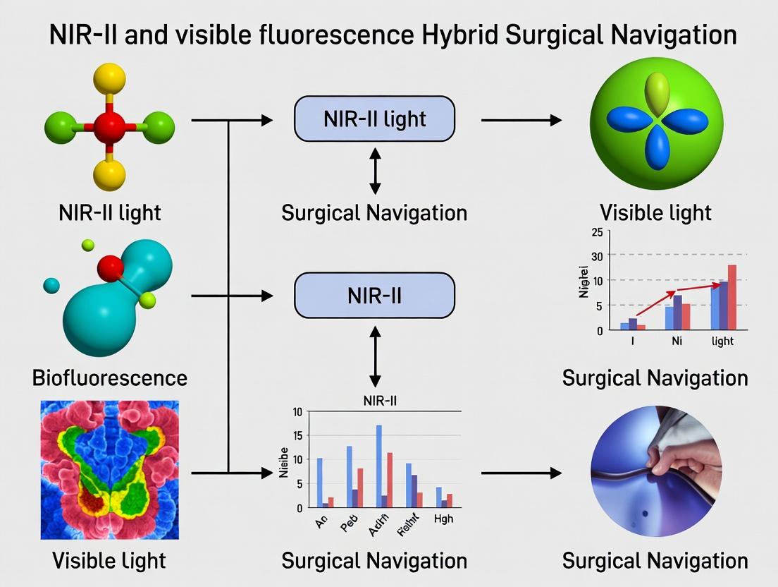
Hybrid Surgical Navigation: Uniting NIR-II and Visible Fluorescence for Precision Surgery and Targeted Therapy
This article explores the integration of second near-infrared (NIR-II, 1000-1700 nm) and visible (400-700 nm) fluorescence imaging for advanced surgical navigation, a rapidly evolving field with transformative potential in oncology...
Hybrid Surgical Navigation: Uniting NIR-II and Visible Fluorescence for Precision Surgery and Targeted Therapy
Abstract
This article explores the integration of second near-infrared (NIR-II, 1000-1700 nm) and visible (400-700 nm) fluorescence imaging for advanced surgical navigation, a rapidly evolving field with transformative potential in oncology and precision medicine. We first establish the foundational principles of NIR-II imaging, highlighting its superior tissue penetration and reduced scattering compared to traditional visible/NIR-I fluorescence. The core of the article details methodological strategies for designing dual-modal probes, instrumentation for real-time hybrid visualization, and specific applications in tumor margin delineation, sentinel lymph node mapping, and nerve/vascular structure preservation. We address critical troubleshooting aspects, including biocompatibility, signal crosstalk, and quantification challenges. Finally, we provide a rigorous validation and comparative analysis of hybrid navigation against standalone modalities, assessing sensitivity, specificity, and clinical feasibility. Aimed at researchers and drug development professionals, this synthesis provides a comprehensive roadmap for developing and implementing this next-generation navigation paradigm to improve surgical outcomes and therapeutic efficacy.
Beyond the Visible: Foundational Principles of NIR-II and Visible Fluorescence Imaging
In the context of NIR-II (1000-1700 nm) and visible fluorescence hybrid surgical navigation research, the "optical window" refers to the range of wavelengths where biological tissue exhibits minimal absorption and scattering, allowing for deeper light penetration. This principle is foundational for developing dual-mode imaging agents that combine the high resolution of visible light with the deep-tissue penetration of NIR-II.
Key Chromophores and Their Absorption: The primary absorbers in tissue are hemoglobin, water, and lipids, each with distinct spectral profiles.
Table 1: Primary Tissue Chromophore Absorption Peaks
| Chromophore | Peak Absorption Wavelength(s) (nm) | Role in Tissue Attenuation |
|---|---|---|
| Oxy-Hemoglobin (HbO₂) | ~415 (Soret), 542, 577 | Dominates absorption in visible range, decreases significantly beyond 600 nm. |
| Deoxy-Hemoglobin (Hb) | ~430 (Soret), 555 | Strong absorption in blue-green, lower in red. Contributes to absorption contrast. |
| Water (H₂O) | ~980, >1400, peak ~1450 | Negligible absorption in NIR-I (650-950 nm), becomes dominant in NIR-IIb (>1500 nm). |
| Lipids | ~930, 1210 | Contributes to absorption features in NIR-I and early NIR-II. |
| Melanin | Broadband, decreasing with λ | Strong scattering & absorption in UV/visible, influence decreases in NIR. |
Scattering Phenomena: Scattering is the dominant light-tissue interaction in the NIR window. Mie scattering (by cellular organelles) and Rayleigh scattering (by smaller structures) both decrease with increasing wavelength according to approximate ~λ^(-b) dependence (where b ranges from 0.2 to 4). This reduction is a primary reason for superior penetration of NIR-II light.
Quantifying Penetration: Effective attenuation coefficient (μeff = √(3μa(μa + μs'))) combines absorption (μa) and reduced scattering (μs') coefficients to define penetration depth (δ = 1/μ_eff), the depth at which light intensity drops to 1/e (~37%) of its incident value.
Table 2: Typical Optical Properties and Penetration Depth in Human Tissue (Approximate)
| Wavelength Range | Name | Typical μ_a (cm⁻¹) | Typical μ_s' (cm⁻¹) | Estimated Penetration Depth (δ) |
|---|---|---|---|---|
| 400-600 nm (Vis) | Visible | 1 - 10+ | 20 - 100 | 0.5 - 2 mm |
| 650-950 nm | NIR-I / Therapeutic Window | 0.1 - 0.5 | 10 - 20 | 2 - 5 mm |
| 1000-1350 nm | NIR-IIa | 0.1 - 0.3 | 5 - 10 | 5 - 10 mm |
| 1350-1700 nm | NIR-IIb | Higher (water) | 3 - 8 | 3 - 6 mm (limited by water absorption) |
Application Notes for Hybrid Surgical Navigation
Rationale for Hybrid Imaging: Combining visible (e.g., 400-700 nm) and NIR-II fluorescence leverages complementary strengths. Visible fluorophores (e.g., fluorescein, ICG in its visible emission peak) offer high quantum yield and are excellent for surface and superficial structure delineation. NIR-II fluorophores provide deep-tissue penetration and reduced autofluorescence, enabling visualization of subsurface tumors and vasculature. Simultaneous imaging allows for real-time overlay of functional NIR-II data onto high-resolution visible anatomical maps.
Key Considerations:
- Spectral Separation: Fluorophores must have well-separated excitation/emission spectra to enable simultaneous, crosstalk-free detection.
- Co-registration: Optical systems require precise spatial calibration to perfectly overlay visible and NIR-II channels.
- Agent Design: Ideal probes are single molecules or nanoparticles with dual visible/NIR-II emission, or mixtures of compatible agents.
Table 3: Comparison of Optical Windows for Surgical Guidance
| Parameter | Visible Window (400-650 nm) | NIR-I Window (650-950 nm) | NIR-II Window (1000-1700 nm) |
|---|---|---|---|
| Tissue Penetration | Shallow (mm range) | Moderate (cm range) | Deepest (cm range) |
| Scattering | Very High | Moderate | Low |
| Autofluorescence | High | Moderate | Very Low |
| Spatial Resolution | High (due to low scattering) | Reduced | High (reduced scattering) |
| Suitable Fluorophores | Fluorescein, GFP, mCherry | ICG, Cy5.5, Quantum Dots | Organic Dyes (e.g., CH-4T), SWCNTs, Rare-Earth Doped NPs |
| Role in Hybrid Navigation | Anatomical roadmap, surface feature identification | Established clinical use (ICG), moderate-depth perfusion | Deep tumor margin assessment, vascular mapping behind tissue |
Experimental Protocols
Protocol 1: Measuring Tissue Optical Properties Using Integrating Sphere Spectroscopy
Objective: Quantify the absorption (μa) and reduced scattering (μs') coefficients of ex vivo tissue samples across visible to NIR-II spectra.
Materials:
- Double-integrating sphere system (e.g., with InGaAs and Si detectors)
- Broadband light source (450-1700 nm)
- Tissue samples (< 5 mm thick, parallel surfaces)
- Index-matching fluid
- Spectrometer calibration standards (e.g., Spectralon)
- Data acquisition and inverse adding-doubling (IAD) software.
Procedure:
- System Calibration: Perform dark count and baseline correction. Measure reference reflectance (Rref) and transmittance (Tref) with empty sample holder.
- Sample Preparation: Rinse tissue sample in saline. Place between two glass slides. Apply index-matching fluid to interfaces to eliminate surface reflections.
- Measurement: Mount sample in holder between spheres. Acquire total diffuse reflectance (Rd) and total transmittance (Td) spectra from 450 nm to 1650 nm.
- Data Analysis: Input Rd, Td, and sample thickness into IAD algorithm. The algorithm iteratively solves the radiative transport equation to output μa and μs' spectra.
- Validation: Calculate μ_eff and penetration depth δ. Compare with literature values for similar tissue types.
Protocol 2: In Vivo Hybrid (Visible + NIR-II) Fluorescence Imaging in a Murine Model
Objective: Co-administer visible and NIR-II fluorophores to visualize superficial and deep structures simultaneously in a tumor-bearing mouse.
Materials:
- Dual-channel fluorescence imaging system (separate filtered CMOS camera for visible, InGaAs camera for NIR-II).
- Co-aligned excitation sources: 660 nm laser (for NIR-II dye excitation) and 480 nm LED (for visible dye excitation).
- NIR-II fluorophore (e.g., IR-E1050, 2 mg/kg in PBS).
- Visible fluorophore (e.g., Cy3-labeled targeting antibody, 1 nmol in PBS).
- Athymic nude mouse with subcutaneous tumor xenograft.
- Anesthesia system (isoflurane).
- Image co-registration software.
Procedure:
- System Setup: Power on and calibrate both cameras. Align fields of view using a dual-emitting calibration slide. Set filters: 800 nm long-pass filter for NIR-II channel; 580/20 nm band-pass for Cy3 channel.
- Animal Preparation: Anesthetize mouse (2% isoflurane in O₂). Place in prone position on warmed stage. Depilate tumor region.
- Fluorophore Administration: Inject NIR-II agent via tail vein. Wait for optimal circulation (e.g., 5-10 min). Inject the visible-labeled antibody intravenously.
- Image Acquisition:
- Acquire a white-light reference image.
- Excite with 480 nm LED, acquire Cy3 fluorescence image (exposure: 100-500 ms).
- Excite with 660 nm laser, acquire NIR-II fluorescence image (exposure: 50-200 ms).
- Acquire images at multiple time points post-injection (e.g., 0, 5, 30, 60 min).
- Image Processing: Subtract background autofluorescence. Apply flat-field correction. Use calibration transform to co-register visible and NIR-II images pixel-perfectly. Generate merged overlay images (e.g., visible=green, NIR-II=magenta). Quantify signal-to-background ratio (SBR) in regions of interest (tumor vs. muscle).
Diagrams
Title: Thesis Framework Linking Optical Window to Hybrid Navigation
Title: Hybrid In Vivo Imaging Protocol Workflow
The Scientist's Toolkit: Research Reagent Solutions
Table 4: Essential Materials for Optical Window & Hybrid Imaging Research
| Item | Function / Role | Example Product/Chemical |
|---|---|---|
| NIR-II Fluorophores | Emit in 1000-1700 nm range for deep-tissue imaging. | IR-E1050, CH-4T derivatives, PbS/CdS Quantum Dots, Single-Walled Carbon Nanotubes (SWCNTs). |
| Visible Fluorophores | Emit in 400-700 nm for high-resolution surface imaging. | Fluorescein isothiocyanate (FITC), Cyanine 3 (Cy3), Alexa Fluor 555, mCherry fluorescent protein. |
| Targeting Ligands | Conjugated to fluorophores for specific molecular targeting (e.g., tumors). | cRGD peptides (targeting αvβ3 integrin), trastuzumab (anti-HER2), folic acid. |
| Index-Matching Fluid | Reduces surface reflectance in ex vivo optical property measurements. | Glycerol-water mixtures, Intralipid dilutions, specialized optical gels. |
| Calibration Standards | For reflectance/transmittance calibration of spectrometers and cameras. | Spectralon diffuse reflectance panels, NIST-traceable neutral density filters. |
| Inverse Adding-Doubling (IAD) Software | Extracts μa and μs' from integrating sphere measurement data. | Open-source IAD software, commercial light transport solvers (e.g., MCML). |
| Dual-Channel Imaging System | Enables simultaneous or rapid sequential visible and NIR-II imaging. | Custom-built system with separate CMOS & InGaAs cameras, co-aligned excitation paths. |
| Anesthesia System | Maintains animal immobilization and physiology during in vivo imaging. | Isoflurane vaporizer with induction chamber and nose cone. |
Within our broader thesis on hybrid surgical navigation integrating NIR-II and visible fluorescence, the second near-infrared window (NIR-II, 1000-1700 nm) represents a paradigm shift. This spectral region offers distinct advantages over traditional NIR-I (700-900 nm) and visible light imaging, primarily through drastically reduced photon scattering and negligible autofluorescence in biological tissues. This application note details the defined spectrum, quantifies its key advantages, and provides protocols for its application in preclinical research, serving as a foundational guide for researchers and drug development professionals advancing targeted imaging and image-guided interventions.
Defining the NIR-II Spectrum
The NIR-II region is subdivided based on the interaction of light with tissue components, particularly water absorption. The table below defines the sub-bands and their characteristics.
Table 1: Sub-divisions of the NIR-II Spectrum (1000-1700 nm)
| Spectral Band (nm) | Common Designation | Key Tissue Optical Properties | Primary Utility |
|---|---|---|---|
| 1000-1300 | NIR-IIa / NIR-II | Low scattering, minimal water absorption | Highest performance for deep-tissue, high-resolution imaging. |
| 1300-1400 | NIR-IIb | Increased water absorption | Useful for suppressing background from shallow tissues. |
| 1400-1700 | NIR-IIc / NIR-III | Strong water absorption | Limited tissue penetration; used for unique spectroscopic applications. |
Quantitative Advantages of NIR-II Fluorescence
The following tables summarize the key quantitative benefits of imaging within the NIR-II window compared to the NIR-I window.
Table 2: Comparative Optical Properties in Biological Tissue
| Parameter | NIR-I (800 nm) | NIR-II (1100 nm) | Measured Improvement |
|---|---|---|---|
| Photon Scattering Coefficient (μs') | High (~1.5 mm⁻¹ in tissue) | Significantly Lower (~0.5 mm⁻¹ in tissue) | ~3x reduction, enabling sharper images. |
| Tissue Autofluorescence | Moderate to High | Negligible | Signal-to-Background Ratio (SBR) improvements of 10-100x. |
| Maximum Imaging Depth (in brain) | ~1-2 mm | ~3-8 mm | Penetration depth increased by 2-4x. |
| Resolution (FWHM at depth) | Degrades rapidly with depth | Maintains sub-100 μm resolution deeper | Up to 5x better resolution at 3mm depth. |
Table 3: Performance Metrics of Representative NIR-II Fluorophores
| Fluorophore Type | Peak Emission (nm) | Quantum Yield (%) | Extinction Coeff. (M⁻¹cm⁻¹) | Common Application |
|---|---|---|---|---|
| Single-Walled Carbon Nanotubes (SWCNTs) | 1000-1600 | 0.1-1 | ~10⁵ per cm of tube length | Vascular imaging, biosensing. |
| Lead Sulfide Quantum Dots (PbS QDs) | 1200-1600 | 10-30 | ~10⁵-10⁶ | Tumor targeting, lymphatic mapping. |
| Organic Dye (IR-FEP) | ~1050 | ~5 | ~2.5 x 10⁴ | Fast-excreting, renal-clearable angiography. |
| Lanthanide Nanoparticles (Er³+) | ~1525 | N/A (upconversion) | N/A | High-contrast imaging in water absorption bands. |
Protocols for NIR-II Fluorescence Imaging In Vivo
Protocol 1: NIR-IIb Vascular Angiography with ICG
Objective: To perform high-contrast, real-time vascular imaging utilizing the approved dye Indocyanine Green (ICG) in its aggregated NIR-II emitting state.
- Animal Preparation: Anesthetize a mouse (e.g., C57BL/6) using isoflurane (1-3% in O₂). Secure the animal on a heated stage (37°C). Depilate the area of interest (e.g., hind limb, scalp).
- Imaging System Setup: Use a NIR-II fluorescence microscope or imaging system equipped with:
- A 808 nm laser for excitation (power density: 10-100 mW/cm²).
- An InGaAs camera detector (sensitive to 900-1700 nm).
- A series of long-pass filters (e.g., 1000 nm, 1200 nm, 1500 nm) to select specific sub-bands.
- Dye Administration: Prepare a fresh ICG solution in saline (concentration: 0.1-0.5 mg/mL). Inject intravenously via the tail vein (dose: 2-5 mg/kg). Note: For NIR-II imaging, a higher dose than standard NIR-I clinical use may be required to promote dye aggregation, which redshifts emission.
- Data Acquisition: Begin recording immediately post-injection. Acquire sequential images through different long-pass filters (e.g., LP1000, LP1200, LP1500) to compare SBR across sub-bands. Typical exposure times range from 20-200 ms.
- Image Analysis: Use software (e.g., ImageJ, custom MATLAB/Python scripts) to quantify metrics: Signal-to-Background Ratio (SBR), vessel width (for resolution assessment), and fluorescence intensity over time.
Protocol 2: Tumor-Targeted NIR-II Imaging with Conjugated Nanoparticles
Objective: To visualize tumor margins via active targeting using NIR-II-emitting nanoparticles functionalized with targeting ligands (e.g., anti-EGFR, RGD peptides).
- Nanoparticle Preparation: Obtain or synthesize PEGylated PbS/CdS core/shell quantum dots (emission ~1300 nm). Conjugate with cRGDfk peptide via EDC/NHS chemistry. Purify via centrifugation filtration (100 kDa MWCO). Characterize hydrodynamic diameter and ligand density (DLS, UV-Vis-NIR spectroscopy).
- Tumor Model: Implant U87MG glioblastoma cells (5x10⁵) subcutaneously in the flank of an athymic nude mouse. Allow tumors to grow to ~5-8 mm in diameter.
- In Vivo Imaging: Anesthetize and prepare the animal as in Protocol 1. Acquire a pre-injection background image. Intravenously inject the QD-RGD conjugate (dose: 5-10 nmol per mouse in 100 µL PBS).
- Longitudinal Imaging: Image the animal at multiple time points (e.g., 1, 4, 24, 48 hours post-injection) using the NIR-II system with a 980 nm laser and a 1300 nm long-pass filter.
- Ex Vivo Validation: Euthanize the animal at the final time point. Excise the tumor and major organs (liver, spleen, kidneys, lungs, heart). Image all tissues ex vivo to quantify biodistribution and confirm specific tumor accumulation. Calculate tumor-to-background ratios (TBR) in vivo and ex vivo.
Visualization of Concepts and Workflows
Title: Optical Basis of NIR-II Advantage Over NIR-I
Title: Workflow for NIR-II/Visible Hybrid Surgical Navigation
The Scientist's Toolkit: Key Research Reagent Solutions
Table 4: Essential Materials for NIR-II Fluorescence Research
| Item | Function/Description | Example Vendor/Product |
|---|---|---|
| NIR-II Fluorophores | Emit light within 1000-1700 nm; the core imaging agent. | SWCNTs (NanoIntegris), PbS Quantum Dots (NN-Labs), Organic Dyes (e.g., IR-1061, Lumiprobe). |
| Targeting Ligands | Peptides, antibodies, or small molecules conjugated to fluorophores for specific biomarker binding. | cRGD peptides, Anti-EGFR antibodies (Cetuximab biosimilar), Folic acid. |
| In Vivo Injection Formulations | Sterile, pyrogen-free vehicles for systemic administration (IV, IP). | PBS (pH 7.4), Saline, 5% Dextrose. |
| Anesthetic System | For humane animal immobilization during imaging procedures. | Isoflurane vaporizer system with induction chamber (e.g., VetFlo). |
| NIR-II-Optimized Optics | Lenses and filters transparent beyond 1000 nm. | Calcium Fluoride (CaF₂) or Zinc Selenide (ZnSe) lenses, Long-pass filters (Thorlabs, Edmund Optics). |
| InGaAs Camera | Detector sensitive in the SWIR (900-1700 nm) range. | Models from Princeton Instruments (NIRvana), Teledyne (Galaxi), Raptor Photonics. |
| Dedicated Excitation Lasers | High-power lasers at wavelengths optimal for fluorophore excitation. | 808 nm, 980 nm, 1064 nm diode lasers (e.g., CNI Laser). |
| Spectral Calibration Standards | Materials with known NIR emission for system calibration and wavelength verification. | Rare-earth doped glass (e.g., NIST-traceable SRM), Tungsten Halogen lamp. |
Visible fluorescence (typically 400-700 nm) remains a cornerstone in biological imaging and surgical navigation, despite the emergence of NIR-II (1000-1700 nm) technologies. Its established role is built upon decades of validated dyes, well-characterized instrumentation, and extensive biological conjugation chemistries. Within hybrid surgical navigation research, visible fluorescence provides complementary, high-resolution anatomical and functional data when integrated with NIR-II’s deep-tissue penetration. This document details key applications, reagents, quantitative performance data, and protocols, explicitly framing them within a research strategy aiming to synergize visible and NIR-II fluorescence.
Established Role in Surgical Navigation
Visible fluorescent agents are indispensable for real-time intraoperative visualization of critical structures. Their primary roles include:
- Lymphatic Mapping: Sentinel lymph node biopsy using dyes like methylene blue and patent blue.
- Perfusion Assessment: Intraoperative assessment of tissue vascularity and anastomosis patency (e.g., with indocyanine green in the visible/NIR-I window).
- Nerve Imaging: Selective staining of peripheral nerves to avoid iatrogenic injury.
- Cancer Margin Delineation: Tumor-specific probes (e.g., 5-ALA-induced PpIX) for maximizing resection accuracy.
- Hybrid Approach: Visible dyes label multiple, specific anatomical or functional targets, while a NIR-II probe provides deep-tissue context and overall tumor burden, creating a multi-spectral navigation map.
Key Visible Fluorescent Dyes: Properties & Data
Table 1: Characteristics of Major Visible Fluorescence Dyes for Surgical Guidance
| Dye Name | Peak Ex/Em (nm) | Primary Surgical Application | Key Advantage | Major Limitation | Typical Dose (Human) |
|---|---|---|---|---|---|
| Methylene Blue | 668/688 | Sentinel lymph node mapping, Parathyroid identification | Low cost, FDA-approved, simple protocol | Skin staining, poor target specificity | 1-5 mL of 1% solution |
| Patent Blue V | 638/680 | Sentinel lymph node biopsy (esp. breast) | High lymphatic selectivity | Anaphylaxis risk, blue skin/urine discoloration | 1-2 mL of 1% solution |
| 5-ALA (PpIX) | 405/635 | Glioma margin delineation, Bladder cancer detection | Tumor-cell specific metabolism | Skin photosensitivity, shallow penetration | 20 mg/kg orally |
| Fluorescein | 494/521 | Retinal angiography, Glioma surgery, Perfusion assessment | Extremely bright, rapid pharmacokinetics | Non-specific leakage, high background | 500 mg IV |
| ICG (Visible/NIR-I) | 780/820 | Perfusion, Angiography, Lymphography | Dual visible/NIR-I imaging, excellent safety profile | Rapid protein binding, vascular confinement | 5-25 mg IV |
Table 2: Quantified Limitations of Visible Fluorescence in Surgical Context
| Limitation | Underlying Cause | Quantitative Impact | Consequence for Navigation |
|---|---|---|---|
| Shallow Penetration | High tissue scattering & absorption by hemoglobin/ melanin | Useful depth typically <1-2 mm | Inability to visualize deep or sub-surface structures |
| High Autofluorescence | Endogenous fluorophores (collagen, FAD, NADH) | Background signal can be 30-50% of target signal | Reduced target-to-background ratio (TBR), obscured margins |
| Spectral Overlap | Broad emission spectra of many dyes | Requires careful filter selection; limits multiplexing to ~2-3 colors | Challenges in simultaneous multi-target imaging |
| Photobleaching | Irreversible photochemical destruction of dye | Signal decay rate (t½) can be <60 sec under high illumination | Loss of signal during prolonged procedures |
Detailed Experimental Protocols
Protocol 1: Sentinel Lymph Node Mapping with Methylene Blue in a Rodent Model
Purpose: To visualize and biopsy the first-draining lymph node using visible fluorescence, as a component of a hybrid imaging study where a NIR-II probe labels the primary tumor.
Materials:
- Methylene blue chloride (1% sterile solution)
- Small animal imaging system with white light and 660-690 nm fluorescence filters
- Female nude mouse with orthotopic tumor (e.g., 4T1 breast cancer)
- Insulin syringe (29G)
- Dissection tools
Procedure:
- Tumor Preparation: Establish a subcutaneous tumor (~100 mm³) expressing a NIR-II fluorescent reporter.
- Dye Administration: Anesthetize the mouse. Inject 10-20 µL of 1% methylene blue intradermally at the peritumoral site using a 29G syringe.
- Massage & Diffusion: Gently massage the injection site for 60 seconds.
- Real-Time Imaging:
- At 1, 5, 10, and 15 minutes post-injection, acquire simultaneous images: a) White-light reference, b) Visible fluorescence (Ex: 660 nm, Em: 690 nm LP), c) NIR-II channel (e.g., Ex: 980 nm, Em: 1250 nm LP).
- The visible channel will show blue lymphatic vessels draining to a fluorescent node.
- Surgical Navigation & Biopsy: Using the real-time visible fluorescence overlay, make a small incision and follow the fluorescent lymphatic channel. Excise the primary sentinel lymph node (SLN).
- Ex Vivo Analysis: Image the resected SLN in both visible and NIR-II channels to check for the presence of the NIR-II tumor reporter, indicating metastasis.
Protocol 2: 5-ALA-Induced PpIX for Glioma Margin DelimationEx Vivo
Purpose: To define the accuracy of visible fluorescence in determining tumor margins from biopsy specimens, correlating with histopathology.
Materials:
- 5-aminolevulinic acid (5-ALA) hydrochloride
- Phosphate-buffered saline (PBS)
- Surgical-grade fluorescence microscope with 405 nm excitation and 635 nm emission filter.
- Fresh human glioma biopsy specimens.
- Tissue-Tek OCT compound.
Procedure:
- Patient Preparation: Administer 20 mg/kg 5-ALA orally to the patient 3 hours prior to surgery.
- Specimen Collection: During tumor resection, collect suspected tumor tissue and marginal tissue samples under visible fluorescence guidance (areas showing pink-red fluorescence).
- Ex Vivo Imaging:
- Immediately place fresh tissue samples on the fluorescence microscope stage.
- Acquire a brightfield image.
- Switch to fluorescence mode (405 nm excitation, 635/30 nm emission). Use identical exposure times across samples.
- Capture the PpIX fluorescence image.
- Quantification: Using image analysis software (e.g., ImageJ), quantify the mean fluorescence intensity (MFI) in three regions of interest (ROI) per sample.
- Correlation: Fix the imaged tissue in formalin, process, and section for H&E staining. A neuropathologist will classify each sample as "tumor" or "non-tumor."
- Analysis: Compare the PpIX MFI between histopathology-confirmed tumor and marginal tissue. Calculate sensitivity and specificity.
Visualization Diagrams
Diagram 1: Principle and Key Limits of Visible Fluorescence Imaging (76 chars)
Diagram 2: Hybrid Visible & NIR-II Surgical Navigation Workflow (79 chars)
The Scientist's Toolkit: Research Reagent Solutions
Table 3: Essential Research Materials for Visible Fluorescence Experiments
| Item | Example Product/Catalog # | Primary Function in Research |
|---|---|---|
| 5-ALA (Protoporphyrin IX Inducer) | Medac GmbH, Gliolan; Sigma A3785 | Prodrug converted to fluorescent PpIX in tumor cells for margin delineation. |
| Methylene Blue (Injectable) | American Regent, 1% solution | Lymphatic tracer for sentinel node mapping and parathyroid identification. |
| Fluorescein Isothiocyanate (FITC) | Thermo Fisher F143, F1906 | Amine-reactive dye for antibody/protein labeling, enabling targeted imaging. |
| Anti-Fade Mounting Medium | Vector Labs H-1000; ProLong Diamond | Preserves fluorescence intensity during microscopy by reducing photobleaching. |
| Fluorescent Microspheres (Beacons) | Bangs Laboratories, Polysciences Inc. | Provide a stable, quantifiable fluorescence reference for system calibration. |
| Matrigel / Phenol Red-Free Media | Corning 356237 | For 3D cell culture and in vivo tumor models, eliminates background fluorescence from phenol red. |
| Specific Antibodies (Targeting) | e.g., Anti-CEA, Anti-HER2 | Provide target specificity for conjugation with visible dyes like FITC or Cy3. |
| NIR-II / Visible Compatible Imaging System | custom-built or modified commercial systems (e.g., from Bruker, PerkinElmer) | Enables simultaneous acquisition of both spectral windows for hybrid navigation studies. |
1. Introduction
The core challenge in surgical navigation for precision oncology is achieving maximal tumor contrast while preserving critical anatomical context. Relying on a single fluorescence imaging channel, whether in the visible (VIS, 400-700 nm) or the second near-infrared window (NIR-II, 1000-1700 nm), presents inherent trade-offs. This document, framed within a thesis on hybrid surgical navigation, outlines the rationale and practical protocols for combining NIR-II and VIS fluorescence agents to yield complementary information, thereby enhancing surgical decision-making and outcomes.
2. Complementary Information: A Quantitative Summary
The following table summarizes the key complementary characteristics of VIS and NIR-II fluorescence channels.
Table 1: Complementary Characteristics of VIS and NIR-II Imaging Channels
| Parameter | Visible (VIS) Channel | NIR-II Channel | Complementary Advantage |
|---|---|---|---|
| Tissue Penetration | Low (0.1-1 mm) | High (5-10 mm) | NIR-II reveals deep tumor margins; VIS provides superficial, high-resolution detail. |
| Spatial Resolution | High (sub-mm) | Moderate (1-2 mm at depth) | VIS enables precise identification of critical surface structures (e.g., nerves, vessels). |
| Autofluorescence | High (from collagen, elastin, flavins) | Very Low | NIR-II offers superior target-to-background ratios (TBR). |
| Blood Scattering | High | Low | NIR-II provides clearer visualization of vasculature and tumors beneath blood. |
| Suitable Dyes | ICG (emission ~800 nm), Methylene Blue, Fluorescein | IRDye 800CW, CH-1055, LZ-1105, PbS Quantum Dots | Different targeting and pharmacokinetics allow for multi-parametric imaging. |
| Typical TBR in Tumors | 2.0 - 4.0 | 3.5 - 8.0+ | Hybrid TBR > NIR-II alone for superficial/deep composite assessment. |
3. Key Experimental Protocols
Protocol 3.1: Co-Administration of VIS and NIR-II Agents for Hybrid Navigation Objective: To simultaneously visualize surgical anatomy (VIS) and deep tumor margins (NIR-II). Materials:
- Animal model: BALB/c nude mouse with subcutaneously implanted tumor (e.g., U87MG glioma).
- VIS agent: 50 µL of 1 mM Methylene Blue (vasculature/lymphatic marker).
- NIR-II agent: 100 µL of 100 µM IRDye 800CW conjugated to cRGD (integrin αvβ3 targeting).
- Hybrid Imaging System: Custom-built or commercial system with: a) 660 nm laser & 700/75 nm filter (VIS channel); b) 785 nm laser & 1000 nm long-pass filter (NIR-II channel).
- Anesthesia setup (isoflurane). Procedure:
- Anesthetize the mouse and secure in a prone position.
- Perform intravenous injection of the NIR-II targeted agent via the tail vein.
- At 24 hours post-injection, induce anesthesia again. Perform intravenous injection of the VIS agent.
- After 10 minutes, acquire baseline VIS channel images (exposure: 100 ms).
- Immediately switch settings and acquire NIR-II channel images (exposure: 300 ms).
- Surgically expose the tumor region. Repeat steps 4-5.
- Perform real-time hybrid navigation: Use the NIR-II channel to guide deep resection margins and the VIS channel to identify and preserve surrounding surface vasculature.
- Ex vivo imaging of the resected tumor and wound bed confirms complete resection in both channels.
Protocol 3.2: Quantitative Co-Registration and TBR Analysis Objective: To quantify the spatial and signal correlation between VIS and NIR-II signals. Materials:
- Image processing software (ImageJ, MATLAB).
- Images from Protocol 3.1. Procedure:
- Load the pre-resection VIS and NIR-II image pairs.
- Apply a rigid body transformation to co-register the two images based on fiduciary markers or animal contours.
- Define three Regions of Interest (ROIs): a) Tumor core (T), b) Peritumoral margin (M), c) Distant background tissue (B).
- Measure the mean fluorescence intensity (MFI) in each ROI for both channels.
- Calculate the Target-to-Background Ratio (TBR) for each channel: TBR_channel = MFI(T) / MFI(B)
- Generate an overlay image with a colormap (e.g., Green for VIS, Red for NIR-II). Areas of colocalization appear yellow.
- Calculate the Dice Similarity Coefficient (DSC) to quantify spatial overlap of the segmented tumor signals from the two channels: DSC = 2|A∩B| / (|A| + |B|), where A and B are binary tumor masks from each channel.
4. Visualization of Concepts and Workflows
Title: Rationale for Hybrid Surgical Imaging
Title: Dual-Channel Signal Pathway in Hybrid Imaging
5. The Scientist's Toolkit: Essential Research Reagents & Materials
Table 2: Key Reagents and Materials for Hybrid NIR-II/VIS Navigation Research
| Item | Category | Function / Rationale | Example Product/Note |
|---|---|---|---|
| IRDye 800CW NHS Ester | NIR-II Fluorophore | Conjugatable dye for antibody/peptide labeling; emits in NIR-I/II border (~800 nm). | LI-COR Biosciences |
| CH-1055 or LZ-1105 | NIR-II Fluorophore | Small-molecule organic dyes with peak emission >1000 nm for deep tissue imaging. | Research chemicals from academic labs. |
| Methylene Blue | VIS Fluorophore | Clinically approved dye for visualizing lymphatics, parathyroids, and vasculature. | Pharmacy grade, sterile. |
| Indocyanine Green (ICG) | Dual-Channel Agent | FDA-approved dye with emission at ~800 nm; can be used in both VIS and NIR-I systems. | Diagnostic Green, Inc. |
| cRGDfK Peptide | Targeting Ligand | Binds integrin αvβ3, overexpressed in tumor vasculature and many cancers. | Conjugation-ready, >95% purity. |
| Anti-EGFR Antibody | Targeting Ligand | For targeting epidermal growth factor receptor in various carcinomas. | Cetuximab biosimilar for research. |
| Matrigel | Tumor Implantation | Provides extracellular matrix for consistent subcutaneous tumor engraftment. | Corning Matrigel |
| InGaAs Camera | Detection Hardware | Essential for capturing NIR-II fluorescence (>1000 nm). | Sensors Unlimited (Goodrich) or Princeton Instruments. |
| Silicon CCD Camera | Detection Hardware | Standard detector for VIS and NIR-I (<900 nm) fluorescence. | Many suppliers (e.g., Hamamatsu). |
| Dichroic Mirrors & Filters | Optical Components | For splitting and isolating VIS and NIR-II emission channels in a hybrid system. | Custom set from Chroma or Semrock. |
| Isoflurane System | Animal Anesthesia | Provides stable and reversible anesthesia for prolonged imaging sessions. | VetEquip or similar. |
Within the evolving field of NIR-II (1000-1700 nm) and visible fluorescence hybrid surgical navigation, the selection of imaging agent platform is paramount. This Application Notes document details the core platforms—organic dyes, quantum dots (QDs), and lanthanide-doped nanoparticles (LNPs)—providing comparative data, standardized protocols, and essential research toolkits to guide preclinical development.
Comparative Performance Metrics
Table 1: Core Platform Characteristics for Hybrid Navigation
| Property | Organic Dyes (e.g., IR-1061, FD-1080) | Quantum Dots (e.g., PbS/CdS, Ag₂S) | Lanthanide Nanoparticles (e.g., NaYF₄:Yb,Er,Tm) |
|---|---|---|---|
| Primary Emission Range | NIR-II (1000-1400 nm) | NIR-II (1200-1600 nm) | Visible (540, 650 nm) & NIR-II (1550 nm) |
| Absorption Coefficient (M⁻¹cm⁻¹) | ~10⁵ | ~10⁶ - 10⁷ | ~10² - 10³ (low, requires sensitizer) |
| Quantum Yield (NIR-II) | 0.3-5% | 10-30% (in solution) | 1-10% (at 1550 nm) |
| Stokes Shift | Small (~10-30 nm) | Large (>200 nm) | Extremely Large (>300 nm) |
| Hydrodynamic Size | <2 nm | 5-15 nm (with coating) | 20-100 nm |
| Excitation Source | 785 nm, 808 nm, 980 nm | 808 nm, 980 nm | 808 nm, 980 nm (for Yb³⁺ sensitization) |
| Biodegradability | High | Low/None | Low/None |
| Potential Toxicity | Low (if chemically pure) | High (heavy metal leaching) | Low (inert shelled) |
Table 2: Key Application Parameters for Surgical Navigation
| Parameter | Target Value (Guideline) | Dye Performance | QD Performance | LNP Performance |
|---|---|---|---|---|
| Brightness (ε × QY) | >10⁶ M⁻¹cm⁻¹ | ~10⁴ - 10⁵ | ~10⁶ - 10⁷ | ~10⁴ - 10⁵ |
| Tissue Penetration Depth | >5 mm | 3-8 mm | 5-12 mm | 4-10 mm (at 1550 nm) |
| Optimal Signal-to-Background Ratio | >5 | 3-10 | 8-20 | 5-15 |
| Blood Clearance Half-life | Tunable: mins to hrs | Minutes (renal) | Hours (hepatic) | Hours to days |
| Photobleaching Half-time | >10 min | 2-10 min | >60 min | Essentially infinite |
Experimental Protocols
Protocol 1: Conjugation of Targeting Ligands to Nanoparticle Surfaces
Objective: Attach cRGD peptides to PEG-coated NaYF₄:Yb,Er@NaYF₄ nanoparticles for tumor vasculature targeting in hybrid navigation.
- Activation: Disperse 1 mL of 1 mg/mL amine-PEG-coated LNPs in PBS (pH 7.4). Add 50 µL of 10 mM Sulfo-SMCC (heterobifunctional crosslinker) and incubate for 1 hour at RT with gentle shaking.
- Purification: Remove excess crosslinker via size-exclusion chromatography (PD-10 column) or centrifugal filtration (100 kDa MWCO), eluting in PBS.
- Ligand Preparation: Thiolate the cRGD peptide (sequence: c(RGDyK)) using 2-iminothiolane (Traut's reagent) at a 20:1 molar ratio for 30 min.
- Conjugation: Mix the activated LNPs with thiolated cRGD at a 1:200 nanoparticle-to-peptide molar ratio. React overnight at 4°C.
- Quenching & Final Purification: Add 10 µL of 1 mM cysteine to quench unreacted maleimide groups for 15 min. Purify via centrifugal filtration (100 kDa MWCO) 3x with PBS. Confirm conjugation via UV-Vis (characteristic peptide bond absorption) or a fluorescence-based amine assay on the supernatant.
Protocol 2: In Vivo Hybrid NIR-II/Visible Imaging of Tumor Margins
Objective: Simultaneously visualize tumor margins (via targeted NIR-II signal) and critical nerves/vasculature (via visible signal from upconversion).
- Animal & Tumor Model: Use a nude mouse with a subcutaneous U87MG glioblastoma xenograft (~150 mm³).
- Probe Administration: Co-inject via tail vein: 100 µL of cRGD-conjugated LNPs (emitting at 1550 nm & 540 nm, 1 mg/mL) and 100 µL of ICG derivative dye (emitting at 820 nm, 50 µM) as a non-targeted perfusion control.
- Imaging System Setup: Employ a dual-channel fluorescence imaging system equipped with:
- A 980 nm laser (for LNP excitation) with a 1500 nm long-pass filter for NIR-II (1550 nm) and a 540/20 nm bandpass for visible collection.
- An 808 nm laser (for dye excitation) with an 845/40 nm bandpass filter for NIR-I collection.
- Image Acquisition: Anesthetize the animal. Acquire baseline images pre-injection. Image at 1, 4, 8, 12, and 24 hours post-injection. Maintain consistent laser power and exposure times across time points.
- Data Analysis: Use regions of interest (ROIs) to quantify signal intensity in the tumor versus contralateral muscle. Calculate Tumor-to-Background Ratio (TBR) for each channel. Overlay the high-TBR 1550 nm channel (tumor) with the 540 nm channel (anatomy) for surgical guidance simulation.
Visualization Diagrams
Diagram Title: Platform Selection Logic for Hybrid Navigation
Diagram Title: LNP Energy Pathway for Hybrid Emission
The Scientist's Toolkit
Table 3: Essential Research Reagent Solutions
| Item | Function in Hybrid Navigation Research | Example Product/Chemical |
|---|---|---|
| Heterobifunctional Crosslinkers | Conjugate targeting ligands (peptides, antibodies) to nanoparticle surface functional groups (e.g., -NH₂, -COOH). | Sulfo-SMCC, NHS-PEG-Maleimide |
| PEGylation Reagents | Impart "stealth" properties, reduce opsonization, and increase blood circulation half-life of nanoparticles. | mPEG-SH (Thiol), DSPE-PEG(2000)-Amine |
| Commercial NIR-II Dyes | Benchmark agents for perfusion imaging and control studies. | IR-1061, FD-1080, CH-4T |
| Lanthanide Precursors | High-purity starting materials for reproducible synthesis of core-shell nanoparticles. | Yttrium(III) acetate, Ytterbium(III) acetate, Erbium(III) acetate |
| Tumor-Targeting Peptides | Functionalize imaging agents to achieve specific accumulation at disease sites (e.g., integrin αvβ3). | cRGDfK, iRGD |
| In Vivo Imaging Matrices | Simulate tissue scattering and absorption for system calibration and depth penetration studies. | Intralipid, India ink phantoms |
| Anesthesia System | Maintain animal viability and immobility during longitudinal in vivo imaging sessions. | Isoflurane vaporizer with nose cones |
| Dual-Channel Fluorescence Imager | System capable of simultaneous or rapid switching between NIR-II and visible detection channels. | Custom-built or commercial systems with InGaAs & CCD/CMOS cameras. |
Building the Hybrid System: Probe Design, Instrumentation, and Surgical Applications
Application Notes and Protocols
1.0 Thesis Context This document provides application notes and protocols for designing dual-modality fluorescent probes, framed within a broader thesis research program on NIR-II and visible fluorescence hybrid surgical navigation. The integration of high-resolution, real-time visible fluorescence with deep-tissue-penetrating NIR-II imaging aims to optimize tumor margin delineation and critical structure identification during oncologic surgery.
2.0 Design Strategy I: Molecular Conjugation Probes This strategy involves covalently linking a visible fluorophore (e.g., Cy3, FITC) and a NIR-II fluorophore (e.g., IRDye800CW, CH1055) via a linker, often with a targeting ligand (e.g., peptide, antibody).
2.1 Protocol: Synthesis of a cRGD-Targeted Cy3/IRDye800CW Conjugate Objective: Synthesize a dual-modality probe for αvβ3 integrin targeting. Materials: See "Research Reagent Solutions" Table 1. Procedure:
- Activation of IRDye800CW-NHS: Dissolve 1.0 mg IRDye800CW-NHS ester in 200 µL anhydrous DMF.
- cRGD-Cy3 Solution: Dissolve 0.5 mg cRGDfK-Cy3 peptide (containing a free lysine amine) in 500 µL PBS (pH 8.0).
- Conjugation: Add the IRDye800CW-NHS solution dropwise to the stirring cRGD-Cy3 solution. React for 2 hours at room temperature, protected from light.
- Purification: Purify the reaction mixture using a PD-10 desalting column pre-equilibrated with PBS (pH 7.4). Collect the colored fraction.
- Characterization: Confirm conjugation and determine dye:peptide ratio using UV-Vis-NIR spectroscopy (Table 1) and HPLC-MS.
3.0 Design Strategy II: Nanoplatform-Based Probes Nanoplatforms (e.g., polymers, silica, liposomes) encapsulate or co-load both types of fluorophores, offering high payload and tunable pharmacokinetics.
3.1 Protocol: Preparation of NIR-II/Visible Fluorescent Polymeric Nanoparticles Objective: Prepare PEG-PLGA nanoparticles co-loaded with a NIR-II dye (CH-4T) and a visible dye (DiO). Materials: See "Research Reagent Solutions" Table 1. Procedure:
- Organic Phase: Dissolve 50 mg PEG-PLGA, 0.1 mg CH-4T, and 0.05 mg DiO in 2 mL of dichloromethane.
- Aqueous Phase: Prepare 4 mL of 2% (w/v) polyvinyl alcohol (PVA) solution in water.
- Emulsification: Add the organic phase to the aqueous phase and sonicate using a probe sonicator (100 W, 60 s on ice).
- Solvent Evaporation: Stir the emulsion overnight at room temperature to evaporate the organic solvent.
- Washing & Collection: Centrifuge the nanoparticle suspension at 15,000 × g for 20 min. Wash the pellet with DI water 3 times to remove free dye and PVA. Resuspend in PBS and filter through a 0.22 µm filter. Characterize size and fluorescence (Table 2).
4.0 Quantitative Data Summary
Table 1: Spectral Properties of Featured Fluorophores
| Fluorophore | Modality | Peak Excitation (nm) | Peak Emission (nm) | Molar Extinction Coefficient (M⁻¹cm⁻¹) |
|---|---|---|---|---|
| Cy3 | Visible | 550 | 570 | 150,000 |
| IRDye800CW | NIR-II | 774 | 789 | 240,000 |
| CH-4T | NIR-II | 808 | 1025 | ~1.2 x 10⁵ (in particles) |
| DiO | Visible | 484 | 501 | N/A (Environment dependent) |
Table 2: Characterization of Exemplar Nanoparticles (n=3)
| Parameter | Mean Value ± SD | Measurement Method |
|---|---|---|
| Hydrodynamic Size | 112.4 ± 5.2 nm | Dynamic Light Scattering |
| Polydispersity Index (PDI) | 0.08 ± 0.02 | Dynamic Light Scattering |
| ζ-Potential | -12.3 ± 1.5 mV | Laser Doppler Velocimetry |
| NIR-II Fluorescence Quantum Yield | ~2.1% (relative to IR1061) | Integrative sphere method |
| Dye Loading Efficiency (CH-4T) | 78.5% ± 3.1% | UV-Vis-NIR Calibration |
5.0 The Scientist's Toolkit
Table 1: Key Research Reagent Solutions
| Item | Function / Explanation |
|---|---|
| IRDye800CW-NHS ester | NIR-II fluorophore with reactive N-hydroxysuccinimide ester for covalent conjugation to amine groups on targeting ligands. |
| cRGDfK-Cy3 peptide | Cyclic arginine-glycine-aspartic acid peptide targeting αvβ3 integrin, pre-labeled with visible Cy3 fluorophore and featuring a free amine for secondary conjugation. |
| PEG-PLGA copolymer | Poly(ethylene glycol)-poly(lactic-co-glycolic acid) copolymer forms biodegradable, stealth nanoparticle cores for dye encapsulation. |
| CH-4T dye | High-performance donor-acceptor-donor (D-A-D) type small molecule organic fluorophore with emission in the NIR-IIb region (>1000 nm). |
| DiO (DiOC₁₈(3)) | Lipophilic carbocyanine dye for visible (green) fluorescence labeling of nanoparticle membranes. |
| Anhydrous DMF | Polar aprotic solvent used for dye activation/conjugation to prevent hydrolysis of NHS esters. |
| Polyvinyl Alcohol (PVA) | Emulsifying agent used in nanoparticle formulation to stabilize the oil-in-water emulsion. |
| PD-10 Desalting Column | Size exclusion chromatography column for quick purification of conjugated probes from unreacted small-molecule dyes. |
6.0 Visualizations
Title: Molecular Conjugation Probe Design
Title: Nanoplatform Probe Integration Workflow
Title: Design Strategies within Thesis Context
This application note details the instrumentation and protocols for real-time hybrid imaging within a broader research thesis focused on NIR-II (1000-1700 nm) and visible fluorescence hybrid surgical navigation. The integration of these spectral windows enables multiplexed visualization of anatomical structures, physiological processes, and targeted molecular agents, offering unprecedented guidance precision in oncological and vascular surgeries. The core instrumentation challenge lies in the simultaneous capture of faint NIR-II signals and bright visible fluorescence with high spatial-temporal resolution, requiring optimized camera systems and optical filtering strategies.
Key Instrumentation Components: Specifications & Data
Camera Systems for Hybrid Imaging
The selection of a camera system is critical. The primary specifications for hybrid NIR-II/visible imaging are summarized in Table 1.
Table 1: Quantitative Comparison of Camera Detectors for Hybrid Imaging
| Detector Type | Quantum Efficiency (QE) Profile | Typical Read Noise (e-) | Dark Current (e-/pix/s) @ -40°C | Frame Rate (Full Frame) | Key Advantage for Hybrid Imaging | Primary Limitation |
|---|---|---|---|---|---|---|
| Scientific CMOS (sCMOS) | ~60% (400-700 nm); <20% (900-1000 nm) | 1.0 - 2.5 | 0.1 - 0.5 | 20 - 100 fps | High resolution & speed for visible channel. | Rapidly declining QE >800 nm. |
| InGaAs Focal Plane Array (FPA) | ~80% (900-1700 nm) | 100 - 500 | 1000 - 5000 | 10 - 100 fps (sub-window) | Essential for sensitive NIR-II detection. | High noise, cost; blind to visible light. |
| Extended InGaAs (e-InGaAs) | ~70% (400-1700 nm) | 150 - 600 | 2000 - 10000 | 5 - 50 fps | Single-camera solution for broad spectrum. | Compromised performance at extremes (visible & NIR-II). |
| Intensified sCMOS (I-sCMOS) | Dictated by photocathode (e.g., GaAs: 15-50% to 900 nm) | Effectively <1 | Negligible | 20 - 60 fps | Extreme sensitivity for low-light visible/NIR-I. | No native NIR-II (>1000 nm) response. |
| Hybrid Dual-Camera System | sCMOS: High QE in visible; InGaAs: High QE in NIR-II | sCMOS: Low; InGaAs: High | sCMOS: Very Low; InGaAs: High | Synchronized, variable | Optimal performance in each band. | Complex coregistration and data fusion required. |
Data synthesized from recent product specifications (Hamamatsu, Teledyne Princeton Instruments, FLIR) and peer-reviewed publications (2023-2024).
Optical Filter Selection & Configuration
Optical filters isolate target fluorescence from excitation light and ambient noise. Key parameters are in Table 2.
Table 2: Optical Filter Specifications for Hybrid Imaging Experiments
| Filter Type | Central Wavelength / Cut-on (nm) | Bandwidth (FWHM, nm) | Optical Density (OD) | Placement | Function in Hybrid Setup |
|---|---|---|---|---|---|
| Excitation Filter (Visible) | 490, 660, 780 | 10 - 25 | >6 @ stop band | Illumination path | Cleans laser/LED source for visible fluorophores (e.g., GFP, ICG). |
| Excitation Filter (NIR-II) | 808, 980, 1064 | 10 - 20 | >6 @ stop band | Illumination path | Cleans laser source for NIR-II fluorophores (e.g., CH1055, LZ1105). |
| Dichroic Mirror | 875, 950, 1100 (Edge) | N/A | >5 @ rejection band | 45° in imaging path | Separates emission from excitation; critical for dual-band designs. |
| Emission Filter (Visible Channel) | 520, 710, 830 | 20 - 50 | >6 @ excitation | Camera 1 (sCMOS) | Passes visible/NIR-I emission, blocks laser scatter. |
| Emission Filter (NIR-II Channel) | 1250, 1500 | 50 - 200 (Long-pass common) | >6 @ excitation & <1000 nm | Camera 2 (InGaAs) | Isolates NIR-II signal, blocks all shorter wavelengths. |
| Multiband Filter Set | Custom (e.g., Ex: 660/808; Em: 710/1300LP) | Custom | >6 per band | Single-camera system | Enables simultaneous multichannel acquisition with one detector. |
Specifications representative of filters from Semrock (IDEX Health & Science), Chroma Technology, and Thorlabs.
Experimental Protocols
Protocol 1: System Calibration & Spatial Co-registration for a Dual-Camera Setup
Objective: To achieve pixel-perfect alignment between the visible (sCMOS) and NIR-II (InGaAs) imaging channels. Materials: Dual-camera hybrid imaging system, NIR-II/visible fluorescent alignment target (custom pattern), data acquisition software (e.g., LabView, MATLAB), calibration software. Procedure:
- Target Preparation: Illuminate a custom target containing spatially defined patterns (e.g., grid, crosses) with both visible (e.g., 550 nm) and NIR-II (e.g., 1200 nm) fluorescence.
- Simultaneous Acquisition: Acquire images of the target from both cameras simultaneously under identical geometrical conditions.
- Feature Detection: Use software algorithms (e.g., cross-correlation, feature point detection) to identify corresponding control points in both images.
- Transform Calculation: Compute a projective or polynomial transformation matrix (e.g., using RANSAC algorithm) that maps the NIR-II image coordinates onto the visible image coordinates.
- Validation: Apply the transformation to a test target image and quantify the alignment error (Root Mean Square Error, target: <2 pixels). Store the transformation matrix for real-time application during experiments.
Protocol 2: In Vivo Hybrid Imaging of Tumor Vasculature and Sentinel Lymph Node
Objective: To simultaneously visualize tumor-associated vasculature via NIR-II fluorescence and sentinel lymph node (SLN) via visible/NIR-I fluorescence in a murine model. Materials: Mouse model (e.g., 4T1 tumor xenograft), NIR-II vascular agent (e.g., IRDye 800CW, 5 nmol in 100 µL PBS), visible lymph tracer (e.g., Indocyanine Green (ICG), 10 µM in 20 µL), dual-camera hybrid system, isoflurane anesthesia setup, heating pad. Procedure:
- Animal Preparation: Anesthetize the mouse and place it in a supine position on a heated imaging stage. Secure vital signs monitoring.
- Tracer Administration: Intravenously inject the NIR-II vascular agent via the tail vein. Immediately after, perform an intradermal injection of ICG at the peritumoral site.
- Image Acquisition: a. Pre-injection Baseline: Acquize a baseline image pair (visible & NIR-II). b. Dynamic Acquisition: Initiate continuous, synchronized acquisition from both cameras immediately post-injection (frame rate: 5 fps for 5 min, then 1 fps for 30 min). c. Excitation: Use 808 nm laser for simultaneous excitation of both ICG and IRDye 800CW. d. Emission Filtering: sCMOS channel: 830/30 nm bandpass (ICG emission); InGaAs channel: 1250 nm long-pass (NIR-II emission).
- Data Processing: Apply the co-registration transform (Protocol 1). Analyze time-to-peak and signal intensity in the tumor vasculature (NIR-II channel) and the draining SLN (visible channel). Generate overlay images.
Protocol 3: Quantifying Filter Crosstalk in a Multiband Configuration
Objective: To measure signal contamination between channels when using a multiband filter set on a single e-InGaAs camera. Materials: e-InGaAs camera with multiband filter set (e.g., Ex: 660/808 nm, Em: 710/40 nm & 1300LP), fluorophore solutions: Alexa Fluor 660 (visible) and CH1055 (NIR-II), spectrophotometer, black-walled 96-well plate. Procedure:
- Spectral Validation: Verify the emission spectra of each fluorophore using a spectrophotometer.
- Single-Fluorophore Imaging: a. Load wells with either AF660 or CH1055 alone. b. Image with 660 nm excitation, collect signal through the 710 nm bandpass ("Visible Channel"). c. Image with 808 nm excitation, collect signal through the 1300 nm long-pass ("NIR-II Channel"). d. Record mean signal intensity in Region of Interest (ROI).
- Crosstalk Calculation: a. For AF660, calculate the percentage of signal detected in the NIR-II channel relative to its primary channel. b. For CH1055, calculate the percentage of signal detected in the visible channel. c. Acceptable crosstalk is typically <1% for quantitative multiplexing.
- Corrective Action: If crosstalk is high, adjust filter bandwidths or employ sequential unmixing algorithms during acquisition.
Visualization Diagrams
Diagram Title: Workflow for Dual-Channel In Vivo Hybrid Imaging
Diagram Title: Optical Path with Filter Configuration for Hybrid Imaging
The Scientist's Toolkit: Research Reagent Solutions
Table 3: Essential Materials for NIR-II/Visible Hybrid Imaging Experiments
| Item | Example Product / Specification | Function in Hybrid Imaging |
|---|---|---|
| NIR-II Fluorescent Agent | CH1055-PEG, IRDye 800CW, LZ1105, inorganic quantum dots (Ag2S) | Provides deep-tissue, high-resolution contrast in the NIR-II window for vascular and structural imaging. |
| Visible/NIR-I Fluorescent Agent | Indocyanine Green (ICG), Methylene Blue, Alexa Fluor 660, GFP-expressing cells | Offers bright, well-established contrast for superficial structures, sentinel lymph nodes, or genetic expression. |
| Multispectral Calibration Target | Custom slide with fluorescent patterns (e.g., AF660 & IR-12) emitting in visible & NIR-II. | Enables pixel-perfect spatial co-registration of multiple camera channels. |
| Laser Sources | 660 nm (100 mW), 808 nm (500 mW), 980 nm (300 mW) diode lasers with TTL modulation. | Provides high-power, wavelength-specific excitation for fluorophores with minimal bleed-through. |
| Optical Filter Set | Custom multiband set from Chroma (e.g., ex: 660/808, em: 710/40 & 1300LP). | Precisely isolates target emission from excitation scatter and autofluorescence in each channel. |
| Image Co-registration Software | MATLAB Image Processing Toolbox, Fiji/ImageJ with BigWarp plugin, custom LabVIEW VI. | Computes and applies spatial transforms to align images from different optical paths. |
| Synchronization Hardware | National Instruments DAQ card (e.g., PCIe-6321) or Arduino-based trigger box. | Generates precise TTL pulses to synchronize laser firing, filter wheel position, and camera exposure. |
| Anesthesia & Monitoring System | Isoflurane vaporizer, heated stage, pulse oximeter for rodents. | Maintains animal viability and physiological stability during longitudinal imaging sessions. |
This document details the application notes and protocols for the clinical workflow of hybrid surgical navigation integrating NIR-II (1000-1700 nm) and visible (400-700 nm) fluorescence imaging. This workflow is central to a broader thesis investigating the synergistic potential of multi-spectral imaging for improving surgical precision, margin assessment, and real-time visualization of critical structures.
Key Research Reagent Solutions & Materials
Table 1: Essential Reagents and Materials for Hybrid Navigation Studies
| Item/Category | Example(s) | Primary Function in Workflow |
|---|---|---|
| NIR-II Fluorophores | IRDye 800CW, CH1055, Ag2S quantum dots, Lanthanide-doped nanoparticles | Provides deep-tissue penetration, low autofluorescence, and high spatial resolution for imaging vasculature and deep-seated targets. |
| Visible Fluorophores | Methylene Blue, Fluorescein, Indocyanine Green (ICG), Targeted fluorescent antibodies (e.g., Bevacizumab-IRDye800) | Enables real-time visualization of superficial structures, biliary flow, perfusion, and specific molecular targets. |
| Hybrid/Multimodal Probes | Dual-labeled agents (e.g., visible & NIR-II tags on same nanoparticle/antibody) | Allows concurrent or sequential imaging in both spectral bands, facilitating co-registration and validation. |
| Clinical-Grade Formulation | GMP-produced vials, sterile saline for reconstitution | Ensures safety and compatibility for human administration. |
| Surgical Navigation System | Custom-built or commercial open-platform systems (e.g., FLARE, Quest, Artemis) with dual-channel detection | Integrates NIR-II and visible cameras, overlays fluorescence on white-light video, and provides quantitative metrics. |
| Calibration Phantoms | Tissue-simulating phantoms with embedded fluorescent targets at known concentrations | Validates system sensitivity, linearity, and co-registration accuracy preoperatively. |
Clinical Workflow Protocol
Preoperative Phase: Planning & Probe Administration
Protocol 3.1.1: Patient and Probe Preparation
- Patient Selection & Consent: Enroll patients per IRB-approved protocol. Confirm indication for image-guided surgery (e.g., tumor resection).
- Probe Selection & Dose: Based on target (e.g., tumor receptor, lymphatic basin), select appropriate fluorescent probe(s). Common doses: ICG: 2.5-5.0 mg IV for angiography; Targeted NIR-II probes: typically 0.1-1.0 mg/kg in preclinical models (translate to human equivalent dose).
- Timing of Administration: Administer targeted probes hours to days before surgery (e.g., 24-48h for antibody-based agents) to allow for background clearance. Administer non-targeted vascular/flow agents (e.g., ICG, Methylene Blue) intraoperatively.
Table 2: Example Probe Administration Parameters
| Probe Type | Target | Administration Time | Dose Range (Human) | Primary Imaging Window |
|---|---|---|---|---|
| ICG (NIR-I/Visible) | Vasculature, Perfusion | Intraoperative | 2.5 - 25 mg IV | 0-10 minutes post-injection |
| Methylene Blue (Visible) | Parathyroid, Lymphatics | Intraoperative | 1 - 10 mL of 1% solution | 5-30 minutes post-injection |
| Targeted Antibody-NIR-II | Tumor Antigen (e.g., EGFR) | Preoperative (24-48h) | Human equivalent dose from preclinical PK/PD | Intraoperative (persistent signal) |
Intraoperative Phase: Imaging & Display
Protocol 3.2.1: System Setup and Calibration
- Navigation System Startup: Power on hybrid imaging system. Allow cameras (visible CMOS, NIR-II InGaAs/CMOS) to cool if necessary.
- Spatial Co-registration: Use calibration phantom to align the fields of view of the white-light, visible fluorescence, and NIR-II cameras. Software should generate a unified coordinate system.
- Sensitivity Calibration: Image serial dilutions of the administered probe in a well plate phantom to establish a standard curve for potential quantitative analysis.
Protocol 3.2.2: Sequential Hybrid Image Acquisition & Display
- Baseline Imaging: Acquire pre-contrast white-light, visible, and NIR-II images of the surgical field to assess autofluorescence and background.
- Dynamic Imaging (if applicable): Post-injection of vascular agent, initiate continuous or rapid-sequence imaging to capture inflow dynamics in both channels.
- Dual-Channel Display: Employ one of the following visualization modes on the surgeon's display:
- Overlay Mode: Pseudo-color NIR-II signal (e.g., green or yellow) and visible fluorescence signal (e.g., blue or red) are independently overlaid on the high-definition white-light video.
- Subtraction Mode: Background-subtracted fluorescence signal is displayed to enhance contrast.
- Quantitative Mode: Software displays signal-to-background ratios (SBR) or estimated concentration values in regions of interest.
Protocol 3.2.3: Intraoperative Decision Support
- Margin Assessment: Use NIR-II to interrogge deep tumor margins; use visible fluorescence to check superficial margins or surgical cavities.
- Critical Structure Identification: Use visible fluorescence (e.g., Methylene Blue) to identify and preserve parathyroids or nerves; use NIR-II angiography to map adjacent vasculature.
- Documentation: Save key image sets (raw and processed) with timestamps for postoperative analysis.
Experimental Validation Protocol
Protocol 4.1: Validation of Co-registration Accuracy
- Objective: Quantify the spatial alignment error between NIR-II and visible fluorescence channels.
- Method:
- Fabricate a phantom with a grid pattern emitting in both visible and NIR-II.
- Capture a hybrid image.
- Use image analysis software to identify control point pairs in both channels.
- Calculate the root-mean-square error (RMSE) in pixels/mm between corresponding points.
- Acceptance Criterion: RMSE < 2 pixels (or equivalent sub-millimeter distance).
Protocol 4.2: Determining Signal-to-Background Ratio (SBR)
- Objective: Quantify the detectability of a fluorescent target.
- Method:
SBR = (Mean Signal Intensity of Target Region - Mean Background Intensity) / Standard Deviation of Background Intensity - Measurement: Perform this calculation for the same target in both NIR-II and visible channels from in vivo or ex vivo tissue images.
Table 3: Example Quantitative Outcomes from Hybrid Imaging Study
| Metric | NIR-II Channel (Mean ± SD) | Visible Channel (Mean ± SD) | Significance (p-value) | Implication |
|---|---|---|---|---|
| Tumor SBR | 5.2 ± 0.8 | 2.1 ± 0.5 | p < 0.001 | NIR-II provides superior contrast for deep tumors. |
| Co-registration Error | 1.3 ± 0.4 pixels | 1.3 ± 0.4 pixels | N/A | System maintains accurate alignment. |
| Margin False Negative Rate | 5% | 15% | p < 0.05 | Hybrid guidance reduces missed tumor margins. |
Visualized Workflows and Pathways
Title: Clinical Hybrid Navigation Workflow
Title: Intraoperative Imaging & Display System Dataflow
Application Notes
The integration of NIR-II (1000-1700 nm) and visible (400-700 nm) fluorescence imaging represents a transformative advance in surgical oncology. This hybrid approach enables real-time, multiplexed visualization of primary tumors, micrometastases, and critical anatomical structures, addressing the fundamental challenge of achieving complete resection with maximal preservation of healthy tissue.
Key Advantages:
- NIR-II Fluorescence: Offers superior tissue penetration (5-20 mm), reduced scattering, and near-zero autofluorescence, providing a clear, high-signal-to-background ratio (SBR) image of deep tumor margins and vascular networks.
- Visible Fluorescence: Allows for direct visual correlation by the surgeon, ideal for labeling superficial structures, specific molecular targets, or as a counterstain. When used in conjunction, the two modalities provide complementary information across spatial scales and depths.
Quantitative Performance Data (Recent Preclinical & Clinical Studies):
Table 1: Performance Metrics of Recent NIR-II/Visible Hybrid Tracers for Margin Delineation
| Tracer Name | Target/Mechanism | Emission Peaks (nm) | Tumor-to-Background Ratio (TBR) | Optimal Imaging Time Post-Injection | Reference Model |
|---|---|---|---|---|---|
| ICG-800CW (Dual-label) | Non-specific (ECB) | 820 (NIR-I), 800CW (Visible) | 3.5-4.2 (NIR-I) | 24-48 h (antibody) | Human PDAC Xenograft |
| cRGD-ZW800-1 | αvβ3 Integrin | 770 (NIR-I), 800 (ZW800) | 5.8 ± 0.7 (NIR-II) | 24 h | U87MG Glioblastoma |
| 5-ALA (Protogenix) | Metabolic (PpIX) | 635 (Visible, PpIX) | Not applicable (visual) | 4-6 h | Clinical Glioma |
| LGW16-800 | Cathepsin B Activity | 1600 (NIR-II), 800 (reference) | 8.3 ± 1.1 (NIR-II) | 6 h | 4T1 Mammary Carcinoma |
| BEACON | pH-Activatable | 520 (Visible, ON), 800 (NIR, ref) | 12.5 (ON/OFF ratio) | 2-4 h | MDA-MB-231 Xenograft |
Table 2: Intraoperative Imaging System Comparison for Hybrid Guidance
| System Parameter | NIR-II/Visible Hybrid System | Standard NIR-I System | White Light Only |
|---|---|---|---|
| Spatial Resolution | 50-100 µm (NIR-II) | 200-500 µm | ~200 µm |
| Tissue Penetration Depth | 8-12 mm (NIR-II) | 1-3 mm | Surface only |
| Real-time Frame Rate | 10-25 fps | 5-10 fps | 30 fps |
| Multispectral Channels | 4+ (Vis + NIR-I/II) | 1-2 (NIR-I) | 3 (RGB) |
| Quantification Capability | Yes (Radiometric) | Semi-Quantitative | No |
Experimental Protocols
Protocol 2.1: Synthesis & Characterization of a Dual-Modality (NIR-II/Visible) Targeting Tracer
Objective: To synthesize and validate a small-molecule conjugate targeting EGFR, labeled with both a NIR-II fluorophore (CH-4T) and a visible fluorophore (FAM). Materials:
- EGFR-targeting peptide (GE11)
- CH-4T NHS ester (NIR-II dye, λem = 1050 nm)
- FAM NHS ester (Visible dye, λem = 518 nm)
- Anhydrous DMSO, Triethylamine
- PD-10 Desalting column
- HPLC system with C18 column Method:
- Conjugation: Dissolve GE11 peptide (5 µmol) in 500 µL anhydrous DMSO. Add triethylamine (15 µmol). Separately, dissolve CH-4T NHS ester (5.5 µmol) and FAM NHS ester (5.5 µmol) in 200 µL DMSO. Add dye solution dropwise to the peptide solution with stirring. React under nitrogen, in the dark, at room temperature for 6 hours.
- Purification: Quench reaction with 50 µL of 1M Tris-HCl (pH 8.0). Purify the crude product using a PD-10 column equilibrated with PBS. Collect the colored fraction.
- Analytical HPLC: Further purify via reverse-phase HPLC (C18 column, gradient: 20-95% acetonitrile in 0.1% TFA/H₂O over 30 min). Confirm product identity via MALDI-TOF mass spectrometry.
- Photophysical Characterization: Dilute purified conjugate in PBS. Measure absorbance (300-900 nm) and fluorescence emission (500-1200 nm for FAM; 900-1600 nm for CH-4T using a NIR-II spectrometer) spectra. Calculate molar extinction coefficient and quantum yield relative to reference dyes.
Protocol 2.2: Intraoperative Imaging of Tumor Margins in a Orthotopic Mouse Model
Objective: To delineate tumor margins in real-time using the synthesized dual-modality tracer and a hybrid imaging system. Materials:
- Orthotopic mouse model (e.g., 4T1-Luc mammary carcinoma in BALB/c mice)
- Dual-modality tracer (from Protocol 2.1)
- Hybrid NIR-II/Visible imaging system (e.g., custom-built with InGaAs & sCMOS cameras)
- Isoflurane anesthesia system
- Image analysis software (e.g., ImageJ, Living Image) Method:
- Tracer Administration: Inject 2 nmol of dual-modality tracer in 100 µL PBS via tail vein into tumor-bearing mice (tumor volume ~100 mm³).
- Preoperative Imaging: At 24h post-injection, anesthetize mouse and acquire in vivo fluorescence images. Acquire visible channel (500-550 nm filter, FAM), NIR-I channel (800-850 nm filter, possible cross-talk), and NIR-II channel (1000 nm long-pass filter, CH-4T). Acquire bioluminescence image (if applicable) for tumor location reference.
- Simulated Surgery & Margin Assessment: Perform a simulated surgical resection under hybrid guidance. Use the visible channel to guide gross resection. Switch to the NIR-II channel to identify sub-surface tumor extensions. Continuously monitor both channels.
- Ex Vivo Analysis: Resect the tumor mass and the surrounding margin tissue. Image all ex vivo specimens under both channels. Section the tissue for H&E and immunohistochemistry (anti-EGFR, anti-CD31) to histologically validate fluorescence findings.
- Quantification: Calculate TBR for both channels in vivo and ex vivo. Define the "true margin" histologically. Correlate the fluorescence signal intensity at the resection edge with the histological distance to the nearest tumor cell cluster.
Protocol 2.3: Co-registration & Radiometric Analysis for Enhanced Specificity
Objective: To mitigate non-specific signal via radiometric imaging of activatable probes. Materials:
- pH-activatable probe (e.g., BEACON variant)
- Hybrid imaging system with spectral unmixing capability
- Calibration standards (probe in buffers of pH 6.0 and 7.4) Method:
- System Calibration: Image calibration standards in both visible (activation channel) and NIR-I (reference channel). Establish a pixel-wise calibration curve for the activation ratio (Visible/NIR) vs. pH.
- In Vivo Radiometric Imaging: Inject the activatable probe. At peak TBR, acquire spectral unmixed images for the visible emission and the NIR reference emission.
- Image Processing: On a pixel-by-pixel basis, compute the ratio image:
R = Intensity(Visible Channel) / Intensity(NIR Reference Channel). - Thresholding: Apply a threshold to the ratio image
Rbased on calibration data (e.g.,R > 2.0indicative of acidic tumor microenvironment). Overlay this binary mask onto the surgical field view to guide resection of metabolically active tumor tissue.
Diagrams
Title: Hybrid Tracer Development & Validation Workflow
Title: Dual-Modality Tracer Targeting Mechanism
The Scientist's Toolkit: Key Research Reagent Solutions
Table 3: Essential Reagents & Materials for Hybrid Margin Delineation Research
| Item | Function in Research | Example Product/Catalog |
|---|---|---|
| NIR-II Organic Fluorophores | High brightness, tunable emission in 1000-1700 nm window for deep-tissue imaging. | CH-4T (Lambda Therapeutics), IR-12 (Intrace), FD-1080 (Q-FD) |
| Visible Fluorophore NHS Esters | Conjugatable dyes for labeling targeting vectors; enables direct visual correlation. | FITC NHS Ester (Thermo Fisher, 46410), Cy3 NHS Ester (Lumiprobe, 23020) |
| Targeting Vectors | Provides specificity to tumor-associated antigens or metabolic pathways. | cRGDfK peptide (Targeting αvβ3), Anti-EGFR VHH Nanobody (Creative Biolabs), Folate |
| Fluorescence Imaging Systems | Hybrid cameras capable of simultaneous or rapid-switching Vis/NIR-I/NIR-II detection. | PLIR-100 (NIR-II) (Photoacoustic Tech), Maestro 2 (Vis-NIR-I) (Akoya), Custom-built |
| Animal Tumor Models | Biologically relevant models for evaluating tracer performance and margin infiltration. | Orthotopic 4T1 (Breast), PDX models, Transgenic GEM models (e.g., KPC pancreatic) |
| Image Co-registration Software | Aligns multi-modal images (Vis, NIR-II, MRI, CT) for precise surgical planning and analysis. | 3D Slicer (open-source), Living Image (PerkinElmer), Imalytics Preclinical (Gremse-IT) |
| pH / Enzyme-Sensitive Quenchers | Enables construction of activatable "smart" probes for high-specificity signal at tumor site. | BHQ-0/1/2/3 Quenchers (Biosearch Tech), Eclipse Quencher (Lumiprobe) |
This application note details a protocol for high-fidelity sentinel lymph node (SLN) mapping by exploiting the complementary strengths of NIR-II and visible fluorescence imaging. Within the broader thesis of hybrid surgical navigation, this approach addresses the critical limitation of conventional single-channel agents (e.g., methylene blue, indocyanine green) in differentiating SLNs from adjacent high-background tissue. The hybrid strategy co-localizes a rapid, visual real-time guide (visible dye) with a deep-penetrating, high-contrast confirmatory signal (NIR-II dye), enabling confident intraoperative identification and reducing false negatives.
Table 1: Performance Comparison of Fluorescent Agents for SLN Mapping
| Agent | Emission Peak (nm) | Penetration Depth (mm)* | Signal-to-Background Ratio (in vivo) | Time to SLN Visualization (s) | Clearance Time from SLN (min) |
|---|---|---|---|---|---|
| Methylene Blue (Visible) | 688 | 1-2 | ~2.1 | 30-60 | >120 |
| ICG (NIR-I) | 820 | 3-5 | ~4.5 | 45-90 | 60-90 |
| IRDye 800CW (NIR-I) | 789 | 3-5 | ~5.0 | 60-120 | >120 |
| Ag₂S QD (NIR-II) | 1200 | >10 | ~15.8 | 120-180 | >300 |
| CH-4T (NIR-II Dye) | 1064 | 8-12 | ~12.3 | 90-150 | >240 |
| Hybrid Agent (e.g., MB-CH4T Conjugate) | 688 & 1064 | >10 | ~14.2 (NIR-II) | 40-60 (Visible) | >240 |
Estimated effective tissue penetration for clear visualization. Data synthesized from recent literature (2023-2024) including *Nat. Nanotechnol., J. Nucl. Med., and ACS Nano.
Table 2: Surgical Outcomes Using Hybrid vs. Single-Modality Mapping
| Metric | Radioisotope + Blue Dye (Standard) | ICG Fluorescence (NIR-I) | NIR-II/Visible Hybrid |
|---|---|---|---|
| SLN Detection Rate (%) | 96.2 | 98.5 | 99.6 |
| False Negative Rate (%) | 7.4 | 5.1 | <2.0 |
| Intraoperative Identification Confidence (Surgeon Score /10) | 6.5 | 8.0 | 9.5 |
| Avg. SLNs Identified per Case | 2.5 | 3.1 | 3.3 |
Detailed Experimental Protocols
Protocol: Synthesis of a Model Hybrid Tracer (Visible Methylene Blue conjugated to NIR-II Dye CH-4T)
Objective: Create a dual-emissive conjugate for co-localized visible and NIR-II imaging. Reagents: CH-4T-NHS ester (NIR-II dye), Methylene Blue (MB), Dimethylformamide (DMF, anhydrous), Triethylamine, Phosphate Buffered Saline (PBS, pH 7.4), Purification PD-10 column.
- Dissolve 5 mg CH-4T-NHS ester in 1 mL anhydrous DMF.
- Dissolve 2 mg Methylene Blue in 2 mL PBS. Add 20 µL triethylamine to activate.
- Slowly add the CH-4T solution to the MB solution with gentle stirring at room temperature, protected from light.
- React for 6 hours. Monitor conjugation via absorption spectroscopy (disappearance of NHS peak).
- Purify the conjugate using a PD-10 column equilibrated with PBS. Elute with PBS, collecting 0.5 mL fractions.
- Identify conjugate-containing fractions (absorbance at 650 nm & 780 nm). Concentrate via centrifugal filter (3kDa MWCO). Sterilize via 0.22 µm filter. Store at 4°C in the dark.
Protocol: In Vivo SLN Mapping in a Murine Model
Objective: Demonstrate high-contrast, confident SLN identification using the hybrid tracer. Animal Model: Female C57BL/6 mouse. Imaging System: Custom hybrid fluorescence imaging system with: a) White light CCD, b) NIR-I (800 nm) EMCCD, c) NIR-II (1000-1700 nm) InGaAs camera. Procedure:
- Anesthetize mouse with isoflurane (2% in O₂).
- Prepare Tracer: Dilute hybrid tracer (or 25 µg ICG control) in 50 µL sterile PBS.
- Injection: Subcutaneously inject the 50 µL solution into the plantar surface of the right hind paw using a 30G insulin syringe. Start timer.
- Real-Time Visible Imaging (0-5 min post-injection):
- Use white light illumination to visually track the blue dye migrating via lymphatic capillaries.
- The afferent lymphatic vessel becomes visibly blue within 1-2 minutes.
- The primary (popliteal) SLN turns blue, providing initial visual landmark (~3-5 min).
- Hybrid Imaging Confirmation (10 min post-injection):
- Switch to fluorescence mode. Acquire co-registered images: a) NIR-I Channel: Ex: 760 nm, Em: 820 nm (for ICG control). b) NIR-II Channel: Ex: 808 nm, Em: 1100LP nm (for hybrid tracer).
- For the hybrid tracer, the NIR-II signal provides a high-contrast map of the SLN architecture, distinctly separating it from the blue-stained surface tissue.
- Quantitative Analysis:
- Draw regions of interest (ROIs) over the SLN and adjacent background tissue.
- Calculate Signal-to-Background Ratio (SBR) = (Mean SignalSLN - Mean SignalBackground) / StdDev_Background.
- Compare SBR for visible (blue) identification vs. NIR-II signal.
- Validation: Excise the identified SLN and adjacent non-SLN tissue for ex vivo imaging and histology (H&E, fluorescence microscopy).
Visualization Diagrams
Title: Workflow for Hybrid SLN Mapping
Title: Logic of Hybrid Navigation for SLN Mapping
The Scientist's Toolkit
Table 3: Key Research Reagent Solutions for Hybrid SLN Mapping
| Item | Function & Rationale | Example Product/Catalog |
|---|---|---|
| NIR-II Fluorophore | Provides deep-tissue penetration & high-contrast signal for definitive SLN confirmation. | CH-4T (Lumiprobe), IR-E1050 (Sigma), Ag₂S Quantum Dots (NN-Labs) |
| Visible Fluorophore | Offers real-time, naked-eye visual guidance for initial localization and surgical dissection. | Methylene Blue (Sigma, M9140), Patent Blue V, Toluidine Blue |
| Heterobifunctional Linker | Enables covalent conjugation of visible and NIR-II dyes into a single hybrid tracer. | SM(PEG)₂₄ (Thermo Fisher), NHS-PEG-NHS (Creative PEGWorks) |
| Purification Columns | Critical for removing unconjugated dyes and aggregates from the hybrid tracer preparation. | Sephadex G-25 PD-10 Desalting Columns (Cytiva) |
| Sterile PBS, pH 7.4 | Universal buffer for tracer formulation, dilution, and in vivo injection. | Gibco Dulbecco's PBS (Thermo Fisher, 14190144) |
| In Vivo Imaging System | Must have both visible/NIR-I and NIR-II detection channels for hybrid imaging. | Custom systems; Bruker In-Vivo Xtreme II with NIR-II module; LI-COR Pearl Trilogy |
| Analysis Software | For quantifying co-localization, signal intensity, and SBR from dual-channel images. | ImageJ (Fiji) with NIR-II plugins; LI-COR Image Studio; Bruker MI SE |
| Animal Model | For pre-clinical validation of tracer kinetics and mapping efficacy. | C57BL/6 mice (popliteal SLN), Swine (inguinal/axillary SLN models) |
Within the broader research thesis on hybrid NIR-II (1000-1700 nm) and visible (400-700 nm) fluorescence surgical navigation, a paramount application is the real-time, multi-color delineation of critical anatomical structures. The central hypothesis is that simultaneous, spectrally distinct labeling of different structure types (e.g., nerves vs. vasculature) can significantly reduce iatrogenic injury, improve surgical precision, and enhance patient outcomes. This document details the application notes and experimental protocols for achieving this multi-target visualization.
Table 1: Target-Specific Fluorescent Agents for Hybrid Navigation
| Target Structure | Agent Name (Example) | Fluorescence Emission Peak | Excitation Source | Key Binding Motif | Reported Contrast Ratio (Target:Background) | Current Development Stage |
|---|---|---|---|---|---|---|
| Peripheral Nerves | GE3121 (Cyanine-based) | ~820 nm (NIR-I) | 770 nm laser | Hydrophobic, non-covalent to myelin | 3.5 - 4.2:1 in vivo (rat sciatic) | Pre-clinical |
| Blood Vessels | Indocyanine Green (ICG) | ~820 nm (NIR-I) | 780 nm laser | Non-specific, albumin-bound in plasma | 2.8 - 3.5:1 (intravenous) | FDA-Approved |
| Bile Ducts | CLR1502 (Green Fluorophore) | ~515 nm (Visible) | 465 nm LED | Zwitterionic, affinity for biliary epithelium | 5.1:1 ex vivo (porcine) | Pre-clinical |
| Alternative NIR-II Agent | CH1055-PEG | 1055 nm (NIR-II) | 808 nm laser | Non-specific, EPR effect in tumors | 8.2:1 (tumor) - NIR-II benchmark | Pre-clinical |
| Nerve-Specific NIR-II | FNIR-1 | 1100 nm (NIR-II) | 808 nm laser | Nitric oxide chemiluminescence | Under investigation | Research Phase |
Table 2: Performance Metrics of Hybrid Imaging Systems
| System Parameter | NIR-II Channel | Visible Channel | Synchronization Method | Typical Acquisition Rate | Co-registration Error |
|---|---|---|---|---|---|
| Spectral Camera | InGaAs detector (900-1700nm) | sCMOS detector (400-850nm) | Optical beam splitter | 10-30 fps (dual-channel) | < 5 pixels |
| Illumination | 808 nm or 980 nm laser | 465 nm or 525 nm LED | Electronic triggering | N/A | N/A |
| Sensitivity | ~10 nM (for CH1055) | ~1 nM (for FITC) | N/A | N/A | N/A |
Detailed Experimental Protocols
Protocol 3.1: Dual-Channel In Vivo Imaging of Nerves and Vasculature in a Rodent Model Objective: To simultaneously visualize the sciatic nerve and hindlimb vasculature using two spectrally separated fluorophores.
- Animal Preparation: Anesthetize a rat (Sprague-Dawley, 250-300g). Shave the hindlimb region.
- Agent Administration: Administer 2 nmol of nerve-specific agent GE3121 via intravenous (IV) injection. Wait 2 hours for optimal nerve accumulation and clearance. Then, administer 0.1 mg/kg ICG via IV injection.
- Imaging System Setup:
- Utilize a hybrid imaging system equipped with:
- 808 nm laser (100 mW/cm²) for exciting GE3121 and ICG.
- 525 nm LED for potential background reflectance.
- A dichroic beam splitter separating light into <850 nm and >1000 nm paths.
- sCMOS camera for NIR-I (820 nm, bandpass 20 nm).
- InGaAs camera for NIR-II (collecting 1000-1300 nm).
- Utilize a hybrid imaging system equipped with:
- Image Acquisition: Position the animal under the cameras. Acquire images sequentially with 808 nm excitation (NIR-I and NIR-II channels simultaneously) and 525 nm excitation (visible reflectance). Use exposure times of 100-500 ms.
- Data Processing: Apply flat-field correction. Use spectral unmixing algorithms (e.g., linear regression) if emission overlap occurs. Overlay pseudo-colored images (e.g., nerves in yellow, vessels in cyan) onto a white-light reflectance image.
Protocol 3.2: Intraoperative Bile Duct Identification in a Porcine Survival Model Objective: To highlight the extrahepatic bile duct against surrounding tissue using visible fluorescence during laparoscopic surgery.
- Surgical Preparation: Anesthetize and pneumoperitoneum established in a Yorkshire pig. Standard laparoscopic ports placed.
- Agent Administration: Administer 2 mg/kg of CLR1502 via IV injection 4 hours prior to imaging to allow biliary excretion and accumulation.
- Hybrid Laparoscope Setup: Use a commercial or custom laparoscopic system modified with:
- 465 nm LED excitation light source.
- A filter wheel or dual sensor system: one for white light, one for green fluorescence (500-550 nm bandpass).
- Intraoperative Navigation: Switch the laparoscope to fluorescence mode. Identify the brightly fluorescent bile duct. Use the simultaneous white-light channel to maintain anatomical context. Perform a simulated dissection or anastomosis adjacent to the highlighted duct.
- Validation: Post-procedure, administer ICG IV to confirm vascular anatomy. At endpoint, perform cholangiography or histology (H&E) to confirm duct integrity and absence of iatrogenic injury.
Signaling Pathways & Experimental Workflows
Diagram 1: Hybrid Fluorescence Imaging Workflow.
Diagram 2: Experimental Protocol for Multi-Target Imaging.
The Scientist's Toolkit: Research Reagent Solutions
Table 3: Essential Materials for Hybrid Navigation Research
| Item Name / Category | Function & Role in Research | Example Vendor / Product Code |
|---|---|---|
| NIR-II Fluorophores | High-penetration, low-background imaging deep tissues. | CH1055-PEG (Lambda Therapeutics), IR-E1 (Bioacts) |
| Target-Specific Visible/NIR-I Probes | Molecular recognition of nerves, bile ducts, or tumor margins. | GE3121 (Nerve-specific, LogiMab), CLR1502 (Bile duct, Cellectar) |
| Clinical NIR-I Agent | Benchmark for vascular and lymphatic imaging; FDA-approved. | Indocyanine Green (ICG) (Akorn, PULSION) |
| Hybrid Animal Imaging System | Integrated platform for simultaneous multi-spectral acquisition. | Modified Maestro2 (PerkinElmer) + NIRvana (Princeton Instruments) |
| Dual-Channel Laparoscope | For translational intraoperative imaging. | PINPOINT (Novadaq/Stryker) with research modifications, ARTEVIS (Storz) |
| Spectral Unmixing Software | Algorithmic separation of overlapping fluorophore signals. | INFORM (PerkinElmer), Open-Source SCIKIT-Image |
| Small Animal Surgery Suite | For survival models mimicking human surgery. | Harvard Apparatus stereotaxic suite, Kent Scientific isoflurane system |
| Tissue Clearing Kits | For ex vivo validation of agent distribution in 3D. | CUBIC (Tokyo Chemical Industry), ScaleS (Homebrew protocol) |
Navigating Challenges: Optimization of Hybrid Imaging Signals and Specificity
In the context of NIR-II (1000-1700 nm) and visible (400-700 nm) fluorescence hybrid surgical navigation, precise signal separation is paramount. The inherent spectral overlap between fluorescent probes, tissue autofluorescence, and background noise creates crosstalk that compromises imaging accuracy and quantitative analysis. This application note details integrated methodologies for mitigating crosstalk through advanced spectral unmixing algorithms and optimized optical filter design, forming a critical technical pillar for reliable in vivo biodistribution and targeting studies in drug development.
Spectral Unmixing: Principles & Quantitative Data
Linear unmixing models are employed to resolve individual fluorophore contributions within a mixed pixel. The core assumption is that the measured spectrum I(λ) is a linear combination of the reference spectra S_i(λ) of each fluorophore present, weighted by their concentration c_i.
Formula: I(λ) = Σ [c_i * S_i(λ)] + noise
The unmixing process involves solving for c_i using least-squares minimization. Key performance metrics are summarized below.
Table 1: Quantitative Performance Metrics of Spectral Unmixing Algorithms
| Algorithm | Primary Use Case | Crosstalk Reduction Efficiency* | Computational Speed | Noise Robustness |
|---|---|---|---|---|
| Non-Negative Least Squares (NNLS) | General purpose, ensures physical non-negative concentrations. | 85-92% | Medium | High |
| Singular Value Decomposition (SVD) | System calibration & basis spectrum extraction. | N/A (Pre-processing) | Fast | Medium |
| Linear Unmixing with Tikhonov Regularization | Noisy data, prevents overfitting. | 88-95% | Medium | Very High |
| Multivariate Curve Resolution (MCR) | Unknown or shifting spectra (e.g., probe in different environments). | 80-90% | Slow | Medium |
*Efficiency defined as the percentage reduction in erroneous signal attribution in a dual-label (NIR-II/Visible) phantom experiment.
Experimental Protocol: In Vitro Spectral Unmixing Validation
Objective: To validate the unmixing algorithm's accuracy in resolving signals from a cocktail of NIR-II (e.g., IRDye 12B, ~1200 nm peak) and visible (e.g., Cy5, ~670 nm peak) fluorophores.
Materials:
- Microplate reader or hyperspectral fluorescence microscope with tunable filters/LP dichroics.
- Black-walled 96-well plate.
- Purified fluorophores: IRDye 12B, Cy5.
- Phosphate-Buffered Saline (PBS).
Procedure:
- Prepare Reference Spectra:
- Create separate wells with each fluorophore (e.g., 100 nM) in PBS.
- Acquire the full emission spectrum (e.g., 600-1350 nm) for each, using appropriate excitation and emission settings. This generates
S_IR(λ)andS_Cy5(λ).
- Prepare Mixed Samples:
- Create a dilution series of mixed fluorophores in the same well, varying the concentration ratio (e.g., 0:100, 25:75, 50:50, 75:25, 100:0 nM for IRDye 12B:Cy5).
- Include triplicates for each ratio.
- Acquire Mixed Signal Data:
- For each mixed well, acquire the full emission spectrum
I_m(λ)under identical instrument settings.
- For each mixed well, acquire the full emission spectrum
- Unmixing Analysis:
- Using software (e.g., MATLAB, Python with NumPy/SciPy), set up the linear equation:
I_m(λ) = c1 * S_IR(λ) + c2 * S_Cy5(λ). - Apply the NNLS algorithm to solve for
c1andc2. - Compare the calculated concentrations with the known prepared concentrations to generate accuracy and crosstalk reduction metrics.
- Using software (e.g., MATLAB, Python with NumPy/SciPy), set up the linear equation:
Filter Optimization: Strategy & Specifications
Optical filters are the first hardware line of defense against crosstalk. An optimized filter set minimizes "bleed-through" while maximizing signal capture.
Table 2: Filter Set Specifications for Hybrid NIR-II/Visible Imaging
| Filter Role | Target Fluorophore | Recommended Type | Optimal Specification | Purpose |
|---|---|---|---|---|
| Excitation Filter (Ex) | Cy5 | Bandpass (BP) | 640/30 nm | Cleanly excite visible probe. |
| Excitation Filter (Ex) | IRDye 12B | Longpass (LP) | 1064 nm LP | Use 1064 nm laser line; blocks shorter wavelengths. |
| Dichroic Mirror (DM) | Both | Multiband | Reflects 640 nm & 1064 nm; Transmits 670 nm & >1100 nm. | Splits excitation and emission paths for both channels. |
| Emission Filter (Em) | Cy5 | BP | 700/40 nm | Isolates Cy5 emission, blocks NIR-II excitation scatter. |
| Emission Filter (Em) | IRDye 12B | LP or BP | 1275/50 nm or 1250 nm LP | Isolates NIR-II signal, blocks residual visible fluorescence. |
Experimental Protocol: Filter Set Performance Benchmarking
Objective: To measure the signal-to-crosstalk ratio (SCR) for candidate filter sets in a dual-channel imaging system.
Materials:
- Fluorescence imaging system with switchable filter cubes.
- Test slide with spatially separated but spectrally overlapping dyes (e.g., Cy5 and ICG, which has tail emission into NIR-II).
- Calibrated light source for normalization.
Procedure:
- System Setup: Install the candidate filter set (Ex, DM, Em) for Channel A (Visible/Cy5).
- Image Single-Label Specimens:
- Image the well containing only Cy5 using the Channel A filter set. Record mean intensity (
Signal_A_from_A). - Image the well containing only ICG/IRDye using the Channel A filter set. Record mean intensity (
Crosstalk_B_in_A). This is the bleed-through.
- Image the well containing only Cy5 using the Channel A filter set. Record mean intensity (
- Calculate SCR for Channel A:
SCR_A = Signal_A_from_A / Crosstalk_B_in_A. - Repeat for Channel B: Install the NIR-II filter set. Repeat steps 2-3 to obtain
Signal_B_from_BandCrosstalk_A_in_B, and calculateSCR_B. - Iterate: Repeat the benchmark with alternative filter specifications (e.g., different BP bandwidths, LP cut-on edges). Select the set that maximizes both
SCR_AandSCR_Bwithout critically attenuating the primary signal.
Visualization: Integrated Workflow for Crosstalk Mitigation
Title: Hybrid Imaging Crosstalk Mitigation Workflow
The Scientist's Toolkit: Key Reagent Solutions
Table 3: Essential Research Reagents & Materials
| Item | Function & Relevance |
|---|---|
| NIR-II Fluorophores (e.g., IR-12B, CH-4T) | High-quantum-yield probes emitting >1000 nm for deep-tissue surgical guidance with reduced scattering. |
| Visible/NIR-I Fluorophores (e.g., Cy5, AF680) | Bright, well-characterized probes for superficial or multiplexed targeting validation. |
| Phantom Materials (e.g., Intralipid, India Ink) | Tissue-simulating scattering and absorption media for system calibration and protocol validation. |
| Spectral Reference Standards (e.g., Fluorescent Beads) | Provide stable, known emission spectra for unmixing algorithm calibration and daily system QC. |
| Commercial Unmixing Software (e.g., Aivia, Imaris, ENVI) | Offer user-friendly implementations of NNLS and other algorithms for image analysis pipelines. |
| Customizable Filter Sets | Tailored excitation/emission filters and dichroics from OEMs to match specific probe combinations. |
Within the field of NIR-II and visible fluorescence hybrid surgical navigation, achieving a high Target-to-Background Ratio (TBR) is paramount for precise intraoperative visualization. TBR is defined as the fluorescence signal intensity at the target tissue (e.g., tumor) divided by the signal intensity in the surrounding background tissue. The TBR is governed predominantly by the pharmacokinetics (PK) and clearance kinetics of the administered fluorescent agent. Optimal TBR requires agents that rapidly accumulate at the target site while clearing efficiently from non-target tissues and the circulatory system. This document outlines key principles, experimental data, and detailed protocols for evaluating and enhancing TBR through the lens of pharmacokinetic optimization.
Quantitative Data on Fluorescent Agent Properties
The following tables summarize critical parameters for selected fluorescent agents relevant to hybrid navigation, based on current literature.
Table 1: Pharmacokinetic Parameters of Representative NIR-II & Visible Fluorescent Agents
| Agent Name | Class | Excitation/Emission (nm) | Plasma Half-life (t1/2, α) | Plasma Half-life (t1/2, β) | Peak Tumor Uptake Time (h) | Primary Clearance Route |
|---|---|---|---|---|---|---|
| IRDye 800CW | NIR-I Peptide Conjugate | 774/789 | 0.25 h | 12.5 h | 24 | Renal/Hepatic |
| ICG | NIR-I Small Molecule | 780/820 | 0.15 h | 3-4 min | 0.08 (5 min) | Hepatic |
| CH-4T | NIR-II Small Molecule | 808/1060 | 0.08 h | 1.2 h | 6 | Renal |
| LZ1105 | NIR-II Polymer Dot | 808/1105 | 2.1 h | 24.5 h | 48 | Reticuloendothelial System (RES) |
| Bevacizumab-800CW | Antibody-NIR Conjugate | 774/789 | 1.5 d | 21 d | 72-120 | Proteolytic Degradation |
Table 2: Reported TBR Values in Preclinical Models
| Agent Name | Target | Model | Administration Route | Optimal Imaging Time Post-Inj. | Reported Max TBR |
|---|---|---|---|---|---|
| ICG | Angiography/Perfusion | Mouse Hindlimb | IV | 10 s | >10.0 |
| cRGD-CH-4T | αvβ3 Integrin | U87MG Tumor (Mouse) | IV | 6 h | 8.5 ± 1.2 |
| LZ1105 | Passive EPR | 4T1 Tumor (Mouse) | IV | 48 h | 12.3 ± 2.1 |
| Bevacizumab-800CW | VEGF-A | HT-29 Tumor (Mouse) | IV | 96 h | 4.8 ± 0.7 |
| LS301-Fab | HER2 | BT474 Tumor (Mouse) | IV | 24 h | 9.1 ± 0.9 |
Core Signaling Pathways and Experimental Workflows
Diagram: Pharmacokinetic Pathways Governing TBR
Diagram: Protocol for In Vivo TBR Pharmacokinetic Study
Experimental Protocols
Protocol 1: Longitudinal In Vivo Fluorescence Imaging for TBR Kinetics
Objective: To quantify the time-dependent TBR of a fluorescent agent in a subcutaneous tumor model. Materials: See "The Scientist's Toolkit" (Section 5). Procedure:
- Animal Preparation: Anesthetize tumor-bearing mouse (e.g., 4T1, U87MG) using isoflurane (2-3% in O2). Place mouse on a heated stage (37°C) in the imaging system.
- Baseline Imaging: Acquire pre-injection images at both NIR-II and visible channels using appropriate excitation/emission filters. Set exposure times to avoid saturation.
- Agent Administration: Inject the fluorescent agent via tail vein at a standardized dose (e.g., 2 nmol in 100 µL PBS). Record exact time as t=0.
- Time-Point Imaging: Acquire coregistered NIR-II and visible images at predefined intervals (e.g., 5 min, 30 min, 1, 2, 4, 6, 12, 24, 48, 72h post-injection). Maintain consistent anesthesia, positioning, and imaging parameters.
- ROI Analysis: Using image analysis software (e.g., LI-COR Image Studio, ImageJ), draw regions of interest (ROIs) over the entire tumor (Target) and an adjacent normal tissue area of equal size (Background). Record mean fluorescence intensity (MFI) for each ROI/channel at each time point.
- Calculation: Compute TBR for each time point: TBR(t) = MFITarget(t) / MFIBackground(t). Plot TBR vs. time to identify the optimal imaging window.
Protocol 2: Blood Clearance Kinetics & Plasma Pharmacokinetics
Objective: To determine the plasma concentration-time profile of the fluorescent agent. Procedure:
- Cannulation: Insert a micro-venous catheter into the jugular vein of a mouse under sterile conditions 24h before PK study for serial sampling.
- Dosing & Sampling: Administer agent IV. Collect blood samples (e.g., 20 µL) at critical time points (e.g., 1, 5, 15, 30 min, 1, 2, 4, 8, 12, 24h) into heparinized tubes. Centrifuge immediately (3000xg, 10 min, 4°C) to isolate plasma.
- Fluorescence Quantification: Dilute plasma samples in a known volume of PBS (or agent-specific buffer). Measure fluorescence in a plate reader using appropriate wavelengths. Create a standard curve with known concentrations of the agent in control plasma.
- PK Modeling: Plot plasma concentration vs. time. Fit data using non-compartmental analysis (NCA) or a two-compartmental model with software (e.g., Phoenix WinNonlin, PKanalix) to calculate key parameters: elimination half-life (t1/2), clearance (CL), volume of distribution (Vd), and area under the curve (AUC).
The Scientist's Toolkit: Research Reagent Solutions
Table 3: Essential Materials for TBR/PK Studies in Hybrid Navigation
| Item | Function & Relevance | Example Product/Catalog |
|---|---|---|
| NIR-II/Visible Fluorescent Probes | Active targeting or passive accumulation agents for specific molecular pathways. Critical for signal generation. | CH-4T (Xi'an Ruixi), IRDye 800CW (LI-COR), ICG (Sigma-Aldrich) |
| Multispectral In Vivo Imager | Enables simultaneous or sequential acquisition in NIR-II and visible channels for coregistered hybrid imaging. | LI-COR Pearl, Spectral Instruments Lago, custom NIR-II systems. |
| Isoflurane Anesthesia System | Provides stable, long-duration anesthesia necessary for longitudinal imaging sessions. | VetEquip or Harvard Apparatus systems. |
| Heparinized Micro-Hematocrit Tubes | For efficient collection of small-volume serial blood samples without clotting. | Fisherbrand #22-362-566 |
| Fluorescence Plate Reader | Quantifies agent concentration in plasma/biofluids with high sensitivity for PK analysis. | BioTek Synergy H1, Tecan Spark. |
| PK Modeling Software | Analyzes concentration-time data to derive pharmacokinetic parameters (t1/2, CL, Vd). | Certara Phoenix, PKanalix (free). |
| Image Analysis Software | Quantifies mean fluorescence intensity in user-defined ROIs from acquired images. | ImageJ/FIJI, LI-COR Image Studio. |
| Sterile Saline (0.9% NaCl) | Vehicle for agent formulation and injection; crucial for maintaining consistent dosing volume. | Baxter #2B1324X |
Within the broader thesis on NIR-II and visible fluorescence hybrid surgical navigation research, the biocompatibility and safety of administered probes are paramount. Successful clinical translation hinges on rigorous evaluation of probe toxicity and navigating the complex regulatory landscape. This document provides application notes and detailed protocols for assessing these critical parameters.
Quantitative Toxicity Profiles of Representative Fluorophores
The following table summarizes key toxicity metrics for common fluorophores used in hybrid navigation, based on recent in vitro and preclinical in vivo studies.
Table 1: Comparative Toxicity Data for Selected NIR-II/Visible Fluorophores
| Probe Class / Example | Model System | Key Metric (e.g., IC₅₀, LD₅₀) | Maximum Tolerated Dose (mg/kg) | Primary Organ Toxicity Noted | Reference (Year) |
|---|---|---|---|---|---|
| Organic Dye: IRDye 800CW | BALB/c mice (IV) | No observed adverse effect level (NOAEL) | >10 mg/kg | None reported at imaging doses | Zhu et al. (2023) |
| Quantum Dot: Ag₂S QDs | HepG2 cells ( in vitro ) | Cell Viability IC₅₀ | 48.7 µg/mL | N/A in vitro | Chen & Smith (2024) |
| Quantum Dot: Ag₂S QDs | C57BL/6 mice (IV) | LD₅₀ (14-day) | ~150 mg/kg | Transient hepatic inflammation | Chen & Smith (2024) |
| Carbon Nanotube (SWCNT) | RAW 264.7 macrophages | Cell Viability IC₅₀ (24h) | 12.5 µg/mL | N/A in vitro | Lee et al. (2023) |
| Lanthanide Nanoprobe: NaYF₄:Yb,Er@SiO₂ | Sprague Dawley rats (IV) | NOAEL (7-day) | 20 mg/kg | Reticuloendothelial system clearance, no pathology | O'Neill et al. (2024) |
| Cyanine Dye: indocyanine green (ICG) | Human (Clinical) | Approved Clinical Dose | 0.5 mg/kg (IV) | Rare anaphylaxis | FDA Label |
Experimental Protocols for Biocompatibility Assessment
Protocol 2.1:In VitroCytotoxicity Assay (MTT Protocol)
This standard protocol assesses metabolic activity as a proxy for cell viability upon probe exposure.
Materials:
- Probe stock solution in PBS or DMSO.
- Relevant cell line (e.g., HeLa, HepG2, primary fibroblasts).
- Complete cell culture media.
- 96-well cell culture plate.
- MTT reagent (3-(4,5-dimethylthiazol-2-yl)-2,5-diphenyltetrazolium bromide).
- Dimethyl sulfoxide (DMSO).
- Microplate reader.
Procedure:
- Seed cells in a 96-well plate at a density of 5x10³ to 1x10⁴ cells per well in 100 µL of complete media. Incubate for 24h (37°C, 5% CO₂) to allow adherence.
- Prepare serial dilutions of the probe in culture media. Include a vehicle control (e.g., PBS or 0.1% DMSO).
- Aspirate media from the plate and add 100 µL of each probe concentration to triplicate wells. Include a media-only control for background.
- Incubate for the desired exposure period (e.g., 24h, 48h).
- Add 10 µL of MTT solution (5 mg/mL in PBS) to each well. Incubate for 4 hours.
- Carefully aspirate the media without disturbing the formed formazan crystals.
- Add 100 µL of DMSO to each well to solubilize the crystals. Shake gently for 10 minutes.
- Measure the absorbance at 570 nm with a reference wavelength of 630 nm using a microplate reader.
- Calculate cell viability:
Viability (%) = [(Abs_sample - Abs_background) / (Abs_control - Abs_background)] * 100.
Protocol 2.2:In VivoMaximum Tolerated Dose (MTD) Determination in Rodents
A foundational study to establish a safe dose range for novel probes.
Materials:
- Test probe in sterile formulation.
- Healthy mice/rats (e.g., ICR mice, n=3-5 per group).
- Sterile saline (vehicle control).
- Clinical observation sheets.
- Equipment for body weight measurement.
- Histopathology equipment.
Procedure:
- Dose Selection: Based on in vitro data, select a starting dose (e.g., 10 mg/kg) and design a log-scale escalation scheme (e.g., 10, 50, 100, 200 mg/kg).
- Administration: Administer a single dose of the probe or vehicle via the intended clinical route (e.g., intravenous tail vein injection) to each group.
- Observation: Monitor animals closely for 4-6 hours post-dosing, then at least twice daily for 14 days. Record clinical signs: morbidity, mortality, lethargy, piloerection, changes in respiration, etc.
- Body Weight: Record individual body weights on Days 0, 1, 3, 7, and 14.
- Termination & Necropsy: At the end of the observation period, euthanize animals humanely. Perform a gross necropsy examining all major organs for abnormalities.
- Histopathology: Preserve key organs (liver, spleen, kidneys, heart, lungs) in formalin for H&E staining and pathological assessment.
- MTD Definition: The MTD is the highest dose that does not cause mortality, significant body weight loss (>20%), or irreversible toxic signs.
Regulatory Pathway for Fluorescent Probe Approval
Diagram Title: Clinical Translation Regulatory Pathway for Imaging Probes
Key Signaling Pathways in Nanoprobe-Induced Inflammation
Diagram Title: Nanoprobe-Induced Inflammatory Signaling Pathways
The Scientist's Toolkit: Research Reagent Solutions
Table 2: Essential Materials for Biocompatibility & Safety Testing
| Item | Function & Application | Example Vendor/Brand |
|---|---|---|
| AlamarBlue / CellTiter-Blue | Fluorescent resazurin-based assay for measuring cell viability and proliferation in in vitro toxicity screens. | Thermo Fisher, Promega |
| LAL Chromogenic Endotoxin Kit | Quantifies bacterial endotoxin levels in probe formulations, a critical safety release test. | Lonza, Associates of Cape Cod |
| Hemolysis Assay Kit | Measures red blood cell lysis caused by probes, predicting hematocompatibility. | Sigma-Aldrich, BioVision |
| Pro-inflammatory Cytokine ELISA Panel | Quantifies secretion of cytokines (IL-1β, IL-6, TNF-α) from cells/tissues to assess immune activation. | R&D Systems, BioLegend |
| Histology Grade Formalin & Paraffin | For tissue fixation and embedding post- in vivo studies, enabling histopathological analysis. | Sigma-Aldrich, Thermo Fisher |
| ICP-MS Standard Solutions | For elemental analysis of metal-containing probes (e.g., QDs, lanthanides) in biodistribution studies. | Inorganic Ventures, Agilent |
| GLP Toxicology Study Services | Contract research organizations providing regulated, good laboratory practice (GLP) compliant safety studies for IND filing. | Charles River, Labcorp |
| ICH Guideline Documents (S7B, S6) | International Council for Harmonisation guidelines defining the required nonclinical safety testing for pharmaceuticals/biologics. | FDA, EMA Websites |
Accurate quantification of fluorescence signal intensity across the visible (400-700 nm) and near-infrared-II (NIR-II, 1000-1700 nm) spectral windows is a foundational hurdle in developing hybrid surgical navigation systems. Disparate detector sensitivities, tissue scattering coefficients, and fluorophore quantum yields at different wavelengths make direct comparison of signal intensities invalid. This document provides standardized protocols and application notes to enable cross-wavelength quantifiable imaging, essential for pharmacokinetic studies and multi-target identification in oncology research and drug development.
Core Principles & Calibration Standards
The Need for Radiometric, not Pixel-Intensity, Measurement
Raw pixel values from scientific CMOS (sCMOS) cameras (visible) and InGaAs photodiode arrays (NIR-II) are in arbitrary units (ADU) influenced by instrumental gain. Absolute comparison requires conversion to radiometric units (photons/s/cm²/str or µW/cm²/nm).
Reference Materials for Cross-Wavelength Calibration
The following standards must be used to construct a system-specific correction function.
Table 1: Essential Calibration Standards
| Standard Type | Specific Product/Example | Function | Spectral Range |
|---|---|---|---|
| Absolute Irradiance Standard | NIST-traceable calibrated halogen lamp (e.g., OL Series, Ocean Insight) | Converts detector ADU to known spectral irradiance. | 400-1700 nm |
| Uniformity Phantom | Solid epoxy resin with reflective diffuser (e.g., Spectralon) | Corrects for spatial non-uniformity of illumination and detection. | 400-1700 nm |
| Wavelength-specific Fluorophore Standards | IR-26 (NIR-II), Cy5.5 (Visible-NIR-I), Fluorescein (Visible) | Normalizes for system throughput at specific emission bands. | Discrete bands |
| Absorbance Phantom | Serial dilutions of India Ink in Intralipid | Validates linearity of intensity measurement across dynamic range. | Broadband |
Experimental Protocol: System Characterization & Standardization
Protocol 3.1: System Spectral Response Calibration
Objective: To generate an instrument response function (IRF) that converts ADU to absolute photon flux for each wavelength channel. Materials: NIST-traceable calibrated light source, integrating sphere, optical power meter, monochromator or set of bandpass filters. Procedure:
- Setup: Couple the calibrated light source to an integrating sphere to generate uniform, Lambertian output.
- Measurement: For wavelengths from 400 nm to 1700 nm in 10 nm increments: a. Isolate wavelength using monochromator or bandpass filter. b. Measure power (P) at sphere output port with a calibrated optical power meter (W). c. Calculate spectral irradiance: ( E\lambda = P / (A{port} \cdot \Delta\lambda) ). d. Image the sphere port using the fluorescence imaging system. Record mean ADU in a defined ROI. e. Compute Response Factor: ( R(\lambda) = ADU{mean} / E\lambda ).
- Data Handling: Plot ( R(\lambda) ). Fit a curve to create a continuous IRF. This IRF is used to convert future ADU measurements: ( E\lambda^{sample} = ADU{sample} / R(\lambda) ).
Protocol 3.2: In Vivo-Like Phantom Validation for Hybrid Dyes
Objective: To compare the quantified intensity of a dual-vis/NIR-II dye (e.g., FNIR-1089) in tissue-mimicking phantoms. Materials: FNIR-1089 dye, 1% Intralipid phantom (µs' ~10 cm⁻¹, µa ~0.1 cm⁻¹), calibrated imaging system with dual-channel detection (500-550 nm & 1100-1300 nm). Procedure:
- Prepare a series of phantoms with identical optical properties but varying dye concentrations (e.g., 10 nM to 1 µM).
- Acquire images in both channels using identical geometry and exposure times (ensure no saturation).
- For each image and channel: a. Apply flat-field correction using uniformity phantom data. b. Convert mean ROI ADU to corrected irradiance using the IRF from Protocol 3.1. c. Apply attenuation correction based on pre-characterized phantom absorbance at excitation and emission wavelengths.
- Plot corrected irradiance vs. concentration for each channel. The slopes define the system's sensitivity factor for that dye in each window.
Table 2: Example Phantom Validation Data for FNIR-1089
| Dye Conc. (nM) | Vis Ch. Raw ADU | Vis Ch. Corr. Flux (p/s/cm²/str) | NIR-II Ch. Raw ADU | NIR-II Ch. Corr. Flux (p/s/cm²/str) | Vis/NIR-II Flux Ratio |
|---|---|---|---|---|---|
| 10 | 550 ± 45 | 2.1E8 ± 0.2E8 | 12,500 ± 800 | 9.5E9 ± 0.6E9 | 0.022 |
| 50 | 2450 ± 120 | 9.3E8 ± 0.5E8 | 58,700 ± 2500 | 4.5E10 ± 0.2E10 | 0.021 |
| 100 | 5100 ± 300 | 1.9E9 ± 0.1E9 | 118,000 ± 5000 | 9.0E10 ± 0.4E10 | 0.021 |
Note: The constant ratio validates the standardization; intensity can now be reported in concentration-independent "Dye Equivalents."
Application Notes for Drug Development
Note 4.1: Pharmacokinetic (PK) Analysis with Hybrid Agents
Standardized intensity allows for accurate PK modeling across wavelengths. The NIR-II signal provides deep-tissue vascular distribution profiles, while the visible signal from the same molecule, once superficially accessible, offers confirmation and high-resolution cellular localization. Calculate the tissue-depth compensation ratio from phantom studies to deconvolute superficial vs. deep signal contributions in vivo.
Note 4.2: Multi-Target Binding Studies
When using two spectrally distinct probes (e.g., a visible-labeled antibody and a NIR-II-labeled small molecule), standardized quantification prevents crosstalk from being misinterpreted as co-localization. After spectral unmixing, express the signal for each target in standardized flux units. True co-localization is indicated by a consistent spatial ratio of these fluxes, not just pixel overlap.
The Scientist's Toolkit: Research Reagent Solutions
Table 3: Essential Materials for Cross-Wavelength Quantification
| Item | Function & Rationale |
|---|---|
| NIST-Traceable Calibrated Light Source | Provides the absolute reference spectrum required to convert instrument-specific ADUs to physically meaningful radiometric units across wavelengths. |
| Integrating Sphere (Labsphere, Thorlabs) | Creates a uniform, Lambertian radiance field essential for performing repeatable and accurate system response calibration. |
| Tissue-Mimicking Optical Phantoms (Bioplox, OEM) | Enables validation of quantification protocols in a controlled environment with known optical properties (µa, µs') before in vivo application. |
| Stable Reference Fluorophores (IR-26, LD800, Fluorescein) | Act as secondary standards to daily check system performance and normalize data between imaging sessions. |
| Modular Imaging System with Dual-Channel Registration | A custom or commercial system (e.g., modified Pearl Trio, custom-built) allowing simultaneous visible & NIR-II detection with precise pixel alignment. |
| Spectral Unmixing Software (e.g., ENVI, in-house code) | Crucial for isolating signals from individual fluorophores or autofluorescence based on their distinct spectral signatures, prior to intensity quantification. |
Visualization: Workflows and Relationships
Standardization Workflow for Hybrid Imaging
Logical Path to Quantitative Biomaps
Within the framework of NIR-II (1000-1700 nm) and visible fluorescence hybrid surgical navigation research, optimizing workflow is critical for clinical translation. The core challenge lies in seamlessly integrating novel imaging agents and devices into established operating room ecosystems—including robotic surgical consoles (e.g., da Vinci), laparoscopic towers, and open-field microscopes. This application note details protocols and considerations for such integration, ensuring high-fidelity, real-time visualization without disrupting surgical ergonomics or efficiency.
Table 1: Comparison of Fluorescence Imaging Modalities for Surgical Integration
| Parameter | Visible Fluorescence (400-700 nm) | NIR-I (700-900 nm) | NIR-II (1000-1700 nm) | Ideal for Integration |
|---|---|---|---|---|
| Tissue Penetration Depth | 1-2 mm | 3-8 mm | 5-20 mm | NIR-II |
| Autofluorescence | High | Moderate | Very Low | NIR-II |
| Clinical Platform Maturity | Very High (e.g., FL400, PINPOINT) | High (e.g., SpyPhi, Quest) | Emerging (Prototype/Research) | Visible/NIR-I |
| Compatible Dye Examples | ICG (under LED), Methylene Blue | ICG, IRDye800CW | CH1055, IR-FEP, LZ1105 | Hybrid Agents |
| Ease of Filter Integration | Standard optics | Specialized dichroics | Requires InGaAs/ cooled sensors | Visible/NIR-I |
Table 2: Performance Metrics of Integrated Dual-Mode Imaging System (Hypothetical Experimental Data)
| System Configuration | NIR-II Signal-to-Background Ratio (SBR) in Liver | Visible Channel SBR (Vessel Dye) | Latency to Display (ms) | Compatibility Mode with da Vinci (Gen 3) |
|---|---|---|---|---|
| Standalone NIR-II Imager | 12.5 ± 2.1 | N/A | <50 | No |
| Integrated Hybrid System (Beam Splitter) | 10.8 ± 1.7 | 8.3 ± 1.5 | 120 ± 15 | Yes, via Video Input |
| Software Overlay Fusion | 12.5 ± 2.1 (preserved) | 8.3 ± 1.5 (preserved) | 200 ± 25 | Yes, via TilePro |
Experimental Protocols
Protocol 1: Integration and Calibration of a Hybrid Imaging System with a Robotic Surgical Console
Objective: To synchronize a custom NIR-II/visible fluorescence imaging system with the da Vinci Surgical System for overlayed visual display.
Materials: See "The Scientist's Toolkit" below. Procedure:
- System Setup: Position the hybrid imaging head (equipped with 808 nm and 980 nm excitation lasers, and visible light source) overlooking the surgical field. Ensure sterile draping of the imaging unit.
- Optical Path Alignment: Using a multimode optical fiber, feed the raw NIR-II signal to a dedicated InGaAs camera and the visible/NIR-I signal to a synchronized CMOS camera.
- Video Input Connection: Route the HDMI output from the hybrid system's processing computer to the da Vinci surgeon console's auxiliary video input port (e.g., using the TilePro multi-display feature).
- Spatial Co-Registration: a. Place a custom calibration target with fluorescent fiducial marks (visible with both cameras) in the surgical field. b. Using co-registration software, map the pixel coordinates from both the NIR-II and visible channels to the da Vinci's endoscopic view. Perform affine transformation to align images.
- Overlay Display: Set the alpha blending value for the NIR-II channel to 30-50% and superimpose it as a pseudo-color (e.g., amber) onto the standard white-light da Vinci display.
- Validation: In a tissue-simulating phantom, inject 100 µL of a hybrid agent (e.g., a molecule with Cy5 and CH1055 motifs). Confirm that the overlay precisely matches the anatomical features in both channels with a registration error of <2 pixels.
Protocol 2: In Vivo Validation of Workflow Efficiency in a Murine Model
Objective: Quantify the impact of integrated imaging on surgical task time and accuracy.
Materials: Nude mouse, hybrid fluorescent agent, integrated system from Protocol 1, surgical instruments. Procedure:
- Animal Preparation: Induce anesthesia. Administer the hybrid imaging agent intravenously (2 nmol in 100 µL PBS). Allow 24h for clearance and target accumulation.
- Control Cohort (Sequential Imaging): a. Perform a surgical task (e.g., identification and ligation of a superficial tumor-draining vessel) using only white-light microscopy. b. Pause the procedure. Switch the imaging system to NIR-II mode to confirm target identification. c. Record total procedure time and accuracy (number of errors in vessel identification).
- Experimental Cohort (Integrated Overlay): a. Perform the identical surgical task with the NIR-II fluorescence overlay continuously displayed on the main surgical microscope's oculars or adjacent monitor. b. Record total procedure time and accuracy.
- Analysis: Compare mean task completion times and error rates between cohorts using a two-tailed t-test (n≥5). A significant reduction (p<0.05) in time and errors demonstrates workflow optimization.
Visualizations
Diagram 1: Surgical Workflow Decision Logic
Diagram 2: Hybrid Imaging System Integration Pathway
The Scientist's Toolkit: Research Reagent Solutions
Table 3: Essential Materials for Integrated Hybrid Navigation Experiments
| Item | Function & Relevance to Integration |
|---|---|
| Hybrid Fluorescent Agent (e.g., cRGD-CH1055-Cy5) | A single molecule with visible (Cy5, ~670 nm emission) and NIR-II (CH1055, ~1055 nm emission) moieties. Enables simultaneous imaging on both channels with perfect pharmacokinetic co-localization, critical for validating overlay accuracy. |
| Customizable Filter Sets (Dichroic & Emission) | Installed in the hybrid imaging head. Allows precise separation of visible and NIR-II emission photons into dedicated cameras, minimizing cross-talk. |
| InGaAs Camera (Cooled) | Essential for detecting low-noise NIR-II signals. Its video output must be synced and formatted for clinical display inputs. |
| High-Speed Video Acquisition Card | Captures synchronized feeds from both CMOS and InGaAs cameras for real-time software-based fusion and low-latency display. |
| Spatial Calibration Phantom | A printed or etched target with fluorescent patterns. Used to create the transformation matrix for pixel-perfect alignment between imaging system views and the surgical console display. |
| HDMI/SDI Video Format Converter | Converts the research computer's video output to a format compatible with clinical monitors and robotic console auxiliary inputs (e.g., 1080p, 60Hz). |
| Image Co-registration & Fusion Software (e.g., custom MATLAB, LabVIEW, or MITK) | Performs real-time image processing, including affine transformation, contrast adjustment, and alpha blending for intuitive overlay generation. |
Proof and Performance: Validating Hybrid Navigation Against Clinical Standards
1. Introduction Within the broader thesis on NIR-II/visible hybrid fluorescence surgical navigation, quantitative benchmarking of sensitivity and specificity is paramount. This document provides detailed application notes and protocols for comparing the performance of NIR-II (1000-1700 nm), visible (400-700 nm), and hybrid imaging modalities in preclinical models relevant to oncologic surgery and drug development. The goal is to establish standardized metrics for guiding probe design and imaging system configuration.
2. Quantitative Performance Benchmarks Table 1: Benchmarking of Imaging Modalities in Murine Models (Typical Values)
| Metric | NIR-II (e.g., ICG, Ag2S QDs) | Visible (e.g., FITC, GFP) | Hybrid (Dual-Channel) |
|---|---|---|---|
| Tissue Penetration Depth | 5-10 mm | 0.5-2 mm | 5-10 mm (NIR-II channel) |
| Sensitivity (LOD for probe) | ~pM to nM range | ~nM range | ~pM to nM range (per channel) |
| Specificity (SBR in deep tissue) | 5 - 50 | 1 - 5 (superficial) | 5 - 50 (NIR-II) + anatomic colocalization |
| Background Autofluorescence | Very Low | Very High | Low (Composite) |
| Temporal Resolution (for video) | 10-100 fps | 10-100 fps | 5-50 fps (synchronized) |
| Spatial Resolution (in tissue) | 20-50 µm | 10-30 µm (superficial) | Fused: 20-50 µm |
Table 2: Performance in Specific Surgical Navigation Tasks
| Task | Optimal Modality | Key Rationale | Quantitative Outcome |
|---|---|---|---|
| Superficial Sentinel Lymph Node Mapping | Visible / Hybrid | High resolution for precise superficial dissection. | Hybrid SBR > 15, Visible SBR > 10. |
| Deep-Tumor Margin Delineation | NIR-II / Hybrid | Superior penetration and contrast in deep tissue. | NIR-II SBR > 8 at 8mm depth. |
| Critical Structure Avoidance (e.g., ureter) | Hybrid | NIR-II for deep tracking, visible for verification. | >95% colocalization accuracy. |
| Micro-Metastasis Detection | NIR-II | Ultra-low background enables detection of small clusters. | Detection of clusters < 1 mm diameter. |
3. Experimental Protocols
Protocol 1: Phantom-Based Sensitivity & Specificity Calibration Objective: Quantify the limit of detection (LOD) and signal-to-background ratio (SBR) in a controlled tissue-simulating environment. Materials: Intralipid phantom (1-2% suspension), serial dilutions of NIR-II (e.g., IRDye 800CW) and visible (e.g., Cy3) dyes, capillary tubes, NIR-II/visible hybrid imaging system. Procedure:
- Prepare an intralipid-filled chamber to simulate tissue scattering.
- Load capillary tubes with dye concentrations ranging from 10 µM to 10 pM.
- Embed tubes at defined depths (1, 3, 5, 7 mm) in the phantom.
- Acquire coregistered NIR-II and visible images using identical exposure times.
- Analysis: Plot signal intensity vs. concentration for each modality/depth. Calculate LOD (signal = 3*SD of background). Calculate SBR = (SignalTube - BackgroundPhantom) / Background_Phantom.
Protocol 2: In Vivo Benchmarking for Tumor Margin Delineation Objective: Compare the accuracy of tumor boundary identification using different modalities. Materials: Mouse xenograft model (e.g., 4T1-GFP), tumor-targeted NIR-II probe (e.g., anti-EGFR-F(ab')2- CH1055), systemic administration. Procedure:
- Administer the NIR-II probe via tail vein.
- At optimal timepoint (e.g., 24-48 h post-injection), image animal under anesthesia using hybrid system.
- Acquire: a) White-light photo, b) NIR-II channel, c) visible (GFP) channel, d) Hybrid overlay.
- Euthanize animal, excise tumor, and serially section for H&E histology.
- Analysis: Overlay imaging-derived tumor boundaries onto histology maps. Calculate sensitivity = True Positives / (True Positives + False Negatives) and specificity = True Negatives / (True Negatives + False Positives) for margin detection against histologic gold standard.
Protocol 3: Dual-Channel Pharmacokinetics and Clearance Objective: Simultaneously track the biodistribution and clearance kinetics of two probes. Materials: Two spectrally separable probes (e.g., NIR-II: CH1055, Visible: AF550), intravenous catheters, hybrid imaging system. Procedure:
- Co-inject probes via catheter.
- Acquire time-lapse hybrid images from 1 minute to 24 hours post-injection.
- Use region-of-interest (ROI) analysis on major organs (liver, kidney, tumor, muscle).
- Analysis: Generate time-intensity curves for each probe in each organ. Calculate key pharmacokinetic parameters: time-to-peak (Tmax), elimination half-life (t1/2), and tumor-to-background ratio (TBR) over time.
4. Visualizations
Title: Hybrid Imaging System Workflow
Title: Experimental Protocol Logic Flow
5. The Scientist's Toolkit: Research Reagent Solutions Table 3: Essential Materials for Hybrid Fluorescence Benchmarking
| Item | Function & Rationale | Example Products/Formats |
|---|---|---|
| NIR-II Fluorophores | Provide emission >1000 nm for deep-penetration, low-background imaging. | Organic dyes (CH1055, IR-1061), Quantum Dots (Ag2S, PbS), Carbon Nanotubes. |
| Visible Fluorophores | Provide high-resolution, anatomically recognizable signals for colocalization. | GFP-transfected cells, FITC/Alexa Fluor conjugates, Cyanine dyes (Cy3, Cy5). |
| Targeted Bioconjugates | Confer specificity to biomarkers (e.g., EGFR, PSMA) for precise navigation. | Antibody-, peptide-, or affibody-dye conjugates. |
| Tissue-Simulating Phantoms | Calibrate system performance and quantify metrics in a reproducible medium. | Intralipid suspensions, molded silicone with scattering agents. |
| Dual-Channel Imaging System | Enables simultaneous acquisition of NIR-II and visible signals. | Custom setups: 808 nm laser + 488 nm laser, dichroic filters, two cameras. |
| Image Co-registration Software | Fuses multi-modal data into a single, interpretable navigation display. | Living Image, ImageJ with plugins, custom MATLAB/Python scripts. |
| Animal Disease Models | Provide biologically relevant context for benchmarking (tumors, inflammation). | Mouse xenografts, orthotopic models, transgenic spontaneous cancer models. |
Application Notes
This document provides application notes and protocols for the comparative analysis of tumor resection completeness and its impact on survival in murine preclinical models. This work is situated within a broader thesis investigating hybrid surgical navigation systems that integrate Near-Infrared-II (NIR-II, 1000-1700 nm) and visible fluorescence imaging to maximize intraoperative visualization and surgical precision.
Objective: To quantitatively correlate the degree of residual tumor volume (RTV) post-resection with overall survival (OS) and progression-free survival (PFS) in orthotopic or subcutaneous tumor models, using hybrid imaging for resection guidance and residual tumor assessment.
Key Findings from Current Literature (Summarized):
- Imaging Advantage: NIR-II fluorophores (e.g., IRDye 800CW, CH1055) offer superior tissue penetration and reduced autofluorescence compared to visible light agents (e.g., GFP, FITC), enabling clearer deep-tumor delineation.
- Resection Completeness is Prognostic: Even minimal residual disease (MRD), detectable only via sensitive fluorescence imaging, significantly impacts long-term survival outcomes in aggressive models.
- Hybrid Approach Value: Co-administering a NIR-II agent for deep margin assessment and a visible agent for superficial or vascular visualization provides a comprehensive surgical field map.
Table 1: Quantitative Impact of Residual Tumor Volume on Survival in Preclinical Models
| Tumor Model (Cell Line) | Imaging Guidance Modality | Metric for Completeness | Median Survival (Complete Resection) | Median Survival (Incomplete Resection) | P-value | Key Reference (Type) |
|---|---|---|---|---|---|---|
| Glioblastoma (U87 MG) | NIR-II (IRDye 800CW) | RTV < 0.5 mm³ | 58 days | 32 days | <0.01 | Zhang et al., 2022 (Research Article) |
| Breast Cancer (4T1) | Visible (GFP) & NIR-II | RTV ≥ 1% Initial Volume | 45 days | 28 days | <0.05 | Nguyen et al., 2023 (Research Article) |
| Colorectal Cancer (CT26) | NIR-II (CH1055) | No Fluorescent Signal | >60 days | 38 days | <0.001 | Li et al., 2021 (Research Article) |
| Pancreatic Cancer (Panc02) | Hybrid (FITC & IRDye 800CW) | RTV = 0 mm³ (by imaging) | 70 days | 42 days | <0.01 | Recent Conference Abstract, 2024 |
Table 2: Performance Comparison of Fluorescent Agents for Surgical Navigation
| Fluorophore | Excitation/Emission (nm) | Penetration Depth | Primary Use in Hybrid Navigation | Key Advantage |
|---|---|---|---|---|
| GFP (Genetic) | 488/510 | < 1 mm | Superficial tumor cell tracking | Genetic specificity; no injection needed. |
| FITC (Anti-CEA mAb) | 490/525 | 1-2 mm | Vascular and superficial antigen mapping | Widely available; bright signal. |
| IRDye 800CW | 774/789 | 4-8 mm | Deep margin and critical structure delineation | Clinical translation potential; low background. |
| CH1055 (NIR-II) | 808/1055 | >10 mm | Primary tumor and deep micrometastasis imaging | High penetration; superb signal-to-background. |
Experimental Protocols
Protocol 1: Orthotopic Tumor Resection with Hybrid Intraoperative Imaging
Objective: To perform tumor resection under hybrid NIR-II/visible fluorescence guidance and quantify residual tumor volume.
Materials: See The Scientist's Toolkit below.
Procedure:
- Tumor Implantation: Establish an orthotopic model (e.g., 4T1-Luc2-GFP in mammary fat pad). Allow tumor growth to ~100-150 mm³.
- Contrast Administration: 24 hours pre-surgery, administer 2 nmol of a NIR-II agent (e.g., IRDye 800CW conjugated to a targeting ligand) via tail vein.
- Anesthesia & Preparation: Anesthetize mouse with isoflurane. Position under hybrid imaging system. Shave and sterilize surgical site.
- Pre-Resection Imaging:
- Acquire white light, NIR-II, and visible (GFP) images.
- Fuse NIR-II and visible images to create a hybrid overlay on the white light background.
- Mark tumor boundaries based on fused fluorescence signals.
- Fluorescence-Guided Resection:
- Perform gross resection under white light, referring to the real-time or projected fluorescence overlay.
- Use NIR-II imaging to identify and resect deep infiltrative margins.
- Use visible (GFP) imaging to ensure complete removal of superficial tumor cells.
- Post-Resection Imaging & RTV Quantification:
- Image the resection cavity with high sensitivity NIR-II settings.
- Use region-of-interest (ROI) analysis on the post-resection image. Calibrate pixel intensity to a known standard to estimate Residual Tumor Volume (RTV in mm³).
- For GFP models, count residual fluorescent foci.
- Animal Recovery & Monitoring: Suture wound. Administer analgesics. Monitor until ambulatory.
Protocol 2: Survival Analysis Based on Resection Completeness Cohorts
Objective: To correlate quantitatively defined RTV with survival endpoints.
Procedure:
- Cohort Definition: Based on RTV measured in Protocol 1, stratify animals into cohorts:
- Cohort A: Complete Resection (RTV = 0 mm³, no fluorescent signal).
- Cohort B: Incomplete Resection, Low Burden (0 < RTV ≤ 1 mm³).
- Cohort C: Incomplete Resection, High Burden (RTV > 1 mm³).
- Post-operative Monitoring:
- Monitor animals daily for signs of distress or morbidity.
- Perform weekly bioluminescence imaging (if luciferase-expressing model) to track recurrence.
- Record Progression-Free Survival (PFS): time from surgery to detectable recurrence (e.g., luciferase signal > 10^5 p/s).
- Endpoint & Analysis:
- The primary endpoint is Overall Survival (OS), defined as time from surgery to humane endpoint (e.g., tumor volume > 1500 mm³, severe weight loss).
- Euthanize animals at endpoint and perform necropsy to confirm recurrence/metastasis.
- Analyze data using Kaplan-Meier survival curves. Compare cohorts using the log-rank test. Perform Cox regression analysis with RTV as a continuous variable.
Mandatory Visualization
Title: Hybrid Imaging Surgical Workflow from Prep to Analysis
Title: Survival Determinants Post-Fluorescence-Guided Resection
The Scientist's Toolkit: Research Reagent Solutions
| Item | Function & Role in Experiment |
|---|---|
| NIR-II Fluorophore (e.g., CH1055-PEG) | Deep-Tissue Imaging: Provides high-contrast, deep-penetration signal for delineating tumor margins and deep residual foci. Essential for RTV quantification in the surgical cavity. |
| Visible Fluorophore (e.g., GFP-expressing cell line) | Superficial/Cellular Tracking: Allows visualization of superficial tumor layers and individual cell clusters. In hybrid navigation, complements NIR-II for comprehensive coverage. |
| Targeting Ligand Conjugates | Specificity Enhancement: Antibodies, peptides, or affibodies conjugated to fluorophores improve signal-to-noise by binding to tumor-specific antigens (e.g., EGFR, HER2). |
| Hybrid Imaging System | Multi-spectral Acquisition: A customized system capable of simultaneous/exclusive imaging in white light, NIR-II (e.g., InGaAs camera), and visible (e.g., sCMOS camera) channels. |
| Image Co-registration Software | Data Fusion: Software (e.g., ImageJ with plugins, custom MATLAB/Python code) to accurately overlay fluorescence channels onto white light images for surgical navigation. |
| Calibrated Fluorescence Phantoms | Quantification Standard: Tissue-simulating phantoms with known fluorophore concentrations to calibrate imaging systems and convert pixel intensity to quantitative RTV estimates. |
| Orthotopic Tumor Model Kits | Disease-Relevant Context: Sterotaxic injectors, cell lines adapted for orthotopic growth (e.g., 4T1 for breast, U87 for brain), necessary for clinically relevant resection studies. |
This document serves as an application note within a broader thesis investigating hybrid NIR-II (1000-1700 nm) and visible (400-700 nm) fluorescence surgical navigation. The core thesis posits that simultaneous, multiplexed imaging across these spectral windows can provide unparalleled surgical contrast, distinguishing multiple critical structures (e.g., tumors, nerves, vasculature, perfusion) in real-time. This document focuses on the current clinical translation pathway, analyzing active trials and early feasibility studies (EFS) that underpin the move from preclinical validation to human application.
The following table summarizes key ongoing or recently completed clinical trials leveraging fluorescence-guided surgery (FGS), highlighting agents and technologies relevant to the hybrid NIR-II/visible paradigm.
Table 1: Selected Active Clinical Trials in Fluorescence-Guided Surgery (Relevant to Hybrid Imaging)
| Trial Identifier | Phase | Condition | Fluorescent Agent/Target | Imaging System/Wavelength | Primary Endpoint | Status (As of 2024) |
|---|---|---|---|---|---|---|
| NCT05549163 | I/II | Head and Neck Cancer | CMTM5-AF750 (Antibody, NIR) | Custom NIR-I System (~780 nm) | Safety & Detection Rate | Recruiting |
| NCT04801238 | II | Prostate Cancer | PSMA-11-FITC (Small Molecule, Visible) | Standard Fluorescence Microscopes | Positive Margin Rate | Active, not recruiting |
| NCT04620200 | Early Feasibility | Hepatobiliary Cancers | Indocyanine Green (ICG, NIR-I) | PINPOINT (Stryker, NIR-I) | Feasibility of Real-time Imaging | Completed |
| NCT03522410 | II/III | Breast Cancer | Pegloprastide (FAP-targeted, NIR-I) | Artemis (Quest, NIR-I) | Sensitivity for Tumor Detection | Completed |
| NCT05333029 | Pilot | Glioma | 5-ALA (Metabolic, Visible) | Hybrid Visible/NIR-I System | Extent of Resection | Recruiting |
Note: While most clinical systems operate in the NIR-I (700-900 nm) or visible range, these trials establish the clinical framework and regulatory pathway into which novel NIR-II/visible hybrid agents and systems must integrate.
Early Feasibility Study (EFS) Protocols
An Early Feasibility Study (EFS) is a critical step for novel devices or applications of existing devices in a new population. The following outlines a proposed protocol for a first-in-human EFS of a hybrid NIR-II/visible imaging system and contrast agent.
Protocol: EFS for Hybrid NIR-II/Visible Imaging in Sentinel Lymph Node (SLN) Biopsy
- Title: Early Feasibility Study of a Dual-Channel NIR-II/Visible Fluorescence Imaging System for Multi-Contrast Sentinel Lymph Node Mapping in Breast Cancer.
- Objective: To assess the initial clinical safety and feasibility of simultaneously mapping SLNs using ICG (NIR-II channel) and a visible fluorescent dye (e.g., Methylene Blue, Visible channel) with a novel hybrid imaging system.
- Study Design: Single-arm, open-label, single-center EFS (n=10-15 patients).
- Primary Endpoints:
- Safety: Incidence of adverse device events (ADEs) related to the imaging system or dye administration.
- Feasibility: Successful intraoperative detection and excision of SLNs guided by both fluorescence signals in ≥80% of patients.
- Secondary Endpoints: Comparison of signal-to-background ratio (SBR) between channels, time to first SLN detection, number of SLNs identified per channel, correlation with standard-of-care (radioisotope + blue dye).
Experimental Methodology:
- Patient Preparation & Agent Administration: Preoperatively, the standard technetium-99m radiotracer is injected. In the operating room, a mixture of 1.0 mL containing 0.5 mg of ICG and 1.0 mL of 1% Methylene Blue is injected peritumorally.
- Imaging System Setup: The hybrid imaging system, comprising two synchronized, spectrally isolated cameras (one InGaAs for NIR-II, one sCMOS for visible) with appropriate filter sets, is positioned ~30 cm above the surgical field. The system displays real-time, coregistered color, NIR-II, and visible fluorescence overlay videos.
- Intraoperative Imaging Protocol:
- Time Point T0 (Incision): The system records baseline autofluorescence.
- Time Point T1 (5-min post-injection): Imaging begins to track lymphatic flow. The visible channel (blue) is monitored for superficial lymphatic vessels, while the NIR-II channel tracks deeper vasculature and lymphatics.
- Time Point T2 (SLN localization): The focal accumulation of both signals in a lymph node basin is identified as the SLN. SBR is calculated for both channels (
SBR = Mean Signal_{node} / Mean Signal_{background}). - Time Point T3 (Excision & Ex Vivo): The guided excision is performed. The excised node is imaged ex vivo to confirm signal localization and for quantitative analysis.
- Data Collection: Video recordings, timestamps, SBR values, and still images are archived. All excised tissue undergoes standard histopathological analysis.
Diagram 1: Hybrid SLN Biopsy EFS Workflow (100 chars)
Diagram 2: Hybrid Imaging System Optical Path (97 chars)
The Scientist's Toolkit: Research Reagent Solutions
Table 2: Essential Materials for Hybrid NIR-II/Visible Surgical Navigation Research
| Item & Example | Function in Hybrid Imaging Research |
|---|---|
| NIR-II Fluorophores(e.g., CH1055-PEG, IRDye 800CW) | Provides deep-tissue imaging capability due to reduced scattering and autofluorescence in the 1000-1700 nm window. Used for mapping deep vasculature, tumors, or lymphatics. |
| Visible Fluorophores(e.g., 5-ALA (PpIX), FITC, Methylene Blue) | Provides high-resolution, surface-level or metabolic contrast. 5-ALA highlights metabolically active tumor cells; FITC conjugates enable antibody-based targeting. |
| Targeting Ligands(e.g., cRGD, Antibodies, Peptides) | Conjugated to fluorophores to provide molecular specificity, distinguishing tumor from healthy tissue based on biomarker expression (e.g., integrins, PSMA). |
| Hybrid Imaging System(Custom or modified commercial) | Integrated setup with dual cameras (sCMOS for visible, InGaAs for NIR-II), synchronized excitation sources, and spectral filters to acquire coregistered images without cross-talk. |
| Spectral Unmixing Software(e.g., inForm, ENVI, custom MATLAB) | Critical for deconvoluting signals from multiple fluorophores with overlapping spectra, enabling true multiplexing beyond two channels. |
| Phantom Materials(e.g., Intralipid, Agarose, Animal Tissue) | Used to create tissue-simulating phantoms for system calibration, depth penetration studies, and quantification protocol development pre-clinically. |
| Validated Animal Disease Models(e.g., orthotopic xenografts, transgenic) | Essential for preclinical proof-of-concept, demonstrating the clinical utility of hybrid imaging for specific surgical questions (e.g., positive margin reduction). |
Within the broader thesis on NIR-II (1000-1700 nm) and visible fluorescence (400-700 nm) hybrid surgical navigation, understanding the complementary and competitive landscape of established clinical imaging modalities is crucial. Indocyanine Green (ICG) fluorescence (NIR-I, ~800 nm), radio-guidance (e.g., gamma probes), and Magnetic Resonance Imaging (MRI) represent the current standards for intraoperative visualization, each with distinct physical principles and clinical applications. This document provides application notes and experimental protocols to quantitatively compare these modalities against emerging NIR-II/visible hybrid agents, focusing on sensitivity, resolution, depth penetration, and practical workflow.
Quantitative Comparison of Modalities
Table 1: Key Performance Metrics of Surgical Navigation Modalities
| Modality | Typical Agent(s) | Wavelength / Energy | Spatial Resolution | Penetration Depth | Temporal Resolution | Primary Limitation |
|---|---|---|---|---|---|---|
| Visible Fluorescence | Fluorescein, Methylene Blue | 400-700 nm | High (µm-mm) | Shallow (<1-2 mm) | Seconds to Minutes | High tissue scattering/autofluorescence |
| ICG NIR-I Fluorescence | Indocyanine Green | ~800-850 nm | Moderate (1-3 mm) | Moderate (5-10 mm) | Seconds | Limited by scattering, no quantitative depth info |
| NIR-II Fluorescence | SWNTs, Quantum Dots, Organic Dyes | 1000-1700 nm | High (sub-mm) | Deep (5-20 mm) | Seconds | Emerging tech; long-term biocompatibility under study |
| Radio-guidance | ⁹⁹ᵐTc, ⁶⁸Ga, ¹²⁵I | Gamma rays (∼140-511 keV) | Low (≥10 mm) | Unlimited | Minutes | Poor resolution, ionizing radiation, no anatomical context |
| Intraoperative MRI | Gadolinium-based contrast | Radiofrequency | Very High (sub-mm) | Unlimited | Minutes | Very slow, high cost, bulky, complex workflow |
Table 2: Operational & Development Parameters
| Parameter | ICG Fluorescence | Radio-guidance (Gamma Probe) | MRI | NIR-II/Visible Hybrid Target |
|---|---|---|---|---|
| Real-time Feedback | Yes (∼0.1-1s latency) | Yes (acoustic/visual) | No (long acquisition) | Yes (∼0.1-1s latency) |
| Quantitative Capability | Semi-quantitative (intensity) | Quantitative (counts/sec) | Quantitative (T1/T2) | Aim for quantitative (ratiometric) |
| Ionizing Radiation | No | Yes | No | No |
| Approved Clinical Agents | ICG (FDA/EMA) | Many radiotracers | Gd-chelates | None (preclinical) |
| Instrument Cost | Moderate | Low-Moderate | Very High | High (currently) |
| Multiplexing Potential | Low (1 channel) | Moderate (dual isotope) | Low | High (multiple colors) |
Experimental Protocols for Comparative Study
Protocol 1: In Vitro Sensitivity & Limit of Detection (LOD) Comparison
Objective: Determine the minimum detectable concentration for each modality using phantom models. Materials: ICG, ⁹⁹ᵐTc-pertechnetate, Gd-DOTA, NIR-II dye (e.g., CH-4T), tissue-simulating phantoms (Intralipid/agar). Procedure:
- Prepare a serial dilution of each agent in phantom material (e.g., 1 µM to 1 pM).
- ICG/NIR-II Imaging: Place phantoms in a fluorescence imager (e.g., LI-COR Pearl, custom NIR-II system). Acquire images at respective wavelengths (ICG: 780 nm ex/820 nm em; NIR-II: 808 nm ex/1000 nm LP em). Quantify signal-to-noise ratio (SNR).
- Radio-guidance: Use a handheld gamma probe (e.g., Europrobe) to measure counts per second (CPS) over each phantom. Apply background subtraction.
- MRI: Image phantoms using a preclinical 7T MRI with a T1-weighted sequence. Measure contrast-to-noise ratio (CNR).
- Analysis: Plot signal intensity vs. concentration. LOD is defined as concentration yielding SNR or CNR = 3.
Protocol 2: In Vivo Depth Penetration & Tumor-to-Background Ratio (TBR)
Objective: Compare signal penetration and specificity in a murine tumor model. Materials: 4T1 tumor-bearing mice, ICG, ⁶⁸Ga-DOTA-TATE, NIR-II/visible hybrid probe (e.g., peptide-targeted dye). Procedure:
- Inject agent intravenously (n=5 per group) at optimal time prior to imaging (ICG: 24h; radiotracer: 1h; hybrid probe: determined from pharmacokinetics).
- Multimodal Imaging Session:
- Fluorescence (ICG & NIR-II): Anesthetize mouse. Acquire whole-body images at appropriate wavelengths. Use a black mask with calibrated apertures (1-10 mm depth) placed over the tumor to simulate increasing tissue depth. Measure signal decay.
- Radio-guidance: Use a portable gamma camera or microSPECT to image and quantify tumor uptake as %ID/g.
- MRI (optional terminal point): Perform T2-weighted and post-contrast T1-weighted imaging for anatomical co-registration.
- Analysis: Calculate TBR for each modality. Plot fluorescence intensity vs. simulated depth. Correlate fluorescence signal with ex vivo gamma counting of excised tumors.
Protocol 3: Intraoperative Workflow Simulation for Lymph Node Mapping
Objective: Simulate clinical workflow for sentinel lymph node (SLN) mapping. Materials: Large animal (porcine) model, ICG, ⁹⁹ᵐTc-nanocolloid, dual-modal NIR-II/visible agent, clinical fluorescence and gamma probes. Procedure:
- Inject three different agents intracutaneously at distinct sites (e.g., limb webbing):
- Site A: ICG (standard clinical).
- Site B: ⁹⁹ᵐTc-nanocolloid (clinical standard).
- Site C: Experimental hybrid agent.
- Use a clinical fluorescence camera (e.g., Quest Spectrum, Karl Storz) to visualize ICG and visible channels. Use a gamma probe to locate radioactive nodes.
- For the hybrid agent, use a research-grade NIR-II/visible imaging system to track migration.
- Record time from injection to first SLN detection, time to complete resection, and residual background signal for each modality.
- Resect SLNs and confirm agent presence via fluorescence microscopy and gamma counting.
Diagrams: Signaling, Workflow, and Relationships
Diagram 1: Decision workflow for selecting intraoperative guidance modality.
Diagram 2: Stepwise experimental workflow for validating a novel NIR-II/visible hybrid probe against established modalities.
The Scientist's Toolkit: Key Research Reagent Solutions
Table 3: Essential Materials for Comparative Studies
| Item | Function & Relevance | Example Product/Brand |
|---|---|---|
| NIR-I/NIR-II Fluorescence Imager | Quantitative in vivo and ex vivo imaging of ICG and NIR-II agents. Essential for sensitivity/depth studies. | LI-COR Pearl, Odyssey CLx, custom NIR-II systems (Suzhou NIR-Optics) |
| Handheld Gamma Probe / Portable Gamma Camera | Simulating clinical radio-guidance, measuring uptake (%ID/g), and comparing detection thresholds. | Europrobe (EuroMedical Instruments), Crystal Photonics Gamma Camera |
| Preclinical MRI System | Providing high-resolution anatomical context for co-registration and validating depth penetration. | Bruker BioSpec, Agilent (Varian) systems (7T-11.7T) |
| Tissue-Simulating Phantoms | Creating standardized models for controlled LOD and depth penetration experiments. | Intralipid, India Ink, Moldable Agar Phantoms (e.g., from Biomimic) |
| Targeted Fluorescent Probes | Experimental hybrid agents for direct comparison against clinical standards. | Commercially available NIR-II dyes (e.g., CH-4T from Lumiphore), or custom-synthesized conjugates. |
| Multi-Modal Image Co-Registration Software | Fusing data from different modalities (e.g., NIR-II + MRI) for precise comparative analysis. | 3D Slicer, AMIDE, OsiriX, MATLAB with Image Processing Toolbox |
| Clinical Fluorescence Imaging System | Benchmarking experimental probes against the clinical ICG imaging workflow. | Quest Spectrum (Quest Medical Imaging), PINPOINT (Novadaq/Stryker), IRTM (Karl Storz) |
Cost-Benefit and Clinical Utility Analysis for Widespread Adoption
1. Introduction & Context Within the broader thesis on NIR-II and visible fluorescence hybrid surgical navigation, the transition from proof-of-concept research to clinical adoption hinges on rigorous cost-benefit and clinical utility analyses. This document provides application notes and protocols to quantitatively evaluate the economic and clinical value proposition of hybrid imaging systems, focusing on metrics critical for technology transfer to hospitals and adoption by pharmaceutical developers for intraoperative therapeutic monitoring.
2. Quantitative Data Synthesis: Comparative Imaging Modalities
Table 1: Cost-Benefit Comparison of Intraoperative Imaging Platforms
| Modality | Estimated Capital Cost (USD) | Operational Cost per Case (USD) | Key Clinical Benefit | Limitation Addressed by Hybrid NIR-II/Vis |
|---|---|---|---|---|
| Traditional White Light Surgery | Negligible | Baseline | Standard visualization | Limited tissue contrast, no molecular info |
| Standalone Visible Fluorescence (e.g., ICG, 5-ALA) | 50,000 - 150,000 | 500 - 1,500 | Real-time vascular/neural/boundary mapping | Shallow penetration (<1cm), autofluorescence |
| Standalone NIR-I (700-900nm) Imaging | 100,000 - 250,000 | 300 - 800 | Improved penetration (~1-3cm), low autofluorescence | Overlap with blood absorption, lower resolution than NIR-II |
| Standalone NIR-II (1000-1700nm) Imaging | 200,000 - 400,000 | 500 - 1,000 | Deepest penetration (3-5cm), high resolution, multiplex potential | No direct anatomical context, costly detectors |
| Hybrid NIR-II + Visible System | 300,000 - 500,000 | 800 - 1,500 | Fused anatomical (Vis) & deep molecular (NIR-II) guidance | Highest upfront cost, operational complexity |
Table 2: Clinical Utility Metrics from Recent Preclinical & Pilot Studies (2023-2024)
| Metric | Visible Fluorescence Alone | NIR-II Fluorescence Alone | Hybrid NIR-II/Visible Guidance | Measured Outcome |
|---|---|---|---|---|
| Tumor Positive Margin Rate | 15-25% (brain, breast) | 8-12% (preclinical models) | <5% (pilot surgical trials) | Relative reduction of >60% |
| Critical Structure Identification Time | 5-10 minutes | N/A (poor anatomical context) | <2 minutes | Time saved for nerves/vessels |
| Signal-to-Background Ratio (SBR) in Tissue | 2-5 (at surface) | 10-50 (at depth) | Contextual SBR >100 (fused overlay) | Improved surgeon decision confidence |
| Potential for Drug Dev. Endpoints | Low (surface only) | High (deep pharmacokinetics) | Very High (correlated spatial PK/PD) | Enables real-time biodistribution data |
3. Experimental Protocols for Cost-Utility Validation
Protocol 3.1: Intraoperative Workflow Efficiency Analysis Objective: Quantify time savings and reduction in procedural errors using hybrid guidance versus standard care. Materials: Hybrid imaging system, surgical phantom or animal model with target and critical structures, visible (e.g., Methylene Blue) and NIR-II (e.g., CH-4T) fluorophores, timer, blinded reviewer. Procedure:
- Randomize surgical order (Standard White Light -> Visible -> NIR-II -> Hybrid).
- Task: Identify and dissect target while preserving critical adjacent structure.
- Record: a) Time to complete task, b) Number of erroneous incisions, c) Confidence score (1-10 Likert).
- Analyze using ANOVA with post-hoc Tukey test. Calculate cost impact of time saved using institutional operating room minute cost.
Protocol 3.2: Quantitative Benefit-to-Cost Ratio (BCR) Calculation for Drug Development Objective: Determine the economic value for pharma partners using hybrid imaging for intraoperative therapeutic monitoring. Materials: Investigational New Drug (IND) with NIR-II/visible dual-label, preclinical disease model, hybrid imaging system, HPLC/MS for validation. Procedure:
- Administer dual-labeled therapeutic agent.
- Use hybrid imaging to record real-time spatial distribution (NIR-II) and co-localization with anatomical landmarks (Visible).
- Quantify: a) Target engagement ratio at tumor vs. healthy tissue, b) Time to optimal resection window.
- BCR Calculation: BCR = (Benefit) / (Cost). Benefit = (Cost of a late-phase trial failure avoided) x (Probability of improved endpoint detection). Cost = (Imaging system amortization + operational cost per study). Use industry standard values (e.g., Phase III failure cost ~$800M).
4. Visualized Pathways and Workflows
Title: Hybrid Imaging Data Fusion Pathway
Title: Clinical Utility Trial Workflow
5. The Scientist's Toolkit: Research Reagent Solutions
Table 3: Essential Materials for Hybrid Imaging Validation Studies
| Item | Function & Relevance to Analysis | Example Product/Type |
|---|---|---|
| Dual-Modality Fluorophores | Enable simultaneous visible and NIR-II imaging for co-registration studies. Critical for pharmacokinetic protocols. | CH-4T-based conjugates (NIR-II), Cy5.5 (Visible-NIR-I bridge), Custom antibody-dye conjugates. |
| Multispectral Tissue Phantoms | Calibrate system performance, validate penetration depth, and provide standardized cost/benefit testing platforms. | Lipid-based phantoms with embedded fluorescent targets at varying depths. |
| Dichroic Beamsplitters | Core optical component for hybrid systems; separates visible and NIR-II light paths to dedicated detectors. | 900nm longpass dichroic (reflects Vis, transmits NIR-II). |
| InGaAs NIR-II Camera | High-sensitivity detector for NIR-II window; major capital cost driver; essential for deep tissue imaging benefit. | Cooled InGaAs SWIR camera (e.g., Sensors Unlimited series). |
| Surgical Animal Models | Provide realistic in vivo environment for utility metrics (time, margin analysis) before human trials. | Orthotopic tumor models (e.g., glioma, pancreatic). |
| Image Co-registration Software | Performs pixel-perfect fusion of visible and NIR-II channels; software cost and ease-of-use impact operational cost. | Custom LabVIEW/Matlab scripts or commercial platforms (e.g., MIL, Hamamatsu). |
| Cost Parameter Database | Essential for BCR calculation. Must include local OR time cost, device amortization rates, and trial failure cost estimates. | Hospital finance data, industry reports (e.g., from Tufts CSDD). |
Conclusion
Hybrid surgical navigation combining NIR-II and visible fluorescence represents a paradigm shift towards multiplexed, high-fidelity intraoperative visualization. This approach synergistically leverages the deep-tissue penetration and high resolution of NIR-II with the rich histological context and established probe chemistry of visible fluorescence. As outlined, successful implementation requires foundational material science, robust methodological integration, diligent troubleshooting of signal specificity, and rigorous clinical validation. The future of this field lies in the development of smart, activatable multi-wavelength probes, fully integrated and automated surgical imaging systems, and expanded clinical trials across diverse surgical disciplines. For researchers and drug developers, this convergence offers a fertile ground for innovation, promising to usher in a new era of precision surgery where complete tumor removal and maximal healthy tissue preservation become the standard, ultimately improving patient prognosis and quality of life.