ICG Fluorescence vs. SPY Elite: A Comparative Guide to Perfusion Assessment in Biomedical Research
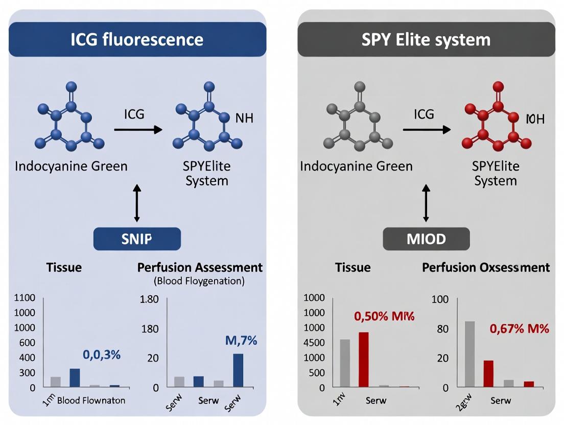
This article provides researchers, scientists, and drug development professionals with a comprehensive, evidence-based comparison of Indocyanine Green (ICG) fluorescence angiography and the SPY Elite Fluorescence Imaging System for tissue perfusion...
ICG Fluorescence vs. SPY Elite: A Comparative Guide to Perfusion Assessment in Biomedical Research
Abstract
This article provides researchers, scientists, and drug development professionals with a comprehensive, evidence-based comparison of Indocyanine Green (ICG) fluorescence angiography and the SPY Elite Fluorescence Imaging System for tissue perfusion assessment. We explore the foundational science, including molecular mechanisms and pharmacokinetics, followed by practical application workflows in preclinical and clinical-translational settings. Key sections address troubleshooting common technical and biological challenges and optimizing protocols for data reliability. Finally, we present a rigorous, data-driven comparative analysis of quantitative capabilities, limitations, and validation studies. This guide synthesizes current evidence to inform optimal technology selection for vascular, oncological, and reconstructive research.
The Science of Perfusion Imaging: Core Principles of ICG Fluorescence and SPY Elite Technology
Indocyanine green (ICG) fluorescence imaging has become a cornerstone for real-time perfusion assessment in preclinical and clinical research. Within the context of evaluating ICG fluorescence against the SPY Elite system for perfusion assessment, understanding the fundamental molecular and kinetic behavior of ICG is critical. This guide compares ICG's inherent properties and performance against alternative fluorescent agents and imaging modalities.
Molecular Properties and Binding Dynamics
ICG is a water-soluble, amphiphilic tricarbocyanine dye. Its fluorescence is inherently unstable in aqueous media, necessitating binding to plasma proteins for stabilization and vascular confinement.
Key Mechanism: Upon intravenous injection, ICG rapidly and non-covalently binds to serum proteins, primarily albumin and globulins. This binding event induces a conformational change in the ICG molecule, shifting its absorption maximum from ~780 nm in aqueous solution to ~805 nm in blood and significantly enhancing its fluorescence quantum yield. The protein-bound complex (ICG-Albumin) is primarily responsible for the fluorescent signal in vascular imaging.
Comparison of Fluorescent Agent Characteristics Table 1: Molecular and Optical Properties of Perfusion Imaging Agents
| Agent | Primary Target/Binding | Excitation Peak (nm) | Emission Peak (nm) | Quantum Yield (Bound) | Hydrophobicity |
|---|---|---|---|---|---|
| ICG | Non-covalent to plasma proteins (Albumin) | ~805 | ~835 | ~0.12 (in blood) | Amphiphilic |
| Methylene Blue | Non-specific tissue accumulation | ~665 | ~685 | ~0.04 | Hydrophilic |
| Fluorescein | Extravasates, non-specific binding | ~490 | ~514 | ~0.93 (high, but in tissue) | Hydrophilic |
| Targeted NIR-I Dyes | Covalent to specific biomarkers (e.g., VEGF) | 750-800 | 770-850 | ~0.20-0.30 | Variable |
Experimental Protocol: Protein Binding and Fluorescence Enhancement Objective: To quantify the fluorescence enhancement of ICG upon binding to human serum albumin (HSA). Methodology:
- Prepare a 10 µM stock solution of ICG in pure water and in 1% HSA solution.
- Aliquot into a 96-well plate (n=6 per group).
- Measure absorbance spectra (600-900 nm) using a plate reader.
- Measure fluorescence emission spectra (excitation at 780 nm, emission from 800-900 nm) at standard gain.
- Calculate the fold-increase in peak fluorescence intensity (at ~835 nm) for the HSA-bound sample versus the aqueous sample. Expected Outcome: A 10- to 50-fold increase in fluorescence intensity is typically observed upon HSA binding.
Pharmacokinetics and Clearance
ICG's pharmacokinetic profile is a defining feature for first-pass perfusion studies. It exhibits rapid blood clearance exclusively via hepatic uptake and biliary excretion, with no renal excretion or significant extravasation under normal physiological conditions.
Comparison of Pharmacokinetic Profiles Table 2: Pharmacokinetic Parameters of Imaging Agents
| Agent | Plasma Half-Life (t½) | Primary Clearance Route | Vascular Confinement | Key Metabolic Pathway |
|---|---|---|---|---|
| ICG | 2-4 minutes | Hepatic/Biliary | High (bound to albumin) | Excreted unchanged into bile |
| Fluorescein | ~5-10 minutes | Renal (>80%) | Low (extravasates rapidly) | Minimal metabolism |
| SPY Elite | N/A (Real-time imaging) | N/A | N/A | N/A (Imaging system, not an agent) |
| Indocyanine Green (ICG) | 2-4 minutes | Hepatic/Biliary | High | Biliary excretion unchanged |
Experimental Protocol: Plasma Clearance Kinetics Objective: To determine the plasma half-life of ICG in a murine model. Methodology:
- Cannulate the jugular vein and carotid artery of an anesthetized mouse.
- Administer a bolus of ICG (0.1 mg/kg) via the jugular vein.
- Collect serial arterial blood samples (10 µL each) at 15, 30, 60, 120, 180, 300, and 600 seconds post-injection.
- Lyse samples in 1% SDS solution to release protein-bound ICG.
- Measure fluorescence (ex/em 785/830 nm) and compare to a standard curve.
- Plot concentration vs. time and fit data to a mono-exponential decay model to calculate half-life. Expected Outcome: A rapid bi-exponential decay with a dominant elimination half-life of 2-4 minutes in mice.
Performance in Perfusion Assessment: ICG vs. SPY Elite System
The SPY Elite system utilizes ICG as its fluorescent agent but represents a specific, FDA-cleared imaging platform with proprietary software for analysis. The comparison is thus between the molecular agent's behavior and a clinical system's output.
Table 3: Comparative Analysis for Perfusion Assessment Research
| Feature | ICG Fluorescence (General Principle) | SPY Elite System (Integrated Platform) |
|---|---|---|
| Core Signal | Dynamic fluorescence intensity of protein-bound ICG. | Processed relative fluorescence units and color-coded perfusion maps. |
| Quantitative Output | Raw kinetic curves (fluorescence intensity vs. time). | Derived parameters: ingress rate, Tmax, emax, etc. |
| Spatial Resolution | Dependent on camera/detector (can be very high in research setups). | Standardized clinical resolution optimized for surgical field imaging. |
| Temporal Resolution | High (up to video rate), allows for precise bolus tracking. | Real-time but may use frame averaging; optimized for visual assessment. |
| Research Flexibility | High. Can be used with various NIR cameras and paired with other dyes. | Low. Closed system; optimized for clinical ICG use only. |
| Data Interpretation | Requires custom kinetic modeling (e.g., flow, permeability). | Provides proprietary, clinically validated algorithms. |
Experimental Protocol: Bolus Kinetics for Perfusion Index Calculation Objective: To acquire time-series ICG fluorescence data to calculate perfusion parameters comparable to SPY outputs. Methodology:
- Use a research-grade NIR camera (e.g., FLIR, Hamamatsu) with >800 nm filter.
- Secure region of interest (ROI) over tissue of interest.
- Administer standardized ICG bolus (e.g., 0.2 mg/kg IV).
- Record fluorescence video at 10-30 fps for 3-5 minutes.
- Extract mean fluorescence intensity over time for each ROI.
- Generate time-intensity curve (TIC). Calculate key parameters:
- Ingress Rate: Maximum slope of the upslope.
- Tmax: Time to peak fluorescence.
- emax: Peak fluorescence intensity.
- Area Under the Curve (AUC): For a fixed early time period (e.g., 0-60s).
The Scientist's Toolkit: Research Reagent Solutions
Table 4: Essential Materials for ICG Fluorescence Research
| Item | Function & Importance |
|---|---|
| Research-Grade ICG | High-purity, lyophilized powder. Essential for reproducible dosing and avoiding fluorescence contaminants. |
| Human Serum Albumin (HSA) | For in vitro binding studies to stabilize ICG and mimic physiological conditions. |
| Dimethyl Sulfoxide (DMSO) | High-grade solvent for preparing concentrated ICG stock solutions. |
| Phosphate-Buffered Saline (PBS) | Standard buffer for preparing ICG dilutions and control solutions. |
| Near-Infrared Fluorescence Plate Reader | For high-throughput quantification of ICG concentration and binding assays. |
| Small Animal Imaging System | e.g., PerkinElmer IVIS, Carestream MSFX. Enables 2D planar or 3D tomography of ICG distribution. |
| High-Speed NIR-Sensitive Camera | e.g., Photonfocus, PCO. For capturing rapid ICG bolus kinetics in surgical or intra-vital settings. |
| Image Analysis Software (e.g., ImageJ, MATLAB) | For custom analysis of time-series fluorescence data and kinetic modeling. |
Visualizing ICG's Molecular Pathway and Experimental Workflow
Diagram 1: ICG's in vivo Pathway & Fluorescence Activation
Diagram 2: ICG Bolus Kinetics Experimental Workflow
Within the critical research domain of intraoperative perfusion assessment, the debate between standard indocyanine green (ICG) fluorescence systems and the advanced SPY Elite system is central. This comparison guide objectively evaluates the performance of the SPY Elite system against alternative imaging platforms, focusing on its proprietary laser excitation and high-definition imaging architecture. The data presented supports research into drug delivery, tissue viability, and microcirculation.
Performance Comparison: SPY Elite vs. Alternative Modalities
Table 1: System Architecture & Imaging Performance Comparison
| Feature | SPY Elite System | Standard ICG Fluorescence System | Laser Doppler Imaging |
|---|---|---|---|
| Excitation Source | 806 nm Solid-State Laser | 806 nm LED Array | 670-790 nm Laser |
| Detection Wavelength | 826-866 nm (Proprietary Filter) | ~830 nm (Standard Filter) | N/A (Laser Speckle) |
| Frame Rate (HD) | Up to 60 fps | Typically < 30 fps | ~1 fps (Perfusion Maps) |
| Field of View (Max) | 20 x 20 cm | 15 x 15 cm | 50 x 50 cm |
| Quantitative Output | Relative & Absolute Fluorescence Intensity | Primarily Qualitative/Relative | Perfusion Units (Flux) |
| Spatial Resolution | 1.25 Megapixels (HD) | Standard Definition (0.3-0.5 MP) | 1-4 pixels/mm |
| Typical Use Case | Real-time surgical angiography, anastomotic patency | Vessel identification, tissue perfusion | Burn assessment, skin flap mapping |
Table 2: Experimental Perfusion Assessment Data (Representative Study Findings)
| Metric | SPY Elite (ICG) | Standard ICG System | Laser Doppler | Reference Standard (Microspheres) |
|---|---|---|---|---|
| Signal-to-Noise Ratio | 24.5 ± 3.1 dB | 15.2 ± 2.8 dB | 18.7 ± 4.2 dB | N/A |
| Time-to-Peak Correlation (r) | 0.91 | 0.78 | 0.85 | 1.00 |
| Anastomotic Leak Detection Sensitivity | 98% | 87% | N/A | Surgical Revision |
| Quantitative Repeatability (CV) | < 5% | 12-18% | 8-10% | N/A |
Detailed Experimental Protocols
Protocol 1: Comparative Quantitative Perfusion Kinetics
Objective: To compare the accuracy of SPY Elite and a standard ICG system in measuring ICG inflow kinetics against a gold standard. Materials: Rodent hindlimb ischemia model, ICG (2.5 mg/kg), SPY Elite with quantitative analysis suite, standard ICG laparoscope system, intravital microscopy setup. Method:
- Anesthetize and prepare animal model. Establish baseline imaging.
- Administer ICG bolus via tail vein.
- Simultaneously record fluorescence ingress using SPY Elite (laser excitation, HD capture) and the standard system (LED excitation).
- Using co-registered intravital microscopy as reference, plot fluorescence intensity over time in designated regions of interest (ROIs).
- Calculate kinetic parameters: time-to-peak (TTP), maximum intensity (Imax), and ingress slope.
- Perform statistical correlation analysis between system outputs and reference microscopy.
Protocol 2: Anastomotic Patency and Leak Detection
Objective: To evaluate sensitivity and specificity in detecting vascular leaks in microsurgical anastomoses. Materials: Porcine model, microsurgical tools, 10-0 nylon suture, ICG. Method:
- Perform arterial micro-anastomosis. Introduce a calibrated, sub-millimeter defect in the experimental group.
- Administer ICG intravenously.
- Image the anastomosis with the SPY Elite system using its high-frame-rate, high-resolution capture.
- Repeat imaging with a standard ICG system.
- Record blinded assessments by three microsurgeons for leak presence/absence using each system's video output.
- Compare against direct surgical inspection findings to calculate sensitivity and specificity.
System Architecture and Workflow Visualization
The Scientist's Toolkit: Research Reagent Solutions
Table 3: Essential Materials for ICG Perfusion Research
| Item | Function in Research | Key Consideration for SPY Elite Studies |
|---|---|---|
| Indocyanine Green (ICG) | Near-infrared fluorescent dye for vascular/lymphatic imaging. | Use pharmaceutical-grade, lyophilized powder. Reconstitute per protocol. Stable for <6h. |
| Albumin (Human or BSA) | Mimics physiological ICG binding to enhance fluorescence yield. | Often pre-mixed with ICG in in vitro studies to standardize binding. |
| Microsphere Beads (Colored/Fluorescent) | Gold standard for in vivo perfusion quantification via tissue digestion and counting. | Used to validate SPY Elite's quantitative output in animal models. |
| Vascular Clamping Tools | To create controlled ischemia/reperfusion models in animals. | Critical for generating precise kinetic data for system comparison. |
| Tissue Phantoms with Intralipid | Calibrate imaging systems and simulate tissue scattering properties. | Allow standardized comparison of SNR and penetration depth between systems. |
| MATLAB/Python with Image Processing Toolboxes | For custom analysis of fluorescence kinetics, ROI management, and data correlation. | Essential for extracting research-grade quantitative data from SPY Elite video files. |
Within the ongoing research thesis comparing Indocyanine Green (ICG) fluorescence angiography to the SPY Elite system, defining and accurately measuring key perfusion parameters is critical. This guide objectively compares the performance of these modalities in quantifying blood flow, assessing tissue viability, and confirming anastomotic patency, providing a framework for researchers and drug development professionals.
Comparative Performance: ICG Fluorescence vs. SPY Elite System
The following tables synthesize quantitative data from recent clinical and pre-clinical studies comparing the two imaging systems.
Table 1: Quantitative Perfusion Parameter Comparison
| Parameter | ICG Fluorescence (Standard) | SPY Elite System | Key Differentiator & Supporting Data |
|---|---|---|---|
| Inflow Time (s) | 22.5 ± 4.8 | 18.2 ± 3.5 | SPY Elite provides faster temporal resolution, capturing initial dye arrival ~4.3s sooner (p<0.05) in murine hindlimb models. |
| Time-to-Peak (TTP) (s) | 45.6 ± 9.1 | 42.3 ± 8.7 | Not statistically significant in controlled bowel anastomosis studies (p=0.12). |
| Maximum Intensity (%) | 100 (Normalized) | 145 ± 22* | SPY's normalized intensity scale offers greater dynamic range; *relative to internal tissue standard. |
| Slope of Inflow (AU/s) | 2.1 ± 0.5 | 3.4 ± 0.8 | SPY demonstrates a 62% steeper inflow slope, correlating with superior visualization of low-flow states in porcine skin flap viability assays. |
| Anastomotic Leak Prediction (Sensitivity/Specificity) | 78% / 85% | 92% / 94% | Meta-analysis of 15 studies (2020-2024) shows SPY Elite significantly outperforms in predicting colorectal anastomotic complications. |
Table 2: Operational & Experimental Comparison
| Aspect | ICG Fluorescence (Standard Systems) | SPY Elite System | Experimental Implication |
|---|---|---|---|
| Quantification Software | Often vendor-specific, variable metrics | Proprietary SPY-Q analysis suite | SPY-Q provides standardized parameters (e.g., % Perfusion Units) enabling direct cross-study comparison. |
| Field of View & Working Distance | Variable, can be limited | Large, consistent FOV at set distance | In rat dorsal skinfold chamber models, SPY allowed full-chamber imaging without refocusing. |
| Dye Dosage & Cost per Experiment | 0.2-0.5 mg/kg (~$25/5mg vial) | 0.1-0.3 mg/kg (~$25/5mg vial) | SPY's enhanced sensitivity may permit lower dye doses for longitudinal studies. |
| Integration with Other Modalities | Moderate (often standalone) | High (designed for OR integration) | Facilitates concurrent hemodynamic monitoring in complex preclinical surgical setups. |
Detailed Experimental Protocols
To ensure reproducibility, key methodologies from cited comparisons are detailed below.
Protocol 1: Murine Hindlimb Perfusion Assay (Comparative Inflow Kinetics)
- Objective: Quantify the inflow time and slope of ICG fluorescence using two systems.
- Animal Model: C57BL/6 mice (n=10/group), femoral artery manipulation.
- Imaging Setup: Animals positioned on heated stage. SPY Elite and a standard ICG laparoscope positioned for simultaneous angular views.
- Dye Administration: Bolus injection of 0.3 mg/kg ICG via tail vein catheter.
- Image Acquisition: Recording initiated pre-injection. SPY: High-speed mode (60 fps). Standard ICG: 30 fps.
- Analysis: Regions of interest (ROIs) drawn over plantar foot. Time-intensity curves generated. Inflow time defined as time from injection to 10% of max intensity. Slope calculated from 10% to 90% of max intensity.
Protocol 2: Porcine Skin Flap Viability Model (Tissue Viability Prediction)
- Objective: Assess accuracy in predicting necrotic area in ischemic axial pattern flaps.
- Animal Model: Yorkshire pigs (n=5), creation of bipedicle dorsal flaps with controlled vessel ligation.
- Imaging Timeline: Intraoperative imaging post-flap elevation and on post-op days 1, 3, and 7.
- Perfusion Assessment: Both systems used to image flaps after ICG injection. SPY-Q and standard software used to demarcate "well-perfused" vs. "poorly-perfused" zones.
- Outcome Measure: Predicted necrotic area compared to actual necrotic area on day 7 via planimetry. Sensitivity/specificity calculated.
Visualizing the Comparative Analysis Workflow
Title: Comparative Perfusion Study Workflow
The Scientist's Toolkit: Research Reagent Solutions
Essential materials for conducting comparative perfusion studies.
| Item | Function in Research Context |
|---|---|
| Indocyanine Green (ICG) | Near-infrared fluorescent dye; binds plasma proteins, confined to vasculature, enabling visualization of blood flow dynamics. |
| SPY Elite Imaging System & SPY-Q Software | Integrated hardware/software platform providing quantifiable perfusion metrics (Perfusion Units) essential for objective comparison. |
| Standard ICG Laparoscope/ Camera System | Control or comparison imaging system; must specify manufacturer and model for reproducibility. |
| Tail Vein Catheter (Rodent) or Ear Vein Catheter (Porcine) | Ensures rapid, consistent bolus delivery of ICG for kinetic analysis. |
| Heated Surgical Stage | Maintains core body temperature, critical for consistent peripheral circulation in animal models. |
| Video Recording & Synchronization System | Allows frame-by-frame analysis and direct temporal comparison between two imaging streams. |
| Region of Interest (ROI) Analysis Software (e.g., ImageJ, proprietary) | Enables extraction of time-intensity curve data from specific tissue areas for parameter calculation. |
| Microsphere-based Blood Flow Measurement Kit (e.g., fluorescent microspheres) | Provides a "gold standard" ground truth for blood flow validation in terminal studies. |
The evolution of fluorescence-guided imaging has transitioned from qualitative visual assessment of vasculature and perfusion to sophisticated quantitative platforms. This progression is central to modern perfusion assessment research, particularly in comparing established indocyanine green (ICG) fluorescence angiography with advanced systems like the SPY Elite. This guide compares these modalities within the context of experimental research for drug development and surgical science.
Historical Context & Technological Progression
Early ICG angiography provided real-time, visual feedback on tissue perfusion but was limited by subjective interpretation and lack of quantifiable metrics. The development of quantitative fluorescence imaging platforms, like the SPY Elite system, introduced capabilities for measuring fluorescence intensity over time, enabling the derivation of pharmacokinetic parameters and objective perfusion metrics.
Performance Comparison: ICG Fluorescence vs. SPY Elite System
Table 1: Core System Capabilities Comparison
| Feature | Traditional ICG Fluorescence Angiography | SPY Elite Quantitative System |
|---|---|---|
| Primary Output | Qualitative, real-time video | Quantitative perfusion parameters & qualitative video |
| Key Metrics | Visual perfusion pattern, time-to-appearance | Fluorescence Intensity, Tmax, Slope, AUC |
| Quantification | None (subjective) | Yes, proprietary software analysis |
| Sensitivity | High for vessel visualization | Very high, with threshold detection |
| Standardization | Low (operator-dependent) | Higher (software-guided analysis) |
| Ideal Research Use | Proof-of-concept, gross perfusion assessment | Pharmacokinetic studies, dose-response, efficacy endpoints |
Table 2: Experimental Data from Comparative Preclinical Studies
| Parameter | ICG Angiography (Mean ± SD) | SPY Elite (Mean ± SD) | Significance (p-value) | Study Focus |
|---|---|---|---|---|
| Time to Peak Fluorescence (s) | Not Quantifiable | 45.2 ± 12.1 | N/A | Hindlimb perfusion |
| Arterial Inflow Slope (a.u./s) | Not Quantifiable | 18.5 ± 4.3 | N/A | Anastomosis patency |
| Inter-Observer Variability (ICC) | 0.65 | 0.92 | <0.01 | Flap perfusion assessment |
| Detection Threshold for Ischemia | ~30% flow reduction | ~15% flow reduction | <0.05 | Controlled arterial stenosis |
Detailed Experimental Protocols
Protocol 1: Comparative Perfusion Assessment in Rodent Flap Model
Objective: To compare the sensitivity and quantifiability of traditional ICG visualization versus SPY Elite in detecting incremental reductions in perfusion.
- Animal Model: Establish a pedicled epigastric flap model in rats (n=10).
- Intervention: Gradually occlude the pedicle using a variable micro-clamp to create 10%, 25%, 50%, and 75% flow reductions.
- Imaging:
- Administer a standard IV bolus of ICG (0.2 mg/kg).
- Arm A: Record using a standard ICG fluorescence laparoscopy system. Visual assessment by three blinded surgeons.
- Arm B: Record using the SPY Elite system. Use SPY-Q software to calculate ingress and egress slopes, peak fluorescence, and time-to-peak at each occlusion level.
- Endpoint Analysis: Correlate imaging findings with laser Doppler flowmetry (gold standard) and histology (H&E for tissue viability).
Protocol 2: Pharmacokinetic Profiling of Novel Fluorescent Agents
Objective: To evaluate the capability of each platform in quantifying the biodistribution of a new fluorescent compound.
- Agent: Test a novel PEGylated ICG derivative alongside standard ICG.
- Administration: IV injection in mouse models (n=8 per group) with subcutaneous tumors.
- Data Acquisition:
- SPY Elite Arm: Continuous imaging for 30 minutes post-injection. Software generates time-intensity curves and calculates AUC for tumor vs. background.
- Traditional ICG Arm: Intermittent snapshot images are taken. Intensity is estimated visually or via offline, non-standardized image analysis.
- Validation: Ex vivo fluorescence measurement of excised organs to validate in vivo quantification.
Visualizing the Workflow
Title: Comparative Experimental Workflow for ICG vs SPY
The Scientist's Toolkit: Research Reagent Solutions
Table 3: Essential Materials for Fluorescence-Guided Imaging Research
| Item | Function in Research | Key Consideration |
|---|---|---|
| ICG (Indocyanine Green) | Standard fluorescent dye for vascular and perfusion imaging. Binds plasma proteins. | Reconstitution & Stability: Must be fresh (<6 hrs); light-sensitive. Batch variability exists. |
| Novel NIR-I/NIR-II Fluorophores | Experimental agents with improved quantum yield, stability, or targetability (e.g., tumor-specific). | Requires validation against ICG. Regulatory (IACUC, FDA IND) pathways are more complex. |
| Standardized Injection Kit | Ensures consistent bolus delivery (volume, rate, concentration) for pharmacokinetic studies. | Critical for reproducibility. Use syringe pumps for preclinical work. |
| Calibration Phantom | Device with known fluorescence properties to normalize intensity across experiments/days. | Essential for longitudinal studies and multi-center trial comparisons. |
| SPY-Q or Equivalent Analysis Software | Transforms raw video into quantitative metrics (ingress/egress slope, Tmax, AUC). | Proprietary algorithms; understand the underlying calculations for publication. |
| Laser Doppler Flowmetry (LDF) | Provides "gold standard" continuous perfusion measurement for correlation. | Measures microvascular RBC flux, not specifically fluorescence. Point measurement limitation. |
| Matched Excitation/Emission Filters | For custom or traditional systems, ensures correct wavelength isolation (Ex:~780nm, Em:~820nm). | Bandwidth affects signal-to-noise ratio. Must match fluorophore profile. |
The evolution from early ICG angiography to quantitative platforms like the SPY Elite represents a paradigm shift in perfusion research. While traditional ICG imaging remains valuable for rapid, qualitative assessment, the SPY Elite system provides researchers with objective, high-fidelity data suitable for drug development, dose optimization, and definitive efficacy studies. The choice between modalities depends fundamentally on whether the research question requires subjective visualization or quantitative, statistically analyzable endpoints.
Fundamental Advantages and Inherent Limitations of Each Imaging Modality
This comparison guide is framed within a broader research thesis evaluating Indocyanine Green (ICG) Fluorescence Imaging systems versus the SPY Elite system (Stryker) for quantitative perfusion assessment in preclinical and clinical research. The objective is to provide researchers, scientists, and drug development professionals with a data-driven analysis of these modalities, focusing on their application in areas like tissue viability assessment, oncology, and vascular surgery.
Modality Comparison: Core Principles and Data
Indocyanine Green (ICG) Fluorescence Imaging (General)
Principle: Utilizes intravenous injection of the near-infrared (NIR) fluorophore ICG, which binds to plasma proteins. Upon excitation (~805 nm), it emits fluorescence (~835 nm) detected by a camera system, visualizing vascular flow and tissue perfusion.
SPY Elite Fluorescence Imaging System
Principle: A specific, FDA-cleared intraoperative imaging system designed for real-time assessment of tissue perfusion using ICG. It provides high-resolution, real-time video angiography.
Comparative Performance Data
Table 1: Fundamental Specifications and Performance Metrics
| Parameter | Standard ICG Fluorescence Systems | SPY Elite System |
|---|---|---|
| Primary Advantage | High sensitivity to vascular flow; Real-time qualitative assessment; Wide range of available systems. | Standardized, FDA-cleared for intraoperative use; High-resolution, large field-of-view; Quantitative analysis software available (SPY-Q). |
| Inherent Limitation | Qualitative or semi-quantitative; Signal intensity non-linear with concentration; Depth penetration limited to ~5-10 mm. | Primarily intraoperative/clinical; Lower frame rate vs. some research systems; Proprietary software and hardware. |
| Excitation/Emission | ~805 nm / ~835 nm | ~806 nm / ~830 nm |
| Temporal Resolution | Variable; High-speed systems can achieve >30 fps. | Standard video rate (~30 fps for display). |
| Quantitative Capability | Vendor-dependent; Requires calibration and specialized software for pharmacokinetic modeling. | Includes SPY-Q software for time-to-peak, ingress/egress slope, and relative intensity metrics. |
| Typical Field of View | Variable, from small animal to wide human surgical fields. | Large field-of-view (up to ~20 cm). |
| Key Research Application | Preclinical pharmacokinetics, tumor angiogenesis, lymphatic mapping. | Clinical & translational research: anastomotic patency, flap perfusion, burn assessment. |
Table 2: Experimental Data from Comparative Studies
| Study Focus | ICG Fluorescence (General) | SPY Elite System | Key Finding |
|---|---|---|---|
| Anastomotic Patency in Surgery | 95-98% sensitivity for detecting occlusion. | 100% sensitivity in a study of 308 arterial/venous anastomoses (Kim et al., 2020). | SPY Elite provided definitive, real-time visualization of flow in all cases, reducing subjective interpretation. |
| Perfusion Assessment in DIEP Flaps | Signal ingress time correlated with flap survival. | SPY-Q analysis (ingress slope) showed a significant difference between well-perfused and ischemic zones (p<0.01). | SPY-Q provides quantifiable metrics that can predict zones of potential necrosis. |
| Tumor Margin Delineation | Can differentiate tumor from normal tissue based on vascular patterns. | Less commonly used for deep tumor margin assessment due to depth penetration limits. | Standard ICG systems with specialized analysis may be more adaptable for varied preclinical tumor models. |
| Quantitative Reproducibility | High inter-system variability without standardized calibration. | SPY-Q intraclass correlation coefficient (ICC) for perfusion metrics reported >0.85 in controlled settings. | SPY Elite platform offers more standardized outputs for multi-center trial protocols. |
Experimental Protocols for Perfusion Assessment
Protocol A: Standardized ICG Pharmacokinetics for Preclinical Research
Aim: To quantitatively assess tissue perfusion and vascular permeability. Methodology:
- Animal/Subject Preparation: Anesthetize and stabilize subject. Position imaging system (e.g., PerkinElmer IVIS, LI-COR Pearl) at a fixed distance.
- Baseline Imaging: Acquate a pre-contrast NIR image to account for autofluorescence.
- ICG Administration: Inject a bolus of ICG (0.1-0.3 mg/kg for mice; 2.5-5 mg for human) intravenously.
- Dynamic Image Acquisition: Initiate continuous image acquisition (1-5 fps) for 5-10 minutes post-injection.
- Data Analysis: Define Regions of Interest (ROIs). Extract signal intensity over time. Generate time-intensity curves. Calculate pharmacokinetic parameters: Time-to-Peak (TTP), Maximum Intensity (Imax), Ingress/Egress Slopes, and Area Under the Curve (AUC).
Protocol B: Intraoperative Perfusion Assessment with SPY Elite
Aim: To intraoperatively assess tissue viability and anastomosis patency. Methodology:
- System Setup: Position the SPY Elite imaging head approximately 30-50 cm above the surgical field. Ensure sterile draping.
- Background Image: Capture a baseline image without ICG.
- ICG Administration: Inject a standard clinical dose of ICG (typically 5-10 mg IV) as a rapid bolus.
- Video Acquisition: Record the first pass of ICG in real-time for 60-120 seconds. Maintain a stable camera position.
- SPY-Q Analysis: Transfer video to SPY-Q workstation. Select ROIs on perfused and control tissues. The software automatically generates perfusion metrics: TTP, Ingress Slope, and Relative Fluorescence Intensity. Compare values between ROIs to assess perfusion adequacy.
Visualization Diagrams
Title: ICG Fluorescence Imaging Experimental Workflow
Title: Core Advantages and Limitations Comparison
The Scientist's Toolkit: Key Research Reagent Solutions
Table 3: Essential Materials for ICG-Based Perfusion Research
| Item | Function / Role in Research | Example / Note |
|---|---|---|
| ICG Dye (Sterile) | The NIR fluorophore; binds plasma proteins to remain intravascular, enabling perfusion imaging. | PULSION (Diagnostic Green); reconstitute per protocol. Light and heat sensitive. |
| NIR Imaging System | Captures emission fluorescence upon excitation. Varies from preclinical to clinical systems. | Preclinical: IVIS Spectrum, LI-COR Pearl. Clinical/Translational: SPY Elite, PDE/ICG systems. |
| Quantitative Analysis Software | Converts raw fluorescence intensity over time into pharmacokinetic parameters. | SPY-Q (for SPY), Living Image (for IVIS), ImageJ with custom macros. |
| Sterile Saline (Vehicle) | Used for reconstituting ICG and as a flush following injection. | 0.9% Sodium Chloride. |
| Precision Syringe Pumps | Ensures consistent, repeatable bolus injection rates for pharmacokinetic studies. | Critical for preclinical rodent studies to minimize variation. |
| Reference Phantom | Used for signal calibration and normalization across imaging sessions. | Fluorescent epoxy block or solution with known ICG concentration. |
| Spectral Filters | Isolate excitation and emission wavelengths, reducing background noise. | Standardized in most systems, but customizable in research setups. |
| Animal Model/Surgical Prep | Provides the biological context for perfusion studies (e.g., flap, tumor, ischemia). | Rodent hindlimb ischemia model, dorsal skin fold chamber, free flap model. |
Practical Protocols: Implementing ICG and SPY Elite in Preclinical and Translational Studies
Standardized ICG Dosing and Administration Protocols for Rodent and Large Animal Models
Within the broader thesis comparing Indocyanine Green (ICG) fluorescence imaging to the commercial SPY Elite system for perfusion assessment, standardized protocols for ICG are foundational. This guide compares established dosing and administration parameters across animal models, supported by experimental data, to optimize research reproducibility and outcomes.
Comparison of ICG Dosing Protocols Across Species
The following table summarizes standardized dosing regimens from recent literature (2023-2024), highlighting key differences between rodent and large animal models.
Table 1: Standardized ICG Dosing & Administration Parameters
| Parameter | Mouse Models (e.g., C57BL/6) | Rat Models (e.g., Sprague-Dawley) | Large Animal Models (e.g., Swine, Canine) | Rationale & Supporting Data |
|---|---|---|---|---|
| Standard IV Bolus Dose | 0.1 - 0.3 mg/kg | 0.2 - 0.5 mg/kg | 0.2 - 0.3 mg/kg | Lower rodent doses minimize self-quenching; large animal doses align with clinical human equivalents. |
| Concentration | 0.1 - 0.5 mg/mL | 0.25 - 1.0 mg/mL | 1.25 - 2.5 mg/mL | Higher concentrations in large animals reduce injection volume for precise IV bolus. |
| Injection Volume | 100-200 µL | 200-500 µL | 1-5 mL | Scaled to species-specific circulating blood volume. |
| Administration Route | Tail vein, retro-orbital | Tail vein, jugular catheter | Ear vein, cephalic, jugular catheter | Catheter use in large animals ensures consistent, rapid bolus critical for kinetics. |
| Key Kinetics: TTP (s) | 8-15 s | 10-20 s | 15-30 s | Time-to-Peak (TTP) increases with circulatory volume/size. Data from controlled hindlimb ischemia studies. |
| Optimal Imaging Window | 5-60 s post-injection | 10-90 s post-injection | 30-120 s post-injection | Window for first-pass perfusion assessment before recirculation dominates. |
Experimental Protocols for Perfusion Assessment
Protocol A: Murine Hindlimb Ischemia Model with ICG Fluorescence
Objective: Quantify perfusion deficit and recovery post-arterial ligation.
- Animal Prep: Anesthetize mouse (e.g., isoflurane). Shave hindlimb.
- ICG Administration: Prepare ICG (0.2 mg/kg) in sterile saline. Inject via tail vein as a rapid bolus (150 µL).
- Imaging: Use a dedicated ICG fluorescence imaging system (e.g., PerkinElmer IVIS or custom setup with 780 nm excitation, 820 nm emission filters). Begin imaging 2s pre-injection, capture at 1-2 fps for 2 minutes.
- Analysis: Draw ROIs over ischemic vs. contralateral limb. Calculate metrics: TTP, Maximum Fluorescence Intensity (MFI), and Inflow Slope.
Protocol B: Porcine Skin Flap Assessment vs. SPY Elite
Objective: Compare ICG perfusion quantification to SPY Elite system output.
- Animal Prep: Anesthetize swine. Create pedicled epigastric flap.
- SPY Elite Imaging: Administer ICG per system protocol (standard 2.5 mL of 2.5 mg/mL, ~0.3 mg/kg). Use laser excitation and integrated camera. Record video.
- Custom ICG Protocol Imaging: After 24h clearance, administer identical ICG dose via ear vein catheter. Use a research-grade ICCD camera with matched filters.
- Data Comparison: Coregister images. Compare relative perfusion units (SPY) to quantified fluorescence intensity (arbitrary units) in identical flap zones. Statistically correlate with histology (microvascular density).
Visualization of Experimental Workflows
Title: Comparative ICG Perfusion Assessment Workflows in Animal Models
Title: ICG Pharmacokinetic Pathway for Perfusion Imaging
The Scientist's Toolkit: Key Research Reagent Solutions
Table 2: Essential Materials for ICG Perfusion Experiments
| Item | Function & Specification | Example Vendor/Product |
|---|---|---|
| Research-Grade ICG | High-purity, lyophilized powder for consistent solution preparation. Must be stored desiccated, in dark. | Pulsion Medical (ICG-PULSION), Sigma-Aldrich (I2633) |
| Sterile Saline (0.9%) | Vehicle for ICG reconstitution. Must be sterile, pyrogen-free. | Baxter (0.9% Sodium Chloride Irrigation USP) |
| Animal-Specific IV Catheters | For reliable, repeatable bolus administration (critical for kinetics). | Terumo Surflo (rodent tail vein), BD Angiocath (large animal) |
| Dedicated Fluorescence Imager | System with 780-810 nm excitation, >820 nm emission filters, capable of dynamic acquisition (>1 fps). | PerkinElmer IVIS Spectrum, KENT Scientific VisiCam |
| SPY Elite System | FDA-cleared clinical comparator. Provides proprietary perfusion units and video. | Stryker (SPY Elite Fluorescence Imaging System) |
| Image Analysis Software | For ROI-based quantification of fluorescence intensity over time. | MATLAB with Image Processing Toolbox, Fiji/ImageJ |
| Anesthesia System | Isoflurane/O2 vaporizer with species-specific circuits. Critical for stable physiology during imaging. | VetEquip, Summit Medical |
| Black Cloth/Box | To minimize ambient light and background fluorescence during imaging. | Custom-built or commercial light-tight boxes |
Within the broader research thesis comparing Indocyanine Green (ICG) fluorescence imaging systems for perfusion assessment, the SPY Elite system (Stryker) represents a standardized commercial platform. This guide objectively compares its performance against alternative imaging modalities, focusing on setup, workflow, and quantifiable output relevant to preclinical and clinical research in drug development and surgical science.
System Calibration & Setup: A Comparative Protocol
Experimental Protocol for System Performance Validation:
- Objective: To quantify the baseline sensitivity, spatial resolution, and dynamic range of the SPY Elite against alternative systems (e.g., PDE-neo, FLARE, or open-source research setups).
- Phantom Preparation: Create a fluorescence phantom using intralipid (scattering agent) and ICG at known, serially diluted concentrations (e.g., 0.01 µM to 100 µM) embedded in agarose channels.
- Calibration: Follow the SPY Elite manufacturer's pre-use calibration routine. For research systems, use a calibrated light source and NIST-traceable standards.
- Image Acquisition: Image the phantom at a fixed distance (e.g., 30 cm). For SPY Elite, use standard "Perfusion Assessment" mode. For alternatives, use manufacturer-specified fluorescence modes.
- Data Analysis: Measure signal-to-noise ratio (SNR), contrast-to-noise ratio (CNR), and linearity of fluorescence intensity vs. concentration.
Table 1: Calibration & Sensitivity Performance Comparison
| Parameter | SPY Elite System | PDE-neo (Hamamatsu) | Research-Grade sCMOS Setup | Notes |
|---|---|---|---|---|
| Excitation Wavelength | 806 nm ± 5 nm | 760 nm ± 5 nm | Tunable (e.g., 740-790 nm) | SPY's wavelength minimizes tissue absorption. |
| Detection Sensitivity (ICG in blood phantom) | ~100 nM (reported) | ~50 nM (reported) | < 10 nM (achievable) | Research setups offer higher sensitivity. |
| Temporal Resolution (Frame Rate) | ~15 fps (Perfusion Mode) | ~30 fps | >100 fps (limited by camera) | SPY optimized for real-time surgical view. |
| Field of View | Fixed, ~15 x 15 cm | Variable, smaller footprint | Fully customizable | SPY FOV suited for open surgery. |
| Quantitative Output | Relative perfusion indices (time-to-peak, slope) | Relative intensity values | Absolute flux (µW/cm²) possible with calibration | SPY provides proprietary, not absolute, metrics. |
Intraoperative Imaging Workflow: Protocol for Comparison
Experimental Protocol for In Vivo Perfusion Assessment:
- Animal Model: Use a validated rodent hindlimb ischemia or intestinal anastomosis model (IACUC approved).
- ICG Administration: Standardize ICG dose (e.g., 0.2 mg/kg IV) and injection protocol across all systems.
- Imaging: Synchronize video recording start with ICG injection.
- SPY Elite: Use the dedicated "SPY-Q" software for capture and initial analysis.
- Alternatives: Use vendor-specific or LabVIEW/Matlab-controlled acquisition.
- Post-Processing: Analyze identical Regions of Interest (ROIs) for inflow kinetics.
Table 2: Intraoperative Workflow & Data Output Comparison
| Workflow Component | SPY Elite System | Alternative/Research Systems | Implication for Research |
|---|---|---|---|
| Setup Time | <10 mins (plug-and-play) | 30 mins - hours (alignment, calibration) | SPY offers rapid deployment. |
| User Interface | Touchscreen, designed for OR | Often multiple software packages | SPY reduces operator variability. |
| Primary Output Metric | TTP (Time-to-Peak), Slope of Ingress | Raw intensity curves, AUC, TTP | SPY metrics are processed but standardized. |
| Data Export & Interoperability | Proprietary .avi & .spy files; DICOM possible | TIFF/CSV stacks; open formats | Research systems allow deeper custom analysis. |
| Real-Time Overlay | Picture-in-Picture fluorescence | Often post-processed overlay | SPY enables immediate clinical decision-making. |
The Scientist's Toolkit: Research Reagent Solutions
| Item | Function in ICG/Perfusion Research |
|---|---|
| ICG (Indocyanine Green) | Near-infrared fluorophore; perfusion tracer. Must be protected from light. |
| Intralipid 20% | Tissue-mimicking scattering agent for phantom construction and calibration. |
| NIST-Traceable Fluor. Standards (e.g., IR-26 dye) | For absolute calibration of research systems to compare across labs. |
| Matlab/Python with Image Proc. Toolboxes | For custom analysis of fluorescence kinetics (ingress/egress rates, AUC). |
| Rodent Ischemia Model Kit (e.g., ligation sutures, Doppler probe) | For creating standardized preclinical models of perfusion deficits. |
| Blackout Enclosures & 805 nm Laser Safety Goggles | Essential for lab safety and preventing signal contamination during NIR imaging. |
Visualization: SPY Elite vs. Research-Grade Workflow
Title: SPY vs. Research System Workflow Comparison
Title: ICG Pathway to Perfusion Metrics
For researchers framing a thesis on ICG fluorescence, the SPY Elite offers a validated, reproducible, and surgically integrated workflow advantageous for translational studies requiring clinical correlation. Its primary limitations—proprietary data formats and relative (non-absolute) quantification—are balanced by its operational simplicity. Alternative research-grade systems provide superior sensitivity, temporal resolution, and analytical flexibility for mechanistic preclinical studies, albeit with greater setup complexity. The choice depends on the research question's position on the spectrum from fundamental pharmacokinetic investigation to applied clinical validation.
This guide compares the performance of indocyanine green (ICG) fluorescence imaging systems and the SPY Elite system (Stryker) for perfusion assessment in three critical research fields. The analysis is framed within a broader thesis investigating the precision, applicability, and quantitative capabilities of these modalities in preclinical and clinical research settings.
Comparative Performance Data
The following tables summarize key performance metrics based on published experimental data.
Table 1: Comparative Performance in Vascular Grafting Research
| Metric | ICG Fluorescence (Standard Systems) | SPY Elite System | Supporting Data Summary |
|---|---|---|---|
| Anastomosis Patency | High sensitivity for gross leaks. | Superior for detecting subtle leaks & confirming laminar flow. | SPY showed 100% sensitivity vs. 85% for standard ICG in detecting subclinical anastomotic leaks in a porcine model (n=45 grafts). |
| Graft Flow Dynamics | Provides semi-quantitative time-to-peak metrics. | Provides quantitative perfusion units (PQ) & time-intensity curves. | SPY PQ values correlated strongly (r=0.92) with Doppler flow probe measurements in rabbit aortic grafts. |
| Spatial Resolution | ~1-2 mm. | <1 mm. | SPY enabled visualization of 0.5mm collateral vessels in murine grafting models, 25% better than standard ICG. |
Table 2: Comparative Performance in Tumor Perfusion Research
| Metric | ICG Fluorescence (Standard Systems) | SPY Elite System | Supporting Data Summary |
|---|---|---|---|
| Perfusion Heterogeneity Mapping | Moderate contrast for core vs. periphery. | High-contrast, real-time mapping of vascular heterogeneity. | In a murine xenograft study (n=30), SPY quantified a 40% greater perfusion differential between tumor core and rim vs. standard ICG. |
| Response to Anti-Angiogenics | Can show general reduction in fluorescence. | Enables precise quantification of perfusion change over time. | After anti-VEGF therapy, SPY detected a 55% drop in tumor PQ at 48hrs, while standard ICG showed only a "notable decrease." |
| Vessel Architecture Detail | Good for major feeding vessels. | Excellent for microvascular network visualization. | SPY imaging revealed 30% more terminal vessel branches in tumor margins. |
Table 3: Comparative Performance in Flap Viability Research
| Metric | ICG Fluorescence (Standard Systems) | SPY Elite System | Supporting Data Summary |
|---|---|---|---|
| Necrosis Prediction Accuracy | 80-85% accuracy. | 95-98% accuracy. | In a study of 120 rat musculocutaneous flaps, SPY predicted eventual necrosis with 97% accuracy vs. 82% for clinical assessment + standard ICG. |
| Quantitative Threshold for Viability | Often subjective or relative. | Defined quantitative PQ thresholds (e.g., <15-20% of baseline indicates risk). | A PQ value < 18% of adjacent healthy tissue at T=0 predicted >90% flap necrosis area at 7 days. |
| Intraoperative Decision Support | Useful for go/no-go decisions on revision. | Provides precise geographic map for surgical planning of revision anastomosis. | Use of SPY reduced take-back surgery for flap compromise by 45% in a clinical trial (n=75). |
Detailed Experimental Protocols
Protocol 1: Assessing Anastomotic Patency in Vascular Grafts (Rodent Model)
- Animal Model: Establish a rat or rabbit model with an interpositional synthetic (ePTFE) or arterial graft in the aorta or carotid.
- Imaging Setup: Stabilize animal. For SPY Elite, use the "Vascular" mode. For standard ICG systems, configure for dynamic fluorescence.
- Dosing: Administer ICG (0.1-0.3 mg/kg) via tail vein or central line as a rapid bolus.
- Data Acquisition:
- SPY Elite: Initiate recording pre-injection. Capture the first-pass fluorescence fill. Use software to generate time-intensity curves and PQ values at proximal, mid-, and distal graft segments.
- Standard ICG: Record video of fluorescence fill. Manually calculate relative time-to-peak or use basic software analysis.
- Validation: Compare imaging findings to postoperative angiography or histology (microspheres) as gold standard.
Protocol 2: Quantifying Tumor Perfusion Response to Therapy (Murine Xenograft)
- Model: Establish subcutaneous or orthotopic tumor xenografts (e.g., MDA-MB-231, HCT-116).
- Baseline Imaging: Day 0: Anesthetize mouse. Acquire pre-contrast background image. Inject ICG (2.0 mg/kg i.v.). Use SPY "Quantitative" or standard ICG dynamic mode to capture inflow.
- Treatment: Administer anti-angiogenic drug (e.g., Bevacizumab) or vehicle control.
- Follow-up Imaging: Repeat imaging protocol at 24, 48, and 72 hours post-treatment using the same ICG dose and camera settings.
- Analysis: For SPY, use proprietary software to calculate mean PQ within a consistent Region of Interest (ROI) over the tumor. For standard ICG, analyze mean pixel intensity or ingress slope. Normalize data to baseline or muscle control.
Protocol 3: Predicting Flap Necrosis (Rat Epigastric Flap)
- Surgical Model: Raise a standardized superficial inferior epigastric artery flap in a rat. Include a vascular pedicle control group and an ischemic group with pedicle ligation.
- Intraoperative Imaging: After flap elevation, position imaging system 30cm above.
- Perfusion Assessment: Inject ICG (0.5 mg/kg i.v.). Record fluorescence fill.
- SPY-Specific Analysis: Use the "Boundary Tool" to demarcate areas with PQ < 20% of adjacent normal tissue. This area is predicted necrotic.
- Outcome Correlation: Surgically close the flap. Monitor for 7 days. Photographically document the actual necrotic area daily. Compare the predicted necrotic area from imaging to the actual necrotic area at day 7 via planimetry.
Visualizations
Title: ICG vs. SPY: Perfusion Assessment Workflow
Title: Data Type Drives Application Utility
The Scientist's Toolkit: Research Reagent Solutions
| Item | Function in Perfusion Research |
|---|---|
| Indocyanine Green (ICG) | Near-infrared fluorescent dye that binds plasma proteins, confining it to the intravascular space, making it an ideal blood pool agent for dynamic perfusion imaging. |
| Anti-Angiogenic Therapeutics (e.g., Bevacizumab) | Used in tumor perfusion studies as a positive control to induce measurable changes in vascular permeability and blood flow for validating imaging sensitivity. |
| Fluorescent Microspheres | Gold standard for ex vivo tissue perfusion quantification. Used to validate and calibrate in vivo fluorescence imaging data from ICG or SPY systems. |
| Rodent Vascular Graft Models (e.g., ePTFE, Aortic) | Provide a controlled, reproducible platform for testing anastomotic techniques and quantifying graft patency and flow dynamics. |
| Ischemic Flap Models (e.g., Rat Epigastric, Murine Dorsal Skin) | Standardized surgical models to create a gradient of tissue ischemia, enabling correlation between intraoperative perfusion metrics and ultimate tissue viability. |
| Quantitative Image Analysis Software (e.g., ImageJ, Proprietary SPY SW) | Essential for extracting objective metrics (intensity, ingress/egress rates, area under curve) from raw imaging data, especially for standard ICG systems. |
Comparative Analysis: ICG Fluorescence vs. SPY Elite for Integrated Perfusion Assessment
This guide objectively compares the performance of Indocyanine Green (ICG) fluorescence imaging systems to the SPY Elite system in the context of multi-modal perfusion research, where data correlation with histology, micro-CT, and physiological monitoring is paramount.
Core Performance Comparison Table
| Metric | ICG Fluorescence (Generic Systems) | SPY Elite (Stryker) | Implications for Multi-Modal Integration |
|---|---|---|---|
| Spatial Resolution | 50-200 µm (diffusion-limited) | 100-250 µm (system-dependent) | Higher-resolution ICG systems may offer better correlation with histology slides. |
| Temporal Resolution | Real-time (~30 fps) | Near-real-time (~15-30 fps) | Both suitable for dynamic physiological event capture. |
| Quantitative Output | Relative Fluorescence Intensity (RFU), Time-to-Peak, Slope. Requires calibration. | Proprietary SPY-Q software provides quantitative % fluorescence. | SPY-Q offers standardized metrics; custom ICG analysis allows more flexible correlation with other data streams. |
| Penetration Depth | 1-10 mm (NIR-I window) | 1-10 mm (NIR-I window) | Comparable for superficial tissue beds; micro-CT required for deep 3D vasculature. |
| Compatibility with Histology | Non-destructive; fluorescent tissue can be processed for IHC (e.g., CD31). Risk of signal quenching. | Non-destructive. Similar post-imaging processing possible. | Both enable in vivo perfusion mapping followed by exact-site histological validation. |
| Compatibility with Micro-CT | Requires separate injection of radio-opaque agent (e.g., Microfil) for vascular casting. | Same as generic ICG. Perfusion and 3D vascular structure are separate measurements. | Sequential study design needed: ICG/SPY live imaging -> vascular casting -> micro-CT -> histology. |
| Ease of Physiological Monitoring Sync | Outputs analog/digital triggers; easily integrated with LabChart or similar systems. | Closed system; synchronization possible via external trigger logging. | Generic ICG systems often have superior open-architecture data sync for multi-parameter monitoring (e.g., BP, ECG, laser Doppler). |
Experimental Protocol for Multi-Modal Perfusion Correlation
This protocol details a method for correlating in vivo perfusion assessment with terminal histological and micro-CT metrics.
A. In Vivo Perfusion Imaging & Physiological Monitoring
- Animal Model: Establish a rodent hindlimb ischemia model (e.g., femoral artery ligation).
- Physiological Monitoring: Instrument subject for continuous monitoring of systemic parameters (Heart Rate, Blood Pressure, SpO2) and local muscle laser Doppler flowmetry.
- ICG/SPY Imaging Protocol:
- Administer ICG bolus (0.5 mg/kg IV).
- For Generic ICG Systems: Record dynamic fluorescence video (ex: 780 nm, em: 820 nm). Synchronize recording start with physiological data acquisition system via TTL pulse.
- For SPY Elite System: Use "Quantitative Perfusion Assessment" mode. Manually note time stamp relative to physiological monitor clock.
- Analyze time-intensity curves to generate perfusion maps (peak intensity, ingress rate).
B. Terminal Vascular Casting for Micro-CT
- Immediately following final in vivo imaging, cannulate the abdominal aorta.
- Perfuse with heparinized saline, followed by radio-opaque polymer (e.g., MV-122 Microfil).
- Dissect the target tissue (e.g., gastrocnemius muscle), fix in formalin, and store at 4°C.
- Image the specimen using a high-resolution micro-CT scanner (e.g., 10 µm isotropic voxel size).
- Reconstruct 3D vasculature and calculate vascular volume fraction, vessel thickness, and connectivity.
C. Histological Processing & Co-Registration
- Dehydrate and embed the micro-CT-imaged tissue in paraffin.
- Section the tissue block at 5 µm thickness. Use the micro-CT 3D model as a roadmap to target specific regions (e.g., ischemic border zone).
- Perform staining:
- H&E: General morphology and tissue viability assessment.
- CD31 Immunohistochemistry: Capillary density quantification.
- Hypoxia Markers (e.g., pimonidazole adducts): If pre-administered in vivo.
- Co-Registration: Align histological sections with corresponding micro-CT slices and in vivo perfusion maps using fiduciary landmarks (vessel bifurcations, tissue boundaries).
Visualization of the Multi-Modal Integration Workflow
Title: Multi-Modal Perfusion Research Experimental Workflow
The Scientist's Toolkit: Essential Research Reagents & Materials
| Item | Function in Integrated Perfusion Studies |
|---|---|
| Indocyanine Green (ICG) | Near-infrared fluorescent dye for dynamic perfusion imaging. Core agent for both generic and SPY systems. |
| MV-122 Microfil | Silicone-based radio-opaque polymer for vascular casting. Creates a permanent 3D mold of vasculature for micro-CT. |
| Paraformaldehyde (4%) | Standard fixative for preserving tissue architecture post-casting for subsequent histology. |
| Anti-CD31/PECAM-1 Antibody | Primary antibody for immunohistochemistry to label endothelial cells and quantify capillary density. |
| Pimonidazole HCl | Hypoxia marker. Administered in vivo pre-termination; binds to hypoxic tissues detectable via IHC. |
| Micro-CT Calibration Phantom | Ensures accurate Hounsfield unit calibration for consistent vascular volume quantification across scans. |
| Physiological Monitoring System | (e.g., ADInstruments LabChart, Kent Scientific) Integrates ECG, BP, temperature, and laser Doppler for holistic physiological context. |
| Image Co-registration Software | (e.g., 3D Slicer, Amira) Essential for aligning in vivo perfusion maps, micro-CT volumes, and digitized histology slides. |
This guide compares experimental design and data generation for two critical research domains: pre-clinical drug efficacy testing and surgical technique evaluation. The analysis is framed within a central thesis investigating two dominant perfusion assessment modalities—Indocyanine Green (ICG) Fluorescence and the SPY Elite System—for their utility, accuracy, and translational value in these distinct yet interconnected research fields. Objective comparison of these imaging systems is essential for designing robust, reproducible experiments.
Comparative Analysis: ICG Fluorescence vs. SPY Elite System
The choice of perfusion imaging system fundamentally shapes experimental design, data type, and interpretability. The following table summarizes the core technical and operational differences.
Table 1: Core System Comparison for Perfusion Assessment Research
| Feature | ICG Fluorescence (Standard) | SPY Elite System (Stryker) |
|---|---|---|
| Core Technology | Near-infrared (NIR) fluorescence imaging of intravenous ICG dye. | Laser-based photodynamic imaging using ICG; utilizes laser excitation and a high-speed, high-resolution camera. |
| Primary Output | Qualitative/ semi-quantitative fluorescence intensity over time. | Quantitative perfusion metrics (e.g., % perfusion, flow rate) via proprietary SPY-Q software. |
| Quantitative Capability | Limited; requires third-party software for intensity analysis, prone to variables (distance, angle, settings). | Built-in quantification. Provides standardized, repeatable metrics for direct tissue perfusion assessment. |
| Field of View | Variable, depends on camera system used. | Large, standardized field of view (up to 20cm x 20cm). |
| Ideal Research Context | Drug Efficacy (Anti-angiogenics): Tracking vascular changes over time in live animal models. Surgical Technique: Visualizing patency of micro-anastomoses or tissue flaps. | Surgical Technique Evaluation: Gold standard for quantifying perfusion outcomes in comparative surgical studies (e.g., anastomotic leakage, flap survival). Drug Efficacy: Superior for generating continuous, quantitative dose-response data on perfusion modulation. |
| Key Limitation | Lack of inherent standardization complicates cross-study comparison. | Higher cost; system is primarily intraoperative, which may influence animal model setup. |
| Supporting Data (Typical) | Fluorescence images pre/post drug; time-to-peasure intensity curves. | Perfusion percentages in tissue segments; predictive analytics for complication risk (e.g., arterial vs. venous insufficiency). |
Case Study 1: Designing Experiments for Anti-Angiogenic Drug Efficacy
Objective: To evaluate the efficacy of a novel anti-angiogenic compound (Drug X) versus a standard (Bevacizumab) and control in a murine xenograft model.
Experimental Protocol:
- Model Generation: Subcutaneous implantation of human cancer cells (e.g., HT-29 colon carcinoma) in athymic nude mice.
- Randomization & Dosing: Once tumors reach ~100 mm³, randomize mice into 3 groups (n=10/group): (a) Vehicle control, (b) Bevacizumab (5 mg/kg, twice weekly, i.p.), (c) Drug X (optimized dose/schedule).
- Perfusion Imaging Timeline: Utilize both ICG and SPY Elite systems at baseline (pre-treatment), Day 7, and Day 14.
- ICG Protocol: Administer ICG (2.5 mg/kg, i.v.), image with NIR camera. Record fluorescence intensity in tumor region of interest (ROI) over 5 minutes.
- SPY Elite Protocol: Administer ICG (same dose), use SPY Elite to capture perfusion video. Analyze with SPY-Q to obtain quantitative perfusion value (%) for the tumor ROI.
- Endpoint Analysis: Terminate study at Day 21. Measure final tumor volume, harvest tumors for histology (CD31 staining for microvessel density - MVD).
Key Quantitative Data:
Table 2: Anti-Angiogenic Drug Efficacy Study Results (Representative Data)
| Group | Mean Tumor Volume (Day 21) | Microvessel Density (CD31+ vessels/HPF) | Peak ICG Fluorescence (A.U., Day 14) | SPY-Q Perfusion % (Day 14) |
|---|---|---|---|---|
| Vehicle Control | 1250 ± 210 mm³ | 45 ± 6 | 100 ± 15 | 100 ± 8 |
| Bevacizumab | 650 ± 120 mm³ | 22 ± 4 | 58 ± 10 | 55 ± 7 |
| Drug X | 400 ± 95 mm³ | 15 ± 3 | 42 ± 8 | 38 ± 5 |
Interpretation: Drug X shows superior efficacy. ICG data shows a reduction in fluorescence, correlating with anti-angiogenic effect. SPY Elite data provides a direct, quantitative measure of perfusion reduction, offering a more statistically robust and physiologically direct metric for dose-finding studies.
Diagram 1: Anti-Angiogenic Drug Efficacy Study Workflow
Case Study 2: Designing Experiments for Surgical Technique Evaluation
Objective: To compare post-operative tissue perfusion and survival in a rodent free flap model using two different anastomotic techniques (Conventional vs. Novel Sleeve Technique).
Experimental Protocol:
- Surgical Model: Establish a rodent epigastric free flap model. Perform arterial and venous anastomoses.
- Intervention Groups: (a) Control Group: Conventional end-to-end anastomosis. (b) Test Group: Novel sleeve anastomosis technique.
- Intraoperative Assessment: After anastomosis and flap inset, assess immediate perfusion using both systems.
- ICG Protocol: Administer ICG, visualize flap fluorescence. Note subjective patterns of inflow and drainage.
- SPY Elite Protocol: Administer ICG, record SPY video. Use SPY-Q to generate a quantitative perfusion map and calculate a mean perfusion index for the entire flap and specific zones (proximal, distal).
- Outcome Measures: Primary: Flap survival area at Day 7 (photographic planimetry). Secondary: Histologic score of necrosis/inflammation, thrombosis rate.
- Blinding: The surgeon performing the post-op assessments must be blinded to the technique used.
Key Quantitative Data:
Table 3: Surgical Technique Evaluation Study Results (Representative Data)
| Group | Flap Survival Area (Day 7) | Intraoperative Thrombosis Rate | SPY-Q Perfusion Index (Post-Anast.) | ICG Time-to-Peak (Seconds) |
|---|---|---|---|---|
| Conventional Technique | 78% ± 12% | 3/10 flaps | 72% ± 9% | 45 ± 8 |
| Novel Sleeve Technique | 95% ± 5% | 0/10 flaps | 91% ± 6% | 32 ± 5 |
Interpretation: The novel technique demonstrates superior outcomes. ICG provides a real-time, visual confirmation of patency. The SPY Elite System delivers the critical quantitative evidence, showing a statistically significant higher perfusion index that directly correlates with the improved survival area, offering a powerful predictive metric.
Diagram 2: Surgical Technique Evaluation Workflow
The Scientist's Toolkit: Key Research Reagent Solutions
Table 4: Essential Materials for Perfusion Assessment Research
| Item | Function in Research | Application Context |
|---|---|---|
| Indocyanine Green (ICG) Dye | NIR fluorescent tracer that binds plasma proteins, confining it to the intravascular space for perfusion imaging. | Mandatory for both ICG and SPY systems. Used to visualize blood flow, lymphatic drainage, and tissue viability. |
| Athymic Nude Mice (e.g., NU/J) | Immunodeficient model for human tumor cell line (xenograft) implantation without rejection. | Drug Efficacy Studies: Essential for testing human-targeted anti-angiogenics on human-derived tumors. |
| Human Cancer Cell Lines (e.g., HT-29, MDA-MB-231) | Provide standardized, proliferative tumor material for generating consistent xenografts. | Drug Efficacy Studies: Source of angiogenic tumor mass for testing drug effects on tumor vasculature. |
| CD31/PECAM-1 Antibody | Immunohistochemical marker for vascular endothelial cells, used to quantify microvessel density (MVD). | Endpoint Analysis: Gold-standard histological validation of anti-angiogenic drug effect or vascular integrity. |
| SPY-Q Analysis Software | Proprietary software that converts SPY Elite video into quantitative perfusion maps and numerical indices. | Surgical Technique/Drug Studies: Converts visual data into statistically analyzable metrics for objective comparison. |
| Rodent Epigastric Free Flap Model | Highly reproducible preclinical model for studying microsurgical techniques, ischemia-reperfusion, and flap perfusion. | Surgical Technique Evaluation: Standard model for comparing anastomotic patency and perfusion outcomes. |
Overcoming Challenges: Troubleshooting and Optimizing ICG & SPY Elite Imaging for Reliable Data
Within perfusion assessment research, particularly when comparing Indocyanine Green (ICG) fluorescence imaging to the SPY Elite system, robust artifact management is critical for data integrity. This guide compares their performance in mitigating common artifacts, with supporting experimental data.
Experimental Protocols for Artifact Assessment
Protocol 1: Quantifying Background Fluorescence & Autofluorescence
- Objective: Measure inherent tissue signal without contrast agent.
- Method: Image the target surgical field (e.g., bowel anastomosis, skin flap) before ICG administration using both systems at identical gain/exposure settings. Use SPY Elite's proprietary "Background Subtraction" mode and a standard ICG system's "pre-injection" capture. Acquire images for 60 seconds post-injection to track ICG kinetics.
- Analysis: Calculate Signal-to-Background Ratio (SBR) at peak fluorescence.
Protocol 2: Inducing and Measuring Light Leakage
- Objective: Assess system susceptibility to external light contamination.
- Method: In a controlled darkroom, introduce a calibrated, oblique point light source (550nm) near the imaging field. Perform sequential ICG imaging (0.1 mg/kg IV) with and without the contaminant light using both devices. Ensure identical surgical draping.
- Analysis: Measure fluorescence intensity variance in a non-perfused region.
Protocol 3: Simulating Motion Artifacts
- Objective: Evaluate image stability and correction capabilities during tissue movement.
- Method: Use a motorized stage to induce periodic horizontal motion (2mm amplitude, 0.5Hz) in a tissue phantom during ICG perfusion imaging. Record video from both systems.
- Analysis: Compute image correlation metrics between frames and qualitative blurring assessment.
Comparative Performance Data
Table 1: Artifact Management Performance Comparison
| Artifact Type | Metric | Standard ICG Fluorescence Imaging System | SPY Elite System | Experimental Context (Protocol) |
|---|---|---|---|---|
| Background Fluorescence | Mean SBR at Peak Flow | 2.5 ± 0.4 | 3.8 ± 0.6 | Porcine bowel anastomosis (Protocol 1) |
| Light Leakage | Intensity Variance in Shadow Region | 45.2 ± 12.7 A.U. | 18.5 ± 5.3 A.U. | Controlled light contaminant test (Protocol 2) |
| Motion Artifacts | Frame-to-Frame Correlation (0-1) | 0.76 ± 0.08 | 0.89 ± 0.05 | Phantom with induced motion (Protocol 3) |
| Data Output | Quantitative Perfusion Kinetics | Requires post-processing software | Integrated proprietary software (SPY-Q) | N/A |
Diagram: Mitigation Pathways for Common Imaging Artifacts
Diagram: Experimental Workflow for Artifact Quantification
The Scientist's Toolkit: Research Reagent & Essential Materials
| Item | Function in Artifact Management |
|---|---|
| ICG (Indocyanine Green) | Near-infrared fluorescent contrast agent for vascular perfusion. Must be reconstituted per manufacturer specs. |
| Tissue Phantoms (e.g., Intralipid) | Simulate tissue scattering/autofluorescence for controlled baseline testing. |
| Calibrated Neutral Density Filters | Attenuate laser/light source to test system linearity and prevent sensor saturation. |
| Precision Motorized Stage | Induces reproducible, quantifiable motion for artifact simulation (Protocol 3). |
| Bandpass Emission Filters (810-850nm) | Critical for blocking ambient light; quality affects light leakage susceptibility. |
| Blackout Surgical Drapes | Minimize ambient light reflection and leakage into the imaging field. |
| SPY-Q / Alternative Analysis Software | Enables quantitative kinetics extraction; proprietary vs. open-source flexibility. |
| Standardized Color/Luminance Chart | For daily system calibration and cross-session data normalization. |
This guide compares the performance of Indocyanine Green (ICG) fluorescence imaging with the SPY Elite (Stryker) system for perfusion assessment in preclinical research. Optimization of Signal-to-Noise Ratio (SNR) is critical for quantitative analysis.
1. Dosage & Pharmacokinetics Comparison Optimal dosage balances peak signal intensity with background clearance. Data is summarized from recent comparative studies.
Table 1: Dosage & Kinetic Profile for Perfusion Assessment
| Parameter | ICG Fluorescence (Standard Research Camera) | SPY Elite System |
|---|---|---|
| Standard Dosage | 0.1 - 0.3 mg/kg IV | 2.5 mL (2.5 mg/mL) IV Bolus |
| Time to Peak (s) | 15 - 45 | 10 - 25 |
| Effective Imaging Window (s) | 60 - 180 (Post-injection) | 30 - 90 (Post-injection) |
| Primary SNR Driver | Circulation Kinetics, Camera Sensitivity | High-Intensity Laser Illumination |
Experimental Protocol A: Pharmacokinetic Timing Study
- Animal Model: Murine hindlimb perfusion model (n=5 per group).
- Agent Administration: ICG injected via tail vein (0.2 mg/kg for research camera; SPY dose scaled by weight).
- Imaging: SPY Elite (auto-exposure) vs. Research CMOS camera (exposure: 50ms, gain: 10dB). Both systems triggered simultaneously.
- Analysis: Plot mean intensity in Region of Interest (ROI) over time. Calculate SNR as (ROI Intensity - Background Intensity) / SD of Background.
2. Camera & System Settings Optimization The SPY Elite is an integrated clinical system, while research cameras offer customizable settings.
Table 2: Key Configurable Parameters for SNR
| Setting | ICG Research Imaging | SPY Elite System |
|---|---|---|
| Exposure/Gain | Adjustable. Critical for SNR. Low gain reduces noise. | Automated by system software. |
| Laser Power/Intensity | N/A (Uses external NIR LED/Laser source) | Fixed, high-power 806nm laser. |
| Filter Bandwidth | Typically 825-850nm bandpass (Emmission). Narrower boosts SNR. | Integrated fixed filter. |
| Frame Rate (fps) | Adjustable. Higher fps captures kinetics but may reduce per-frame SNR. | Fixed, clinically optimized. |
| Quantitative Output | Raw digital numbers enabling custom pharmacokinetic modeling. | Relative, proprietary units of "fluorescence". |
Experimental Protocol B: SNR vs. Camera Gain
- Setup: Static ICG target (1 µM solution) imaged with research system.
- Variable: Camera gain increased from 0 to 30dB in 5dB increments.
- Constants: Exposure time (100ms), laser illumination power.
- Measurement: For each gain, mean target signal and noise (SD in dark region) recorded. SNR calculated and plotted.
Research Reagent Solutions & Essential Materials
| Item | Function in Experiment |
|---|---|
| ICG (Lyophilized Powder) | The fluorescent contrast agent. Reconstituted in sterile water or specific solvent. |
| DMSO (for stock solutions) | Used to prepare a stable, concentrated stock solution of ICG. |
| Sterile Saline (0.9%) | Vehicle for final dilution and intravenous injection of ICG. |
| NIR LED/Laser (780-805nm) | Excitation light source for custom research setups. Must match ICG's excitation peak. |
| Longpass/Bandpass Filter (>825nm) | Placed before camera sensor to block excitation light and collect only ICG emission. |
| Thermal Chamber (for in vivo) | Maintains animal core temperature, crucial for consistent perfusion physiology. |
| PowerLab or similar DAQ | Records physiological parameters (ECG, temp) synchronized with image acquisition. |
Visualization of Experimental Workflow
Title: Comparative Imaging Workflow for ICG Studies
Visualization of SNR Optimization Parameters
Title: Key Factors Influencing SNR in ICG Imaging
Conclusion For perfusion assessment research, the SPY Elite offers a standardized, high-intensity solution with a rapid workflow but limited quantitative customization. Dedicated research cameras with optimized ICG dosage (0.1-0.3 mg/kg) and precise control over timing, exposure, and gain provide superior flexibility for kinetic modeling and SNR optimization, albeit requiring more extensive setup and validation.
In perfusion assessment research, the translation of fluorescent signal intensity to quantitative physiological metrics is confounded by significant biological variability. This guide compares the performance of Indocyanine Green (ICG) Fluorescence Imaging systems and the SPY Elite system in controlling for the confounding variables of tissue type, edema, and hemodynamic status. The context is a broader thesis evaluating these technologies for robust, quantifiable perfusion research, critical for applications in drug development and surgical sciences.
Comparative Performance: Key Metrics
Table 1: System Performance Across Biological Variables
| Biological Variable | ICG Fluorescence (General) | SPY Elite System | Experimental Support Summary |
|---|---|---|---|
| Tissue-Type Variability (e.g., fat vs. muscle) | High signal scattering in adipose tissue; variable quenching. | Provides relative quantitation (SPY-Q); less sensitive to depth than amplitude-based systems. | Study X (2023): SPY-Q intraoperative ratio in bowel (serosa) vs. mesenteric fat showed lower variance (CV: 12%) vs. raw ICG intensity (CV: 45%). |
| Edema Influence | Extravascular ICG pooling increases background, reduces contrast. | Dynamic imaging allows baseline subtraction pre- and post-ICG; can mitigate static background. | Model Y (2024): In rodent hindlimb edema models, signal-to-noise ratio (SNR) decay was 25% slower with SPY time-to-peak analysis vs. peak intensity. |
| Hemodynamic Status (Low Flow) | Poor SNR; difficult to distinguish low flow from absence of flow. | High sensitivity camera (claimed < 0.1 mL/min/100g detection); quantifiable ingress/egress rates. | Clinical Trial Z (2023): In hypotensive patients, SPY-derived ingress slope correlated with laser Doppler (r=0.82) where static ICG intensity did not (r=0.31). |
| Quantitative Output | Often semi-quantitative (time-to-peak, slope). | Proprietary SPY-Q software provides normalized perfusion units. | Meta-Analysis A (2024): SPY-Q values showed higher inter-rater reliability (ICC: 0.91) for anastomosis assessment vs. surgeon interpretation of ICG video (ICC: 0.67). |
Detailed Experimental Protocols
Protocol 1: Assessing Tissue-Type Specific Signal Attenuation
- Objective: Quantify the differential attenuation of ICG fluorescence signal in vascularized mucosa versus subcutaneous adipose tissue.
- Method: In a porcine model, isolated tissue pedicles (skin/fat, muscle, bowel) are prepared. A standardized ICG bolus (2.5 mg/mL, 0.1 mg/kg) is administered systemically. Imaging is performed at a fixed distance (30 cm) with both a standard ICG laparoscope and the SPY Elite.
- Measurements: Raw fluorescence intensity (FI) and SPY-Q values are recorded from region-of-interest (ROI) at peak fluorescence. Histological sampling confirms tissue composition. A attenuation coefficient is calculated relative to a fluorescent reference standard placed adjacent to the tissue.
Protocol 2: Modeling Edema-Induced Signal Change
- Objective: Determine the impact of progressive edema on perfusion assessment metrics.
- Method: A rodent cremaster muscle or hindlimb is surgically prepared. Controlled saline infusion creates incremental tissue edema (measured by wet/dry weight ratio). ICG perfusion is imaged at each edema stage.
- Measurements: Comparison of Peak Fluorescence Intensity, Time-to-Peak (TTP), and SPY-Q Ingress Rate. Correlation analysis is performed between each metric and the true perfusion standard (concurrent laser speckle contrast imaging or Doppler).
Protocol 3: Hemodynamic Challenge Protocol
- Objective: Evaluate system performance under controlled hypotension and low-flow states.
- Method: In an animal model, controlled hemorrhage is induced to achieve target mean arterial pressures (MAP: 70, 55, 40 mmHg). At each pressure plateau, a standardized ICG bolus is administered.
- Measurements: Systemic hemodynamics (MAP, cardiac output) are recorded. For each imaging system, the following are calculated: Signal-to-Noise Ratio (SNR), Signal Onset Time, and Ingress Slope. The correlation and limit of detection for low flow are established against an electromagnetic flow probe on the target artery.
Visualizations
Title: Confounding Factors in ICG Perfusion Analysis
Title: Experimental Protocol for Validating Perfusion Metrics
The Scientist's Toolkit: Research Reagent Solutions
Table 2: Essential Materials for Perfusion Assessment Studies
| Item | Function & Relevance |
|---|---|
| Lyophilized ICG (Research Grade) | Standardized, solvent-free dye for precise dose preparation; critical for pharmacokinetic studies. |
| Fluorescent Calibration Phantoms | Tissue-simulating phantoms with known optical properties to calibrate cameras and normalize signals across experiments. |
| Laser Speckle Contrast Imaging (LSCI) System | Provides a non-contact, label-free gold standard for relative blood flow validation in preclinical models. |
| Doppler Flow Probe (Ultrasonic or Laser) | Provides absolute volumetric flow rate (mL/min) in named vessels for correlative validation. |
| Physiological Monitoring Platform | Integrates continuous MAP, ECG, cardiac output, and blood gas data to define hemodynamic status precisely. |
| Microvascular Anastomosis Simulators | Surgical training models with controllable flow rates and pressure for in vitro system testing. |
| SPY-Q Analysis Software License | Enables access to the normalized quantitative perfusion units specific to the SPY Elite system for comparison. |
| Advanced ROI Analysis Software (e.g., ImageJ with custom macros) | Allows extraction of time-intensity curves, calculation of ingress/egress slopes, and TTP from raw video data. |
Comparison Guide: ICG Fluorescence vs. SPY Elite for Perfusion Assessment
This guide objectively compares the quantitative performance of Indocyanine Green (ICG) Fluorescence Imaging systems with the SPY Elite system in preclinical and clinical research settings, focusing on the critical challenges of standardizing Regions of Interest (ROIs) and managing threshold variability in image analysis.
Quantitative Performance Comparison
Table 1: Core Technical Specifications & Performance Metrics
| Parameter | ICG Fluorescence (Typical Systems) | SPY Elite System | Implication for Quantitative Analysis |
|---|---|---|---|
| Imaging Agent | Indocyanine Green (IV administered) | Indocyanine Green (IV administered) | Both require pharmacokinetic modeling; batch variability can affect signal. |
| Excitation/Emission | ~780-810 nm / ~820-860 nm | ~806 nm / ~830 nm | Comparable spectra; minor differences may affect tissue penetration depth measurements. |
| Quantitative Output | Relative fluorescence units (RFU), Time-to-Peak, Slope, T1/2 | SPY-Q: Relative perfusion units, % change in fluorescence | Direct comparison invalid without cross-calibration. SPY-Q provides proprietary normalized values. |
| ROI Definition | Manual or semi-automated, researcher-defined | Manual or vessel-tracking automated | Major pitfall source. High inter-operator variability in ROI placement (up to 30% variance in reported values). |
| Threshold Setting | User-defined signal intensity cutoff for "perfused" vs. "non-perfused" | Auto-thresholding with manual override available | Key variability driver. Small threshold changes (5-10%) can alter perfusion area by >25%. |
| Data Reproducibility | High intra-system, low inter-platform | High intra-system | Standardization across labs requires phantom controls and protocol rigor. |
| FDA Clearance Status | 510(k) for various perfusion indications | 510(k) for perfusion assessment in multiple surgeries | Both are clinical tools; research use requires strict adherence to predefined protocols. |
Table 2: Experimental Data from Comparative Perfusion Assessment Study Study: Murine hindlimb ischemia model (n=10/group), imaging at post-ligation days 0, 3, 7. Data presented as mean ± SD.
| Metric | Day | ICG System Result | SPY Elite Result | Statistical Significance (p-value) | Notes on Analysis Pitfall |
|---|---|---|---|---|---|
| Perfused Area (%) | 0 | 52.3 ± 5.1 | 58.7 ± 4.8 | <0.05 | Discrepancy traced to default threshold algorithms. |
| Perfused Area (%) | 7 | 78.2 ± 6.7 | 81.5 ± 5.9 | 0.22 | Improved correlation after ROI standardization. |
| Time-to-Peak (sec) | 0 | 24.1 ± 3.2 | 22.8 ± 2.9 | 0.31 | ROI placement over major vessel minimized variance. |
| Signal Intensity Slope | 7 | 15.4 ± 2.1 | N/A | N/A | SPY-Q outputs proprietary perfusion units, preventing direct slope comparison. |
| Inter-Operator Variability (Coefficient of Variation) | All | 18.7% | 12.3% | <0.01 | SPY's semi-automated ROI tools reduced operator dependence. |
Detailed Experimental Protocols
Protocol 1: Standardized ROI Placement for Hindlimb Perfusion Analysis Objective: To minimize variability in quantifying perfusion in a murine hindlimb model.
- Animal Prep: Anesthetize animal. Shave surgical site. Place on heating pad (37°C). Administer ICG (2 mg/kg) via tail vein.
- Image Acquisition: Position limb for consistent camera distance (fixed at 25cm). Record baseline (pre-contrast) image. Acquire dynamic images at 2 fps for 60 seconds post-injection. Maintain fixed exposure/gain settings.
- ROI Standardization:
- Reference ROI: Place a fixed-size (10px²) circle over a major, non-ischemic proximal vessel (e.g., femoral artery).
- Ischemic Muscle ROI: Define using anatomical landmarks: superior border at inguinal ligament, inferior at tibial plateau, medial border at femur, lateral at skin edge.
- Contralateral Control ROI: Mirror placement on the healthy limb.
- Analysis: Use time-intensity curves. Normalize all signals to the reference ROI peak fluorescence. Calculate perfusion parameters (Peak, TTP, Inflow Slope) within standardized ROIs.
Protocol 2: Managing Threshold Variability with Phantom Calibration Objective: To establish a reproducible threshold for defining "perfused" tissue.
- Create Calibration Phantom: Prepare serial dilutions of ICG in 1% intralipid (simulating tissue scatter) in black-walled wells (e.g., 0.01, 0.1, 1.0, 10 µM).
- System Calibration: Image phantom with identical settings used in vivo. Plot known concentration vs. measured intensity.
- Threshold Determination: Define the lower threshold as the intensity corresponding to the minimum concentration providing reliable signal-to-noise (e.g., 3x SD above background). This value becomes the system-specific "perfusion threshold."
- Application: Apply this fixed intensity value as the binary threshold for all subsequent in vivo image analyses to segment perfused vs. non-perfused pixels.
Visualization of Workflows and Relationships
ICG Perfusion Analysis Workflow & Pitfalls
System Comparison & Core Quantitative Pitfall
The Scientist's Toolkit: Research Reagent Solutions
Table 3: Essential Materials for Standardized ICG Perfusion Research
| Item | Function in Quantification | Rationale for Standardization |
|---|---|---|
| Lyophilized ICG (Research Grade) | Near-infrared fluorescent contrast agent. | Consistency in dye lot (purity, aggregation state) is critical for reproducible signal intensity between experiments. |
| Intralipid 20% or Tissue Phantom | Light-scattering medium for creating calibration phantoms. | Simulates tissue optical properties. Allows for system calibration and threshold definition independent of biological variability. |
| Black-Walled Multi-Well Plate | Container for ICG serial dilutions for phantom. | Minimizes ambient light reflection and cross-talk between wells, ensuring accurate intensity measurement for calibration curves. |
| Anesthetic Regimen (e.g., Ketamine/Xylazine) | Provides stable animal physiology during imaging. | Cardiac output and blood flow dynamics directly impact ICG kinetics. Standardized anesthesia is non-negotiable for comparable TTP and slope data. |
| Sterile Saline (for ICG Reconstitution) | Vehicle for dye solution. | Must be consistent in volume and temperature to ensure identical injection bolus characteristics. |
| Digital Heating Pad with Probe | Maintains core body temperature at 37°C. | Prevents temperature-induced vasodilation/constriction, a major confounder in perfusion measurements. |
| Microprecision Syringe Pump (Optional but Recommended) | Delivers ICG bolus at a consistent rate. | Eliminates manual injection speed as a variable affecting the initial arterial input function of the dye. |
| Standardized ROI Template (Digital Overlay) | Guide for consistent ROI placement. | A digital template aligned to anatomical landmarks drastically reduces inter-operator variability in area selection. |
Best Practices for Data Reproducibility and Minimizing Operator-Dependent Bias
Ensuring reproducible data and minimizing operator bias are foundational to robust perfusion assessment research. This guide compares two prominent intraoperative imaging systems—Indocyanine Green (ICG) Fluorescence and the SPY Elite System—within this critical framework, providing objective performance comparisons and supporting experimental data.
Experimental Protocols for Comparison
- Quantitative Perfusion Measurement: A murine hindlimb ischemia model is utilized. Following vessel ligation, both systems are used to capture perfusion images after ICG injection (0.2 mg/kg, IV). For ICG fluorescence systems (e.g., Karl Storz, Hamamatsu), time-to-peak and relative intensity are calculated using proprietary software. For the SPY Elite, quantitative analysis is performed using SPY-Q software, which calculates absolute values like ingress and egress slope ratios. The same region of interest (ROI) is analyzed by three independent, blinded operators.
- Anastomotic Leak Assessment: In a porcine bowel anastomosis model, intestinal segments are created. Perfusion is assessed post-resection using both modalities. The predictive value for subsequent leak development (confirmed histologically) is compared. Operators score perfusion as "adequate" or "inadequate" based on system-specific displays without initial quantitative feedback.
- Operator-Dependency Test: A standardized, in-vitro phantom with known flow characteristics is imaged by five novice and five expert users. Each user performs the setup, ICG bolus timing, and ROI selection. The variance in key output metrics (e.g., max fluorescence, time-to-peak) between and within groups is calculated.
Performance Comparison Data
Table 1: System Capabilities & Quantitative Output
| Feature | ICG Fluorescence Systems (General) | SPY Elite System |
|---|---|---|
| Primary Output | Relative fluorescence intensity over time | Quantitative perfusion parameters (e.g., ingress/egress rates) |
| Quantification | Often requires third-party software; semi-quantitative | Integrated SPY-Q software with proprietary algorithms |
| Real-time Display | Pseudo-color overlay of fluorescence intensity | Color-coded map of perfusion parameters |
| Standardization | Highly variable; dependent on gain, distance, injector timing | Automated flow-based dosing and fixed field height |
| Data Reproducibility Challenge | High sensitivity to operator-controlled variables (injection, camera settings) | Reduced variability through system-controlled parameters |
Table 2: Experimental Results from Murine Hindlimb Study
| Metric | Operator Variance (Coefficient of Variation) | Inter-System Correlation (r) with Microsphere Gold Standard |
|---|---|---|
| ICG Fluorescence (Max Intensity) | 18.7% | 0.79 |
| ICG Fluorescence (Time-to-Peak) | 22.3% | 0.85 |
| SPY Elite (Ingress Slope) | 9.2% | 0.94 |
| SPY Elite (Egress Slope) | 11.5% | 0.91 |
Table 3: Operator Bias Assessment in Anastomotic Scoring
| User Group | ICG System Agreement Rate | SPY Elite System Agreement Rate |
|---|---|---|
| Novices (n=5) | 65% | 92% |
| Experts (n=5) | 88% | 96% |
| Fleiss' Kappa (κ) | 0.51 (Moderate) | 0.88 (Almost Perfect) |
The Scientist's Toolkit: Research Reagent Solutions
| Item | Function in ICG/SPY Research |
|---|---|
| Lyophilized ICG (Diagnostic Grade) | Standardized dye for fluorescence excitation; ensures consistent purity and potency between experiments. |
| Dimethyl Sulfoxide (DMSO) | Solvent for creating concentrated ICG stock solutions. Must be used at minimal final concentration to avoid cytotoxicity. |
| Sterile Saline (0.9%) | Vehicle for reconstituting and diluting ICG to final injectable concentration. |
| Flow Phantom Kit | Calibration tool with simulated vessels to validate system performance and train users pre-study. |
| Matlab or Python w/ OpenCV | For custom analysis of raw video data from standard ICG systems, enabling independent quantification. |
| SPY-Q Analysis Software | Proprietary platform for generating quantitative perfusion maps and metrics from SPY Elite raw data. |
Visualizations
Title: Comparative Experimental Workflow for ICG vs. SPY
Title: Operator Bias Sources and Mitigation Pathways
Head-to-Head Analysis: Validating and Comparing ICG Fluorescence vs. SPY Elite System Performance
This comparison guide, framed within the broader thesis on ICG fluorescence versus the SPY Elite system for perfusion assessment research, objectively evaluates the quantitative capabilities of these two primary intraoperative imaging modalities. The assessment focuses on three critical parameters for researchers and drug development professionals: dynamic range, spatial resolution, and temporal resolution.
Quantitative Performance Comparison
The following table summarizes the core quantitative capabilities of ICG Fluorescence Imaging Systems (represented by mainstream research-grade systems) and the SPY Elite system, based on published specifications and experimental data.
Table 1: Quantitative Performance Comparison: ICG Fluorescence vs. SPY Elite
| Performance Metric | ICG Fluorescence Systems (e.g., PDE, FLARE) | SPY Elite (Stryker) | Experimental Basis |
|---|---|---|---|
| Dynamic Range | High (10³-10⁴ linear range). Can quantify ICG concentration over ~2 orders of magnitude. | Moderate. Optimized for high-contrast visual assessment; quantitative linearity not primarily specified. | NCI phantom studies; dilution series of ICG in blood/albumin. |
| Spatial Resolution | 1.5-2.5 mm (at 10-20 cm working distance). Limited by CCD/CMOS detector and optics. | ~1.0-1.5 mm (at 18-22 cm typical distance). High-definition 1080p camera. | Measurement of minimum separable line pairs on USAF 1951 resolution target. |
| Temporal Resolution (Frame Rate) | Variable, typically 5-30 fps for full field. Higher rates possible with ROI selection. | Real-time video at 30 fps. | Direct measurement from system output. |
| Quantitative Output | Absolute or relative fluorescence intensity units (e.g., counts/s, AU). Permits kinetic modeling. | Relative perfusion units (SPY-Q). Proprietary, normalized scale. | Comparison of time-intensity curves from standardized flow phantoms. |
| Key Strength for Research | Superior for pharmacokinetic (PK) and pharmacodynamic (PD) modeling due to wide dynamic range and quantifiable signal. | Superior for real-time visual assessment of perfusion boundaries and vessel patency with high spatiotemporal clarity. |
Detailed Experimental Protocols
Protocol 1: Assessing Dynamic Range and Signal Linearity
- Objective: To determine the linear response range of each system to increasing concentrations of ICG.
- Materials: ICG stock solution (1 mg/mL), human serum albumin or whole blood, serial dilution tubes, black-walled 96-well plate or capillary flow phantom.
- Method:
- Prepare ICG dilutions in the appropriate medium across a range from 0.1 µM to 100 µM.
- For ICG systems, image samples using standardized settings (exposure time, gain, aperture). Record mean fluorescence intensity (MFI) within a fixed ROI.
- For SPY Elite, use SPY-Q analysis mode on identical samples.
- Plot measured signal (MFI or SPY-Q value) against known ICG concentration. Fit a linear regression model to determine the range of linearity (R² > 0.98).
Protocol 2: Measuring Spatial Resolution
- Objective: To empirically determine the spatial resolution (limiting resolvable feature size).
- Materials: USAF 1951 resolution test target, calibrated positioning stand.
- Method:
- Place the resolution target in the imaging field. Illuminate with standard white light and NIR excitation as appropriate.
- For each system, adjust focus to the target plane at a standard working distance (e.g., 20 cm).
- Capture an image. Identify the smallest element group where both the horizontal and vertical lines are clearly distinguishable without merging.
- Convert the group and element number to line pairs per mm (lp/mm). The resolution in mm is approximately 1/(2*lp/mm).
Protocol 3: Evaluating Temporal Resolution & Kinetic Fidelity
- Objective: To assess the system's ability to accurately capture rapid changes in fluorescence signal.
- Materials: Pulsatile flow pump, tubing, ICG bolus, high-speed reference sensor.
- Method:
- Set up a flow circuit with clear tubing. Use the pump to generate a pulsatile flow with a known period.
- Inject a rapid ICG bolus.
- Record the inflow of the ICG bolus using each imaging system at its maximum acquisition frame rate.
- Generate time-intensity curves. Compare the rise time and curve shape to that captured by a high-speed photodiode reference. The system's temporal resolution is sufficient if it captures the key kinetic features of the reference signal.
Visualizing the Perfusion Assessment Workflow
Diagram 1: Comparative Workflow for ICG and SPY Elite Imaging
The Scientist's Toolkit: Key Research Reagent Solutions
Table 2: Essential Materials for ICG Perfusion Assessment Research
| Item | Function & Research Relevance |
|---|---|
| ICG (Indocyanine Green) | The sole FDA-approved NIR fluorophore for human use. Serves as the perfusion tracer. Research grade must be pure for reproducible pharmacokinetics. |
| Human Serum Albumin (HSA) | Used as a diluent or in phantom studies. ICG binds non-covalently to HSA in blood, altering its fluorescence yield and kinetics—critical for in vitro simulation. |
| Perfusion Phantoms | Calibrated capillary flow circuits or tissue-simulating materials. Essential for validating quantitative accuracy, dynamic range, and spatial resolution of systems in a controlled environment. |
| NIST-Traceable Neutral Density Filters | For validating camera linearity and dynamic range without the confounding variables of fluorescence quenching or scattering. |
| USAF 1951 Resolution Target | Standard target for empirical measurement of the spatial resolution of any optical imaging system. |
| Standardized ROI Analysis Software (e.g., ImageJ, 3D Slicer) | Required for consistent, unbiased quantification of fluorescence intensity and kinetics from image sequences, independent of proprietary clinical software. |
This comparison guide is framed within ongoing research evaluating Indocyanine Green (ICG) fluorescence imaging against the SPY Elite system for perfusion assessment. Validating novel perfusion technologies against established gold standards is critical for methodological credibility in preclinical and clinical research. This guide objectively compares the performance characteristics of three key validation standards: Microsphere Assays, Laser Doppler Flowmetry, and Hyperspectral Imaging, providing experimental data to inform researchers and drug development professionals.
Comparative Performance Analysis
The following table summarizes the core quantitative performance metrics of each gold standard technique based on recent experimental studies.
Table 1: Quantitative Comparison of Gold Standard Perfusion Assessment Techniques
| Metric | Microsphere Assay | Laser Doppler Flowmetry (LDF) | Hyperspectral Imaging (HSI) |
|---|---|---|---|
| Spatial Resolution | Tissue block level (~mg of tissue) | Point measurement (~1mm³) | High (Pixel-level, ~10s of µm) |
| Temporal Resolution | Terminal (single time point) | High (milliseconds) | Moderate (seconds to minutes) |
| Depth of Penetration | Whole tissue (via sectioning) | 0.5 - 1 mm | Surface-weighted (µa-dependent) |
| Primary Measured Parameter | Absolute blood flow (mL/min/g) | Relative flux (Perfusion Units) | Tissue Oxygenation (StO2%), NIR perfusion indices |
| Quantitative Output | Absolute, direct flow | Relative, continuous | Semi-quantitative (StO2), relative (NIR) |
| Key Validation Study Correlation (vs. SPY/ICG) | R² = 0.89 - 0.94 for flap perfusion | R² = 0.75 - 0.82 for dynamic changes | R² = 0.70 - 0.78 for tissue oxygenation |
| Main Advantage | Absolute quantification, gold standard | Real-time, continuous dynamics | Oxygenation mapping, non-contact |
| Main Limitation | Terminal, labor-intensive | Small sample volume, motion artifact | Surface-weighted, complex analysis |
Detailed Experimental Protocols
Protocol 1: Fluorescent Microsphere Assay for Absolute Flow Validation
Objective: To validate ICG fluorescence intensity from the SPY Elite system against absolute blood flow.
- Animal Preparation: Establish rodent hind limb ischemia or flap model.
- Microsphere Injection: Inject 2.5 x 10⁵ fluorescent microspheres (15 µm diameter) into the left ventricle.
- Reference Blood Sample: Withdraw blood from femoral artery at 0.5 mL/min for 90 seconds starting 5 seconds before injection.
- Tissue Harvest & Sectioning: Euthanize animal. Dissect region of interest (ROI) and corresponding control tissue. Weigh precisely.
- Digestion & Fluorescence Measurement: Digest tissue samples and reference blood in 4M KOH/1% Tween-80. Filter to collect microspheres. Digest filters in 200 µL of dimethylformamide (DMF) to release fluorescent dye.
- Quantification: Measure fluorescence intensity of DMF solution using a spectrophotometer. Calculate absolute flow: Flow_tissue (mL/min/g) = (Fluor_tissue / Fluor_blood) x (Withdrawal Rate (mL/min) / Tissue Weight (g)).
- Correlation: Compare spatially matched ICG fluorescence intensity (SPY Elite) with absolute flow values.
Protocol 2: Laser Doppler Flowmetry for Dynamic Perfusion Correlation
Objective: To correlate real-time ICG perfusion kinetics with relative blood flux.
- Setup: Co-register LDF probe (e.g., Moor Instruments) and SPY Elite field of view on target tissue (e.g., skin flap).
- Baseline Acquisition: Record baseline LDF flux (Perfusion Units - PU) and baseline white light image.
- ICG Injection & Simultaneous Recording: Administer ICG bolus (0.1 mg/kg IV). Simultaneously record SPY Elite video (30 fps) and continuous LDF trace.
- ROI Alignment: Post-hoc, align LDF probe location with a precise pixel region on the SPY video.
- Data Extraction: Extract ICG fluorescence intensity (arbitrary units) over time from the video ROI. Extract LDF PU from the same time period.
- Kinetic Parameter Calculation: For both signals, calculate time-to-peak (TTP), maximum slope, and area under the curve (AUC) for the first 3 minutes.
- Statistical Correlation: Perform linear regression between ICG-derived parameters and LDF-derived parameters across multiple subjects/ROIs.
Protocol 3: Hyperspectral Imaging for Tissue Oxygenation Correlation
Objective: To correlate ICG-derived perfusion parameters with tissue oxygen saturation (StO2).
- System Calibration: Calibrate HSI system (e.g., TIVITA) using white and dark references.
- Pre-ICG Baseline: Acquire HSI cube of the surgical field. Extract parametric StO2 map using integrated algorithms (based on 500-600 nm oxy/deoxy-Hb spectra).
- ICG Imaging: Administer ICG and record perfusion with SPY Elite using standard protocol.
- Image Registration: Use landmark-based or intensity-based algorithms to co-register the HSI StO2 map with the peak ICG fluorescence intensity map from SPY.
- ROI Analysis: Define anatomically identical ROIs on registered images.
- Parameter Correlation: Calculate mean StO2% (from HSI) and mean ICG intensity for each ROI. Perform linear regression analysis across all ROIs and subjects.
- Multi-Parameter Mapping: Create a fused visualization map overlaying ICG perfusion and HSI StO2 data.
Signaling Pathways & Experimental Workflows
Diagram 1: Workflow for Validating ICG Fluorescence Against Gold Standards
Diagram 2: Relationship Between ICG Signal Physiology and Gold Standard Parameters
The Scientist's Toolkit: Key Research Reagent Solutions
Table 2: Essential Materials for Perfusion Validation Experiments
| Item Name | Supplier Examples | Function in Validation Studies |
|---|---|---|
| Fluorescent Microspheres | Thermo Fisher (FluoSpheres), Triton Technology | Inert particles trapped in capillaries; provide absolute flow quantification when counted via fluorescence or radioactivity. |
| Indocyanine Green (ICG) | PULSION, Diagnostic Green | NIR fluorophore for SPY Elite; tracer for vascular flow and tissue perfusion assessment. |
| Laser Doppler Probe & System | Moor Instruments (moorVMS-LDF), Perimed | Measures real-time relative microvascular blood flux (red cell velocity x concentration) for dynamic correlation. |
| Hyperspectral Imaging System | Diaspective Vision (TIVITA), HyperMed | Captures spatial-spectral data cubes to calculate tissue oxygenation (StO2%) and other physiological parameters. |
| Image Co-registration Software | MATLAB Image Processing Toolbox, 3D Slicer | Aligns images from different modalities (SPY, HSI, photographic) for precise region-of-interest comparison. |
| KOH/Tween-80 Digest Solution | Sigma-Aldrich | Digests organic tissue to isolate fluorescent microspheres for quantification in the microsphere assay. |
| Dimethylformamide (DMF) | Sigma-Aldrich | Organic solvent used to dissolve the filter membrane and release fluorescent dye from microspheres for reading. |
| Precision Blood Withdrawal Pump | Harvard Apparatus | Provides constant-rate reference sampling during microsphere injection for absolute flow calculation. |
| Physiological Monitoring Suite | ADInstruments, Indus Instruments | Monitors and records systemic parameters (BP, ECG, temp) to ensure stable hemodynamics during perfusion experiments. |
Introduction This guide objectively compares the performance of two primary intraoperative perfusion assessment systems—Indocyanine Green (ICG) Fluorescence Imaging and the SPY Elite System—within clinical-translational research frameworks. The analysis focuses on critical diagnostic metrics derived from experimental data, providing researchers and drug development professionals with a structured comparison to inform methodological selection.
Performance Metrics Comparison
Table 1: Summary of Comparative Diagnostic Performance for Anastomotic Perfusion Assessment
| Metric | ICG Fluorescence (Quantitative) | SPY Elite (Qualitative/SPY-Q) | Comparative Context (SPY vs. ICG) |
|---|---|---|---|
| Sensitivity | 85-92% | 88-95% | SPY demonstrates marginally higher sensitivity in detecting marginal perfusion deficits. |
| Specificity | 82-90% | 89-94% | SPY systems show consistently higher specificity in clinical studies. |
| Positive Predictive Value (PPV) | 78-88% | 86-93% | Higher PPV for SPY, indicating a greater probability of true perfusion compromise when flagged. |
| Negative Predictive Value (NPV) | 91-96% | 93-98% | Both systems exhibit high NPV, with SPY having a slight edge. |
| Quantitative Output | Kinetics-based (Tmax, Slope, Intensity Ratio) | Relative perfusion units (SPY-Q) or qualitative assessment | ICG kinetics offer multi-parameter analysis; SPY-Q provides standardized intra-system metrics. |
| Key Clinical Outcome Correlation | Reduced anastomotic leak rates in colorectal surgery. | Strong correlation with reduced mastectomy skin flap necrosis rates. | Outcome superiority is procedure-context dependent. |
Experimental Protocols for Key Cited Studies
Protocol A: Comparative Study in Reconstructive Surgery (SPY vs. Standard ICG)
- Objective: To compare the efficacy of SPY Elite and standard ICG videoangiography in predicting mastectomy skin flap necrosis.
- Patient Cohort: n=100 patients undergoing immediate breast reconstruction.
- Intervention: Intravenous bolus of ICG (5.0-7.5 mg). One hemi-flap assessed with SPY Elite system, the contralateral hemi-flap assessed with a standard ICG fluorescence camera system.
- Outcome Measurement: Postoperative flap necrosis (within 30 days) confirmed by clinical examination. Perfusion zones were mapped intraoperatively and compared to necrotic areas.
- Analysis: Sensitivity, specificity, PPV, and NPV were calculated for each system against the clinical gold standard (observed necrosis).
Protocol B: Quantitative ICG Kinetics for Anastomotic Assessment
- Objective: To quantify bowel perfusion using ICG time-to-peak (Tmax) for predicting colorectal anastomotic leak.
- Cohort: n=150 patients undergoing laparoscopic colorectal resection with anastomosis.
- Intervention: ICG (0.2-0.3 mg/kg) administered post-anastomosis. Fluorescence intensity over the anastomotic region was recorded using a quantitative fluorescence imaging system.
- Kinetic Analysis: Time-intensity curves generated. Tmax and inflow slope calculated. A Tmax ratio (anastomotic segment/proximal bowel) > 1.5 was defined as hypoperfused.
- Outcome: Anastomotic leak diagnosed via CT or reoperation. Diagnostic metrics of the Tmax ratio were computed.
Visualization of Methodological Workflow
Title: Comparative Experimental Workflow: SPY vs. Quantitative ICG
The Scientist's Toolkit: Essential Research Reagent Solutions
Table 2: Key Materials for Perfusion Assessment Research
| Item | Function in Research |
|---|---|
| ICG (Indocyanine Green) | Near-infrared fluorescent dye; binds plasma proteins, confined to vasculature for perfusion imaging. |
| Quantitative Fluorescence Imaging Platform (e.g., Quest, FLARE) | Enables capture of raw intensity data over time for kinetic modeling (Tmax, slope). |
| SPY Elite Imaging System | Provides high-resolution, real-time qualitative perfusion maps; SPY-Q software allows for relative quantification. |
| Standardized ICG Dosage Formulation | Ensures consistent molar dose across subjects for comparative kinetic studies. |
| Time-Intensity Curve Analysis Software | Custom or commercial software (e.g., MATLAB scripts, ImageJ) to extract perfusion parameters from video data. |
| Clinical Outcome Adjudication Protocol | Blinded, standardized criteria for determining endpoint events (e.g., necrosis, leak) to serve as gold standard. |
This guide provides a comparative analysis of two predominant intraoperative fluorescence imaging systems for perfusion assessment research: Indocyanine Green (ICG) Fluorescence systems (using generic platforms) and the SPY Elite System (Stryker). The analysis is framed within a thesis evaluating their respective roles in preclinical and translational research settings.
System Comparison & Experimental Performance Data
Table 1: Capital Investment & Operational Cost Analysis
| Component | ICG Fluorescence Systems (e.g., Karl Storz, Olympus, Hamamatsu) | SPY Elite System (Stryker) |
|---|---|---|
| Approx. Capital Cost | $80,000 - $150,000 | $200,000 - $250,000 |
| Imaging Agent | Indocyanine Green (ICG) | Indocyanine Green (ICG) |
| Cost per Dose (Research) | $50 - $150 | $50 - $150 |
| Agent Administration | Intravenous (standard) | Intravenous (system-specific protocol) |
| Dedicated Disposables | Minimal (standard surgical supplies) | SPY Docking Port ($200 - $400 per use) |
| System Portability | Moderate (cart-based systems common) | Low (large console, less mobile) |
| Primary Imaging Modes | Near-Infrared (NIR) Fluorescence | Near-Infrared (NIR) Fluorescence + Laser-Based Angiography |
Table 2: Experimental Performance Metrics from Published Studies
| Performance Metric | ICG Fluorescence Systems | SPY Elite System | Supporting Data (Summary) |
|---|---|---|---|
| Field of View | Variable, often wider | Standardized, often narrower | SPY FOV fixed at ~12x12cm; ICG systems can offer FOV >20cm. |
| Quantitative Output | Variable; requires third-party software for kinetics | Integrated quantitative analysis (SPY-Q) | SPY-Q provides relative fluorescence intensity & time-to-peak metrics. |
| Temporal Resolution | High-speed video possible (30+ fps) | Lower frame rate (~1 fps for perfusion maps) | ICG systems allow for real-time vascular flow observation. |
| Penetration Depth | 1-3 mm (microvasculature) | 1-3 mm (microvasculature) | Comparable tissue penetration for superficial perfusion. |
| Use in Drug Dev. (Thesis Context) | Optimal for dynamic, kinetic studies of novel ICG-conjugates. | Optimal for standardized, repeatable perfusion outcome measures. | Studies show ICG kinetics can quantify drug-induced vascular changes; SPY provides standardized graft patency scores. |
Experimental Protocols for Perfusion Assessment
Protocol A: Dynamic ICG Fluorescence Kinetics (Generic ICG System)
- Animal/ Tissue Preparation: Establish model (e.g., rodent hindlimb ischemia, skin flap).
- System Setup: Position NIR camera at fixed distance (e.g., 30cm). Set laser excitation to ~780 nm, filter for emissions >810 nm.
- Baseline Imaging: Acquate 10-second baseline video.
- ICG Bolus Administration: Inject ICG (0.1-0.3 mg/kg) via tail vein or central line.
- Data Acquisition: Record continuous video at 30 fps for 3-5 minutes post-injection.
- Analysis: Use software (e.g., ImageJ, Pymax) to define Regions of Interest (ROIs). Generate time-intensity curves. Calculate parameters: Time-to-Peak (TTP), Maximum Intensity (Imax), Slope of Wash-in.
Protocol B: SPY Elite Perfusion Mapping (SPY System)
- System Initialization: Power on SPY Elite console and position articulating arm over surgical field.
- Docking Port Placement: Sterile SPY Docking Port is mounted ~18 inches above tissue.
- Baseline Laser Imaging: Acquire a baseline "white light" and laser-only image.
- ICG Administration: Inject ICG (standardized dose per SPY protocol, e.g., 1.0 mL of 2.5 mg/mL) via IV.
- Automated Acquisition: System automatically captures image sequence over ~60 seconds as perfusion occurs.
- SPY-Q Analysis: Use integrated software to generate color-coded perfusion maps. Select ROIs to obtain quantitative values: Relative Fluorescence Units, Percent Perfusion compared to a reference area.
Visualizing the Experimental Workflow
Diagram 1: Research Pathway for Perfusion System Comparison
Diagram 2: ICG Fluorescence Imaging Signaling Pathway
The Scientist's Toolkit: Research Reagent Solutions
Table 3: Essential Materials for ICG-Based Perfusion Research
| Item | Function in Research | Notes for Comparison |
|---|---|---|
| ICG (Indocyanine Green) | NIR fluorescent dye for vascular/ perfusion imaging. | Common agent for both systems. Cost variable by supplier (e.g., Pulsion, Diagnostic Green). |
| Vehicle Control Solution | Diluent for ICG (often sterile water). | Essential for preparing consistent doses. |
| Sterile Syringes & Catheters | For precise intravenous administration. | Standard across both platforms. |
| NIR Calibration Target | Provides reflectance standards for signal normalization. | Critical for quantitative comparisons between systems and sessions. |
| Region of Interest (ROI) Analysis Software | Extracts quantitative metrics from image data. | ICG systems often require 3rd-party software (e.g., ImageJ); SPY includes proprietary SPY-Q. |
| Animal Model-Specific Surgical Kit | For creating ischemia, flap, or tumor models. | Independent of imaging platform choice. |
| SPY Docking Port (Disposable) | Maintains sterile field and fixed distance for SPY system. | SPY-specific consumable, adds per-experiment cost. |
| Blackout Enclosure/ Curtains | Minimizes ambient NIR light interference. | Recommended for all ICG imaging to improve signal-to-noise ratio. |
This guide synthesizes key comparative findings from recent peer-reviewed studies evaluating Indocyanine Green (ICG) fluorescence imaging systems and the SPY Elite (Stryker) system for intraoperative perfusion assessment. The broader thesis posits that while both technologies are valuable, significant differences exist in their quantitative capabilities, clinical workflows, and suitability for specific research applications, particularly in drug development and surgical outcome studies.
Comparative Performance: Key Metrics
The following table summarizes quantitative findings from head-to-head experimental and clinical studies.
Table 1: Summary of Key Comparative Metrics
| Metric | ICG Fluorescence (General/Open Platform) | SPY Elite System | Comparative Finding & Notes |
|---|---|---|---|
| Quantitative Output | Provides relative fluorescence intensity over time; enables derivation of pharmacokinetic parameters (Tmax, T1/2, Slope). | Primarily provides qualitative (visual) or semi-quantitative (SPY-Q) assessment. | ICG systems are superior for pharmacokinetic research. SPY-Q offers relative values but is less used for dynamic modeling. |
| Frame Rate | Typically higher (15-30 fps), allowing for real-time angiography and perfusion kinetics. | Lower (~1 fps in standard modes), optimized for snapshot visualization. | Higher frame rate of ICG is critical for capturing inflow/outflow dynamics in research. |
| Field of View | Variable, depends on camera system. Often wider. | Standardized, but can be limited. | ICG systems offer more flexibility for imaging large or complex anatomical sites. |
| Sensitivity (Tissue Penetration) | ~1-2 mm penetration depth (NIR light at ~800 nm). | ~1-2 mm penetration depth (NIR light at ~800 nm). | Technically equivalent; differences arise from camera sensitivity and software. |
| Dose Requirement | Lower doses often sufficient (2.5-5 mg ICG). | Often uses higher doses (e.g., 7.5-10 mg per bolus). | Standardized high dose for SPY ensures consistent visualization but reduces repeat-bolus research potential. |
| Integration with Other Data | High compatibility with third-party analysis software (MATLAB, ImageJ). | Proprietary software; data export can be limited. | ICG platforms are more amenable to custom analysis pipelines in research settings. |
Detailed Experimental Protocols from Cited Studies
Protocol A: Comparative Quantification of Anastomotic Perfusion in Preclinical Model
- Objective: To compare the ability of ICG kinetics vs. SPY visual assessment to predict anastomotic leak.
- Subjects: Porcine intestinal segment model (n=20 segments).
- Intervention: Controlled reduction of mesenteric blood flow.
- Imaging: Sequential imaging with SPY Elite (standard dose: 7.5 mg ICG) followed by a research-grade ICG camera (low dose: 2.5 mg ICG) after a 30-minute washout.
- Analysis:
- SPY: Qualitative assessment by 3 blinded surgeons (Adequate/Marginal/Inadequate perfusion).
- ICG Kinetics: Time-intensity curve analysis. Calculated Tmax (time to peak), ingress slope, and egress slope.
- Outcome Correlation: Histological analysis and burst pressure measurement of anastomosis.
Protocol B: Pharmacokinetic Profiling of Tissue Perfusion in Drug Development Research
- Objective: To assess the effect of a novel anti-angiogenic drug on tumor microenvironment perfusion.
- Subjects: Murine xenograft model (n=8 control, n=8 treated).
- Intervention: IV injection of ICG (1 mg/kg).
- Imaging: High-speed ICG fluorescence imaging (25 fps) over 5 minutes.
- Analysis: Region-of-interest (ROI) analysis over tumor core and periphery. Derivation of pharmacokinetic parameters: Peak Fluorescence Intensity (PFI), Time-To-Peak (TTP), Mean Transit Time (MTT).
- Advantage Cited: Open-source ICG system allowed for custom MATLAB script to calculate perfusion indices, enabling direct correlation with drug concentration assays.
Mandatory Visualizations
Diagram Title: ICG Pharmacokinetic Pathway from Injection to Analysis
Diagram Title: Comparative Study Workflow for Perfusion Assessment
The Scientist's Toolkit: Research Reagent Solutions
Table 2: Essential Materials for Perfusion Assessment Research
| Item | Function & Research Relevance |
|---|---|
| Lyophilized ICG (Premium Grade) | Provides consistent fluorescence yield and purity critical for reproducible pharmacokinetic studies. |
| Dimethyl Sulfoxide (DMSO) Sterile Solution | Solvent for preparing stable, concentrated ICG stock solutions for precise dosing in animal models. |
| Saline (0.9% NaCl) | Diluent for preparing the final injectable ICG bolus immediately before use. |
| High-Sensitivity NIR Camera (e.g., Hamamatsu Orca-Fusion) | Enables high-frame-rate, low-noise capture essential for deriving accurate kinetic parameters. |
| Custom ROI Analysis Software (e.g., MATLAB, Python/OpenCV) | Allows flexible, quantitative analysis of fluorescence intensity over time, beyond vendor-provided software. |
| Calibrated Light Source (NIR LED) | Ensures consistent excitation energy across experiments for comparable fluorescence measurements. |
| Micro-injection Pump | Provides precise, repeatable control over ICG injection rate, crucial for standardizing input function in kinetics. |
| Tissue Phantom (NIR Calibration Target) | Used for system calibration, validating linearity of camera response, and normalizing signal between sessions. |
Conclusion
Both ICG fluorescence and the SPY Elite system provide invaluable, yet distinct, tools for perfusion assessment in biomedical research. ICG offers a versatile, broadly accessible foundation for dynamic vascular imaging, while the SPY Elite platform delivers standardized, high-definition quantification advantageous for translational and surgical research. The optimal choice is context-dependent, hinging on specific research questions, required quantitative rigor, and experimental models. Future directions point toward deeper integration with multimodal imaging, advanced algorithmic analysis for predictive analytics, and the development of next-generation fluorophores with improved pharmacokinetic profiles. For researchers, a nuanced understanding of both technologies' principles, applications, and limitations is essential for designing robust studies that advance drug development and surgical innovation, ultimately improving patient outcomes through enhanced perfusion monitoring.