ICG Pharmacokinetics & Biodistribution in Surgery: A Comprehensive Guide for Translational Researchers
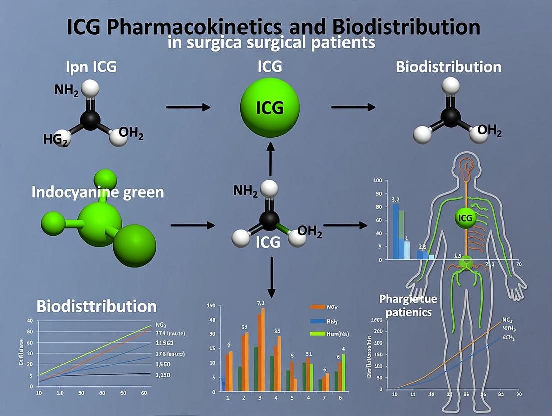
This article provides a detailed examination of indocyanine green (ICG) pharmacokinetics and biodistribution in surgical patients.
ICG Pharmacokinetics & Biodistribution in Surgery: A Comprehensive Guide for Translational Researchers
Abstract
This article provides a detailed examination of indocyanine green (ICG) pharmacokinetics and biodistribution in surgical patients. We explore the foundational science of ICG, including its chemical properties, fluorescence mechanisms, and metabolic pathways. Methodological approaches for real-time intraoperative imaging, dosing protocols, and data acquisition are discussed. The content addresses common challenges such as signal variability, tissue-specific clearance, and optimization strategies for different surgical specialties. Finally, we validate findings through comparative analysis with other imaging agents and highlight the clinical validation and future implications of ICG in precision surgery and drug development.
The Science of ICG: Chemical Properties, Fluorescence, and Metabolic Pathways in Surgical Patients
This whitepaper provides an in-depth technical analysis of the molecular and optical characteristics of Indocyanine Green (ICG) that underpin its preeminent role in clinical surgical imaging. This discussion is framed within the critical context of ongoing research into ICG pharmacokinetics and biodistribution in surgical patients, which directly informs and optimizes its intraoperative application. For researchers and drug development professionals, understanding this structure-function relationship is key to advancing image-guided surgery and developing next-generation agents.
Core Chemical Structure and Its Implications
ICG (C43H47N2NaO6S2) is a tricarbocyanine dye with a amphiphilic structure central to its behavior in vivo.
- Hydrophobic Polycyclic Core: A conjugated heptamethine chain bridging two lipophilic benzoindolium rings. This planar, lipophilic core is responsible for strong NIR absorption and fluorescence, and promotes non-covalent binding to plasma proteins (primarily albumin) and cellular membranes.
- Hydrophilic Sulfonate Groups: Sulfonate substituents on each ring confer water solubility and prevent the dye from crossing intact cellular membranes, confining it initially to the vascular compartment post-injection.
- Chemical Lability: The central polycarbon chain is susceptible to photodegradation and nucleophilic attack by water, glutathione, and other biomolecules, leading to degradation and fluorescence quenching over time—a key factor in its pharmacokinetic profile.
This amphiphilicity dictates its initial vascular confinement, hepatic clearance, and interaction with biological targets, forming the basis for its biodistribution.
Optical Properties: The NIR Window Advantage
ICG's optical profile is ideally matched to biological imaging.
Table 1: Key Optical Properties of ICG in Aqueous Solution (Bound to Albumin)
| Property | Value / Characteristic | Significance for Surgical Imaging |
|---|---|---|
| Peak Absorption | ~780 nm | Minimizes interference from endogenous chromophores (hemoglobin, melanin). |
| Peak Emission | ~820 nm | Falls within the "NIR-I window" (700-900 nm) where tissue scattering and autofluorescence are low. |
| Molar Extinction Coefficient (ε) | ~130,000 L·mol⁻¹·cm⁻¹ | Enables high absorption and bright signal at low concentrations (typical clinical doses: 0.1-0.3 mg/kg). |
| Quantum Yield (in Blood) | ~4-8% (higher when protein-bound) | Sufficient for high-contrast imaging despite quenching in aqueous environments. |
| Fluorescence Lifetime | ~0.3-0.5 ns | Allows for lifetime imaging techniques to differentiate signal from background. |
The shift to longer wavelengths upon protein binding (J-aggregation) and the concentration-dependent quenching are critical considerations for quantitative imaging protocol design.
Link to Pharmacokinetics & Biodistribution in Surgical Patients
ICG's structure-driven behavior defines its pharmacokinetic (PK) phases, which are exploitable for specific surgical applications.
Table 2: Correlating ICG Properties with Clinical PK Phases and Surgical Applications
| PK Phase | Time Post-IV Injection | Dominant Process | Driving Structural Property | Surgical Imaging Application |
|---|---|---|---|---|
| First Pass (Vascular) | 0-3 minutes | Rapid mixing, binding to plasma proteins. | Amphiphilicity: Sulfonates enable solubility; lipophilic core drives albumin binding. | Angiography (e.g., coronary bypass, flap perfusion), tumor delineation via enhanced permeability and retention (EPR) in leaks. |
| Distribution & Clearance | 3-15 minutes | Extravasation in leaky tissues, hepatic uptake. | Protein binding modulates size; lipophilicity facilitates hepatocyte uptake. | Sentinel lymph node mapping, liver segment identification, biliary imaging. |
| Elimination | >15 minutes | Biliary excretion (>95%). | Molecular weight and hepatic metabolism. | Assessment of bile duct patency, liver function testing. |
Understanding patient-specific factors—such as hepatic function, serum albumin levels, and capillary permeability in tumor or inflamed tissue—that alter this PK profile is a core focus of current research to standardize and quantify ICG imaging.
Key Experimental Protocols in ICG Research
Protocol 1: Measuring Protein-Binding Kinetics and Quantum Yield Enhancement
Objective: Quantify the binding affinity of ICG to Human Serum Albumin (HSA) and the resultant fluorescence enhancement. Methodology:
- Prepare a 1 µM ICG solution in phosphate-buffered saline (PBS).
- Titrate with increasing concentrations of HSA (0 to 50 µM).
- For each titration point:
- Record absorption spectrum (600-900 nm). Note the shift from ~780 nm to ~805 nm.
- Record fluorescence emission spectrum (excite at 780 nm, collect 800-850 nm).
- Perform fluorescence quenching titration with a known quencher (e.g., potassium iodide) to calculate Stern-Volmer constants for bound vs. free dye.
- Analyze fluorescence intensity vs. [HSA] using a binding isotherm (e.g., Langmuir) to determine dissociation constant (Kd).
- Calculate relative quantum yield using a reference dye (e.g., IR-26 in DCM) for free ICG and HSA-bound ICG.
Protocol 2: In Vivo Biodistribution and Pharmacokinetic Profiling in a Murine Model
Objective: Characterize the tissue-specific uptake and clearance of ICG. Methodology:
- Administer ICG intravenously (2 mg/kg) to animal models (e.g., mice).
- At predetermined time points (e.g., 1, 5, 15, 30, 60, 120 min), euthanize cohorts (n=5/time point).
- Collect blood plasma and key organs (liver, kidney, spleen, lung, muscle, tumor if applicable).
- Homogenize tissues and extract ICG using a solvent (e.g., DMSO).
- Quantify ICG concentration in extracts using fluorescence plate reader (ex/em: 780/820 nm) against a standard curve.
- Perform non-compartmental PK analysis on plasma data. Generate biodistribution profiles as % Injected Dose per Gram (%ID/g) of tissue.
Signaling Pathways and Experimental Workflows
ICG Pharmacokinetic Pathway and Surgical Applications
Integrated Workflow for ICG Imaging Research
The Scientist's Toolkit: Key Research Reagent Solutions
Table 3: Essential Materials for ICG Pharmacokinetic and Imaging Research
| Item / Reagent | Function / Rationale | Example Vendor / Cat. No. (Illustrative) |
|---|---|---|
| ICG, USP Grade | Clinical reference standard for translational studies. | PULSION Medical Systems; FDA-approved vial. |
| ICG, Analytic Grade (>95% purity) | For precise in vitro assays to avoid impurities affecting optical properties. | Sigma-Aldrich, 12633. |
| Human Serum Albumin (HSA), Fatty Acid Free | Key binding partner for in vitro simulation of plasma behavior. | Sigma-Aldrich, A3782. |
| NIR Fluorescence Imaging System | For in vivo animal or intraoperative imaging (ex: ~750-800 nm, em: ~820 nm). | PerkinElmer IVIS; KARL STORZ OPAL; Hamamatsu Photonics. |
| Fluorescence Spectrophotometer with NIR Detector | Essential for characterizing optical properties in solution. | Horiba Fluorolog; Edinburgh Instruments FLS1000. |
| In Vivo Imaging Animal Model (e.g., nude mouse, hepatic injury model) | For biodistribution and PK studies relevant to surgical conditions. | Charles River Laboratories. |
| Tissue Homogenization Kit & Solvent (DMSO/Methanol) | For efficient extraction of ICG from tissues for quantitative analysis. | Omni International homogenizers; DMSO (Sigma, D8418). |
| Microplate Reader with NIR Filters | High-throughput quantification of ICG in extracted samples. | BioTek Synergy H1. |
| PK/PD Modeling Software | For non-compartmental and compartmental analysis of biodistribution data. | Certara Phoenix WinNonlin. |
ICG's ideal suitability for surgical imaging is a direct consequence of its specific chemical architecture, which yields optimal NIR optical properties and dictates a predictable, exploitable pharmacokinetic profile. Ongoing research into the nuances of its biodistribution in patients with varying pathophysiology is refining its application, moving from qualitative visualization toward quantitative, patient-specific surgical guidance. This synergy between fundamental physico-chemistry and clinical PK research continues to solidify ICG's role as the cornerstone of fluorescence-guided surgery.
This whitepaper provides an in-depth technical analysis of the indocyanine green (ICG) metabolic pathway, central to its role as a pharmacokinetic and surgical imaging probe. Framed within a thesis on ICG biodistribution in surgical patients, it details the molecular mechanisms of hepatic uptake via OATP1B3, cytosolic binding, canalicular excretion by MRP2, and high-affinity plasma protein binding. The guide consolidates current quantitative data, presents validated experimental protocols, and visualizes critical pathways to support research in hepatobiliary function and drug development.
Indocyanine green is a water-soluble, anionic tricarbocyanine dye whose unique pharmacokinetic profile—rapid hepatic extraction and exclusive biliary excretion—makes it an indispensable tool for intraoperative imaging and liver function assessment. Understanding its precise metabolic pathway is critical for interpreting fluorescence-guided surgery data, modeling hepatic transport, and developing derivative agents.
Core Pathway Mechanisms
Plasma Protein Binding
Immediately upon intravenous injection, ICG binds extensively to plasma proteins, primarily albumin and, to a lesser extent, alpha-1 lipoproteins. This binding confines ICG to the vascular compartment initially, preventing extravasation and directing it to the liver.
Key Binding Parameters:
- Primary Carrier: Human Serum Albumin (HSA)
- Binding Constant (Kd): ~0.6 µM
- Number of High-Affinity Sites on HSA: 1-2
Hepatic Uptake
The ICG-albumin complex is transported to the liver sinusoids. Uptake into hepatocytes is mediated primarily by the organic anion-transporting polypeptide 1B3 (OATP1B3, gene SLCO1B3), with potential minor contributions from other transporters like NTCP. This process is energy-independent and driven by concentration gradients.
Cytosolic Binding and Storage
Once inside the hepatocyte, ICG dissociates from albumin and binds to intracellular binding proteins, primarily glutathione S-transferase (GST) and possibly fatty acid-binding protein (L-FABP). This facilitates its transit through the aqueous cytosol to the canalicular membrane.
Biliary Excretion
Excretion across the canalicular membrane into the bile is the rate-limiting step of ICG clearance. This active, ATP-dependent transport is primarily mediated by the multidrug resistance-associated protein 2 (MRP2, gene ABCC2). ICG is then eliminated unchanged in the bile, with no enterophepatic recirculation.
Table 1: Key Pharmacokinetic Parameters of ICG in Humans
| Parameter | Value (Mean ± SD or Range) | Notes |
|---|---|---|
| Plasma Protein Binding | >95% | Primarily to albumin. |
| Distribution Half-life (t½α) | 2-4 min | Represents mixing and initial uptake. |
| Elimination Half-life (t½β) | 3-5 min | Represents biliary excretion phase. |
| Plasma Clearance Rate | 0.14 - 0.21 L/min | Liver blood flow dependent. |
| Hepatic Extraction Ratio | 0.5 - 0.8 | High first-pass extraction. |
| Time to Peak Biliary Excretion | ~10 minutes | Post IV administration. |
| Molecular Weight | 774.96 Da | Anionic, amphiphilic structure. |
| Primary Excretion Route | >97% Biliary | No metabolism; fecal elimination. |
Table 2: Key Transporters in the ICG Pathway
| Transporter | Gene | Location | Role in ICG Pathway | Inhibitors |
|---|---|---|---|---|
| OATP1B3 | SLCO1B3 | Basolateral (Sinusoidal) membrane | Primary hepatic uptake. | Rifampin, Cyclosporine A |
| MRP2 | ABCC2 | Apical (Canalicular) membrane | Primary biliary excretion. | Probenecid, MK-571 |
| NTCP | SLC10A1 | Basolateral membrane | Potential minor uptake route. | Na+ depletion, Myrcludex B |
Experimental Protocols
Protocol: Assessing ICG Plasma Protein Binding (Ultrafiltration)
Objective: To determine the fraction of ICG bound to plasma proteins. Materials: Human plasma, ICG stock solution (1 mg/mL in sterile water), 10 kDa molecular weight cut-off centrifugal ultrafilters, microcentrifuge, spectrophotometer/fluorometer. Procedure:
- Spike ICG into human plasma to a final concentration of 5 µM. Incubate at 37°C for 10 min.
- Load 500 µL of the ICG-plasma mixture into an ultrafiltration device.
- Centrifuge at 2000 x g, 37°C, for 15-20 minutes to obtain protein-free filtrate.
- Measure ICG concentration in the initial plasma mixture (Ctotal) and in the filtrate (Cfree) using absorbance at 780 nm or fluorescence (ex/em ~780/820 nm).
- Calculation: % Bound = [(Ctotal - Cfree) / C_total] x 100.
Protocol:In VitroOATP1B3-Mediated Uptake Assay
Objective: To characterize the kinetic parameters (Km, Vmax) of OATP1B3 for ICG. Materials: HEK293 cells stably expressing OATP1B3 (and mock-transfected controls), uptake buffer (Hanks' Balanced Salt Solution, HBSS), ICG, transport inhibitor (e.g., 100 µM Rifampin). Procedure:
- Culture cells in 24-well plates to 90% confluence.
- Wash cells twice with pre-warmed (37°C) HBSS.
- Uptake Phase: Add HBSS containing a range of ICG concentrations (e.g., 0.5-50 µM) to the wells. Include inhibitor controls. Incubate at 37°C for a precise time (e.g., 2 min).
- Termination: Quickly aspirate the uptake solution and wash cells three times with ice-cold HBSS.
- Lysis: Lyse cells with 0.1% Triton X-100 in water.
- Quantification: Measure ICG fluorescence in the lysate. Normalize to total protein content (BCA assay). Subtract uptake in mock cells to determine OATP1B3-specific transport. Analyze data with Michaelis-Menten kinetics.
Protocol:Ex VivoBiliary Excretion Index (BEI) Assay
Objective: To assess the canalicular excretion function using sandwich-cultured hepatocytes (SCH). Materials: Primary rat or human hepatocytes in sandwich culture, standard and Ca2+-free buffers, ICG, fluorescence microscope/plate reader. Procedure:
- Pre-incubation: Incubate SCH in standard buffer (+Ca2+) to maintain intact bile canaliculi (BC) or in Ca2+-free buffer to disrupt BC junctions for 30 min.
- Uptake: Add ICG (e.g., 5 µM) to both sets of cells. Incubate for 30-60 min at 37°C.
- Wash & Accumulation: Wash cells thoroughly and measure total cellular accumulation (T) via fluorescence.
- Excretion Measurement: For cells with intact BC, a subsequent chase incubation in ICG-free +Ca2+ buffer allows excretion to continue. For cells with disrupted BC, the content of BC is released into the medium upon washing with Ca2+-free buffer.
- Calculation: BEI = [(Tdisrupted - Tintact) / Tdisrupted] x 100. Tdisrupted represents cellular accumulation only, while T_intact represents cellular + biliary accumulation.
Pathway Visualizations
ICG Pathway from Injection to Biliary Excretion
Hepatocyte Transport Mechanism for ICG
The Scientist's Toolkit: Key Research Reagents & Materials
Table 3: Essential Reagents for ICG Pathway Research
| Item | Function/Application | Example/Notes |
|---|---|---|
| Pharmaceutical Grade ICG | Core substrate for in vivo and in vitro studies. | Ensure sterility, high purity (>95%), and prepare fresh solutions protected from light. |
| Human Serum Albumin (HSA) | For studying plasma protein binding kinetics. | Use fatty acid-free HSA for consistent results in binding assays. |
| OATP1B3-Expressing Cell Line | To isolate and study the primary uptake transporter. | HEK293 or MDCKII cells stably transfected with human SLCO1B3. |
| MRP2-Expressing Membrane Vesicles | To study ATP-dependent canalicular transport kinetics. | Commercially available inside-out vesicles from Sf9 or mammalian cells. |
| Specific Transport Inhibitors | To confirm transporter-specific activity. | Rifampin (OATP1B3), Probenecid/MK-571 (MRP2), Myrcludex B (NTCP). |
| Sandwich-Cultured Hepatocytes (SCH) | Gold-standard in vitro model for integrated uptake & biliary excretion. | Primary rat or human hepatocytes cultured between two layers of gelled collagen. |
| Fluorescence Plate Reader | Quantification of ICG in solutions, lysates, or cells. | Requires near-infrared (NIR) capability (excitation ~750-780 nm, emission >800 nm). |
| Clinical-Grade NIR Imaging System | For in vivo surgical or preclinical biodistribution studies. | Systems like the FLARE or PDE; allows real-time visualization of ICG fluorescence. |
This guide provides a technical examination of three foundational pharmacokinetic (PK) parameters—Volume of Distribution (Vd), Clearance (CL), and Half-Life (t1/2)—framed within the context of research into Indocyanine Green (ICG) pharmacokinetics and biodistribution in surgical patients. These parameters are critical for quantifying tissue penetration, elimination mechanisms, and dosing regimens in real-time surgical imaging and hepatic function assessment.
Core Pharmacokinetic Parameters: Definitions and Interrelationships
Volume of Distribution (Vd) is a theoretical volume that relates the total amount of drug in the body to its plasma concentration. It indicates the extent of tissue distribution. A high Vd suggests significant tissue penetration, while a low Vd suggests confinement to the vascular space. For ICG, Vd is expected to be low (~0.05 L/kg) as it binds extensively to plasma proteins and remains primarily intravascular in healthy subjects, but can increase in pathological states.
Clearance (CL) is the volume of plasma from which a substance is completely removed per unit time. It represents the sum of all elimination processes (hepatic, renal, etc.). ICG is exclusively cleared by the liver via active transport into bile, making its clearance a direct marker of hepatic function and blood flow.
Half-Life (t1/2) is the time required for the plasma concentration to decrease by 50%. It is a derived parameter dependent on both Vd and CL, as described by the equation: t1/2 = (0.693 * Vd) / CL
This relationship is fundamental: changes in half-life can result from alterations in distribution or clearance, necessitating careful interpretation in clinical research.
Table 1: Key Pharmacokinetic Parameters for ICG in Surgical Research
| Parameter | Symbol | Typical Value for ICG (Healthy) | Primary Determinants | Significance in Surgical ICG Research |
|---|---|---|---|---|
| Volume of Distribution | Vd | ~0.05 L/kg (Plasma Volume) | Plasma protein binding, capillary permeability, tissue binding. | Quantifies extravasation; increased in sepsis, capillary leak, or liver disease. |
| Clearance | CL | ~0.7-1.0 mL/min/kg | Hepatic blood flow, hepatocyte function, biliary patency. | Gold-standard metric for liver functional reserve pre- and post-resection. |
| Half-Life | t1/2 | ~3-5 minutes | Dependent on Vd and CL. | Guides timing for repeated dosing in fluorescence imaging sequences. |
| Fraction Unbound | fu | <0.01 (Highly bound) | Albumin concentration, competing substances. | Affects clearance rate and susceptibility to changes in protein binding. |
Methodologies for Determining PK Parameters in ICG Studies
Experimental Protocol: Serial Blood Sampling for ICG PK Analysis
- Objective: To determine Vd, CL, and t1/2 of ICG in surgical patients.
- Reagent: Sterile ICG powder (e.g., 25 mg vials), reconstituted with provided solvent.
- Dosing: A precise intravenous bolus (e.g., 0.25 mg/kg) is administered via a central or large peripheral vein.
- Sample Collection: Arterial or venous blood samples are collected at frequent intervals (e.g., 0, 1, 2, 3, 4, 5, 7, 10, 15, 20 minutes post-injection) into heparinized tubes.
- Sample Processing: Plasma is separated by centrifugation. ICG concentration is quantified via spectrophotometry (λmax ~805 nm in plasma) or high-performance liquid chromatography (HPLC) for greater specificity.
- PK Analysis: Concentration-time data are fitted to a pharmacokinetic model (typically a mono- or bi-exponential decay) using non-linear regression software (e.g., Phoenix WinNonlin, NONMEM).
- Clearance (CL) = Dose / AUC (Area Under the concentration-time curve).
- Volume of Distribution (Vd) = CL / Elimination Rate Constant (k) or via the non-compartmental method: Vdss = Dose * AUMC / (AUC)^2 (where AUMC is area under the moment curve).
- Half-Life (t1/2) = 0.693 / k.
Experimental Protocol: Non-Invasive Pulse Densitometry for Real-Time ICG Clearance (e.g., LiMON System)
- Objective: Bedside, real-time measurement of ICG plasma disappearance rate (PDR) and derived parameters.
- Setup: A finger clip or external probe is placed on the patient.
- Dosing: IV bolus of ICG (typically 0.25-0.5 mg/kg).
- Measurement: The probe uses optical densitometry at specific wavelengths to measure ICG concentration transcutaneously for ~15-30 minutes.
- Analysis: The system generates a decay curve and calculates:
- Plasma Disappearance Rate (PDR) (%/min): The percentage decrease in ICG concentration per minute, often reported as a single-value metric of liver function (normal >18%/min).
- t1/2: Calculated from the PDR curve.
- Note: This method provides robust estimates of CL and t1/2 but is less accurate for absolute Vd determination.
The Scientist's Toolkit: Research Reagent Solutions for ICG PK Studies
Table 2: Essential Materials and Reagents
| Item | Function & Specificity in ICG PK Research |
|---|---|
| Medical-Grade ICG (Sterile) | The tracer agent. Must be of injectable grade, protected from light, and used promptly after reconstitution to ensure stability and accurate dosing. |
| Spectrophotometer / Fluorescence Reader | Quantifies ICG concentration in plasma samples. A near-infrared (NIR) capable reader is optimal for direct measurement of ICG's peak absorbance/emission. |
| HPLC System with Fluorescence/NIR Detector | Provides superior specificity for ICG quantification, separating it from metabolites or background chromophores in complex biological matrices. |
| Pulse Densitometry Monitor (e.g., LiMON) | Enables non-invasive, real-time in vivo PK monitoring, crucial for intraoperative and ICU applications without the need for blood draws. |
| Pharmacokinetic Modeling Software | Essential for fitting concentration-time data to compartmental or non-compartmental models to extract precise Vd, CL, and t1/2 values. |
| Heparinized Blood Collection Tubes | Prevents coagulation during rapid serial sampling, ensuring accurate plasma yield for concentration analysis. |
Visualizing Relationships and Workflows
PK Parameter Determination from IV Bolus Data
Two-Compartment Model for ICG Distribution and Clearance
Impact of Surgical Pathologies on ICG PK Parameters
This whitepaper details the critical physiological factors governing the biodistribution of indocyanine green (ICG), a near-infrared fluorescent tracer, in surgical patients. This analysis is a core component of a broader thesis investigating ICG pharmacokinetics to establish predictive models for surgical outcomes, drug delivery optimization, and real-time tissue viability assessment. Precise understanding of these variables is paramount for translating ICG-guided surgery from qualitative imaging to quantitative, patient-specific diagnostics.
Table 1: Impact of Patient-Specific Physiology on ICG Pharmacokinetics
| Factor | Key Parameter | Effect on ICG Kinetics | Typical Quantitative Influence (from Recent Literature) |
|---|---|---|---|
| Hepatic Function | Indocyanine green retention rate at 15 min (ICG-R15) | Directly correlates with hepatic extraction efficiency and plasma clearance rate. Impaired function slows clearance. | Normal: ICG-R15 < 10%. Mild impairment: 10-20%. Severe impairment: > 40%. Clearance half-life can double from ~3 min to >6 min in cirrhosis. |
| Cardiac Output & Blood Flow | Cardiac Index (L/min/m²) | Determines initial mixing and delivery rate to organs. Low output prolongs distribution phase. | A 30% decrease in cardiac index can increase time-to-peak fluorescence in peripheral tissue by 50-100%. |
| Body Composition | Body Surface Area (BSA, m²), Lean Body Mass | Volume of distribution correlates with plasma volume, which is linked to BSA. Affects initial concentration. | Dosing normalized to BSA (e.g., 0.25 mg/kg vs. fixed dose) reduces inter-patient variability in peak intensity by up to 35%. |
| Renal Function | Glomerular Filtration Rate (GFR) | Minimal renal excretion in healthy states. Severe dysfunction can alter plasma protein binding and indirect kinetics. | In anuria, terminal elimination half-life may increase marginally by ~10-15%, primarily due to fluid shifts. |
| Serum Protein Levels | Albumin Concentration (g/dL) | ICG is >95% albumin-bound. Hypoalbuminemia can slightly increase free fraction, altering tissue penetration. | Albumin < 2.5 g/dL can lead to a 20-25% faster initial tissue uptake in some models due to altered binding equilibrium. |
Table 2: Impact of Blood Flow Dynamics on Regional ICG Distribution
| Organ/Region | Flow Characteristic | Effect on ICG Signal Kinetics | Measurable Parameters |
|---|---|---|---|
| Hepatobiliary System | High perfusion, active transport | Rapid uptake, biliary excretion. Signal rises in liver, then in bile ducts/gallbladder. | Time-to-peak (TTP) in liver parenchyma: 60-120s. Hepatic clearance rate (k): 0.2-0.3 min⁻¹. |
| Malignant Tumors | Chaotic, hyper-permeable vasculature (EPR effect) | Enhanced permeability and retention. Slower accumulation and washout vs. normal tissue. | Tumor-to-Background Ratio (TBR): Peaks at 3-10 min post-injection. Washout rate often slower. |
| Ischemic Tissue | Reduced/absent arterial inflow | Delayed arrival, reduced peak intensity, and slower wash-in rate. | Time differential of >10-15s compared to healthy tissue is clinically significant for anastomosis assessment. |
Experimental Protocols for Key Investigations
Protocol A: Measuring Systemic ICG Pharmacokinetics in Surgical Patients
- Objective: To derive patient-specific pharmacokinetic (PK) parameters (clearance, half-life, volume of distribution).
- Materials: See "Scientist's Toolkit" below.
- Method:
- Patient Preparation: Baseline blood draw for albumin, bilirubin, creatinine. BSA calculation.
- ICG Administration: Precisely inject a standardized IV dose (e.g., 0.25 mg/kg) via a peripheral line.
- Serial Blood Sampling: Collect venous blood samples at pre-defined intervals (e.g., 0, 1, 3, 5, 10, 15, 30 minutes post-injection) into heparinized tubes.
- Sample Processing: Centrifuge samples immediately at 3000 rpm for 10 min to separate plasma.
- Quantification: Dilute plasma 1:10 with sterile saline. Measure ICG concentration using a spectrophotometer at 805 nm (absorption peak) against a plasma blank. A standard curve is required.
- PK Modeling: Fit concentration-time data to a two-compartment model using software (e.g., Phoenix WinNonlin) to calculate PK parameters.
Protocol B: Intraoperative Laser Fluorescence Imaging for Regional Biodistribution
- Objective: To visualize and quantify spatial and temporal ICG distribution in real-time.
- Method:
- System Calibration: Calibrate the NIR fluorescence imaging system using a fluorescence reference standard.
- Baseline Imaging: Acquate a pre-injection background image of the surgical field under NIR light.
- ICG Bolus Injection: Administer standardized ICG dose intravenously.
- Continuous Video Acquisition: Record NIR fluorescence video for at least 10-15 minutes post-injection.
- Region of Interest (ROI) Analysis: Using proprietary or open-source software (e.g., ImageJ), define ROIs over target tissues (e.g., tumor, normal liver, bowel anastomosis).
- Kinetic Curve Generation: Extract mean fluorescence intensity (MFI) over time for each ROI. Calculate parameters like TTP, TBR, wash-in/washout rates.
Visualization: Signaling Pathways and Experimental Workflow
Title: ICG Biodistribution and Elimination Pathway
Title: Integrated Protocol for ICG Biodistribution Research
The Scientist's Toolkit: Key Research Reagent Solutions
Table 3: Essential Materials for ICG Biodistribution Research
| Item | Function & Importance | Example/Note |
|---|---|---|
| Pharmaceutical-Grade ICG | The fluorescent tracer agent. Must be reconstituted fresh to maintain fluorescence yield. | PULSION (Diagnostic Green), Verdye. |
| Sterile Water for Injection | Reconstitution solvent. Must be aqueous, non-ionic to prevent ICG aggregation. | 0.9% NaCl can also be used per manufacturer. |
| Human Serum Albumin (HSA) | For in vitro binding studies and calibration standards to mimic physiological conditions. | Essential for creating accurate standard curves in plasma matrix. |
| NIR Fluorescence Imaging System | For real-time, intraoperative visualization and quantification of ICG distribution. | Hamamatsu PDE-Neo, KARL STORZ OPAL, PerkinElmer Fluobeam. |
| Spectrophotometer / Plate Reader | For precise quantification of ICG concentration in blood/plasma samples. | Requires NIR capability (absorption at ~805 nm). |
| Pharmacokinetic Modeling Software | To fit concentration-time data and derive critical PK parameters (CL, Vd, t½). | Phoenix WinNonlin, PK-Sim, open-source R packages (e.g., nlmixr). |
| Image Analysis Software | To extract quantitative kinetic data (MFI, TBR) from fluorescence video sequences. | ImageJ/FIJI with custom macros, proprietary software from imager vendors. |
| Standardized Fluorescence Phantom | For daily calibration and validation of imaging system sensitivity and linearity. | Ensures inter-study data comparability. |
Historical Context and Evolution of ICG from Diagnostic Agent to Surgical Navigator
This whitepaper is framed within a broader thesis investigating the pharmacokinetics (PK) and biodistribution of Indocyanine Green (ICG) in surgical patients. The transition of ICG from a purely diagnostic agent to an intraoperative navigational tool is fundamentally underpinned by a detailed understanding of its PK profile—including its binding to plasma proteins, hepatic clearance, extravasation into tissues, and its unique fluorescence properties when bound to various biomolecules. This evolution is not merely technological but is driven by a deepening comprehension of its in vivo behavior across different pathophysiological states, enabling precise application in oncology, vascular, and reconstructive surgery.
Historical Context and Diagnostic Foundations
Indocyanine Green, a tricarbocyanine dye, was first approved by the FDA in 1959 for diagnostic purposes in hepatic function and cardiac output studies. Its near-infrared (NIR) fluorescence (emission peak ~830 nm) was a latent property not initially exploited.
Key Diagnostic Parameters:
- Peak Absorption: 800 nm in blood.
- Peak Emission: 830 nm in blood.
- Plasma Protein Binding: >95% to albumin and lipoproteins.
- Elimination: Exclusive hepatic excretion into bile, no enterohepatic recirculation.
Table 1: Evolution of ICG Applications Over Time
| Era | Primary Application | Key Mechanism | Limitation |
|---|---|---|---|
| 1960s-1990s | Hepatic Function Assessment | Photometric measurement of plasma clearance rate | No real-time imaging; systemic PK only |
| 1990s-2000s | Angiography (Cardio, Ophthalmic) | NIR fluorescence imaging of vascular flow | Qualitative assessment; 2D imaging |
| 2000s-2010s | Sentinel Lymph Node (SLN) Mapping | Interstitial diffusion and lymphatic uptake | Timing and dose critical; variable PK |
| 2010s-Present | Perfusion & Cancer Navigation | Tumor-specific accumulation (Enhanced Permeability & Retention - EPR) and real-time NIR imaging | Quantification challenges; tissue-depth penetration (~5-10 mm) |
Core Pharmacokinetic Principles Enabling Surgical Navigation
The surgical utility of ICG is predicated on its PK-driven biodistribution.
- Vascular Phase (Seconds-Minutes): ICG remains intravascular, bound to plasma proteins. This enables angiography and perfusion assessment.
- Interstitial Phase (Minutes): Extravasation in permeable tissues (e.g., inflammation, tumors via EPR effect).
- Cellular Phase (Hours): Specific uptake, e.g., by hepatocytes or, when conjugated to targeting moieties, by cancer cells.
Table 2: Quantitative PK Parameters of ICG in Surgical Patients
| Parameter | Normal Value (Adults) | Impact on Surgical Navigation | Source (Recent Study) |
|---|---|---|---|
| Plasma Half-life (T1/2) | 3-5 minutes | Dictates timing for angiography vs. SLN mapping | Pasternak, 2023 J Surg Oncol |
| Volume of Distribution (Vd) | ~0.05 L/kg (confined to plasma) | High contrast for vascular imaging | Ishizawa et al., 2022 Ann Surg |
| Clearance (CL) | 0.5-0.7 L/min | Reduced in liver dysfunction, affects dosing | Desmettre et al., 2021 Pharmaceutics |
| Protein Binding | >95% (Albumin) | Defines its distribution and fluorescence quenching in blood | Zhu et al., 2023 Bioconjug Chem |
Experimental Protocols for Key Research Applications
Protocol 1: Intraoperative Tumor Delineation in Hepatectomy (Based on EPR)
- Patient Preparation: Obtain informed consent. Baseline liver function tests (LFTs).
- ICG Administration: Intravenous bolus of 0.5 mg/kg, administered 24 hours prior to surgery.
- Mechanism: ICG extravasates in hyperpermeable tumor vasculature and is retained, while clearing from normal parenchyma.
- Intraoperative Imaging: Use NIR fluorescence imaging system (e.g., PINPOINT, SPY). Excitation: 760-785 nm, Emission filter: >820 nm.
- Data Capture: Record fluorescence intensity ratios (Tumor:Normal) from region-of-interest (ROI) analysis.
Protocol 2: Sentinel Lymph Node (SLN) Mapping in Breast Cancer
- Preparation: Prepare a 1.25 mg/mL ICG solution in sterile water.
- Administration: Intradermal or parenchymal peritumoral injection of 1-2 mL (total 1.25-2.5 mg) immediately after induction of anesthesia.
- Imaging: Use real-time NIR camera. Track the lymphatic channels from injection site.
- SLN Identification: The first lymph node(s) exhibiting fluorescence are marked as SLNs. Record time-to-visualization and signal intensity.
- Ex Vivo Confirmation: Excised nodes are imaged ex vivo to confirm fluorescence.
Protocol 3: Quantitative Perfusion Assessment in Anastomosis
- Administration: IV bolus of 0.1-0.2 mg/kg ICG after vascular anastomosis is completed.
- Imaging: Continuous video recording under NIR illumination.
- Quantitative Analysis: Use proprietary software (e.g., FLARE, Quest) to generate time-intensity curves. Calculate ingress rate (slope), maximum intensity (Imax), and time-to-peak.
- Outcome Correlation: Perfusion metrics are correlated with clinical outcomes (e.g., anastomotic leak).
Visualization of Key Concepts
Title: ICG Pharmacokinetic Phases Driving Surgical Applications
Title: Protocol for Tumor Delineation Using EPR Effect
The Scientist's Toolkit: Key Research Reagent Solutions
Table 3: Essential Materials for ICG-based Surgical Research
| Item / Reagent | Function & Rationale |
|---|---|
| ICG (Pulmocare, Infracyanine, etc.) | The core NIR fluorophore. Must be reconstituted fresh to avoid aggregation and fluorescence quenching. |
| Human Serum Albumin (HSA) | For in vitro binding studies to simulate plasma conditions and modulate fluorescence yield. |
| Phosphate Buffered Saline (PBS) | Standard vehicle for dilution and control experiments. |
| Dimethyl Sulfoxide (DMSO) | Solvent for creating high-concentration ICG stock solutions for in vitro assays. |
| Lipoprotein Solutions (LDL/HDL) | To study alternative binding partners of ICG and their impact on cellular uptake. |
| Commercial NIR Imaging System (e.g., FLARE, SPY, PDE) | Provides standardized excitation (760-785 nm) and emission (>820 nm) filtering for reproducible imaging. |
| Fluorescence Spectrophotometer | For quantifying ICG concentration in plasma/tissue homogenates and measuring quantum yield. |
| Liquid Chromatography-Mass Spectrometry (LC-MS/MS) | Gold standard for quantifying ICG and potential metabolites in pharmacokinetic studies. |
| Small Animal NIR Imager (e.g., IVIS Spectrum) | For pre-clinical PK/biodistribution studies in rodent models of disease. |
| ImageJ / FIJI with NIR Plugins | Open-source software for quantitative analysis of fluorescence intensity, signal-to-noise ratios, and biodistribution. |
Mastering ICG Imaging Protocols: Dosing, Timing, and Intraoperative Data Acquisition
This whitepaper, framed within a broader research thesis on indocyanine green (ICG) pharmacokinetics and biodistribution in surgical patients, examines the fundamental dichotomy in clinical dosing: weight-based versus fixed-dose protocols. The choice of dosing strategy directly impacts the precision of pharmacokinetic (PK) studies, influences biodistribution patterns, and determines the safety and efficacy margins for diagnostic and therapeutic agents. For researchers and drug development professionals, understanding the quantitative and methodological implications of each approach is critical for designing robust clinical trials and translating findings into standardized clinical practice.
Pharmacokinetic Foundations in Surgical Research
The study of ICG serves as a paradigm for evaluating dosing strategies. ICG is a near-infrared fluorescent tracer used extensively to assess hepatic function, visualize vasculature, and map lymphatic drainage. Its pharmacokinetics are characterized by rapid binding to plasma proteins, exclusive hepatic clearance, and minimal extrahepatic distribution. In surgical patients, variables such as blood volume shifts, altered organ perfusion, and fluid resuscitation can significantly modulate these parameters, making dosing strategy a non-trivial variable in research design.
Comparative Analysis: Weight-Based vs. Fixed-Dose
The core debate centers on whether drug administration should be scaled to an individual's body size (typically weight) or administered as a universal quantity.
Theoretical and Practical Rationale
Weight-Based Dosing aims to normalize drug exposure (e.g., peak plasma concentration, area under the curve) across a population with varying body sizes. It is rooted in the principle that key PK parameters like volume of distribution and clearance often correlate with body weight, especially for drugs distributed in body water or metabolized by processes scaled to size.
Fixed-Dose Protocols administer a standard amount regardless of patient size. This approach is justified when the therapeutic or diagnostic window is wide, when drug distribution is not closely tied to body composition, or when operational simplicity, reduced dosing errors, and streamlined preparation outweigh the benefits of individualization.
Quantitative Data Comparison
Recent clinical studies and meta-analyses provide comparative data on key outcomes for both strategies. The following table synthesizes findings pertinent to imaging agents and drugs used in perioperative settings.
Table 1: Comparative Outcomes of Dosing Strategies in Clinical Studies
| Parameter | Weight-Based Dosing | Fixed-Dose Protocol | Primary Study Context |
|---|---|---|---|
| Inter-Patient PK Variability | Coefficient of Variation (CV) for AUC: 15-25% | CV for AUC: 25-40% | Oncology & Antibiotic Therapies |
| Diagnostic Signal Consistency | Reduced variability in tissue fluorescence intensity (e.g., ICG lymphography) | Increased risk of under-dosing in high-weight patients for signal generation | Fluorescence-Guided Surgery |
| Dosing Error Incidence | Higher (calculation & preparation errors) | Lower | Multi-Center Clinical Trials |
| Operational Efficiency | Lower (requires calculation, specific syringes) | Higher (pre-filled syringes, no calculation) | Emergency & Surgical Settings |
| Cost Implications | Potential drug waste in low-weight patients; variable cost per patient | Predictable, uniform drug cost per patient; may over-dose low-weight patients | Hospital Pharmacy Budgeting |
| Optimal Use Case | Narrow therapeutic index drugs; drugs with strong PK/weight correlation (e.g., ICG for hepatic function quantitation) | Drugs with wide safety margin; target saturation kinetics; qualitative imaging (e.g., ICG for angiography) | Drug Development & Surgical Imaging |
Experimental Protocol for Dosing Strategy Comparison in ICG Research
To empirically compare dosing strategies in a research setting, the following detailed methodology can be employed.
Protocol: A Randomized Crossover Study of ICG Pharmacokinetics with Weight-Based vs. Fixed Dosing in Surgical Patients
Objective: To compare the pharmacokinetic variability and biodistribution profile of ICG administered via weight-based versus fixed-dose protocols in patients undergoing major abdominal surgery.
Patient Population: N=20 adults, BMI range 18-35 kg/m², scheduled for elective hepatic or colorectal resection.
Study Design: Randomized, two-period crossover. Each patient receives both dosing strategies in separate sessions (pre-op and post-op).
Interventions:
- Weight-Based Dose: ICG 0.25 mg/kg of ideal body weight, administered intravenously.
- Fixed Dose: ICG 25 mg flat dose, administered intravenously.
Methodology:
- ICG Administration: Slow IV push over 30 seconds via a dedicated peripheral line.
- Blood Sampling: Serial blood samples (3 mL) collected at times: 0 (pre-dose), 2, 5, 10, 15, 30, 60, 120, 180, and 240 minutes post-injection. Samples are centrifuged, and plasma is stored at -80°C.
- Non-Invasive Fluorescence Imaging: A calibrated fluorescence imaging system (e.g., SPY-PHI) is used to record real-time biodistribution in a region of interest (e.g., liver, surgical field) at 1, 5, 15, 30, 60, and 120 minutes.
- Bioanalysis: Plasma ICG concentration is quantified using a validated high-performance liquid chromatography (HPLC) method with fluorescence detection.
- PK Analysis: Non-compartmental analysis (NCA) is performed to calculate: Area Under the Curve (AUC0-∞), Peak Plasma Concentration (Cmax), Time to Cmax (Tmax), Clearance (CL), and Volume of Distribution (Vd).
- Statistical Analysis: Primary endpoint: Comparison of the coefficient of variation (CV%) for AUC between the two dosing strategies using a mixed-effects model. Secondary endpoints: Comparison of Cmax variability, correlation of PK parameters with body weight, and quantitative analysis of imaging signal uniformity.
Visualizing the Research Decision Pathway
The logical flow for selecting a dosing strategy within a PK study is outlined below.
Diagram 1: Dosing Strategy Selection Logic (100 chars)
The Scientist's Toolkit: Research Reagent Solutions
Table 2: Essential Materials for ICG Pharmacokinetic & Biodistribution Studies
| Item / Reagent | Function & Explanation |
|---|---|
| Indocyanine Green (ICG) | The near-infrared fluorophore; must be of high, injectable grade (e.g., USP). Reconstituted per manufacturer protocol to ensure consistent bioavailability. |
| Certified Reference Standard (ICG) | Highly pure ICG for calibrating bioanalytical assays (HPLC). Essential for validating the accuracy of concentration measurements. |
| Human Plasma/Serum (Pooled) | Used for preparing calibration standards and quality control samples in method development and validation for PK assays. |
| Protein Precipitation Reagents | (e.g., Methanol, Acetonitrile). For deproteinizing plasma samples prior to HPLC analysis, ensuring accurate ICG quantification. |
| HPLC System with Fluorescence Detector | Primary instrument for quantifying plasma ICG concentrations. Requires specific NIR filter sets (ex: ~780 nm, em: ~820 nm). |
| Calibrated Fluorescence Imaging System | (e.g., open-platform NIR cameras like FLARE, or clinical systems like SPY-PHI). For non-invasive, real-time biodistribution and pharmacokinetic imaging. Must be radiometrically calibrated. |
| Pharmacokinetic Modeling Software | (e.g., Phoenix WinNonlin, NONMEM). For performing non-compartmental and compartmental analysis of concentration-time data. |
| Standardized Body Surface Area (BSA) Calculator | Critical for alternative dosing calculations (e.g., BSA-based) often used in oncology for comparison. |
| Pre-filled Syringe Kits (Placebo) | For blinding in randomized trials comparing dosing strategies, ensuring operational parity between weight-based and fixed-dose arms. |
The choice between weight-based and fixed-dose protocols is a fundamental design decision that reverberates through all stages of pharmacokinetic and biodistribution research, particularly in variable populations like surgical patients. Weight-based dosing offers superior normalization of PK parameters and is indispensable for quantitative imaging biomarkers. Fixed-dose protocols provide operational robustness and are sufficient for qualitative endpoints with wide margins. The optimal strategy must be derived from a clear understanding of the agent's pharmacokinetics, the primary study objective, and the practical realities of the clinical environment. Future research should focus on developing adaptive or stratified dosing models that leverage patient-specific factors beyond weight to further individualize and precision-target diagnostic and therapeutic interventions.
This technical guide synthesizes current research on the precise timing intervals required for optimal imaging following the administration of fluorescent contrast agents, with a specific focus on Indocyanine Green (ICG). Framed within a broader thesis on ICG pharmacokinetics and biodistribution in surgical patients, this document provides a rigorous, data-driven framework for researchers and drug development professionals seeking to standardize and optimize in vivo imaging protocols for vascular, lymphatic, and tissue perfusion assessment.
The efficacy of fluorescence-guided surgery and real-time perfusion assessment is fundamentally governed by the pharmacokinetic (PK) and biodistribution profile of the contrast agent. ICG, a near-infrared (NIR) fluorophore, binds rapidly to plasma proteins (primarily albumin) upon intravenous administration, confining it initially to the intravascular space. Its subsequent clearance via hepatic metabolism and biliary excretion creates a dynamic, time-dependent concentration gradient across vascular, interstitial, and lymphatic compartments. This treatise posits that identifying and adhering to critical "admin-to-image" intervals is not merely procedural but central to accurately interpreting imaging data, distinguishing pathological from physiological signal, and deriving quantitative metrics in surgical research.
Core Pharmacokinetic Principles of ICG
ICG's behavior in vivo follows a biphasic model:
- Phase 1 (Vascular Phase): 0-3 minutes post-IV bolus. ICG is predominantly intravascular, enabling angiography and quantitative perfusion analysis (e.g., ingress/egress rates).
- Phase 2 (Extravasation & Lymphatic Phase): 3-60+ minutes. Protein-bound ICG extravasates in areas of increased capillary permeability or is deliberately trafficked into lymphatic vessels via interstitial injection. This phase is critical for lymphatic mapping and assessing tissue viability via relative retention.
The exact timing windows are dependent on route of administration (intravenous vs. interstitial), dose, tissue type, and patient hemodynamic status.
Quantified Admin-to-Image Intervals: A Data Synthesis
The following tables consolidate optimal imaging intervals based on live-searched current clinical and preclinical literature.
Table 1: Intravenous Administration Windows
| Imaging Target | Recommended Admin-to-Image Interval | Key Rationale & PK Basis | Primary Metrics Derived |
|---|---|---|---|
| Macrovasculature (Angiography) | 10-30 seconds | Captures first-pass of high-concentration ICG bolus. | Vessel patency, anatomy, anastomotic leaks. |
| Tissue Perfusion (Capillary Level) | 1-3 minutes | Peak parenchymal enhancement during intravascular phase. | Time-to-peak, slope of inflow/outflow, signal intensity ratio. |
| Sentinel Lymph Node Mapping | Not recommended via IV | IV ICG does not reliably concentrate in lymph nodes. | N/A |
Table 2: Subcutaneous/Interstitial Administration Windows
| Imaging Target | Recommended Admin-to-Image Interval | Key Rationale & PK Basis | Primary Metrics Derived |
|---|---|---|---|
| Lymphatic Vessels (Lymphangiography) | 0-5 minutes (immediate imaging) | Visualizes initial lymphatic capillary uptake and collecting vessels. | Vessel architecture, identification of lymphatic leakage. |
| Sentinel Lymph Node (SLN) | 3-10 minutes (for superficial injection) 15-30 minutes (for deep injection) | Time for ICG-protein complex to transit via afferent lymphatics to nodal basin. | SLN identification rate, signal-to-background ratio. |
Table 3: Timing for Pathophysiological Assessment
| Assessment Goal | Optimal Timing Post-IV | Interpretation Caveat |
|---|---|---|
| Ischemic Tissue Demarcation | 2-4 minutes | Poorly perfused areas show low or delayed signal. Must differentiate from chronic scarring. |
| Hyperemia/Inflammation | 5-10 minutes (late vascular phase) | Increased permeability leads to greater extravasation and signal retention. |
| Tumor Delineation | Variable (often 24h with antibody-ICG conjugates) | Relies on Enhanced Permeability and Retention (EPR) effect of macromolecular agents, not free ICG. |
Detailed Experimental Protocols for Validation
Protocol A: Quantifying Vascular Perfusion Kinetics
Objective: To establish a patient-specific time-intensity curve for tissue perfusion. Methodology:
- Agent Preparation: Reconstitute 25mg ICG in 10mL sterile water. Draw 0.2-0.3 mg/kg into a syringe protected from light.
- Imaging Setup: Position NIR fluorescence camera system (e.g., FLARE, SPY, etc.) at a fixed distance (e.g., 30cm) from the tissue region of interest (ROI). Set acquisition parameters: 800nm excitation, 830nm emission filter, constant gain.
- Synchronized Administration & Acquisition: Initiate high-frame-rate video recording (≥1 fps). Administer ICG as a rapid IV bolus via a dedicated peripheral line followed by a saline flush. Record for 3 minutes continuously.
- Data Analysis: Using proprietary or open-source software (e.g., ImageJ), define ROIs over target tissue and a reference artery. Plot mean fluorescence intensity vs. time. Calculate: Ingress Slope (ΔIntensity/ΔTime to peak), Time-to-Peak (TTP), and Egress Slope.
Protocol B: Standardized Sentinel Lymph Node Mapping
Objective: To reliably identify the primary draining lymph node(s). Methodology:
- Agent Preparation: Dilute 1.25mg ICG in 0.5mL of sterile water and 0.5mL of human serum albumin (HSA) or autologous blood to form pre-complexed ICG:HSA.
- Interstitial Injection: At the anatomical site of interest, administer 0.2-0.5mL of the ICG:HSA solution intradermally or subcutaneously.
- Dynamic Imaging: Begin imaging immediately post-injection. Observe for the appearance of distinct lymphatic channels draining from the injection site.
- Node Identification: Follow the lymphatic channels until the first (sentinel) node is visualized, typically within 3-10 minutes. Mark the overlying skin.
- Ex Vivo Confirmation: After surgical resection, image the excised tissue ex vivo to confirm nodal fluorescence and measure signal-to-background ratios.
Visualizing Workflows & Pharmacokinetic Pathways
Diagram Title: Decision Flow for Admin-to-Image Intervals Based on Route & Target
Diagram Title: ICG Pharmacokinetic Pathway After IV Administration
The Scientist's Toolkit: Key Research Reagent Solutions
| Reagent/Material | Function & Research Application | Key Considerations |
|---|---|---|
| ICG (Indocyanine Green) | Near-infrared fluorophore; the core imaging agent. | Use USP-grade for clinical research. Light and temperature-sensitive. Reconstitute immediately before use. |
| Human Serum Albumin (HSA) | Pre-complex with ICG to standardize size/charge, modulate pharmacokinetics, and enhance lymphatic uptake. | Critical for consistent interstitial injection protocols. Use fatty-acid-free for reproducible binding. |
| Sterile Water for Injection | Standard diluent for ICG reconstitution. | Avoid saline for initial reconstitution (can cause precipitation). |
| NIR Fluorescence Imaging System | Detection and quantification of ICG fluorescence (e.g., FLARE, SPY, PDE). | Must have appropriate excitation (∼780nm) and emission (∼820nm) filters. Calibrate for intensity linearity. |
| Fluorescence Phantom | Calibration tool for daily system performance validation and inter-study standardization. | Contains materials with known fluorescence properties to control for instrument drift. |
| Image Analysis Software (e.g., ImageJ, OsiriX, Proprietary) | Enables quantitative ROI analysis, time-intensity curve generation, and signal-to-background ratio calculation. | Essential for moving from qualitative visualization to quantitative pharmacokinetic data. |
| Light-Shielding Materials (e.g., foil, amber vials) | Protects ICG from photodegradation before and during administration. | Maintaining consistent potency is crucial for dose-response studies. |
This guide is framed within a broader thesis investigating Indocyanine Green (ICG) pharmacokinetics and biodistribution in surgical patients. Accurate quantitative analysis of ICG fluorescence is critical for deriving pharmacokinetic parameters (e.g., clearance rates, distribution volumes) and mapping biodistribution. Optimizing Near-Infrared (NIR) imaging systems is therefore foundational to obtaining reliable, reproducible, and physiologically meaningful data.
Core Imaging Modalities for Quantitative NIR Fluorescence
Quantitative NIR imaging in clinical research primarily utilizes two modalities: planar fluorescence imaging and fluorescence-assisted surgery systems. The choice depends on the research question—pharmacokinetics often requires dynamic planar imaging, while biodistribution mapping may utilize surgical systems.
Table 1: Comparison of Quantitative NIR Fluorescence Modalities
| Modality | Typical Use Case | Quantitative Strength | Key Limitation | Optimal for Thesis Parameter |
|---|---|---|---|---|
| Planar Fluorescence Imaging (e.g., Pearlab, FLARE) | Dynamic, non-contact imaging of a tissue plane. | High temporal resolution for kinetic modeling. | Limited by tissue optical properties (attenuation, scatter). | ICG plasma clearance (T1/2), initial distribution. |
| Portable / Laparoscopic Fluorescence Systems (e.g., Quest, Artemis) | Intraoperative, real-time imaging. | Spatial context for organ-specific uptake. | Variable camera-to-target distance affects signal intensity. | Organ-specific ICG biodistribution (e.g., liver, tumor). |
| Hybrid SPECT/Fluorescence Imaging | Fusion of functional uptake with anatomical/fluorescence data. | Absolute quantification via radiotracer co-registration. | High cost, complex protocol. | Validating fluorescence quantification with gold-standard nuclear imaging. |
Critical System Settings & Calibration for Quantification
Quantitative accuracy depends on rigorous control of system variables and calibration against standards.
Experimental Protocol 1: Daily System Calibration for Quantitative Studies
- Purpose: To correct for instrument drift and convert raw fluorescence units (RFU) to standardized values.
- Materials: A set of stable NIR fluorescent phantoms (e.g., IRDye 800CW or ICG in sealed capillary tubes) with known concentrations spanning the expected dynamic range (e.g., 0.1 nM to 1000 nM).
- Procedure: a. Power on the NIR imaging system and allow the laser/excitation source to stabilize for 30 minutes. b. Set the imaging parameters to a standardized baseline (e.g., laser power: 10 mW/cm², exposure time: 100 ms, F-stop: f/4, gain: low). c. Image the calibration phantoms at a fixed distance (e.g., 20 cm) under uniform illumination. d. For each phantom, measure the mean fluorescence intensity within a fixed region of interest (ROI). e. Generate a standard curve by plotting known concentration vs. measured intensity. Fit with a linear or polynomial function. The ( R^2 ) value must be >0.98. f. Apply this calibration function to all subsequent experimental images acquired with the same settings on that day.
Table 2: Key Imaging Parameters & Optimization Guidelines
| Parameter | Impact on Quantification | Optimization Guideline |
|---|---|---|
| Excitation Power | Linear effect on signal, but high power can cause photobleaching or tissue heating. | Use the lowest power that yields sufficient SNR; keep constant for a study. |
| Exposure Time | Linear effect on signal within non-saturating range. | Adjust to keep target signal within 20-80% of camera's dynamic range; avoid saturation. |
| Camera Gain | Amplifies signal and noise. Reduces linearity. | Keep at minimum (unity gain) for quantitative work; increase only if necessary, and document. |
| Field of View (FOV) & Distance | Signal intensity decays with the inverse square of distance. | Standardize camera-to-subject distance using a physical spacer or laser rangefinder. |
| Filter Selection | Defines excitation/emission bands; affects background and crosstalk. | Use narrow-band filters matching ICG (Ex: ~780 nm, Em: ~820 nm) to minimize autofluorescence. |
Experimental Protocol for In-Vivo ICG Pharmacokinetics
This protocol outlines a standardized method for acquiring quantitative ICG pharmacokinetic data in a surgical research setting.
Experimental Protocol 2: Dynamic Planar Imaging for ICG Pharmacokinetics
- Animal/Patient Preparation: Position subject to allow clear imaging of the region of interest (e.g., torso for hepatic clearance).
- Baseline Image Acquisition: Acquire a pre-injection image set (both white light and NIR fluorescence) using the calibrated settings from Protocol 1.
- ICG Administration: Adminulate a standardized ICG dose (e.g., 0.25 mg/kg for humans, 2 mg/kg for rodents) via rapid intravenous bolus. Precisely record the time of injection.
- Dynamic Image Series: Initiate continuous or rapid serial imaging immediately post-injection. Typical frame intervals: 1 sec for first minute, 5 sec for next 4 minutes, then 30 sec intervals for up to 60 minutes.
- ROI Analysis: Define ROIs over major organs (heart, liver, kidney, target tissue) and a background region. Subtract the pre-injection background from all subsequent frames.
- Data Processing: Plot time-activity curves (TACs) for each ROI. Model data using pharmacokinetic compartment models (e.g., two-compartment model) to extract rate constants (k1, k2) and half-lives.
Diagram Title: Workflow for ICG PK Imaging Analysis
The Scientist's Toolkit: Research Reagent Solutions
Table 3: Essential Research Reagents & Materials for Quantitative ICG Studies
| Item | Function & Rationale |
|---|---|
| Clinical-Grade ICG (e.g., PULSION, Diagnostic Green) | Standardized, sterile dye for human studies. Ensures consistent purity and fluorescence yield. |
| NIR Fluorescent Calibration Phantoms (e.g., Li-Cor) | Stable, standardized references for daily system calibration and cross-study validation. |
| Matrigel or Tissue-Mimicking Phantoms | Simulates optical scattering/absorption of tissue for system characterization and depth quantification studies. |
| Blackout Enclosure or Hood | Eliminates ambient NIR light (from LEDs, windows) which is a major source of background noise. |
| Spectral Unmixing Software (e.g., Optellum, In-Vivo Analyzer) | Separates ICG signal from background autofluorescence or other fluorophores, improving specificity. |
| Co-registration Software (e.g., 3D Slicer, PMOD) | Aligns fluorescence images with CT/MRI data for anatomical localization in biodistribution studies. |
Data Analysis & Normalization Strategies
Raw fluorescence intensity must be normalized to account for non-physiological variables.
Table 4: Common Normalization Methods for Quantitative NIR Data
| Method | Calculation | Corrects For | Use Case |
|---|---|---|---|
| Background Subtraction | ( I{norm} = I{ROI} - I_{Bkg} ) | Camera dark current, ambient light. | All quantitative analyses. |
| Exposure Normalization | ( I{norm} = I{raw} / Exposure Time (ms) ) | Variations in acquisition settings. | Comparing images from different scans. |
| Spatial Flat-Field Correction | ( I{norm} = I{raw} / I_{flat-field} ) | Non-uniform excitation illumination. | Planar imaging over large FOV. |
| Radiometric (Ex-Ref.) | ( I{norm} = I{fluor} / I_{reflect} ) | Tissue optical properties, distance. | Most robust for in-vivo quantification. |
Diagram Title: NIR Signal Normalization Pipeline
Optimizing NIR fluorescence systems for quantitative analysis requires a meticulous, multi-step approach encompassing modality selection, rigorous system calibration, standardized experimental protocols, and sophisticated data normalization. When applied within the context of ICG pharmacokinetics and biodistribution research in surgical patients, these practices transform qualitative imaging into a robust tool for generating reliable, quantitative biological data essential for advancing intraoperative molecular imaging and therapeutic monitoring.
This whitepaper provides a technical guide for acquiring, managing, and analyzing data in the context of Indocyanine Green (ICG) pharmacokinetics (PK) and biodistribution research in surgical oncology. It bridges the gap between real-time intraoperative visualization and the construction of robust quantitative PK models, which are critical for developing image-guided drug delivery systems and dose optimization.
Indocyanine Green (ICG) is a near-infrared (NIR) fluorophore approved by the FDA for clinical imaging. In surgical research, it serves a dual purpose: as a real-time contrast agent for visualizing anatomy (e.g., bile ducts, vascular perfusion) and as a model compound for studying the PK and biodistribution of macromolecules. Its binding to plasma proteins, primarily albumin, mimics the behavior of many therapeutic agents, making it an ideal candidate for translational PK research.
Data Acquisition: From Fluorescence to Quantitative Metrics
Real-Time Imaging Systems
Data acquisition begins with NIR fluorescence imaging systems. These can be laparoscopic/robotic systems (e.g., da Vinci Firefly), open-field cameras (e.g., SPY Elite, Quest), or bespoke research systems.
Core Acquisition Parameters:
- Excitation: 750-800 nm light.
- Emission: Detection >820 nm via a specialized NIR camera.
- Frame Rate: Typically 15-30 fps for real-time visualization; higher for dynamic PK studies.
- Field of View & Distance: Must be standardized for quantitative series.
- Laser Power & Camera Gain: Must be fixed after calibration to allow for intra- and inter-patient comparison.
Quantitative Calibration Protocol
Raw fluorescence intensity (FI) is unitless and system-dependent. Conversion to a quantitative metric like ICG concentration ([ICG]) is essential.
Experimental Protocol: Calibration Phantom Creation
- Materials: Prepare a serial dilution of ICG in whole human blood or 1% albumin solution (e.g., 0.01, 0.1, 1.0, 2.5, 5.0, 10.0 µg/mL).
- Phantom: Place dilutions in identical, optically clear wells embedded in a black plastic block to mimic tissue background and prevent cross-illumination.
- Imaging: Image the phantom at a fixed distance, power, and gain used for clinical imaging.
- Analysis: Extract mean FI from a region-of-interest (ROI) for each well.
- Modeling: Fit a linear or polynomial regression model (FI = a*[ICG] + b) to generate a calibration curve.
Table 1: Example Calibration Data for a Representative Imaging System
| ICG Concentration (µg/mL) | Mean Fluorescence Intensity (A.U.) | Standard Deviation |
|---|---|---|
| 0.0 | 15.2 | 1.1 |
| 0.1 | 18.5 | 1.3 |
| 1.0 | 45.7 | 2.8 |
| 2.5 | 102.3 | 5.1 |
| 5.0 | 195.6 | 8.9 |
| 10.0 | 375.4 | 15.2 |
Calibration Equation: FI = 36.8[ICG] + 16.1 (R² = 0.998)*
Data Management Pipeline
A structured pipeline is vital for handling multimodal data.
Diagram Title: ICG PK Data Management Workflow
Pharmacokinetic Modeling of ICG Data
From Time-Intensity Curves to PK Parameters
Time-intensity curves (TICs) are generated by plotting calibrated [ICG] within a tissue ROI over time. Key empirical parameters include:
- Time-to-Peak (TTP): Seconds/minutes post-injection.
- Maximum Intensity (Imax): µg/mL.
- Initial Slope: Related to perfusion.
- Area Under the Curve (AUC): Related to total exposure.
Compartmental Modeling
Empirical parameters are integrated into formal PK models. ICG kinetics typically follow a 2-compartment mammillary model.
Experimental Protocol: Plasma PK Sampling for Model Validation
- Timing: Concurrent with imaging, collect venous blood samples (e.g., at 0, 30s, 2, 5, 10, 20, 40, 60, 120 min post-IV ICG bolus).
- Processing: Centrifuge samples, isolate plasma.
- Measurement: Quantify [ICG] in plasma using fluorescence spectrophotometry (ex/em: 780/820 nm) against a calibrated standard curve.
- Analysis: Fit bi-exponential decay model: Cp(t) = A·e-αt + B·e-βt, where Cp is plasma concentration.
Table 2: Key PK Parameters from a 2-Compartment Model for ICG
| Parameter | Symbol | Typical Unit | Physiological Relevance |
|---|---|---|---|
| Initial Distribution Half-life | t1/2,α | minutes | Rapid mixing in plasma and distribution into extracellular space. |
| Elimination Half-life | t1/2,β | minutes | Hepatic clearance and biliary excretion. |
| Systemic Clearance | CL | L/min | Measure of hepatic extraction efficiency. |
| Volume of Central Compartment | Vc | L | Approximates plasma volume. |
| Area Under the Curve | AUC0-∞ | µg·min/mL | Total systemic exposure. |
Diagram Title: Two-Compartment PK Model for ICG
The Scientist's Toolkit: Research Reagent Solutions
Table 3: Essential Materials for ICG Pharmacokinetics Research
| Item/Reagent | Function & Rationale |
|---|---|
| Clinical-Grade ICG (e.g., PULSION, Diagnostic Green) | Standardized, sterile NIR fluorophore for human administration. Consistency is critical for PK studies. |
| NIR Fluorescence Imaging System (e.g., Hamamatsu Photonics PDE Neo, Iridium by VisionSense) | Research-grade camera allowing control over acquisition parameters (gain, exposure, filter) essential for quantification. |
| Spectrofluorometer (e.g., Horiba PTI QuantaMaster) | Gold-standard for precise measurement of [ICG] in plasma/serum samples for PK model validation. |
| Black-Sided Calibration Phantom | Custom phantom with wells of known [ICG] for converting camera intensity to concentration. Black walls prevent light scatter. |
| Medical Image Analysis Software (e.g., 3D Slicer, Horos, MATLAB Image Processing Toolbox) | For defining ROIs, segmenting tissues, and extracting time-series intensity data from image stacks. |
| Pharmacokinetic Modeling Software (e.g., Monolix, NONMEM, Phoenix WinNonlin, R/PKsim) | For non-linear mixed-effects modeling, population PK (PopPK) analysis, and deriving parameters with confidence intervals. |
| Data Management Platform (e.g., REDCap, XNAT, custom SQL database) | For HIPAA-compliant storage, management, and linkage of imaging data, PK samples, and patient metadata. |
This technical guide examines the tailoring of indocyanine green (ICG) application protocols across surgical specialties, framed within a broader thesis on ICG pharmacokinetics and biodistribution in surgical patients. The optimization of dosage, timing, and administration routes is critical for maximizing signal-to-noise ratios and achieving specific clinical endpoints, from tumor margin delineation to lymphatic mapping.
The clinical utility of ICG across diverse surgical fields is predicated on a deep understanding of its fundamental pharmacokinetic (PK) and biodistribution profile. ICG binds rapidly to plasma proteins (primarily albumin) upon intravenous administration, confining it to the vascular compartment initially. Its exclusive hepatic clearance and extravasation in areas of increased vascular permeability or lymphatic drainage form the basis for all application-specific protocols.
The following tables summarize key quantitative parameters derived from current clinical research.
Table 1: ICG Dosage and Timing Protocols for Oncologic Surgery
| Application | ICG Dose (mg) | Administration Timing (Pre-Op) | Excitation/Emission (nm) | Key PK Parameter Leveraged |
|---|---|---|---|---|
| Solid Tumor Visualization (e.g., Hepatic) | 5-10 | 24-48 hours | 780/820 | Enhanced Permeability & Retention (EPR) effect in tumor tissue |
| Sentinel Lymph Node Biopsy (Breast) | 1.25-5.0 | 3-30 minutes (peritumoral) | 780/820 | Rapid lymphatic drainage from interstitial space |
| Perfusion Assessment (Anastomosis) | 2.5-7.5 | Intraoperatively (IV bolus) | 780/820 | Real-time vascular flow and tissue perfusion |
Table 2: ICG Protocols in Hepato-Biliary Surgery
| Application | ICG Dose (mg) | Administration Timing | Primary Objective | Biodistribution Phase Targeted |
|---|---|---|---|---|
| Liver Function Reserve (ICG Clearance Test) | 0.5 mg/kg | Pre-op (Day before) | Quantify hepatic uptake & excretion (R15, K) | Plasma clearance & biliary excretion |
| Bile Duct Visualization | 2.5-5.0 | Intraoperatively (IV) | Real-time mapping of biliary anatomy | Hepatocyte uptake & biliary secretion (~15-30 min post-injection) |
| Liver Segment Demarcation | 2.5-5.0 | Intraoperatively (IV) | Visualize portal territory for resection | Parenchymal staining post-portal venous uptake |
Table 3: ICG Protocols in Reconstructive Surgery
| Application | ICG Dose (mg) | Administration Route & Timing | Critical Time Window for Imaging | Parameter Assessed |
|---|---|---|---|---|
| Flap Perfusion Assessment | 5.0-10.0 | IV post-flap elevation | 30-60 seconds post-injection | Inflow kinetics, capillary filling |
| Lymphaticovenous Anastomosis | 0.1-0.5 mL of 0.1% ICG | Intradermal (web spaces) | Immediate - 10 minutes | Lymphatic vessel mapping & dysfunction |
Experimental Protocols for Research Validation
Protocol: Quantifying Tumor-to-Background Ratio (TBR) in Murine Models
Objective: To evaluate optimal dosing and timing for tumor margin delineation. Materials: Murine xenograft model, ICG, NIRF imaging system, analysis software. Method:
- ICG Administration: Inject ICG intravenously via tail vein at doses ranging from 0.1 to 2.0 mg/kg.
- Longitudinal Imaging: Acquire NIRF images at pre-determined time points (5 min, 30 min, 1h, 3h, 6h, 24h, 48h) post-injection.
- Region of Interest (ROI) Analysis: Delineate ROIs over the tumor (T) and adjacent normal tissue (N).
- Quantification: Calculate TBR as Mean Fluorescence Intensity (MFIT) / MFIN for each time point. Plot TBR over time to identify peak.
- Validation: Excise tissues for ex vivo imaging and histology to correlate fluorescence with pathology.
Protocol: Dynamic ICG Clearance for Hepatic Function
Objective: To correlate non-invasive ICG clearance metrics with postoperative liver failure risk. Materials: Pulse dye densitometry (PDD) system or transcutaneous probe, ICG. Method:
- Baseline Measurement: Establish baseline absorbance/fluorescence.
- ICG Bolus: Administer ICG at 0.5 mg/kg as a rapid IV bolus.
- Continuous Monitoring: Record signal decay curve over 15 minutes.
- PK Calculation: Calculate the plasma disappearance rate (K) and retention rate at 15 minutes (R15) using mono-exponential decay models.
- Correlation: Correlate K and R15 with standard serum liver function tests and clinical outcomes.
Visualizing ICG Pathways and Protocols
ICG Pharmacokinetic Pathways & Surgical Applications
Oncologic Tumor Margin Delineation Workflow
The Scientist's Toolkit: Essential Research Reagents & Materials
Table 4: Key Research Reagent Solutions for ICG Surgical Research
| Item/Reagent | Function in Research | Key Considerations for Protocol Design |
|---|---|---|
| Indocyanine Green (ICG) | The active fluorescent chromophore. | Source purity, reconstitution stability (use within 6-10h), light sensitivity. |
| Human Serum Albumin (HSA) | In vitro binding studies to model ICG plasma behavior. | Used to determine binding affinity and quenching effects in solution. |
| Near-Infrared Fluorescence (NIRF) Imaging System | Detection and quantification of ICG signal. | Must specify laser/light source (∼780 nm) and emission filter (∼820 nm). Sensitivity and field of view are critical. |
| Matrigel or Tumor Cell Lines | For creating in vivo xenograft models to study EPR effect. | Choice affects vascular permeability and ICG retention characteristics. |
| Pulse Dye Densitometry (PDD) System | Non-invasive, real-time measurement of plasma ICG concentration for PK studies. | Calibration required; correlates optical density with [ICG]. |
| Fluorophore-Quantifying Software (e.g., ImageJ, proprietary) | To calculate MFI, TBR, signal decay rates (K). | ROI selection consistency is paramount for reproducible data. |
| Lymphatic Mapping Phantoms | In vitro models to optimize injection depth/volume for sentinel node protocols. | Simulates interstitial space and lymphatic drainage. |
The standardization and further refinement of application-specific ICG protocols require continued research rooted in its pharmacokinetics. Future directions include the development of second-generation ICG derivatives with tailored clearance profiles and the integration of quantitative, real-time PK analysis into intraoperative imaging platforms, moving beyond qualitative assessment towards truly personalized surgical guidance.
Solving ICG Imaging Challenges: Signal Variability, Clearance Issues, and Protocol Optimization
Thesis Context: This whitepaper is framed within a broader thesis investigating the complex pharmacokinetics and biodistribution of Indocyanine Green (ICG) fluorescence imaging in heterogeneous surgical patient populations. Understanding and mitigating signal variability from patient-specific confounders is critical for quantitative accuracy.
Pathophysiological Impact on ICG Pharmacokinetics
ICG pharmacokinetics are highly dependent on physiological parameters that are perturbed in the studied conditions. The dye binds extensively to plasma proteins (primarily albumin), is exclusively eliminated by the liver into bile, and its distribution is influenced by body composition.
Table 1: Quantitative Impact of Confounders on Key ICG Parameters
| Confounding Condition | Primary Impact | Effect on Plasma Half-life | Effect on Peak Fluorescence Intensity | Effect on Signal-to-Background Ratio (SBR) |
|---|---|---|---|---|
| Hypoalbuminemia (< 3.5 g/dL) | Reduced plasma protein binding | Decreased (Increased free fraction) | Decreased (Rapid vascular extravasation) | Variable (Increased background noise) |
| Hyperbilirubinemia (> 2.0 mg/dL) | Competitive hepatic uptake & excretion | Markedly Increased (Up to 3-5x normal) | Decreased & Delayed | Reduced (Diminished target accumulation) |
| Obesity (BMI ≥ 30 kg/m²) | Altered volume of distribution; fatty tissue attenuation | Minimally Changed | Decreased (Signal attenuation, volumetric dilution) | Reduced (Lower contrast) |
Core Mechanisms & Signaling Pathways
The hepatic handling of ICG involves specific transport pathways that are directly competed for by bilirubin.
Title: Hepatic ICG Transport & Bilirubin Competition Pathway
Experimental Protocols for Investigating Confounders
Protocol:In VitroBinding Assay for Hypoalbuminemia Simulation
Objective: Quantify the equilibrium binding constant of ICG to human serum albumin (HSA) and the free fraction under varying albumin concentrations.
- Prepare a series of HSA solutions in phosphate-buffered saline (PBS) (pH 7.4) mimicking clinical hypoalbuminemia (0.5 - 4.5 g/dL).
- Add ICG to each solution for a final concentration of 10 µM.
- Incubate at 37°C for 15 minutes.
- Utilize fluorescence quenching titration or ultrafiltration followed by spectrophotometric measurement of free ICG at 780 nm.
- Calculate binding affinity (Kd) using Scatchard or Langmuir isotherm analysis.
Protocol: Pharmacokinetic Study in Hyperbilirubinemic Models
Objective: Characterize the altered pharmacokinetic profile of ICG in the presence of elevated bilirubin.
- Model: Use wild-type rats or TR- rats (lacking functional Mrp2) with induced hyperbilirubinemia via bile duct ligation or bolus bilirubin infusion.
- Administration: Inject ICG intravenously (0.25 mg/kg).
- Sampling: Collect serial blood samples from a venous catheter over 60 minutes.
- Analysis: Measure plasma ICG concentration via fluorescence plate reader (ex/em: 780/820 nm). Fit data to a two-compartment model using software (e.g., Phoenix WinNonlin).
- Key Parameters: Calculate clearance (CL), volume of distribution (Vd), and half-life (t1/2). Compare to control group.
Protocol: Signal Attenuation Mapping in Obese Tissue Phantoms
Objective: Measure the attenuation coefficient of near-infrared (NIR) light through adipose tissue of varying thickness.
- Phantom Construction: Create tissue-simulating phantoms with Intralipid (scattering) and India ink (absorption). Layer with ex vivo porcine adipose tissue (0-50 mm thickness).
- Imaging Setup: Place ICG-filled capillary tubes (simulating vessels) beneath layers. Use a clinical NIR fluorescence imaging system (e.g., SpyPhi, Quest).
- Acquisition: Acquire images at standardized settings (laser power, exposure time, gain).
- Quantification: Measure fluorescence intensity through each thickness. Plot intensity vs. depth and fit to the Beer-Lambert law to derive effective attenuation coefficient (µeff).
Table 2: Example Experimental Data from Signal Attenuation Protocol
| Adipose Layer Thickness (mm) | Mean Fluorescence Intensity (a.u.) | Standard Deviation (a.u.) | Calculated µeff (mm⁻¹) |
|---|---|---|---|
| 0 (Control) | 15,250 | 1,205 | N/A |
| 10 | 8,110 | 745 | 0.064 |
| 20 | 3,455 | 320 | 0.071 |
| 30 | 1,420 | 155 | 0.075 |
| 40 | 580 | 85 | 0.078 |
Title: Integrated Workflow for Signal Variability Research
The Scientist's Toolkit: Research Reagent Solutions
Table 3: Essential Materials for Investigating ICG Signal Variability
| Item | Function/Application | Example/Note |
|---|---|---|
| Clinical-Grade ICG | Fluorescent tracer for in vivo studies. | PULSION (Diagnostic Green); ensure consistent formulation. |
| Human Serum Albumin (Fraction V) | For in vitro binding studies and calibration standards. | Sigma-Aldrich A1653; use high-purity, fatty acid-free. |
| Bilirubin (Unconjugated) | Competitor molecule for hepatic uptake/excretion studies. | Prepare fresh solution in DMSO/alkaline buffer, protect from light. |
| Tissue-Simulating Phantoms | Calibrating imaging systems and attenuation studies. | Homogeneous phantoms with Intralipid & India ink. |
| Near-Infrared Fluorescence Imaging System | Quantitative image acquisition. | Systems with calibrated laser power and spectral filters (e.g., FLARE, Iridium). |
| Pharmacokinetic Modeling Software | Analyzing time-concentration data. | Phoenix WinNonlin, NONMEM, or PKanalix. |
| Multivariate Statistical Package | Analyzing confounding variable interactions. | R, Python (SciPy/Statsmodels), or GraphPad Prism. |
Within research focused on Indocyanine Green (ICG) pharmacokinetics and biodistribution in surgical patients, achieving a high signal-to-noise ratio (SNR) is paramount. The utility of near-infrared (NIR) fluorescence imaging for real-time visualization of vasculature, tumors, and lymphatic drainage is directly compromised by sources of optical noise, primarily tissue autofluorescence and non-specific background fluorescence. This technical guide details current methodologies to suppress these noise sources, thereby enhancing the specificity and quantitative accuracy critical for robust biodistribution data.
The primary challenge in ICG imaging arises from optical noise, which can be categorized as follows:
- Tissue Autofluorescence: Endogenous fluorophores (e.g., collagen, elastin, flavins, porphyrins) emit light upon excitation, primarily in the visible spectrum but with tails extending into the NIR.
- Background Fluorescence: Non-specific binding of ICG to plasma proteins (other than its primary carrier, albumin) or cellular components, or free ICG accumulating in the interstitial space.
- Instrumental Noise: Includes shot noise, dark current of the camera, and ambient light leakage.
Techniques for Noise Reduction
Spectral Unmixing and Optical Filter Optimization
The most effective strategy leverages the distinct spectral properties of ICG versus autofluorescence.
- Principle: Autofluorescence typically has a broad emission spectrum, while ICG has a sharper emission peak near ~830 nm.
- Method: Use a narrow bandpass filter on the emission side (e.g., 825-845 nm) to exclude the majority of autofluorescence. Advanced systems implement multispectral imaging, capturing images at multiple emission wavelengths and using linear unmixing algorithms to isolate the ICG-specific signal.
Table 1: Spectral Characteristics of ICG vs. Common Autofluorophores
| Fluorophore | Primary Excitation (nm) | Primary Emission (nm) | Notes for NIR-I Imaging |
|---|---|---|---|
| ICG (bound to albumin) | ~780-805 nm | ~820-850 nm | Target signal; sharp emission peak. |
| Collagen & Elastin | ~300-400 nm | ~400-550 nm | Broad emission; minimal beyond 750 nm. |
| Flavins (FAD, FMN) | ~450 nm | ~515-550 nm | Broad emission; minimal beyond 700 nm. |
| Porphyrins | ~400-450 nm | ~600-700 nm | Long tail can extend into NIR. |
| Lipofuscin | ~340-390 nm | ~540-700 nm | Broad emission; variable. |
Experimental Protocol: In-Vitro Spectral Unmixing Validation
- Sample Preparation: Prepare wells containing: (a) ICG-albumin complex in buffer, (b) tissue homogenate (e.g., liver), (c) a mixture of (a) and (b).
- Image Acquisition: Using a tunable filter or multiple emission filters (e.g., 800 nm, 820 nm, 840 nm, 860 nm), acquire images of all wells under standardized NIR excitation (e.g., 785 nm laser).
- Data Processing: Use commercial or custom software (e.g., in MATLAB or Python) to perform linear unmixing. Define reference spectra from the pure ICG and pure homogenate wells.
- Validation: The unmixed "ICG channel" from the mixed well should correlate linearly with spiked ICG concentration, with significantly higher SNR than the raw 820 nm channel.
Diagram 1: Spectral Unmixing Workflow
Temporal Gating (Time-Domain Imaging)
Leverages differences in fluorescence lifetime between ICG (∼0.56 ns in blood) and most autofluorophores (typically shorter-lived).
- Principle: After a pulsed laser excitation, the emission from autofluorescence decays faster than that from ICG. By introducing a delay before detection, early-emitted autofluorescence can be excluded.
- Method: Requires time-gated or time-correlated single photon counting (TCSPC) cameras. A delay of 1-2 nanoseconds can significantly improve SNR.
Pharmacokinetic and Formulation Strategies
Optimizing the ICG formulation and imaging timing relative to its pharmacokinetic (PK) phases.
- Principle: Background arises from free ICG and non-specific extravasation. The highest target-to-background ratio (TBR) for vascular structures is during the initial intravascular phase (first few minutes post-injection). For lymphatic mapping, the "wash-in" phase from the interstitium is key.
- Method: For tumor detection, leveraging the enhanced permeability and retention (EPR) effect requires imaging at later time points (e.g., 24-48 hours), but residual circulating ICG must clear.
Table 2: Signal-to-Noise Optimization by PK Phase
| Imaging Target | Optimal PK Phase | Rationale | Key Noise Source |
|---|---|---|---|
| Angiography | Bolus Phase (0-2 min p.i.) | High intravascular concentration. | Minimal if fast imaging. |
| Lymphatic Mapping | Interstitial Wash-in (5-20 min p.i.) | Uptake by initial lymphatics. | Subcutaneous background. |
| Tumor Delineation | EPR Phase (24-48 hr p.i.) | Accumulation in tumor tissue. | Reticuloendothelial system (RES) uptake in liver/spleen. |
| Biliary Anatomy | Hepatobiliary Phase (>30 min p.i.) | Exclusive hepatic clearance. | Hepatic parenchymal signal. |
Experimental Protocol: Determining Optimal Imaging Window for Tumor SNR
- Animal Model: Use an immunocompromised mouse with a subcutaneous tumor xenograft.
- ICG Administration: Inject a standardized dose of ICG (e.g., 2 mg/kg) intravenously.
- Longitudinal Imaging: Acquire NIR fluorescence images of the tumor and a contralateral background region at defined time points: 5 min, 30 min, 2 hr, 6 hr, 24 hr, 48 hr post-injection. Maintain identical camera settings.
- Quantification: Measure mean fluorescence intensity (MFI) in the tumor (T) and background (B) regions of interest (ROIs). Calculate TBR = TMFI / BMFI.
- Analysis: Plot TBR vs. time. The peak TBR defines the optimal imaging window for that specific tumor model.
Quenching and Advanced Probe Design
Moving beyond free ICG to engineered formulations.
- Principle: Quench ICG fluorescence until it reaches the target, or use fluorophores with higher quantum yields and longer emission wavelengths (NIR-II: 1000-1700 nm) where tissue scattering and autofluorescence are drastically lower.
- Method:
- Quenched Probes: ICG encapsulated in carriers or conjugated to quenching molecules that are released/activated by target-specific enzymes (e.g., matrix metalloproteinases).
- NIR-II Dyes: Use of organic dyes (e.g., CH1055) or carbon nanotubes that emit >1000 nm. This is an emerging field with great promise for deep-tissue imaging.
The Scientist's Toolkit: Research Reagent Solutions
Table 3: Essential Reagents and Materials for ICG SNR Research
| Item | Function & Relevance to SNR Optimization |
|---|---|
| Clinical-Grade ICG | Standardized, pure source of fluorophore for PK/biodistribution studies. Variability in generic formulations can affect results. |
| Human Serum Albumin (HSA) | To pre-complex ICG in vitro, creating a more stable and predictably circulating form, reducing non-specific leakage. |
| Phosphate-Buffered Saline (PBS) | Standard vehicle for dilution and control injections. |
| Tissue-Mimicking Phantoms | Contains scattering agents (e.g., Intralipid) and absorbers (e.g., India ink) to calibrate imaging systems and validate unmixing protocols. |
| Specific Protease Inhibitors | Used in ex vivo tissue analysis to prevent degradation of target antigens or activation of probes, preserving signal localization. |
| Commercial Spectral Unmixing Software (e.g., from PerkinElmer, LI-COR, Akoya) | Essential for processing multispectral data and quantitatively isolating ICG signal from background. |
| NIR Blocking Filters | Mounted on room lights to eliminate ambient light contamination during sensitive imaging procedures. |
| Reference Standard (e.g., IRDye 800CW) | A stable, solid-phase fluorescent reference for day-to-day calibration of imaging system intensity and uniformity. |
Diagram 2: ICG PK Phases & SNR Strategy
Optimizing SNR in ICG fluorescence imaging is not a single-step process but a multifaceted strategy integrated into experimental design. For research on ICG pharmacokinetics and biodistribution, the combination of spectral unmixing, precise timing aligned with PK phases, and the emerging use of advanced probe formulations forms the cornerstone of reliable data generation. Implementing these techniques allows researchers to extract quantitative biodistribution metrics with high fidelity, essential for translating fluorescence-guided surgery into a truly quantitative tool for precision oncology and surgical navigation.
Indocyanine green (ICG) is a near-infrared fluorescent tricarbocyanine dye used extensively in surgical and pharmacological research for assessing hepatic function, cardiac output, and fluorescence-guided imaging. Its pharmacokinetics (PK) and biodistribution are primarily governed by hepatic clearance and biliary excretion, with negligible renal elimination. Research into its behavior in patients with renal or hepatic impairment is critical for interpreting imaging data, dosing accurately, and ensuring safety in surgical populations. This whitepaper provides a technical guide for designing and conducting studies to manage the non-standard clearance of ICG and similar compounds in these patient cohorts, framed within a broader thesis on ICG PK/biodistribution research.
Table 1: Key Pharmacokinetic Parameters of ICG in Healthy and Impaired Organ Function
| Parameter | Healthy Subjects | Hepatic Impairment (Child-Pugh B) | Renal Impairment (eGFR <30 mL/min) | Notes |
|---|---|---|---|---|
| Plasma Half-life (t₁/₂) | 2.5 - 4.0 min | Increased to 5.8 - 15.0 min | ~3.0 - 4.5 min (minimal change) | Direct reflection of hepatic extraction efficiency. |
| Plasma Clearance (CL) | 0.54 - 0.66 L/min | Reduced by 50-70% | ~0.50 - 0.65 L/min | Hepatic blood flow and function dependent. |
| Volume of Distribution (Vd) | 0.05 - 0.1 L/kg | Slightly increased (~0.08-0.15 L/kg) | Comparable to healthy | Confined to plasma and interstitium; binds to plasma proteins. |
| Fraction Excreted Unchanged in Urine | <0.001% | <0.001% | <0.001% | Renal impairment does not alter primary elimination route. |
| Primary Elimination Route | Hepatobiliary (~100%) | Impaired, delayed biliary excretion | Hepatobiliary (~100%) | Biliary excretion is rate-limiting in hepatic impairment. |
Table 2: Protocol Adjustments for ICG Dosing Based on Organ Function
| Patient Population | Standard ICG Dose (Imaging) | Recommended Adjusted Dose | Key Monitoring Parameters | Rationale |
|---|---|---|---|---|
| Normal Hepatic/Renal Function | 0.25 - 0.5 mg/kg | No adjustment required. | Plasma disappearance rate (PDR), t₁/₂. | Baseline for comparison. |
| Mild Hepatic Impairment (Child-Pugh A) | 0.25 - 0.5 mg/kg | Consider 25% reduction (0.19-0.38 mg/kg). | PDR, t₁/₂, bilirubin levels. | Moderate reduction in clearance. |
| Moderate-Severe Hepatic Impairment (Child-Pugh B/C) | 0.25 - 0.5 mg/kg | Reduce by 50-75% (0.125-0.25 mg/kg). | Extended t₁/₂, serum ICG retention at 15 min (R15). | Significantly reduced clearance; risk of prolonged fluorescence and saturation. |
| Renal Impairment (Any Stage) | 0.25 - 0.5 mg/kg | No dose adjustment required. | Standard hepatic PK parameters. | Elimination pathway unaffected. |
| Combined Hepato-Renal Impairment | 0.25 - 0.5 mg/kg | Reduce by ≥50% based on hepatic status. | PDR, t₁/₂, R15, renal function markers. | Hepatic impairment is the primary driver for adjustment. |
Experimental Protocols for ICG Pharmacokinetic Studies in Impaired Clearance
Protocol 1: Serial Blood Sampling for Plasma Disappearance Rate (PDR) and Half-life
- Objective: Quantify the rate of ICG clearance from plasma.
- Materials: See "The Scientist's Toolkit" (Section 6).
- Methodology:
- Patient Preparation: Overnight fast, baseline blood draw for hematocrit and bilirubin.
- ICG Administration: Administer weight-based ICG dose via a dedicated peripheral venous line. Flush line with saline.
- Blood Sampling: Draw 3-5 mL venous blood samples from a contralateral line at: T=0 (pre-dose), 2, 4, 6, 8, 10, 12, 15, 20, and 30 minutes post-injection. Use heparinized tubes.
- Sample Processing: Centrifuge samples at 1500 × g for 10 min at 4°C. Separate plasma immediately.
- Analysis: Dilute plasma 1:10 with sterile 1% albumin/saline solution. Measure absorbance at 805 nm (or fluorescence at λex/λem ~780/810 nm) using a spectrophotometer/plate reader. Compare against a standard curve.
- PK Analysis: Plot plasma concentration vs. time. Calculate PDR (%/min) as the initial linear decline slope. Calculate t₁/₂ using non-compartmental analysis (e.g., linear regression of log-concentration vs. time terminal phase).
Protocol 2: Non-Invasive Fluorescence Imaging for Tissue Biodistribution & Retention
- Objective: Assess spatial and temporal distribution of ICG in tissues (e.g., liver, gut) in real-time.
- Materials: NIR fluorescence imaging system, ICG, dosing supplies.
- Methodology:
- Calibration: Calibrate imaging system using fluorescent phantoms of known concentration.
- Baseline Imaging: Acquire pre-injection images at standard NIR settings (exposure, gain).
- ICG Administration & Dynamic Imaging: Administer ICG dose. Record dynamic video or rapid-sequence static images over the region of interest (e.g., abdomen) for 30-60 minutes.
- Quantitative Analysis: Use region-of-interest (ROI) software to quantify mean fluorescence intensity (MFI) over time for liver parenchyma, extrahepatic bile ducts, and background tissue. Generate time-intensity curves.
- Key Metric: Calculate Time-to-Peak Fluorescence in the liver and bile ducts. Note prolonged parenchymal retention in hepatic impairment.
Visualizing Key Concepts and Workflows
Title: ICG Clearance Pathways & Impact of Organ Impairment
Title: Workflow for Studying ICG PK in Clearance-Impaired Patients
The Scientist's Toolkit: Key Research Reagent Solutions
Table 3: Essential Materials for ICG Pharmacokinetic Studies
| Item / Reagent Solution | Function / Rationale |
|---|---|
| Clinical-Grade ICG Powder/Vial | The pharmaceutical-grade tracer agent. Must be reconstituted with sterile water (not saline or solutions with ions) to prevent aggregation. |
| Human Serum Albumin (HSA) Solution (1%) | Used to dilute plasma samples and create standard curves. Mimics physiological protein binding conditions for accurate spectrophotometry. |
| Heparinized Blood Collection Tubes | Prevents coagulation during serial sampling. EDTA can interfere with some assays. |
| Sterile Saline for Injection | For flushing IV lines post-ICG administration to ensure complete dose delivery and maintain line patency. |
| Near-Infrared (NIR) Fluorescence Imaging System | Enables real-time, non-invasive visualization of ICG biodistribution and excretion. Critical for hepatobiliary kinetics. |
| Microplate Reader with NIR Fluorescence/Absorbance Capability | For high-throughput analysis of plasma/serum ICG concentrations from serial samples. |
| Organic Solvent (e.g., Methanol or DMSO) | For potential extraction of ICG from tissue homogenates in preclinical biodistribution studies. |
| Mass Spectrometry (LC-MS/MS) Kit for ICG Quantification | Gold-standard for specific, sensitive quantification of ICG and potential metabolites in complex biological matrices. |
| Pharmacokinetic Modeling Software (e.g., WinNonlin, PK-Solver) | For performing non-compartmental and compartmental analysis of concentration-time data to derive key PK parameters. |
The pharmacokinetics and biodistribution of Indocyanine Green (ICG) in surgical patients are profoundly influenced by the physiological and pathophysiological state of target tissues. This whitepaper addresses the core challenge of obtaining reliable near-infrared fluorescence (NIRF) imaging data in complex tissue beds—specifically adipose, fibrotic, and inflamed tissues—which are frequently encountered in oncologic, reconstructive, and general surgery. Within the broader thesis on ICG dynamics, understanding and overcoming the barriers posed by these tissues is critical for standardizing imaging protocols, interpreting signal quantification accurately, and advancing theranostic applications.
Tissue-Specific Challenges & Quantitative Data
The altered microenvironment of each tissue type presents unique obstacles for ICG-based imaging.
Table 1: Key Challenges for ICG Imaging in Complex Tissue Beds
| Tissue Type | Primary Physicochemical Challenge | Impact on ICG Pharmacokinetics | Typical Signal Artifact |
|---|---|---|---|
| Adipose Tissue | High lipid content; hydrophobic environment. | Increased non-specific partitioning; altered binding kinetics with plasma proteins. | High background signal; reduced target-to-background ratio (TBR). |
| Fibrotic Tissue | Dense extracellular matrix (ECM); reduced vascularity and permeability. | Impaired perfusion and diffusion; hindered macromolecular extravasation. | False-negative results; heterogeneous signal distribution. |
| Inflamed Tissue | Enhanced Permeability and Retention (EPR) effect; enzymatic degradation. | Accelerated accumulation but also rapid clearance/leakage; potential dye degradation. | Overestimation of target mass; non-specific "flare". |
Table 2: Reported Quantitative Metrics from Recent Studies (2022-2024)
| Study Focus | Tissue Model | Key Metric | Control Tissue Value | Target Tissue Value | Implication |
|---|---|---|---|---|---|
| ICG in Obesity Surgery | Human Subcutaneous Fat | Signal-to-Noise Ratio (SNR) at 24h | Muscle: 12.5 ± 2.1 | Adipose: 3.8 ± 1.4 | 67% reduction in SNR complicates margin assessment. |
| Tumor Fibrosis Imaging | Murine PDAC Model | TBR (Tumor:Stroma) | Normal Pancreas: 8.2 | Fibrotic Stroma: 1.9 | Dense stroma attenuates tumor signal by 77%. |
| Imaging Arthritis | Murine Knee Arthritis | Peak Fluorescence Intensity | Healthy Joint: 550 AU | Inflamed Joint: 2150 AU | 4-fold increase due to EPR, not specific binding. |
| ICG Clearance in NASH | Murine NASH Model | Hepatic Clearance Half-life (t½) | Healthy Liver: 2.8 min | Fibrotic Liver: 8.5 min | 3-fold increase indicates impaired hepatocyte function. |
Experimental Protocols for Validation
To generate data as summarized in Table 2, rigorous methodologies are employed.
Protocol 1: Quantifying ICG Retention in Adipose Tissue
Aim: To measure the time-dependent partitioning of ICG into adipocytes.
- Tissue Preparation: Obtain fresh human subcutaneous adipose tissue (from elective surgery) and skeletal muscle (control). Section into 300 mg explants.
- ICG Incubation: Prepare 10 µM ICG in 5% human serum albumin (HSA) solution. Immerse tissue explants in 1 mL of ICG-HSA solution at 37°C.
- Kinetic Sampling: At t = 5, 15, 30, 60, 120 min, remove explants, rinse in ICG-free buffer, and homogenize.
- Extraction & Quantification: Extract ICG from homogenate using methanol. Measure fluorescence (excitation 780 nm, emission 820 nm) with a plate reader. Normalize fluorescence to tissue weight.
- Data Analysis: Fit time-fluorescence curves to a one-phase association model to derive the maximum retention (Fmax) and rate constant (k).
Protocol 2: Imaging ICG Perfusion in Fibrotic Tissue Beds
Aim: To assess the impact of fibrosis on ICG delivery kinetics in vivo.
- Animal Model: Use a validated murine model of liver fibrosis (e.g., CCl4-induced or Stellate Cell activated model).
- ICG Administration: Via tail vein, inject a bolus of ICG (2.5 mg/kg) in sterile saline.
- Real-Time Imaging: Using a clinical-grade NIRF imaging system (e.g., Quest Spectrum), acquire dynamic images at 1 frame/second for 10 minutes post-injection. Maintain consistent camera distance and settings.
- Region of Interest (ROI) Analysis: Define ROIs over fibrotic lesions and adjacent normal parenchyma. Generate time-intensity curves.
- Pharmacokinetic Modeling: Calculate key parameters: Time-to-Peak (TTP), Maximum Intensity (Imax), and Wash-Out Slope. Compare between fibrotic and normal ROIs using AUC analysis.
Protocol 3: Differentiating EPR from Targeted Binding in Inflammation
Aim: To decouple non-specific vascular leakage from specific cellular uptake in inflammation.
- Dual-Tracer Design: Utilize ICG (binds non-specifically to albumin, ~67 kDa) and a similarly sized, non-binding control fluorophore (e.g., IRDye 680RD PEG, 70 kDa).
- Model Induction: Induce sterile inflammation in murine hind limb (e.g., with Zymosan A).
- Co-Injection & Imaging: Co-inject ICG and the control fluorophore intravenously. Perform simultaneous dual-channel NIRF imaging over 60 minutes.
- Image Co-Registration & Analysis: Coregister ICG and control dye channels. Calculate the differential uptake ratio (DUR) at t=60 min: (ICG Signal Inflamed/Control Dye Signal Inflamed) / (ICG Signal Muscle/Control Dye Signal Muscle). A DUR ~1 indicates pure EPR effect; DUR >1 suggests additional ICG-specific retention.
Visualization of Pathways and Workflows
Diagram 1: ICG Pathways in Complex Tissues
Diagram 2: ICG Imaging PK Workflow
The Scientist's Toolkit: Research Reagent Solutions
Table 3: Essential Reagents and Materials for Overcoming Tissue Limitations
| Item | Supplier Examples | Primary Function in Context |
|---|---|---|
| ICG, USP Grade | PULSION Medical, Diagnostic Green | The standard fluorophore; ensure high purity for reproducible PK studies. |
| Human Serum Albumin (HSA), Fatty Acid Free | Sigma-Aldrich, MilliporeSigma | Creates the physiologic ICG-Albumin complex; fatty acid-free minimizes variability. |
| IRDye 680RD PEG | LI-COR Biosciences | A non-binding, size-matched control fluorophore to differentiate EPR from specific uptake. |
| Matrigel (High Concentration) | Corning | In vitro modeling of dense extracellular matrix to study diffusion barriers in fibrosis. |
| Lipid Removal Agent (e.g., LipidSorb) | BioVision | Pre-treatment agent for ex vivo adipose tissue to reduce background fluorescence. |
| Protease Inhibitor Cocktail | Roche, cOmplete | Preserves ICG in inflamed tissue explants by inhibiting enzymatic degradation. |
| Hyaluronidase | STEMCELL Technologies | Enzyme used to temporarily degrade hyaluronic acid in fibrotic stroma to improve tracer penetration. |
| Animal Models: - Leptin-deficient (ob/ob) mice - CCl4-Induced Fibrosis kits - K/BxN Serum-Transfer Arthritis | Jackson Laboratory, Specific Inducers | Genetically or chemically induced models of adipose, fibrotic, and inflamed tissue beds. |
| Clinical NIR Imaging System | Quest Medical Imaging, Stryker, Karl Storz | For translational in vivo imaging; must allow for quantitative, dynamic data acquisition. |
This whitepaper provides an in-depth technical analysis of dosing regimen optimization for Indocyanine Green (ICG) administration, framed within a broader thesis on ICG pharmacokinetics (PK) and biodistribution in surgical patients. The precise manipulation of plasma ICG concentration-time profiles via bolus versus infusion protocols is critical for enhancing the quantitative accuracy of dynamic contrast-enhanced (DCE) imaging techniques. This optimization is fundamental to extracting robust physiological parameters—such as perfusion, vascular permeability, and hepatic function—which inform surgical decision-making and patient-specific therapeutic strategies.
Pharmacokinetic Fundamentals of ICG
ICG is a near-infrared fluorophore that, upon intravenous administration, binds extensively to plasma proteins (primarily albumin) and is exclusively eliminated by hepatocytes into the bile. Its pharmacokinetics are typically described by a two-compartment model:
- Central Compartment: Represents the intravascular space.
- Peripheral Compartment: Represents the interstitial space in well-perfused tissues.
- Elimination: Irreversible clearance from the central compartment via the liver.
The choice of dosing regimen (bolus vs. infusion) directly influences the concentration gradient between compartments, thereby affecting the derived kinetic parameters from DCE imaging.
Bolus vs. Infusion Dosing: A Quantitative Comparison
The two primary dosing strategies offer distinct concentration-time profiles, each with advantages and limitations for DCE analysis.
Table 1: Comparative Analysis of Bolus vs. Infusion Dosing for ICG-DCE
| Parameter | Bolus Dosing (Rapid IV Push) | Controlled Infusion (Constant Rate) |
|---|---|---|
| Plasma [C] Profile | Sharp, high-amplitude peak followed by rapid bi-phasic decay. | Gradual rise to a target steady-state plateau concentration. |
| Key Advantage | High initial contrast-to-noise ratio (CNR). Captures rapid first-pass kinetics for perfusion modeling. | Maintains stable [C], simplifying PK modeling. Reduces artifacts from flow-rate limitations. |
| Primary Limitation | Susceptible to recirculation artifacts. High peak [C] may violate linearity assumptions of models. | Longer data acquisition time required. Lower peak CNR may challenge detection thresholds. |
| Best Suited For | High-temporal-resolution perfusion studies (e.g., tumor blood flow). | Precise quantification of permeability-surface area product (PS) or hepatic extraction fraction. |
| Modeling Complexity | High; requires robust models to handle rapid changes (e.g., Tofts, extended Tofts). | Lower; steady-state allows for simpler compartmental or Patlak analysis. |
| Typical Dose (Research) | 0.05 - 0.1 mg/kg | 0.5 - 2.0 mg/min to achieve target [C] (e.g., 10-20 µg/mL) |
The Role of Dynamic Contrast Enhancement (DCE)
DCE imaging tracks the temporal change in ICG fluorescence intensity within tissues. The acquired time-series data is fit to pharmacokinetic models to extract quantitative physiological parameters.
Table 2: Key Pharmacokinetic Parameters Derived from ICG-DCE
| Parameter | Symbol | Unit | Physiological Interpretation |
|---|---|---|---|
| Blood Flow | F | mL/min/100g | Rate of blood delivery to tissue. |
| Permeability x Surface Area | PS | mL/min/100g | Product of capillary wall permeability and vascular surface area, indicating "leakiness." |
| Extraction Fraction | E | Dimensionless | Fraction of tracer extracted from blood to tissue in a single pass. |
| Volume Fraction of Plasma | vp | % | Fractional volume of blood plasma in tissue. |
| Volume Fraction of Interstitium | ve | % | Fractional volume of extravascular-extracellular space. |
| Hepatic Clearance Rate | CLH | L/min | Volume of plasma cleared of ICG by the liver per unit time. |
Experimental Protocols for DCE Studies
Protocol A: Standardized Bolus Administration for Perfusion Imaging
- Patient Preparation: Establish IV access. Position fluorescence imaging system (e.g., open-field laparoscope, SPY-PHI) over region of interest.
- Baseline Imaging: Acquire pre-contrast images for 30-60 seconds to establish baseline autofluorescence (I₀).
- ICG Administration: Adminulate a standardized dose of 0.1 mg/kg ICG via rapid intravenous push (<5 seconds), followed by a 10 mL saline flush.
- Data Acquisition: Initiate high-frame-rate imaging (≥1 fps) immediately upon administration. Continue acquisition for 10-15 minutes to capture both first-pass and redistribution phases.
- Data Processing: Convert intensity to concentration using a calibration factor. Apply motion correction algorithms. Fit time-concentration curves to a selected pharmacokinetic model (e.g., Tofts model).
Protocol B: Targeted Infusion for Permeability Quantification
- Target Concentration Calculation: Determine desired steady-state plasma concentration (Css). Calculate required infusion rate (IR) using: IR = Css • CLH, where CLH is the estimated hepatic clearance.
- Loading Dose (Optional): To accelerate time to steady-state, a loading bolus may be administered: Dosebolus = Css • Vd, where Vd is the volume of distribution.
- Infusion Setup: Prepare ICG solution in a syringe pump. Connect infusion line to patient IV.
- Initiate Infusion: Start constant-rate infusion at calculated IR (e.g., 1.0 mg/min).
- Imaging & Sampling: Begin continuous imaging. Steady-state is typically reached in 4-5 times the elimination half-life (~10-15 mins for ICG). Verify stability of fluorescence signal in a reference vessel.
- Modeling: During steady-state, the tissue uptake rate is linear. Use the Patlak plot model to calculate PS and vp.
Visualization of Key Concepts
Decision Workflow for ICG Dosing & DCE Analysis
Two-Compartment PK Model for ICG with Dosing Inputs
The Scientist's Toolkit: Essential Research Reagent Solutions
Table 3: Key Materials & Reagents for ICG-DCE Research
| Item | Function & Rationale | Example/Specification |
|---|---|---|
| Pharmaceutical-Grade ICG | The fluorescent tracer agent. Must be of consistent purity and formulation for reproducible PK. | Diagnogreen (Diagnostic Green, Inc.), PULSION (Medical AG). |
| Albumin Solution (Human) | Used for in vitro calibration. Mimics ICG's protein-binding behavior in plasma for creating standard curves. | 4-5% Human Serum Albumin in saline. |
| Fluorescence Calibration Phantom | A physical reference with known optical properties to convert camera intensity units to quantitative fluorophore concentration. | Solid phantoms with embedded ICG at fixed concentrations or liquid well-plate phantoms. |
| Programmable Dual-Syringe Pump | Enables precise administration of both a loading bolus and a sustained infusion for complex hybrid dosing protocols. | Allows independent control of two syringes (e.g., bolus syringe + infusion syringe). |
| Dynamic Range Calibration Kit | A set of fluorescent standards covering the expected in vivo concentration range (ng/mL to µg/mL). | Essential for validating the linearity of the imaging system's response. |
| Motion Stabilization Software | Post-processing algorithm to correct for tissue movement during long DCE acquisitions, which is critical for accurate ROI analysis. | Feature-based or intensity-based image registration algorithms (e.g., in MATLAB, Python OpenCV). |
| Validated PK Modeling Software | Software that implements standard (Tofts, Patlak) and potentially custom compartmental models for parameter estimation. | PMI (Platform for Medical Imaging), MITK, or custom scripts in pharmacokinetic toolkits. |
Validating ICG Performance: Comparative Analysis with Alternatives and Clinical Outcomes Data
This document serves as a technical guide within the broader thesis research context of Indocyanine Green (ICG) pharmacokinetics and biodistribution in surgical patients. The objective is to provide a comparative analysis of the near-infrared (NIR) fluorophore ICG against other clinically relevant NIR agents, focusing on critical parameters for translational research and intraoperative imaging.
Comparative Pharmacokinetics & Physicochemical Properties
The imaging window and clearance profile of a fluorophore are directly governed by its physicochemical properties and subsequent pharmacokinetic behavior.
Table 1: Core Properties of Selected NIR Fluorophores
| Fluorophore | Peak Excitation/Emission (nm) | Molecular Weight (Da) | Plasma Protein Binding | Primary Clearance Route | Hydrophilicity |
|---|---|---|---|---|---|
| ICG | ~780 / ~820 | 775 | High (>95%) to albumin | Hepatobiliary | Amphiphilic |
| Methylene Blue | ~665 / ~685 | 320 | Moderate | Renal | Hydrophilic |
| 5-ALA (PpIX) | ~635 / ~704 | 168 (precursor) | Low | Metabolic | Lipophilic |
| IRDye 800CW | ~774 / ~789 | ~1000-2000 | Variable (conjugate-dependent) | Renal (small) / RES | Dependent on conjugate |
| Fluorescein | ~494 / ~512 | 376 | Moderate (60-80%) | Renal | Hydrophilic |
Safety and Toxicity Profiles
Safety is paramount for clinical translation. Adverse event rates are typically derived from post-marketing surveillance and clinical trials.
Table 2: Comparative Safety and Regulatory Status
| Fluorophore | Approved Indications (FDA/EMA) | Common Dose Range (IV) | Reported Adverse Event Rate | Major Contraindications |
|---|---|---|---|---|
| ICG | Cardiac output, hepatic function, ophthalmic angiography | 0.1 - 5 mg/kg | <0.1% (Anaphylaxis rare) | Iodine/shellfish allergy |
| Methylene Blue | Methemoglobinemia, parathyroid identification | 1 - 2 mg/kg | 1-2% (Mild: urine discoloration) | G6PD deficiency, Serotonin syndrome risk |
| 5-ALA | Visualization of malignant glioma (EMA), bladder cancer (FDA) | 20 mg/kg (oral) | ~15-20% (Photosensitivity, liver enzyme elevation) | Porphyria, hypersensitivity |
| IRDye 800CW | Investigational Only | Varies by conjugate | Under investigation (Generally well-tolerated in trials) | Study-specific |
| Fluorescein | Retinal angiography | 500 mg (standard dose) | ~1-5% (Nausea, vomiting; severe reactions ~1:1900) | History of severe reaction |
Clearance Kinetics and Imaging Windows
Understanding biodistribution and clearance is central to timing intraoperative imaging. The following data synthesizes findings from recent clinical pharmacokinetic studies.
Table 3: Pharmacokinetic Parameters and Optimal Imaging Windows
| Fluorophore | Distribution Half-life (t1/2α) | Elimination Half-life (t1/2β) | Peak Signal Time (Post-IV) | Practical Imaging Window | Key Biodistribution Sites |
|---|---|---|---|---|---|
| ICG | 2-4 minutes | ~150-180 minutes (multiexponential) | 30-60 seconds (vascular); 30-60 min (hepatic/biliary) | Vascular: <5 min; Lymphatic: 5-30 min; Biliary: 15-90 min | Plasma compartment, hepatocytes, bile |
| Methylene Blue | 5-10 minutes | 5-6 hours | 30-60 minutes (tissue accumulation) | Parathyroid: 15-60 min; Sentinel node: 5-30 min | Wide tissue distribution, renal excretion |
| 5-ALA (PpIX) | N/A (prodrug) | PpIX accumulates over hours | 4-6 hours post-oral administration | 2-8 hours post-administration | Proliferating cells (neoplastic) |
| IRDye 800CW | Variable | 10-24 hours (conjugate-dependent) | 1-24 hours (target-dependent) | 1-48 hours (broad, target-dependent) | Blood pool, target antigen, RES (for particulates) |
Experimental Protocols for Comparative Analysis
This section outlines key methodologies relevant to the thesis research on ICG.
Protocol: Quantitative Pharmacokinetic Profiling in a Murine Model
Objective: To determine the plasma clearance kinetics and hepatobiliary excretion rates of ICG compared to a renal-cleared NIR dye (e.g., IRDye 680RD).
Materials:
- ICG (Diagnostic Green, Inc.): Reference standard.
- IRDye 680RD (LI-COR Biosciences): Renal-cleared comparator.
- In Vivo Imaging System (IVIS) (PerkinElmer) or equivalent with spectral unmixing capability.
- C57BL/6 mice (n=8 per group).
- Heparinized capillary tubes for serial blood sampling.
Procedure:
- Dosing: Administer 2.5 nmol of each fluorophore via tail vein injection in separate animal cohorts.
- Temporal Imaging: Anesthetize mice and image at pre-defined time points (0.5, 2, 5, 10, 15, 30, 60, 120, 240, 360 min) using standardized exposure settings.
- Blood Sampling: Collect 10 µL of blood via submandibular puncture at each time point.
- Ex Vivo Analysis: At terminal timepoints (e.g., 10 min, 60 min, 360 min), harvest organs (liver, kidneys, spleen, lung) for ex vivo imaging.
- Data Analysis: Plot fluorescence intensity in Region of Interests (ROIs) over time. Calculate pharmacokinetic parameters (Cmax, t1/2, AUC) using non-compartmental analysis software (e.g., PK Solver).
Protocol: In Vitro Serum Protein Binding Assay
Objective: To quantify the percentage of fluorophore bound to serum proteins using ultrafiltration.
Materials:
- Human Serum Albumin (HSA) solution (40 mg/mL in PBS).
- Fluorophore Stock Solutions.
- Centrifugal Filters (10 kDa MWCO, Amicon Ultra).
- Microplate Reader with NIR fluorescence capability.
Procedure:
- Incubation: Mix 10 µM fluorophore solution with an equal volume of HSA solution. Incubate at 37°C for 30 min.
- Filtration: Load 200 µL of the mixture into a centrifugal filter unit. Centrifuge at 14,000 x g for 10 min.
- Measurement: Measure the fluorescence intensity of the filtrate (unbound fraction) and the initial mixture (total). Use a standard curve for quantification.
- Calculation: % Bound = [1 - (Filtrate Concentration / Total Concentration)] x 100.
Visualization: Pathways and Workflows
Diagram 1: ICG Pharmacokinetic Pathway
Diagram 2: In Vivo PK Study Workflow
The Scientist's Toolkit: Key Research Reagents & Materials
Table 4: Essential Materials for ICG Pharmacokinetics Research
| Item | Function/Benefit | Example Supplier/Catalog |
|---|---|---|
| Clinical-Grade ICG | Ensures purity, consistency, and regulatory compliance for translational studies. | Diagnostic Green, Inc. (PULSION) |
| NIR-Compatible In Vivo Imager | Enables quantitative, longitudinal tracking of fluorophore distribution and clearance. | PerkinElmer IVIS Spectrum, LI-COR Pearl |
| Spectral Unmixing Software | Critical for distinguishing multiple fluorophores or autofluorescence in complex in vivo settings. | Living Image Software (PerkinElmer), Image Studio (LI-COR) |
| Centrifugal Filters (3-10 kDa MWCO) | For separating protein-bound from free fluorophore in protein binding assays. | Amicon Ultra (Merck Millipore) |
| Human Serum Albumin (Fatty Acid Free) | Standardized protein source for in vitro binding and quenching studies. | Sigma-Aldrich (A3782) |
| Pharmacokinetic Modeling Software | Facilitates non-compartmental analysis (NCA) of time-fluorescence data to derive PK parameters. | PK Solver, Phoenix WinNonlin |
| Matrigel or Tumor Cell Lines | For creating subcutaneous tumor xenografts to study targeted vs. passive accumulation. | Corning, ATCC |
| Heparinized Micro-Hematocrit Capillaries | Allows for repeated, low-volume blood sampling in rodent PK studies. | Fisher Scientific |
Correlating Pharmacokinetic Data with Histopathological and Clinical Outcomes
This whitepaper provides a technical guide for the correlation of pharmacokinetic (PK) data with histopathological and clinical endpoints. The methodology is framed within a broader research thesis investigating the pharmacokinetics and biodistribution of Indocyanine Green (ICG) in surgical oncology patients. The objective is to establish quantitative, causal links between dynamic drug/tracer concentrations in tissues (PK), the subsequent biological effects on tissue morphology (histopathology), and the ultimate patient health results (clinical outcomes). This triad is critical for optimizing surgical guidance, dosing regimens, and therapeutic efficacy in personalized medicine.
Core Conceptual Framework and Workflow
The correlation process follows a defined, iterative pipeline from patient administration to integrated analysis.
Diagram Title: Workflow for PK-Histopathology-Clinical Outcome Correlation
Key Experimental Protocols and Methodologies
Protocol for ICG Pharmacokinetic Data Acquisition in Surgery
Objective: To quantify the spatial and temporal distribution of ICG in target tissue and plasma.
- ICG Administration: IV bolus injection (e.g., 5-10 mg) at a standardized time pre-incision.
- Intraoperative Fluorescence Imaging: Using a near-infrared (NIR) camera system.
- Time-Series Imaging: Capture images at fixed intervals (e.g., 0, 30, 60, 90, 120 seconds post-injection; then every 5 mins).
- Quantification: Use region-of-interest (ROI) analysis on software to extract fluorescence intensity (FI) values from tumor, margin, and normal tissue. Convert FI to relative ICG concentration using calibration curves.
- Plasma Sampling: Collect blood samples at critical timepoints (e.g., pre-dose, 1, 3, 5, 10, 30, 60 min post-injection).
- Processing: Centrifuge, separate plasma.
- Quantification: Measure ICG concentration using fluorescence spectrophotometry (λex/λem: ~780/820 nm).
- PK Parameter Calculation: Fit concentration-time data to non-compartmental or compartmental models (e.g., using WinNonlin) to derive:
- AUC (Area Under the Curve)
- C~max~ (Peak concentration)
- T~max~ (Time to C~max~)
- Clearance (CL)
- Terminal half-life (t~1/2~)
Protocol for Correlative Histopathological Analysis
Objective: To obtain quantitative morphological and molecular data from the precisely imaged tissue.
- Tissue Mapping & Sectioning: Create a precise topographic map of the resected specimen. Section tissue blocks corresponding directly to imaged ROIs.
- Standard Staining: Hematoxylin & Eosin (H&E) for general morphology, tumor cellularity, necrosis.
- Immunohistochemistry (IHC): Select markers based on hypothesis (e.g., for ICG: HCC-01 for hepatocellular carcinoma; or pan-cytokeratin for epithelial tumors).
- Digital Pathology & Quantification:
- Scan slides to create whole-slide images (WSI).
- Use image analysis software (e.g., QuPath, HALO) for:
- Tumor Cell Density (% area from H&E/IHC).
- Microvessel Density (MVD) (via CD31/CD34 IHC).
- Marker Expression Scores (H-score, % positivity).
Data Integration and Statistical Correlation Methods
The core challenge is aligning multi-scale, multi-modal datasets.
Spatial Registration
Align histology slides with surgical fluorescence images using fiducial markers or contour-based registration software.
Correlation Analysis
- Primary Correlation: Pearson/Spearman correlation between PK parameters (e.g., AUC~tissue~) and histopathology metrics (e.g., MVD, tumor cell density) within matched ROIs.
- Multivariate Analysis: Multiple linear regression or machine learning (e.g., random forest) to model clinical outcomes (dependent variable) using PK and histopathology parameters as predictors.
Summarized Quantitative Data from Key Studies
Table 1: Exemplar PK-Histopathology Correlations in ICG-Guided Surgery Studies
| Cancer Type | Key PK Parameter (ICG) | Correlated Histopathological Metric | Correlation Coefficient (r/r_s) | P-value | Clinical Outcome Link | Reference Year |
|---|---|---|---|---|---|---|
| Hepatocellular Carcinoma | Tumor-to-Liver Ratio (TLR) | Microvessel Density (CD34) | r_s = 0.78 | <0.001 | Positive margin rate reduction | 2022 |
| Colorectal Liver Mets | Signal-to-Background Ratio (SBR) | Tumor Cellularity (% Area) | r = 0.65 | 0.002 | Improved lesion detection sensitivity | 2023 |
| Pancreatic Cancer | Time-to-Peak (T~max~) in Tumor | Fibrosis Score (Masson's Trichrome) | r_s = -0.71 | 0.001 | Predictor of resection difficulty | 2021 |
| Breast Cancer (Sentinel Node) | AUC in Lymph Node | Metastatic Burden (H&E) | r = 0.82 | <0.001 | High negative predictive value | 2023 |
The Scientist's Toolkit: Essential Research Reagents & Materials
Table 2: Key Research Reagent Solutions for ICG PK-Histopathology Correlation Studies
| Item Name / Category | Function / Purpose | Example Product / Specification |
|---|---|---|
| ICG for Injection | Near-infrared fluorescent tracer for PK/biodistribution studies. | Diagnogreen (ICG-Pulsion), sterile lyophilized powder. |
| NIR Fluorescence Imaging System | Real-time, quantitative acquisition of ICG fluorescence in tissue. | Karl Storz IMAGE1 S, Fluobeam LX, or PDE-neoII. |
| Anti-CD31 / CD34 Antibodies | IHC markers for quantifying microvessel density, a key correlate for ICG uptake. | Rabbit monoclonal anti-CD31 (Clone EP78), ready-to-use IHC formulations. |
| Digital Pathology Scanner | Creates high-resolution whole-slide images for quantitative analysis. | Leica Aperio AT2, Hamamatsu NanoZoomer S360. |
| Quantitative Image Analysis Software | Extracts objective metrics (density, H-score) from histology & fluorescence images. | Indica Labs HALO, QuPath (open-source), Visiopharm. |
| PK/PD Modeling Software | Fits concentration-time data to derive standard PK parameters. | Certara Phoenix WinNonlin, non-compartmental analysis module. |
| RNA Stabilization Solution | Preserves tissue RNA from resected samples for subsequent genomic correlation. | RNAlater Stabilization Solution. |
| Multispectral Imaging Systems | Unmixes autofluorescence from specific ICG signal in complex tissue. | Nuance or Vectra systems for ex vivo specimen analysis. |
Signaling Pathways Linking PK to Tissue Effects
ICG distribution is not passive. Its PK is influenced by and can inform on specific biological pathways.
Diagram Title: Biological Pathways Connecting ICG PK to Histopathology
Indocyanine green (ICG) fluorescence imaging has become a transformative tool in surgical and oncological research, enabling real-time visualization of vascular flow, tissue perfusion, and lymphatic drainage. The core thesis of contemporary research posits that the pharmacokinetics (absorption, distribution, metabolism, excretion) and biodistribution of ICG are not merely passive processes but are dynamically modulated by patient-specific pathophysiological states—including vascular integrity, hepatic function, and tissue inflammation. This variability forms the fundamental challenge for multicenter trials. To derive clinically meaningful and comparable data on ICG dynamics across different research sites, rigorous validation of imaging metrics is paramount. This guide addresses the critical need for standardization, ensuring that quantitative measures of ICG fluorescence intensity, time-to-peak, clearance rates, and spatial distribution are reproducible, reliable, and capable of supporting high-stakes drug development and surgical outcome studies.
Core Technical Principles & Standardization Targets
The quantification of ICG fluorescence is based on the near-infrared (NIR) emission (peak ~830 nm) following excitation (~805 nm). Key pharmacokinetic parameters derived from time-intensity curves include:
- Fluorescence Intensity (FI): Arbitrary units influenced by concentration, tissue depth, and system settings.
- Time-to-Peak (TTP): Seconds from injection to maximum FI in a region of interest (ROI).
- Maximum Intensity (Imax): The peak FI value.
- Rise Time (RT) & Washout Rates: Slopes of the influx and clearance phases.
- Biodistribution Patterns: Qualitative and quantitative assessment of ICG spread within tissues.
Standardization must address pre-analytical (patient prep, ICG formulation), analytical (imaging system, acquisition settings), and post-analytical (data processing, ROI definition) variables.
Experimental Protocols for Validation
Protocol for System Performance Validation (Phantom-Based)
Objective: To ensure consistent performance across different imaging platforms at multiple centers. Materials: NIR fluorescence phantom with embedded targets of known ICG concentration in a scattering matrix (e.g., intralipid). Methodology:
- Preparation: Prepare phantom with ICG concentrations spanning the expected dynamic range (e.g., 0.1 – 100 µM).
- System Setup: Standardize room lighting (dark), camera distance (e.g., 30 cm), field of view, and use manufacturer-specified "standard" imaging mode.
- Acquisition: Capture static images of all phantom targets. Record exposure time, gain, laser power settings.
- Analysis: Plot measured FI vs. known concentration. Calculate linearity (R²), limit of detection (LoD), and uniformity across the field. Deliverable: A site-specific calibration curve and performance certificate.
Protocol for Intra- & Inter-Operator Reproducibility
Objective: To quantify variability introduced by human operators in ROI selection and analysis. Methodology:
- Dataset: Use a set of 10-15 standardized, de-identified ICG perfusion video files (e.g., of organ anastomosis).
- Operators: Multiple trained analysts at each center, blinded to each other's results.
- Task: Each operator defines a predefined anatomical ROI and extracts the time-intensity curve.
- Statistical Analysis: Calculate Intra-class Correlation Coefficient (ICC) for key parameters (TTP, Imax) both within (intra-operator, test-retest) and between (inter-operator) analysts.
Protocol for Multicenter Pharmacokinetic (PK) Data Harmonization
Objective: To align PK data acquisition across different patient populations and imaging hardware. Methodology:
- Standardized Injection: Define exact ICG dose (e.g., 0.1 mg/kg), concentration, injection speed (bolus vs. slow push), and site (peripheral vs. central line).
- Synchronized Imaging: Start video recording 5-10 seconds pre-injection. Maintain a fixed imaging geometry and mode throughout the key PK phase (typically 5-10 minutes).
- Reference Calibrator: Include a small, sterile reference phantom with a fixed ICG concentration in a corner of the field of view (where clinically feasible) to normalize FI values across systems.
- Centralized Processing: Transmit raw video files to a core lab for analysis using a single, validated software algorithm with pre-defined ROI criteria.
Summarized Quantitative Data from Recent Studies
Table 1: Reported Variability in Key ICG Metrics Without Standardization
| Metric | Reported Range in Literature (Multicenter Context) | Primary Source of Variability |
|---|---|---|
| Time-to-Peak (Liver) | 180 - 600 seconds | Injection protocol, ROI definition, hepatic function status |
| Plasma Disappearance Rate (PDR) | 12 - 30 %/min | Analytic method (blood sampling vs. imaging), calibration |
| Maximum Intensity (Artery) | Arbitrary units vary by >1000% | Laser power, camera gain, tissue distance, system model |
| Lymphatic Flow Speed | 0.1 - 0.6 cm/s | ROI selection, patient movement, imaging frame rate |
Table 2: Impact of Standardization Protocols on Data Reproducibility
| Standardization Measure | Parameter Assessed | Coefficient of Variation (Before) | Coefficient of Variation (After) | Reference Study Type |
|---|---|---|---|---|
| Fixed Injection Protocol | TTP in bowel anastomosis | 35% | 18% | Phantom & Clinical Pilot |
| Use of Reference Phantom | Measured FI of 10 µM target | 65% (across systems) | 12% (across systems) | Multicenter Phantom Trial |
| Centralized Core Lab Analysis | Inter-operator ICC for Imax | 0.72 | 0.94 | Retrospective Clinical Trial |
Visualized Workflows and Relationships
Title: ICG Trial Validation Phases
Title: Sources of Variability in ICG Imaging
The Scientist's Toolkit: Essential Research Reagents & Materials
Table 3: Key Reagent Solutions for Multicenter ICG Imaging Research
| Item | Function in Validation | Specification Notes |
|---|---|---|
| Clinical-Grade ICG | The fluorescent tracer agent. | Use only approved, lyophilized formulation. Document lot number and reconstitution time. |
| NIR Fluorescence Phantom | System calibration & performance tracking. | Should mimic tissue scattering/absorption with stable, embedded ICG targets at multiple concentrations. |
| Sterile Reference Calibrator | In-field signal normalization. | Small, sealed container with fixed ICG concentration for background image correction. |
| Standardized Injection Kit | Ensures reproducible bolus delivery. | Pre-filled syringes or precise programmable pumps with fixed flush volumes. |
| Optical Density (OD) Filters | Validation of camera linearity. | Neutral density filters placed between light source and camera to test response to signal attenuation. |
| Central Analysis Software License | Harmonized post-processing. | A single, validated software platform for ROI analysis and PK modeling deployed to a core lab. |
| Data Transfer & Storage Solution | Secure, HIPAA/GCP-compliant handling of large video files. | Cloud-based or physical transfer protocol with encryption and audit trail. |
The Role of ICG as a Model Compound for Novel Theranostic Agent Development
This whitepaper serves as a technical guide, framed within a broader thesis investigating the pharmacokinetics and biodistribution of Indocyanine Green (ICG) in surgical patients. ICG, a near-infrared (NIR) fluorophore approved by the FDA for diagnostic imaging, has emerged as a foundational model for developing next-generation theranostic agents. Its established safety profile, optical properties, and dynamic in vivo behavior provide a critical template for engineering novel compounds that combine diagnostics and therapy. Research within the stated thesis context directly informs this development by quantifying ICG's clearance rates, tissue-specific accumulation, and protein-binding characteristics in human patients, thereby creating a benchmark for novel agent design.
ICG: Core Properties & Pharmacokinetic Data
ICG's utility stems from its physicochemical and pharmacokinetic profile, which is extensively characterized in human surgical studies. Key quantitative data from recent investigations are summarized below.
Table 1: Key Physicochemical & Pharmacokinetic Properties of ICG in Humans
| Property | Value / Description | Significance for Theranostic Development |
|---|---|---|
| Peak Absorption | ~800 nm in blood | Enables deep tissue penetration for imaging. |
| Peak Emission | ~830 nm | Minimizes autofluorescence, enhancing signal-to-noise. |
| Plasma Protein Binding | >95% (primarily to albumin) | Dictates vascular confinement and hepatic clearance pathway. |
| Plasma Half-life (t½) | 3-5 minutes | Rapid clearance allows for repeated imaging but limits therapeutic window. |
| Clearance Route | Hepatic (excreted unchanged in bile) | Defines a primary biodistribution pathway for liver-targeting agents. |
| Standard IV Dose | 0.1 - 0.5 mg/kg | Establishes a safe dosing baseline for conjugate molecules. |
| Quantum Yield in Blood | ~4% (quenched by protein binding) | Highlights need for signal amplification strategies in design. |
Table 2: Quantitative Biodistribution Data from Surgical Fluorescence Imaging Studies
| Tissue / Parameter | Typical Fluorescence Signal Intensity (A.U.)* | Time to Peak Signal (Post-IV) | Notes from Surgical Research |
|---|---|---|---|
| Liver Parenchyma | High | 1-3 minutes | Rapid uptake by hepatocytes. |
| Extrahepatic Bile Duct | Very High | 5-10 minutes | Clear visualization for cholangiography. |
| Sentinel Lymph Nodes | Moderate-High | 10-20 minutes | Dependent on interstitial drainage at injection site. |
| Colorectal Tumor | Variable (Low-Moderate) | 1-5 minutes (via angiography) | Enhanced Permeability and Retention (EPR) effect contributes. |
| Background Tissue | Low | N/A | High contrast achievable due to rapid blood clearance. |
*A.U. = Arbitrary Units, dependent on imaging system.
ICG as a Blueprint: Engineering Novel Theranostic Agents
The pharmacokinetic data from surgical research directly informs the rational design of ICG-derived theranostics. The core strategy involves conjugating or encapsulating ICG with therapeutic cargos or targeting moieties, while aiming to modulate its distribution.
Diagram 1: ICG as a Template for Theranostic Design
Key Experimental Protocols in ICG-Based Research
Protocol: Quantifying ICG Pharmacokinetics in Surgical Patients
Objective: To measure plasma clearance and hepatic uptake rates.
- Patient Preparation: IV line placement. Baseline blood sample.
- ICG Administration: Bolus IV injection of ICG (0.25 mg/kg) via peripheral vein.
- Blood Sampling: Serial blood draws (e.g., at 0, 1, 3, 5, 10, 15, 30 min) into heparinized tubes.
- Sample Processing: Immediate centrifugation (3000 rpm, 10 min, 4°C). Plasma collection.
- Spectrofluorometric Analysis: Dilute plasma 1:100 in phosphate-buffered saline (PBS). Measure fluorescence (Ex/Em: 780/830 nm) against a standard curve of known ICG concentrations in plasma.
- Data Analysis: Plot concentration vs. time. Fit to a two-compartment pharmacokinetic model to calculate t½α (distribution) and t½β (elimination) half-lives.
Protocol: Intraoperative Fluorescence Imaging of Tumor Biodistribution
Objective: To visualize and quantify ICG accumulation in tumors.
- Pre-operative Dose: Administer ICG (5 mg/kg) intravenously 24 hours before surgery.
- Intraoperative Imaging: Use a FDA-cleared NIR fluorescence imaging system (e.g., PINPOINT, SPY).
- System Setup: Set excitation light to ~805 nm, apply appropriate filter for emission >830 nm. Adjust camera gain on non-target tissue to set background.
- Image Acquisition: After tumor exposure, acquire fluorescence and white-light images in the same field of view.
- Quantification: Use proprietary or open-source software (e.g., ImageJ) to define Regions of Interest (ROIs) over tumor and adjacent normal tissue. Calculate Tumor-to-Background Ratio (TBR) = (Mean Fluorescence Intensity of Tumor) / (Mean Fluorescence Intensity of Background).
Key Signaling Pathways in ICG-Mediated Phototherapy
Beyond diagnostics, ICG is a potent photosensitizer for Photodynamic Therapy (PDT) and Photothermal Therapy (PTT). Its activation triggers specific cytotoxic pathways.
Diagram 2: ICG-Mediated Phototherapy Pathways
The Scientist's Toolkit: Key Research Reagent Solutions
Table 3: Essential Materials for ICG-Based Theranostic Research
| Item / Reagent | Function & Role in Research | Example/Supplier |
|---|---|---|
| ICG (Indocyanine Green) | Core fluorophore; diagnostic and phototherapeutic agent. | Diagnostic Green, Inc.; Sigma-Aldrich (I2633) |
| Human Serum Albumin (HSA) | To study protein-binding interactions and create HSA-ICG nanocomplexes. | Sigma-Aldrich (A1653) |
| DSPE-PEG(2000)-Maleimide | A lipid-PEG linker for conjugating targeting peptides/antibodies to nanocarriers encapsulating ICG. | Avanti Polar Lipids (880126P) |
| Amine-Reactive ICG Derivative (e.g., ICG-NHS) | For covalent conjugation to antibodies, peptides, or polymers bearing primary amine groups. | Lumiprobe (22360) |
| Liposome Kit (e.g., DIY Kit) | For encapsulating ICG and drugs into stealth liposomes to modify PK/BD. | Encapsula NanoSciences |
| Near-Infrared Fluorescence Imager | For in vitro and in vivo quantification of biodistribution and therapeutic efficacy. | LI-COR (Odyssey); PerkinElmer (IVIS) |
| Singlet Oxygen Sensor Green (SOSG) | Fluorogenic probe to detect and quantify singlet oxygen production during ICG-PDT studies. | Thermo Fisher Scientific (S36002) |
| Caspase-3/7 Assay Kit | To quantify apoptosis activation following ICG-based theranostic interventions. | Promega (Caspase-Glo 3/7) |
Indocyanine Green remains an indispensable model compound in theranostics development. Detailed pharmacokinetic and biodistribution data from surgical patient research provide the essential framework for engineering advanced conjugates and nanocarriers. By systematically modifying ICG through conjugation, targeting, and encapsulation—guided by the experimental protocols and pathways outlined—researchers can create sophisticated agents with optimized targeting, controlled drug release, and integrated real-time imaging feedback, thereby fulfilling the promise of personalized theranostic medicine.
This whitepaper presents a cost-benefit and workflow analysis of routine indocyanine green (ICG) use in surgery, framed within the broader thesis research on ICG pharmacokinetics and biodistribution in surgical patients. The clinical utility of ICG fluorescence imaging is well-established; however, its economic implications and impact on surgical workflow efficiency require rigorous, data-driven evaluation for sustainable adoption. This analysis integrates pharmacokinetic modeling with health economic principles to provide a framework for researchers and healthcare systems.
Current Quantitative Data on ICG Use: Clinical and Economic Outcomes
The following tables summarize recent data on clinical outcomes, cost components, and workflow metrics associated with ICG use across surgical specialties.
Table 1: Clinical Outcome Metrics from Recent Meta-Analyses (2020-2024)
| Surgical Application | Number of Studies (Patients) | Primary Endpoint Improvement | Reported Effect Size (Risk Ratio or Mean Difference) | Key Pharmacokinetic Factor |
|---|---|---|---|---|
| Hepatic Resection | 18 RCTs (2,450 pts) | Bile Leak Reduction | RR: 0.41 (95% CI: 0.28-0.60) | Hepatobiliary excretion kinetics |
| Colorectal Anastomosis | 12 RCTs (1,780 pts) | Anastomotic Leak Reduction | RR: 0.56 (95% CI: 0.38-0.83) | Tissue perfusion assessment |
| Sentinel Lymph Node Biopsy (Breast) | 25 Studies (4,200 pts) | Sentinel Node Detection Rate | Mean Increase: 8.2% (95% CI: 5.1-11.3%) | Lymphatic drainage patterns |
| Plastic Surgery (Perfusion) | 9 Studies (620 flaps) | Flap Survival/Re-operation | RR for Complications: 0.52 (95% CI: 0.34-0.79) | Cutaneous perfusion timing |
Table 2: Cost-Benefit Analysis Input Parameters (2024 USD)
| Cost Component | ICG-Assisted Procedure | Conventional Procedure | Notes & Variability |
|---|---|---|---|
| ICG Dye Cost per vial (25mg) | $150 - $300 | $0 | Price varies by manufacturer and purchasing agreement. |
| Imaging System Capital Cost | $80,000 - $200,000 (amortized) | $0 | Amortized over 5-7 years. Portable systems lower cost. |
| OR Time Cost (per minute) | +2.5% to +8.0% | Baseline | Added time for dye administration/imaging (5-15 mins). |
| Complication Management Cost (e.g., anastomotic leak) | -$15,000 to -$45,000 | Baseline | Cost avoidance from reduced complications. |
| Length of Stay (Days) | -0.5 to -2.0 days | Baseline | Reduction from fewer complications. |
| Readmission Rate | -3% to -12% absolute reduction | Baseline | Associated cost avoidance. |
Table 3: Workflow Impact Metrics from Observational Time-Motion Studies
| Workflow Phase | Median Time Added (Minutes) | Range (Minutes) | Key Efficiency Modifiers |
|---|---|---|---|
| Pre-operative Setup/Calibration | 3.5 | 1-8 | System integration with existing laparoscopic/robotic stack |
| Intra-operative Administration & Imaging | 7.0 | 3-18 | Surgeon familiarity, standardized protocol |
| Decision-making Pause/Interpretation | 4.0 | 1-10 | Real-time pharmacokinetic knowledge (peak fluorescence timing) |
| Total Added Time | 14.5 | 5-36 | Protocol standardization reduces variability |
Detailed Experimental Protocols for Pharmacoeconomic Research
To generate the data required for robust cost-benefit analysis, the following experimental methodologies are employed within the broader pharmacokinetics thesis.
Protocol 1: Prospective, Randomized Controlled Trial (RCT) with Embedded Economic Evaluation
- Objective: Compare ICG-guided vs. standard surgery for colorectal anastomosis, measuring clinical outcomes, direct/indirect costs, and health-related quality of life (QALYs).
- Patient Population: n=300, adults undergoing elective laparoscopic colorectal resection.
- Randomization: 1:1 allocation, stratified by surgeon and ASA classification.
- ICG Arm Protocol:
- Administer ICG intravenously (0.25 mg/kg) after anastomosis construction.
- Use near-infrared (NIR) imaging system (e.g., PINPOINT or SPY) to assess perfusion.
- Quantify fluorescence intensity at anastomotic site and proximal bowel using region-of-interest (ROI) software.
- Record time from injection to peak fluorescence (Tmax) and signal decay half-life (T1/2) as pharmacokinetic endpoints.
- Decision rule: If relative perfusion (ROI ratio) < 0.5, consider surgical revision.
- Control Arm: Standard white-light assessment of anastomosis.
- Primary Clinical Endpoint: 30-day anastomotic leak rate (clinically/radiologically defined).
- Economic Endpoint: Incremental Cost-Effectiveness Ratio (ICER) in $/QALY gained, calculated from payer perspective over 1-year horizon.
- Cost Data Collection: Track all resource use: OR time, equipment, supplies, ICG dose, hospitalization, re-interventions, readmissions.
- Analysis: Intention-to-treat. Bootstrap resampling (10,000 replicates) for ICER confidence intervals. Sensitivity analysis on key parameters (ICG cost, system amortization).
Protocol 2: Time-Driven Activity-Based Costing (TDABC) of Surgical Workflow
- Objective: Precisely map the ICG-use process and assign minute-level costs.
- Process Mapping:
- Conduct direct observation of 20 ICG-guided procedures.
- Define each step: cart retrieval, system power-on, sterile drape application, dose preparation, intravenous administration, imaging wand use, image interpretation, system shutdown.
- Capacity Cost Rate Calculation:
- Identify all involved resources (personnel: surgeon, scrub nurse, circulator; equipment: imaging system, monitor; space: OR).
- Calculate practical capacity for each resource (e.g., OR minutes available per year).
- Determine total annual cost for each resource (salary + benefits + maintenance + depreciation).
- Compute cost per minute = (Total Annual Cost) / (Practical Capacity in Minutes).
- Time Estimation: For each process step, measure the time consumed by each resource.
- Total Cost of Process: Sum over all steps: (Time used by Resource A * Cost per minute of A) + (Time for Resource B * Cost per minute of B) + ... + (Consumable Cost: ICG vial, drape).
- Integration with PK Data: Correlate workflow delays with suboptimal imaging timing (e.g., imaging before Tmax) to quantify cost of pharmacokinetic misalignment.
Visualization of Key Concepts and Workflows
Title: Link Between ICG Pharmacokinetics and Economic Outcomes
Title: TDABC Workflow Map for ICG-Guided Surgery
The Scientist's Toolkit: Research Reagent Solutions
Table 4: Essential Materials for ICG Pharmacokinetic and Economic Research
| Item | Function in Research | Key Considerations for Experimental Design |
|---|---|---|
| ICG (Indocyanine Green) | The fluorescent tracer for perfusion and biliary imaging. | Source purity critical for consistent PK. Must be reconstituted fresh. Light-sensitive. |
| Near-Infrared (NIR) Imaging Systems (e.g., FLUOBEAM, SPY-PHI, PINPOINT) | Detect ICG fluorescence (excitation ~805 nm, emission ~835 nm). | System choice affects signal quantification. Must calibrate for intensity measurements across studies. |
| Quantitative Analysis Software (e.g., ImageJ with NIR plugins, proprietary system software) | Analyze fluorescence intensity, time-to-peak (Tmax), decay curves, and region-of-interest (ROI) ratios. | Standardized ROI placement and background subtraction protocols are essential for inter-rater reliability. |
| Pharmacokinetic Modeling Software (e.g., WinNonlin, NONMEM, Monolix) | Model ICG distribution/elimination kinetics (e.g., two-compartment model) from serial fluorescence or plasma concentration data. | Allows population PK analysis to identify covariates (e.g., liver function) affecting ICG clearance. |
| Time-Motion Tracking Software (e.g., WorkObservationTimer, custom digital logs) | Record precise timestamps for each step in the surgical workflow during TDABC studies. | Minimizes observer bias. Should be piloted to define consistent process steps. |
| Health Economic Modeling Platforms (e.g., TreeAge Pro, R with 'heemod' package) | Build decision-analytic models (Markov models, decision trees) to calculate ICERs and perform sensitivity analyses. | Model structure must reflect clinical pathway. Inputs should be sourced from primary PK/clinical data where possible. |
| Standardized Data Collection Forms (Electronic) | Capture cost data (resources, quantities, unit prices), PK parameters, and clinical outcomes in a structured format. | Ensures data completeness and quality for economic analysis. REDCap is commonly used. |
Conclusion
The integration of ICG pharmacokinetics and biodistribution knowledge into surgical practice represents a significant advancement in precision imaging. Foundational science provides the basis for understanding its behavior, while robust methodologies enable reliable intraoperative application. Addressing troubleshooting and optimization challenges is crucial for consistent results across diverse patient populations. Finally, rigorous validation and comparative studies solidify ICG's role not just as a surgical tool, but as a critical model compound for translational research. Future directions include the development of ICG-derived theranostic agents, AI-enhanced pharmacokinetic modeling, and its expanded use as a biomarker for real-time tissue viability and metabolic function assessment, paving the way for the next generation of image-guided therapies.