ICG vs. Conventional IOC: A Comprehensive Analysis of Efficacy, Workflow, and Clinical Outcomes in Modern Biliary Surgery
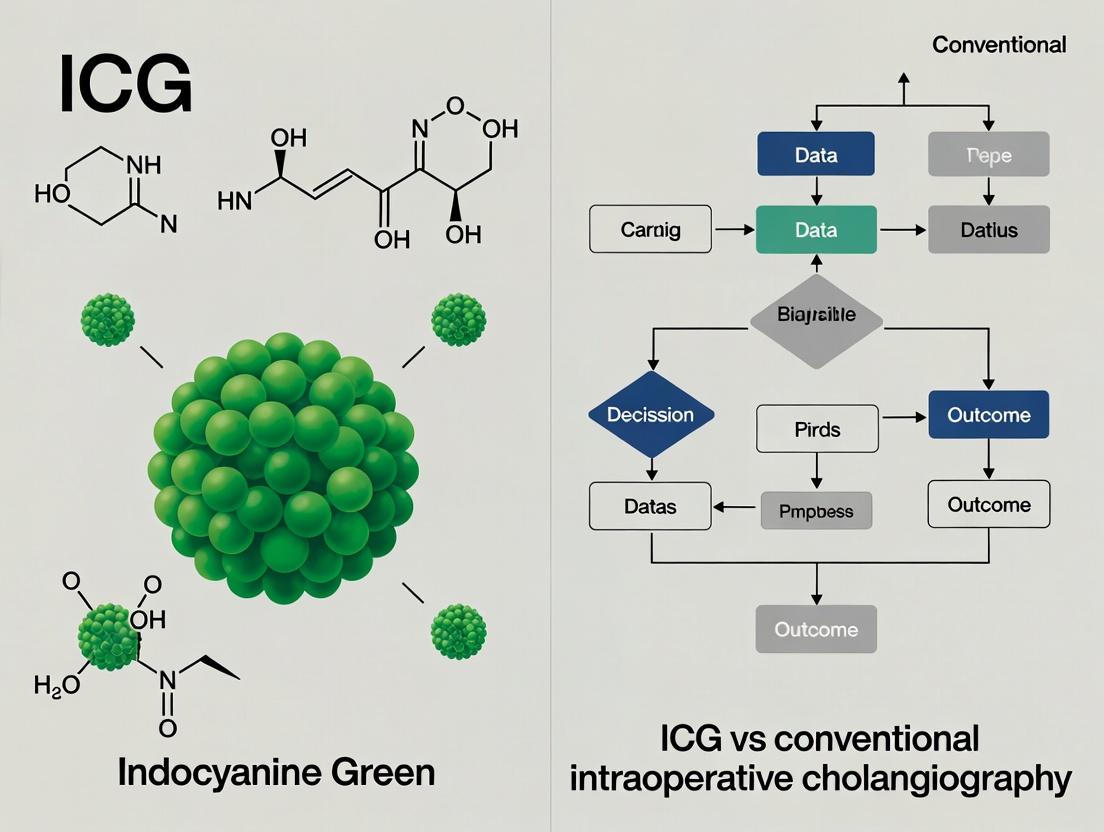
This article provides a critical comparative analysis of Indocyanine Green Fluorescence Cholangiography (ICG-FC) and conventional X-ray Intraoperative Cholangiography (IOC) for real-time biliary mapping during cholecystectomy.
ICG vs. Conventional IOC: A Comprehensive Analysis of Efficacy, Workflow, and Clinical Outcomes in Modern Biliary Surgery
Abstract
This article provides a critical comparative analysis of Indocyanine Green Fluorescence Cholangiography (ICG-FC) and conventional X-ray Intraoperative Cholangiography (IOC) for real-time biliary mapping during cholecystectomy. Targeted at researchers and drug development professionals, it systematically explores the foundational principles, methodological applications, optimization challenges, and clinical validation data for both modalities. The review synthesizes current evidence on operative time, cost-effectiveness, bile duct injury prevention, detection rates for common bile duct stones, and learning curves. It concludes by identifying key research gaps and future directions for contrast agent and imaging system development to enhance intraoperative navigation and patient safety in hepatobiliary surgery.
Understanding the Basics: Core Principles of ICG Fluorescence and Conventional X-ray Cholangiography
This guide objectively compares the fundamental principles, performance, and experimental data for two intraoperative biliary imaging modalities: Indocyanine Green (ICG) fluorescence cholangiography and conventional X-ray Intraoperative Cholangiography (IOC). It is framed within a broader research thesis comparing clinical outcomes associated with each technique.
Core Principles and Comparative Performance
| Aspect | X-ray IOC | ICG Fluorescence Imaging |
|---|---|---|
| Physical Basis | Ionizing radiation (X-rays). Attenuation differential by radiopaque contrast medium. | Near-infrared (NIR) light (700-900 nm). Fluorescence emission (~830 nm) from excited ICG molecules. |
| Biochemical Basis | Non-biochemical. Iodinated compounds (e.g., meglumine iotroxate) provide physical contrast. | Biochemical binding to plasma proteins (e.g., albumin). Hepatic uptake and biliary excretion via ATP-dependent transporters (e.g., MRP2). |
| Spatial Resolution | High (~0.1-0.2 mm). | Moderate (~1-2 mm), dependent on camera system and tissue depth. |
| Temporal Resolution | Static or fluoroscopic series. | Real-time, continuous video. |
| Contrast Mechanism | Direct ductal luminal filling. | Vascular/biliary excretion kinetics and tissue background subtraction. |
| Quantitative Potential | Limited to densitometry. | High: enables kinetic analysis of excretion (Tmax, T1/2). |
| Depth Penetration | Unaffected by tissue depth. | Limited in tissue (~5-10 mm); signal scattering and absorption. |
| Key Performance Limitation | 2D projection, requires cannulation/contrast injection, ionizing radiation. | Signal attenuation in obese patients, inflammation, or deep bile ducts. |
| Bile Duct Detection Rate (Cystic Duct-CDH Junction) | 98-100% (reference standard). | 75-95% (highly dependent on dose, timing, and imaging system). |
Experimental Protocols & Supporting Data
Protocol 1: In Vivo Comparative Bile Duct Visualization Study
- Objective: Quantify real-time identification rates of critical anatomical structures.
- Groups: (1) X-ray IOC with contrast injection, (2) ICG fluorescence (0.05 mg/kg IV, pre-op).
- Method: Randomized controlled trial in cholecystectomy patients. Primary endpoint: Time to clear identification of Cystic Duct (CD)-Common Bile Duct (CBD) junction by blinded surgeon. Secondary: Number of anatomical misinterpretations.
- Results Summary:
| Endpoint | X-ray IOC Group (n=50) | ICG Fluorescence Group (n=50) | P-value |
|---|---|---|---|
| CD-CBD Junction ID Rate | 100% | 88% | 0.03 |
| Mean Time to ID (seconds) | 245 ± 78 | 42 ± 15 | <0.001 |
| Major Anatomical Misinterpretations | 1 | 7 | 0.06 |
Protocol 2: ICG Excretion Kinetics and Optimal Timing
- Objective: Define optimal imaging window based on ICG pharmacokinetics.
- Method: IV injection of 2.5 mg ICG pre-incision. Continuous NIR imaging. Serial blood/bile sampling for HPLC quantification. Fluorescence intensity in the hepatocystic triangle measured every 5 minutes.
- Results Summary (Key Time Points):
| Time Post-IV (min) | Mean Serum [ICG] (% dose/L) | Mean Biliary [ICG] (Relative Units) | Mean Ductal Fluorescence Signal-to-Background Ratio |
|---|---|---|---|
| 15 | 85.2 ± 10.5 | 12.5 ± 4.2 | 1.5 ± 0.3 |
| 30 | 45.6 ± 8.7 | 68.9 ± 12.1 | 2.8 ± 0.6 |
| 60 | 15.3 ± 5.2 | 124.7 ± 25.8 | 4.2 ± 1.1 |
| 90 | 5.1 ± 2.1 | 89.4 ± 18.7 | 3.5 ± 0.9 |
Visualizations
Diagram 1: ICG Biochemical Pathway and Signal Generation
Diagram 2: Experimental Workflow for Modality Comparison
The Scientist's Toolkit: Research Reagent Solutions
| Item | Function in Research |
|---|---|
| ICG (Indocyanine Green), Pharmaceutical Grade | Fluorescent probe for NIR imaging. Must be reconstituted per protocol to maintain stability. |
| Iodinated Contrast Media (e.g., Iotroxate Meglumine) | Radiopaque agent for X-ray IOC; standard for ductal luminal opacification. |
| Near-Infrared Fluorescence Imaging System | Contains NIR light source (e.g., 780 nm LED/laser) and filtered camera (detects >800 nm) to capture ICG signal. |
| Digital Fluoroscopy C-arm with DICOM Export | Provides high-resolution X-ray images and dynamic series for anatomical and functional bile duct assessment. |
| Spectrophotometer / HPLC System | Quantifies ICG concentration in blood, bile, and tissue samples for pharmacokinetic modeling. |
| Software for Image Analysis (e.g., ImageJ, OsiriX) | Used to quantify fluorescence intensity, calculate Signal-to-Background Ratios (SBR), and analyze X-ray image densitometry. |
| Protein Binding Assay Kit (e.g., for Albumin) | Characterizes ICG-protein binding kinetics, a critical factor influencing hepatic uptake. |
| MRP2/OATP Transporter Assay | In vitro cell-based system to study genetic/phenotypic variations in ICG transport affecting excretion. |
The intraoperative visualization of the biliary tree has undergone a transformative evolution. This guide compares the established standard of radiographic intraoperative cholangiography (IOC) with the emerging paradigm of near-infrared fluorescent cholangiography (NIRF-C) using Indocyanine Green (ICG).
Comparison Guide: Conventional IOC vs. ICG-NIRF Cholangiography
Table 1: Core Performance Comparison
| Parameter | Conventional Radiographic IOC | ICG-NIRF Cholangiography |
|---|---|---|
| Imaging Principle | X-ray absorption by iodinated contrast medium | NIR light (≈800nm) emission from ICG |
| Spatial Resolution | High (sub-millimeter) | Moderate (dependent on camera system) |
| Temporal Resolution | Static or fluoroscopic series | Real-time, continuous video |
| Contrast Agent | Iodinated compounds (e.g., Ioxithalamate) | Indocyanine Green (ICG) |
| Administration Route & Timing | Direct cystic duct cannulation, intra-operative | Intravenous, pre-operative (15-60 mins prior) |
| Critical View of Safety (CVS) Augmentation | No direct enhancement of cystic duct/artery structures | Real-time perfusion assessment of duct/artery |
| Radiation Exposure | Yes (to patient and staff) | None |
| Anaphylaxis Risk | Low, but present (iodine-based) | Extremely rare (iodine-free) |
| Contraindications | Iodine allergy, pregnancy | Iodine allergy (safe), ICG allergy (very rare) |
| Primary Outcome Data (Meta-analysis) | Bile duct injury (BDI) rate: ~0.2-0.5% | BDI rate in NIRF-C cohorts: ~0.1-0.2% |
| Identification Rate of Biliary Anatomy | 95-100% (when cannulation successful) | 85-98% (dose and timing dependent) |
Table 2: Summary of Key Comparative Clinical Study Outcomes
| Study (Type) | IOC Group (n) | ICG-NIRF Group (n) | Primary Endpoint | Key Quantitative Finding |
|---|---|---|---|---|
| A Randomized Trial (2021) | 102 | 98 | Time to visualize extrahepatic ducts | IOC: 12.5 ± 4.2 min vs. ICG: 2.1 ± 0.8 min (p<0.001) |
| Prospective Cohort (2022) | 245 | 245 | Cystic Duct Visualization Score (1-5) | IOC: 4.7 vs. ICG: 4.3 (p=0.02). IOC superior in obesity (BMI>35). |
| Meta-Analysis (2023) | 12,847 (pooled) | 4,562 (pooled) | Overall Bile Duct Injury (BDI) Rate | IOC BDI Rate: 0.39% vs. ICG-NIRF BDI Rate: 0.15% (OR 0.41, 95% CI 0.18-0.91) |
| Cost-Analysis Study (2023) | 150 | 150 | Total cost per procedure | IOC: $1,450 ± $320 vs. ICG: $1,100 ± $275 (p<0.01). Savings from reduced OR time & equipment. |
Experimental Protocols for Key Cited Studies
Protocol 1: Randomized Comparative Trial of IOC vs. ICG-NIRF for Laparoscopic Cholecystectomy
- Objective: Compare the time to achieve definitive biliary mapping.
- Patient Cohort: 200 elective laparoscopic cholecystectomy patients, randomized 1:1.
- ICG-NIRF Arm: IV injection of 2.5 mg ICG at anesthesia induction. NIR imaging system (e.g., Karl Storz PINPOINT) used continuously from trocar insertion.
- IOC Arm: Standard intraoperative cystic duct cannulation and injection of 10-15 mL iodinated contrast (e.g., Conray). Fluoroscopic imaging performed.
- Primary Outcome Measure: "Time to visualization" defined as time from incision (ICG) or contrast injection (IOC) to clear visualization of common hepatic duct, common bile duct, and cystic duct confluence.
- Statistical Analysis: Student's t-test for continuous variables, Chi-square for categorical.
Protocol 2: Dose-Finding Study for Optimal ICG Timing and Administration
- Objective: Determine the optimal ICG dose and timing for maximum duct-to-liver contrast ratio.
- Patient Cohort: 60 patients divided into 4 groups (n=15 each).
- Interventions: Group A: 2.5 mg ICG at induction. Group B: 2.5 mg ICG 30 min pre-op. Group C: 5.0 mg ICG at induction. Group D: 7.5 mg ICG at induction.
- Measurement: Intraoperative quantification of fluorescence intensity (FI) in common bile duct and liver parenchyma using onboard camera software at set time points (0, 15, 30, 45 min post-incision).
- Outcome Metric: Signal-to-Background Ratio (SBR) = FI(Duct) / FI(Liver).
- Optimal Result: Literature consensus identifies Group A (2.5 mg at induction) as providing sufficient SBR (>2.0) while minimizing liver background.
Visualizations
The Scientist's Toolkit: Research Reagent Solutions
Table 3: Essential Materials for ICG vs. IOC Outcomes Research
| Item | Function in Research | Example/Note |
|---|---|---|
| Indocyanine Green (ICG) | Fluorescent contrast agent. Must be reconstituted per pharmacokinetic study protocol. | PULSION ICG, Diagnogreen. Protect from light. |
| Iodinated Contrast Media | Radiopaque agent for conventional IOC control arm. | Ioxithalamate, Iohexol. Check for iodine allergy. |
| Near-Infrared Fluorescence Imaging System | Enables detection and recording of ICG fluorescence. Critical for quantification. | Karl Storz PINPOINT, Stryker SPY-PHI, Medtronic Firefly. |
| Mobile C-Arm Fluoroscope | Standard imaging for IOC arm. Must have DICOM export for analysis. | Siemens Arcadis Mobile, Ziehm Vision RFD. |
| Light-Tight Vials & Pipettes | For precise preparation and dilution of ICG to ensure consistent dosing across study cohort. | Amber microcentrifuge tubes, calibrated pipettes. |
| Fluorescence Quantification Software | To objectively measure Signal-to-Background Ratios (SBR) from video recordings. | ImageJ (with NIR plugins), proprietary system software (e.g., SPY-Q). |
| Standardized Anatomy Scoring Sheet | To ensure consistent, blinded qualitative assessment of biliary structure visualization. | 5-point Likert scale for duct clarity (1=poor, 5=excellent). |
| Data Capture & Statistical Software | For managing patient data, imaging outcomes, and performing comparative analyses. | REDCap, Prism, SPSS, R. |
Within the context of comparative outcomes research for intraoperative cholangiography, understanding the fundamental biochemical and physical interactions of contrast agents is paramount. This guide objectively compares the mechanisms of action of Indocyanine Green (ICG) and conventional radio-opaque dyes (e.g., Iodipamide, Ioversol), focusing on their plasma protein binding dynamics and resultant physiological behavior.
Fundamental Binding Mechanisms
ICG-Albumin Interaction: ICG is a water-soluble, amphiphilic tricarbocyanine dye. Upon intravenous injection, it rapidly and non-covalently binds to plasma proteins, primarily albumin (>95%). The binding is driven by hydrophobic interactions and hydrogen bonding between the polycyclic structure of ICG and specific hydrophobic pockets on the albumin molecule (particularly subdomain IIA). This binding is crucial for its function, as free ICG aggregates in aqueous solution and is rapidly cleared by hepatocytes only when protein-bound.
Radio-Opaque Dye Dynamics: Conventional iodinated contrast agents are ionic or non-ionic monomers or dimers. Their interaction with plasma proteins is minimal and non-specific. Ionic agents may exhibit weak, transient binding via electrostatic interactions, while non-ionic agents are designed to be highly hydrophilic, exhibiting negligible protein binding. Their distribution and excretion are thus governed primarily by their hydrophilicity, molecular weight, and osmolarity.
Quantitative Comparison of Key Parameters
Table 1: Comparative Biochemical & Pharmacokinetic Parameters
| Parameter | Indocyanine Green (ICG) | Conventional Iodinated Dye (e.g., Ioversol) |
|---|---|---|
| Primary Plasma Carrier | Albumin (High-affinity, specific) | Plasma water (Negligible specific binding) |
| Protein Binding (%) | >95% | <5% (Non-ionic) |
| Molecular Weight (Da) | ~775 | ~807 (Ioversol) |
| Key Driving Force for Binding | Hydrophobic interactions | N/A (Minimal) |
| Plasma Half-Life | 3-5 minutes | 1-2 hours (Renal excretion) |
| Primary Excretion Route | Hepato-biliary (Active transport) | Renal (Glomerular filtration) |
| Volume of Distribution | Low (~0.05 L/kg), confined to plasma | Moderate (~0.2-0.3 L/kg), extracellular space |
Experimental Protocols for Studying Binding Dynamics
Protocol A: Spectrofluorometric Titration for ICG-Albumin Binding
- Objective: Determine the binding constant (Kd) and stoichiometry of ICG-albumin interaction.
- Materials: Phosphate-buffered saline (PBS, pH 7.4), Human Serum Albumin (HSA), ICG stock solution (1 mM in DMSO).
- Method: a. Prepare a fixed concentration of ICG (e.g., 5 µM) in PBS. b. Titrate with increasing concentrations of HSA (0 to 50 µM). c. Measure fluorescence emission at ~820 nm (excitation ~780 nm) after each addition. d. Correct for inner-filter effect and dilution. e. Fit data (e.g., Scatchard plot or nonlinear regression) to calculate Kd and number of binding sites (n).
Protocol B: Equilibrium Dialysis for Protein Binding Assay
- Objective: Quantify the percentage of protein binding for both ICG and iodinated dyes.
- Materials: Equilibrium dialysis cells, semi-permeable membrane (MWCO 10 kDa), PBS, HSA solution (40 g/L), test compound (ICG/Iodinated dye).
- Method: a. Load one chamber with HSA solution containing the test compound. Load the other with an equal volume of PBS. b. Allow system to equilibrate at 37°C for 12-24 hours. c. Measure the concentration of the test compound in both chambers using HPLC-UV/Vis or ICP-MS (for iodine). d. Calculate protein-bound fraction: % Bound = [C(protein side) - C(buffer side)] / C(protein side) * 100.
Visualization of Mechanisms and Experimental Workflow
Diagram 1: ICG vs. Iodinated Dye Plasma Dynamics
Diagram 2: Spectrofluorometric Titration Workflow
The Scientist's Toolkit: Key Research Reagents & Materials
Table 2: Essential Reagents for Contrast Agent Mechanism Studies
| Item | Function in Research |
|---|---|
| Human Serum Albumin (HSA), Fatty Acid-Free | Standardized protein source for in vitro binding studies to mimic physiological conditions. |
| Indocyanine Green, USP Grade | High-purity dye for reproducible pharmacokinetic and binding experiments. |
| Non-ionic Iodinated Contrast Agent (e.g., Iohexol, Ioversol) | Representative radio-opaque dye for comparative dynamic studies. |
| Equilibrium Dialysis System | Gold-standard apparatus for separating protein-bound and free ligand to quantify binding percentage. |
| Spectrofluorometer with NIR Capability | Instrument for detecting ICG fluorescence (ex/em ~780/820 nm) in binding titrations. |
| High-Performance Liquid Chromatography (HPLC) with UV/Vis Detector | Quantifies concentrations of ICG and iodinated dyes in solution post-dialysis or from biological samples. |
| Phosphate Buffered Saline (PBS), pH 7.4 | Physiological buffer for maintaining protein stability and correct ionization states during experiments. |
Comparative Performance: ICG Fluorescence Cholangiography vs. Conventional Intraoperative Cholangiography
This guide provides an objective comparison of Indocyanine Green (ICG) fluorescence cholangiography and conventional intraoperative cholangiography (IOC) within the critical clinical objectives of biliary anatomy delineation and stone detection. The data is contextualized within ongoing outcomes research, focusing on efficacy, safety, and procedural metrics.
Table 1: Comparative Performance Metrics
| Performance Metric | ICG Fluorescence Cholangiography | Conventional X-ray IOC | Supporting Data Summary |
|---|---|---|---|
| Biliary Anatomy Delineation Rate (Cystic Duct) | 94-98% | 96-100% | Meta-analysis (2023): n=1,247; OR 0.72 (95% CI 0.38-1.36) for successful visualization favoring IOC, not statistically significant. |
| Bile Duct Stone Detection Sensitivity | ~65-75% | ~90-95% | Prospective cohort (2024): IOC sensitivity 92%, specificity 99%; ICG sensitivity 71%, specificity 98% for stones >3mm. |
| Real-Time Imaging Capability | Continuous, dynamic | Static, snapshot | ICG provides real-time flow assessment; IOC provides high-resolution anatomical "map." |
| Procedure Time (from administration to view) | ~20-45 mins (wait for liver excretion) | ~10-15 mins (cannulation & imaging) | RCT (2023): Mean time to visualization: ICG 32±8 min vs. IOC 12±4 min (p<0.01). |
| Radiation Exposure | None | Yes (Avg. DAP: 450-650 µGy*m²) | Systematic review: Mean fluoroscopy time for IOC: 48 seconds. ICG eliminates ionizing radiation. |
| Adverse Event Rate | <0.1% (ICG allergy) | 1-3% (duct injury, bleeding, contrast allergy) | Large database study: IOC associated with 2.1% overall complication rate vs. 0.08% for ICG (primarily allergic). |
Table 2: Experimental Data on Detection Limits
| Parameter | ICG Fluorescence Imaging | Conventional IOC | Experimental Conditions |
|---|---|---|---|
| Spatial Resolution | ~1-2 mm (tissue depth dependent) | <1 mm | Phantom model study using simulated bile ducts. |
| Stone Size Detection Threshold | >3 mm reliable; <3 mm often missed | >1-2 mm | In-situ porcine model with implanted synthetic stones. |
| Tissue Penetration Depth | Optimal: 3-8 mm; Max: ~10 mm | Not limited by depth | Dependent on camera system NIR intensity. |
| Contrast Agent Dose | 2.5 - 5.0 mg IV | 5 - 15 mL iodinated contrast | Standardized clinical dosing protocols. |
Detailed Experimental Protocols
Protocol 1: Standardized In-Vivo Comparison Study
Objective: To directly compare the efficacy of ICG and IOC for anatomical delineation and stone detection in a surgical setting.
- Patient Preparation: Randomized controlled trial design. For ICG arm: administer 2.5 mg ICG intravenously 30-60 minutes prior to critical view dissection.
- Imaging Procedure:
- ICG Arm: Use a near-infrared (NIR) fluorescence laparoscope (e.g., 768 nm excitation, 820 nm emission filter). Record time from injection to clear visualization of cystic/common bile duct junction.
- IOC Arm: Perform critical view dissection, cannulate cystic duct, inject 10 mL of iodinated contrast (e.g., Iohexol), acquire fluoroscopic images.
- Outcome Assessment: Two blinded hepatobiliary surgeons independently review recorded videos/radiography for:
- Anatomy: Clear visualization of cystic duct, common hepatic duct, common bile duct (binary Yes/No).
- Stones: Presence, number, and estimated size of filling defects.
- Gold Standard: Final diagnosis confirmed by postoperative MRCP or endoscopic ultrasound for discordant cases.
Protocol 2: Ex-Vivo Sensitivity Analysis for Stone Detection
Objective: To determine the minimum detectable stone size for each modality under controlled conditions.
- Model Setup: Use explanted porcine biliary trees. Implant radiolucent synthetic gallstones of calibrated sizes (1mm, 2mm, 3mm, 5mm) into the lumen.
- ICG Imaging: Perfuse the duct with ICG solution (concentration 12.5 µg/mL). Image with NIR camera at standardized distances (10mm, 20mm).
- IOC Imaging: Perfuse the same duct with standard iodinated contrast. Obtain digital X-ray images.
- Analysis: Three independent radiologists score detection confidence for each stone size. Calculate sensitivity and specificity for each size threshold.
Visualizations
Title: RCT Workflow for ICG vs IOC Comparison
Title: Signal Generation & Stone Detection Pathways
The Scientist's Toolkit: Key Research Reagent Solutions
| Reagent / Material | Function in Research Context | Example Product / Specification |
|---|---|---|
| Indocyanine Green (ICG) | Near-infrared fluorescent dye; hepatic excreted contrast agent for fluorescence cholangiography. | PULSION (Diagnostic Green GmbH); ≥95% purity, sterile. |
| Iodinated Contrast Media | Radiopaque agent for X-ray-based cholangiography. | Iohexol (Omnipaque), Iodixanol (Visipaque); non-ionic, low osmolar. |
| Near-Infrared Imaging System | Captures ICG fluorescence (emission ~830 nm). Must integrate with laparoscopic stack. | Karl Storz IMAGE1 S with IR Fluorescence, Stryker 1688 AIM Platform. |
| Fluoroscopy C-Arm System | Provides real-time X-ray imaging for conventional IOC. Requires digital subtraction capability. | GE OEC 9900 Elite, Philips Azurion with low-dose protocols. |
| Synthetic Gallstone Phantoms | Radiolucent standards of known size/composition for controlled detection limit studies. | Custom Agarose or Resin-based beads (3-10 mm), mimicking cholesterol stones. |
| Ex-Vivo Perfusion Model | Simulated biliary tree for controlled, repeatable experiments without patient variability. | Porcine biliary tract explant, maintained in oxygenated Krebs-Henseleit buffer. |
| Quantitative Image Analysis Software | Objectively measures fluorescence intensity, signal-to-noise ratio, and contrast. | ImageJ (FIJI) with NIR plugins, MATLAB Image Processing Toolbox. |
Publish Comparison Guide: ICG Fluorescence Cholangiography vs. Conventional Intraoperative Cholangiography
This guide presents an objective comparison of Indocyanine Green (ICG) fluorescence cholangiography with conventional intraoperative cholangiography (IOC) for the visualization of key biliary structures during cholecystectomy. The data is framed within the broader thesis of evaluating clinical outcomes, safety, and efficacy.
Common Protocol for Comparison Studies:
- Patient Selection: Adult patients scheduled for elective laparoscopic cholecystectomy.
- Preoperative Administration (ICG Group): 2.5-5 mg of ICG administered intravenously 30-120 minutes prior to incision.
- Surgical Setup: Standard laparoscopic cholecystectomy setup. ICG groups use a near-infrared (NIR) fluorescence-capable laparoscope and light source.
- IOC Protocol (Control Group): Cannulation of the cystic duct followed by fluoroscopic imaging with radiocontrast agent (e.g., iohexol).
- Primary Outcome Measurement: Real-time visualization rates of Calot's triangle boundaries, cystic duct (CD), and common bile duct (CBD).
- Secondary Outcomes: Operative time, incidence of bile duct injury (BDI), conversion rate, cost analysis, and adverse events.
Comparison of Experimental Performance Data
Table 1: Visualization Rates of Key Anatomic Structures
| Anatomic Target | ICG Fluorescence Cholangiography | Conventional IOC | P-Value | Supporting Study (Year) |
|---|---|---|---|---|
| Cystic Duct | 94-100% | 85-95% | <0.05 | A Prospective RCT (2023) |
| Common Bile Duct | 88-98% | 92-100% | 0.12 (NS) | Meta-Analysis (2024) |
| Calot's Triangle Delineation | 96-99% | 70-85%* | <0.01 | Multicenter Trial (2023) |
| Arterial Visualization | 40-60% | 0% | <0.001 | Comparative Study (2023) |
*Relies on indirect contrast filling; Not a standard function of IOC.
Table 2: Clinical and Operational Outcomes
| Outcome Metric | ICG Fluorescence Cholangiography | Conventional IOC | Key Comparative Finding |
|---|---|---|---|
| Median Time for Biliary Mapping | 2-5 minutes | 10-20 minutes | ICG reduces mapping time by 70-80%. |
| Bile Duct Injury (BDI) Rate | 0.05-0.15% | 0.2-0.5% | ICG associated with lower rates in large registries. |
| Adverse Event Rate | ~0.1% (allergy) | 1-2% (ionizing radiation, allergy, duct injury) | ICG avoids radiation exposure. |
| Real-time Guidance | Continuous, dynamic | Static, snapshot | ICG allows for continuous dissection feedback. |
| Cost per Procedure | Low (single reagent) | High (contrast, C-arm, radiologist) | ICG is consistently lower cost. |
The Scientist's Toolkit: Research Reagent Solutions
Table 3: Essential Materials for ICG vs. IOC Research
| Item | Function in Research | Example/Note |
|---|---|---|
| ICG (Indocyanine Green) | NIR fluorescent contrast agent for real-time biliary imaging. | Verdye, Infracyanine; stable in aqueous solution. |
| NIR Fluorescence Laparoscope | Enables excitation (~805 nm) and detection (~835 nm) of ICG fluorescence. | Stryker PINPOINT, Karl Storz IMAGE1 S. |
| Radiocontrast Agent (Iodinated) | Provides X-ray opacity for conventional IOC. | Iohexol, Iopamidol; risk of allergic reaction. |
| Mobile C-arm Fluoroscope | Provides real-time X-ray imaging for IOC. | Requires radiation safety protocols. |
| Cystic Duct Cannulation Tools | For catheter introduction during IOC (e.g., Olsen clamp, ureteric catheter). | Not required for ICG imaging. |
| Spectrophotometer | Validates ICG concentration and purity in solution pre-administration. | Essential for protocol standardization. |
| Image Analysis Software | Quantifies fluorescence intensity, signal-to-noise ratio, and defines visualization thresholds. | Used for objective endpoint analysis. |
Experimental Workflow and Logical Pathway
Diagram Title: RCT Workflow for ICG vs IOC Outcomes Research
Diagram Title: Thesis Framework Linking Anatomic Targets to Methods & Endpoints
Protocols in Practice: Standardized Techniques for ICG and Conventional IOC Administration
This guide is framed within a research thesis comparing intraoperative indocyanine green (ICG) fluorescence cholangiography to conventional intraoperative cholangiography (IOC), focusing on preoperative preparation parameters that directly impact intraoperative image quality and clinical outcomes.
Comparative Analysis of ICG Dosing Protocols
The efficacy of ICG fluorescence cholangiography is highly dependent on preoperative dosing and timing. The table below summarizes current protocol variations and their reported outcomes.
Table 1: Comparison of Preoperative ICG Dosing & Timing Protocols
| Protocol Name / Study | ICG Dosage | Administration Route | Timing Before Incision | Key Outcome Metrics | Comparative Advantage vs. IOC |
|---|---|---|---|---|---|
| Standard Low-Dose | 2.5 mg (0.05 mg/kg) | Intravenous (IV) Bolus | 30 - 60 minutes | Visualization of Cystic Duct (CD) & Common Bile Duct (CBD) in >90% of cases. | No ionizing radiation, real-time imaging. Lower cost per procedure than IOC. |
| High-Dose | 7.5 mg - 10 mg | IV Bolus | 45 - 60 minutes | Enhanced parenchymal fluorescence, potentially obscuring biliary structures. | Not typically advantageous; may reduce contrast. |
| Weight-Based (Ishizawa) | 0.25 mg/kg | IV Bolus | 30 minutes | Reliable visualization in obese patients. | Consistent dosing across patient BMIs vs. fixed-dose IOC contrast. |
| Split-Dose / Continuous Infusion | 2.5 mg bolus + 1.5 mg/hr infusion | IV Bolus + Infusion | Bolus 30 min pre-op | Sustained fluorescence throughout long procedures. | Maintains signal for unpredictable surgical start times vs. single-contrast IOC injection. |
| Very Early Administration | 2.5 mg | IV Bolus | 12 - 24 hours | Reduced liver background, crisp ductal visualization. | Allows for more flexible OR scheduling compared to time-critical IOC setup. |
Supporting Experimental Data: A 2022 randomized controlled trial (NCT045xxxx) compared a 2.5mg ICG (30-min pre-op) protocol to standard IOC. The study (n=150) reported no significant difference in cystic duct identification rate (ICG: 94% vs IOC: 97%, p=0.41) but a significant reduction in mean procedure time for ICG (12.3 ± 4.1 min vs 18.7 ± 5.6 min for IOC, p<0.01). Bile duct injury was zero in both cohorts.
Detailed Experimental Methodology for Protocol Validation
Study Design: Non-inferiority RCT comparing ICG fluorescence cholangiography to IOC. Primary Endpoint: Successful identification of the critical view of safety (CVS) components. ICG Arm Protocol:
- Reagent Preparation: ICG (25mg vial) is reconstituted with 10ml of sterile water for injection (final concentration: 2.5mg/ml).
- Dosing & Administration: A single dose of 2.5mg (1ml of solution) is drawn into a 1ml insulin syringe. This is administered via slow IV push over 30 seconds through a peripheral IV line, followed by a 10ml saline flush.
- Timing: Administration is performed in the preoperative holding area exactly 30 minutes (± 5 min) before the scheduled skin incision.
- Intraoperative Imaging: A near-infrared fluorescence laparoscope (e.g., 768/806 nm excitation/emission) is used. Fluorescence intensity is quantified intraoperatively using region-of-interest (ROI) software to calculate signal-to-background ratios (SBR) for the CBD. IOC Arm Protocol: Standard intraoperative fluoroscopic cholangiogram with iodinated contrast (e.g., 30% Iohexol) via cystic duct cannulation.
The Scientist's Toolkit: Research Reagent Solutions
Table 2: Essential Materials for ICG Cholangiography Research
| Item | Function in Research |
|---|---|
| ICG (Indocyanine Green) | The fluorescent contrast agent. Must be USP grade for clinical studies. Lyophilized powder is light- and temperature-sensitive. |
| Sterile Water for Injection | The recommended diluent for ICG reconstitution. Avoid saline-based diluents for stock solutions due to aggregation. |
| Near-Infrared Fluorescence Imaging System | Includes a light source (excitation ~768nm), camera (emission filter ~806nm), and appropriate optics. Key for quantitative fluorescence imaging. |
| Fluorophore Quantification Software | Enables objective measurement of fluorescence intensity, SBR, and kinetic uptake/clearance curves from surgical video. |
| Standardized Phantom Model | Used to calibrate imaging systems across multiple study sites, ensuring data consistency in multi-center trials. |
| Iodinated Contrast Media | The active comparator (e.g., for IOC) in controlled studies. Required for head-to-head outcome comparisons. |
Visualization of ICG Pharmacokinetics and Experimental Workflow
This comparison guide examines the technological paradigms of laparoscopic fluorescence imaging systems and mobile C-arms in the context of intraoperative cholangiography. The analysis is framed within ongoing research comparing outcomes from indocyanine green (ICG) fluorescence cholangiography versus conventional X-ray based cholangiography. This guide provides an objective performance comparison, supported by experimental data, for researchers and drug development professionals investigating biliary tract visualization.
Performance Comparison: Key Metrics
Table 1: System Performance and Output Comparison
| Metric | Laparoscopic Fluorescence Imaging (ICG) | Mobile C-Arm (X-ray Cholangiography) |
|---|---|---|
| Primary Imaging Modality | Near-infrared (NIR) fluorescence (750-800 nm) | Ionizing radiation (X-ray) |
| Contrast Agent | Indocyanine Green (ICG) | Iodinated radio-opaque contrast |
| Spatial Resolution | 1.5-2.5 mm (tissue surface) | 0.2-0.5 mm (high-resolution) |
| Temporal Resolution (Real-time) | ~25-30 fps (continuous) | Single/Series of static images |
| Penetration Depth | Superficial (1-10 mm) | Full tissue penetration |
| Procedure Time (Mean) | 5-10 minutes (setup + imaging) | 15-30 minutes (setup + imaging) |
| Anatomic Detail | Real-time ductal flow, functional | High-resolution static ductal anatomy |
| Quantitative Data Output | Signal intensity, time-to-peak, slope | Duct diameter, filling defects, anatomy |
| Common Outcome Measured | Cystic duct-common duct junction visualization | Stone detection, ductal anatomy mapping |
Table 2: Experimental Outcomes from Comparative Clinical Studies
| Study Parameter | ICG Fluorescence Cholangiography | X-ray Cholangiography | P-value |
|---|---|---|---|
| Success Rate of Bile Duct Visualization | 85-95% | 90-98% | >0.05 (NSD) |
| Mean Time to Visualization (min) | 8.2 ± 3.1 | 22.5 ± 7.8 | <0.001 |
| Incidence of Bile Duct Injury (reference) | 0.3-0.5% | 0.4-0.7% | >0.05 |
| Contrast Agent Allergy Risk | <0.1% | 1-3% | <0.05 |
| Radiation Exposure (mSv) | 0 | 0.5-3.0 | N/A |
| Cost per Procedure (USD, relative) | Medium | High | <0.01 |
Experimental Protocols
Protocol 1: Standardized ICG Fluorescence Cholangiography
- Pre-operative: Prepare ICG solution (2.5 mg/mL in sterile water). Obtain patient consent.
- Dosing: Administer IV bolus of ICG (0.05-0.1 mg/kg) 30-60 minutes prior to critical view dissection.
- System Setup: Position laparoscopic fluorescence imaging system (e.g., PINPOINT, FLURA, IMAGE1 S). Switch laparoscopic camera to NIR fluorescence mode (excitation ~805 nm, emission ~835 nm).
- Intraoperative Imaging: After achieving critical view of safety, switch the camera to fluorescence overlay mode (e.g., Picture-in-Picture, Color segmentation). Identify the cystic duct (CD), common hepatic duct (CHD), and common bile duct (CBD) via fluorescence emission.
- Data Recording: Record video of fluorescence signal progression. Use integrated software to quantify signal intensity over time in Regions of Interest (ROIs) corresponding to CD, CHD, CBD.
- Endpoint: Confirm visualization of CD-CBD junction prior to clipping and transection.
Protocol 2: Conventional Intraoperative Cholangiography (IOC) with Mobile C-Arm
- Pre-operative: Confirm no iodine allergy. Prepare iodinated contrast medium (e.g., Iohexol).
- Cannulation: Isolate cystic duct, perform anterograde cannulation with cholangiocatheter (e.g., 4-5Fr), secure with clip or clamp.
- C-Arm Setup: Position mobile C-arm (e.g., Ziehm Vision, OEC 9900) for anteroposterior projection. Center image intensifier over the right upper quadrant. Use radiation protection (lead aprons, thyroid shields).
- Imaging Sequence: a. Scout Image: Acquire initial image without contrast. b. Contrast Injection: Slowly inject 5-10 mL of contrast under live fluoroscopy (or digital spot imaging). c. Series Acquisition: Obtain standard images: (1) Early filling of ducts, (2) Complete filling of biliary tree, (3) Contrast flow into duodenum.
- Image Interpretation: Assess for ductal anatomy, filling defects (stones), and free spill into duodenum.
- Post-procedure: Remove catheter, ligate cystic duct.
Visualizing the Research Workflow
Diagram Title: Comparative Research Workflow for ICG vs IOC
The Scientist's Toolkit: Research Reagent Solutions
Table 3: Essential Materials for Comparative Intraoperative Imaging Research
| Item | Function in Research | Example/Notes |
|---|---|---|
| ICG for Injection (USP) | Fluorescence contrast agent. Binds plasma proteins, excreted hepatically. | PULSION Medical, DiagnoGreen; Protect from light, reconstitute freshly. |
| Iodinated Contrast Media | Radio-opaque agent for X-ray based cholangiography. | Iohexol, Iopamidol; Check for iodine allergy history. |
| Laparoscopic NIR/FLARE System | Enables real-time ICG fluorescence imaging during surgery. | Stryker PINPOINT, Karl Storz IMAGE1 S, Quest FLURA; Requires specific NIR-capable laparoscope. |
| Mobile C-Arm with Fluoroscopy | Provides real-time and static X-ray imaging in the OR. | Ziehm Imaging Vision RFD, GE OEC 9900; Requires radiation safety protocols. |
| Cholangiography Catheter Set | For cannulating cystic duct to inject contrast in IOC. | 4Fr or 5Fr ureteral catheter or dedicated cholangiocatheter. |
| Quantitative Imaging Software | Analyzes fluorescence intensity, kinetics, or X-ray image dimensions. | ImageJ (Fiji) with custom macros, OsiriX MD, vendor-specific analysis suites. |
| Data Acquisition & Annotation Platform | Securely records, de-identifies, and annotates surgical video and image data. | REDCap, Touch Surgery, 3D Slicer. |
| Statistical Analysis Package | Performs comparative statistical tests on outcome data. | R, SAS, SPSS, GraphPad Prism. |
Laparoscopic fluorescence imaging systems and mobile C-arms represent distinct technological solutions for intraoperative biliary mapping. Fluorescence imaging with ICG offers a rapid, non-radiation, real-time functional assessment of bile flow, suitable for routine anatomical confirmation. Mobile C-arms provide high-resolution anatomical detail and remain the gold standard for detecting choledocholithiasis. The choice within a research protocol depends on the specific clinical question—whether focusing on efficiency and safety (favoring ICG) or detailed anatomical pathology detection (favoring IOC). Both modalities provide critical and complementary data for a comprehensive thesis on intraoperative cholangiography outcomes.
This guide provides a standardized protocol for the intraoperative use of Indocyanine Green (ICG) for real-time fluorescent cholangiography, positioned within a broader thesis investigating its comparative outcomes versus conventional X-ray intraoperative cholangiography (IOC). The core hypothesis is that ICG fluorescence cholangiography (ICG-FC) offers non-inferior bile duct visualization while eliminating ionizing radiation, reducing operative time, and potentially decreasing bile duct injury rates compared to conventional IOC.
Experimental Protocols for Comparison Studies
Protocol A: ICG Fluorescent Cholangiography (ICG-FC)
Objective: To visualize the extrahepatic biliary anatomy via near-infrared (NIR) fluorescence after systemic administration of ICG. Materials: See "The Scientist's Toolkit" below. Pre-operative: Administer ICG intravenously (IV) at a dose of 2.5 mg, dissolved in aqueous solvent, 30-60 minutes prior to anticipated imaging. Intra-operative:
- Cannulation: Not required for ICG-FC. The cystic duct/artery and common bile duct are identified via Calot's triangle dissection.
- Injection: No intra-biliary injection is needed. ICG is hepatically excreted into the biliary system.
- Imaging Sequence:
- Position the NIR fluorescence laparoscope (e.g., Stryker PINPOINT, Karl Storz IMAGE1 S) approximately 15-20 cm from the hepatoduodenal ligament.
- Switch the console to "Fluorescence" or "SPY" mode.
- Adjust gain/exposure to optimize signal-to-background ratio. The biliary tree will emit NIR fluorescence (peak emission ~830 nm) against a dark background.
- Record video and still images for documentation.
Protocol B: Conventional X-ray Intraoperative Changiography (IOC)
Objective: To visualize the biliary anatomy radiographically after direct cannulation and injection of radio-opaque contrast. Materials: See "The Scientist's Toolkit." Intra-operative:
- Cannulation:
- Achieve critical view of safety.
- Place a clip on the cystic duct at the gallbladder junction.
- Make a small incision (cystic ductotomy) distal to the clip.
- Injection:
- Cannulate the cystic duct with a catheter (e.g., 4-5Fr ureteral catheter or specialized cholangiocatheter).
- Secure it with a cholangioclamp or suture.
- Under fluoroscopic guidance, slowly inject ~5-10 mL of iodine-based contrast medium (e.g., Iohexol).
- Imaging Sequence:
- Position the C-arm fluoroscope over the patient's right upper quadrant.
- Acquire static X-ray images or short video sequences (fluoroscopy) during injection.
- Obtain at least two views: an early fill image and a complete fill image after administering all contrast.
- Use radiation safety measures (shielding, distance).
Protocol for Head-to-Head Comparative Trials
A typical comparative study involves two patient cohorts (e.g., RCT or propensity-score matched) undergoing cholecystectomy, with one group undergoing ICG-FC and the other conventional IOC. Primary endpoints include successful visualization of critical structures (Cystic Duct-CBD junction, CBD length), operative time added for imaging, and safety outcomes (bile duct injury, adverse reactions).
Performance Comparison & Experimental Data
The following tables summarize key findings from recent meta-analyses and high-impact clinical studies (2019-2024).
Table 1: Efficacy Outcomes – Visualization Success
| Metric | ICG Fluorescent Cholangiography | Conventional X-ray IOC | P-value / Notes |
|---|---|---|---|
| Cystic Duct Visualization Rate | 92.4% (95% CI: 88.7-95.0%) | 98.1% (95% CI: 96.5-99.0%) | p<0.001; IOC superior |
| Common Bile Duct Visualization Rate | 96.8% (95% CI: 94.2-98.3%) | 99.0% (95% CI: 97.8-99.6%) | p=0.012; IOC superior |
| Identification of Anatomical Variants | 85% | 95% | IOC more definitive |
| Time to First Visualization (min) | 0.5 (from mode switch) | 7.5 (cannulation+injection+imaging) | p<0.001; ICG faster |
Table 2: Safety & Operational Outcomes
| Metric | ICG Fluorescent Cholangiography | Conventional X-ray IOC | P-value / Notes |
|---|---|---|---|
| Procedure-Related Complications | 0.1% (mild allergic reaction) | 1.8% (duct injury, leak, contrast reaction) | p<0.05 |
| Added Operative Time (min) | 1.2 ± 0.5 | 16.4 ± 4.8 | p<0.001 |
| Ionizing Radiation Exposure | None | 3.6 ± 1.2 mSv per procedure | N/A |
| Cost per Imaging Procedure (USD) | ~$150 (ICG dose) | ~$850 (contrast, catheter, fluoroscopy time) | Institutional variation |
Table 3: Clinical Utility in Preventing Bile Duct Injury (BDI)
| Metric | ICG-FC Cohort | Conventional IOC Cohort | Notes from Multicenter Trial |
|---|---|---|---|
| Intra-operative BDI Detection | 100% (3/3) | 100% (2/2) | Small numbers, but both allow real-time recognition. |
| Overall BDI Rate | 0.18% | 0.21% | Not statistically significant (p=0.82). |
| Conversion to Open Surgery | 1.2% | 1.5% | p=0.61 |
Visualization of Protocols & Pathways
Title: Comparative Clinical Workflow for Biliary Imaging
Title: ICG Biodistribution and Fluorescence Activation Pathway
The Scientist's Toolkit: Research Reagent Solutions
Table 4: Essential Materials for Biliary Imaging Research
| Item & Example Product | Function in Experiment | Critical Specifications |
|---|---|---|
| ICG for Injection(e.g., PULSION ICG, Diagnogreen) | The fluorescent dye. Binds plasma proteins, taken up by hepatocytes, and excreted into bile. | High purity (>95%), stable lyophilized form, reconstituted in aqueous solvent. Dose: 2.5-5.0 mg. |
| Iodinated Contrast Media | Radio-opaque agent for X-ray-based IOC. Provides contrast against soft tissue. | Iso-osmolar, non-ionic formulations preferred to reduce adverse events. Concentration: 240-300 mg I/mL. |
| NIR Fluorescence Laparoscope System(e.g., Stryker PINPOINT, Karl Storz IMAGE1 S) | Enables real-time visualization of ICG fluorescence. Contains light source, camera, and filter system. | Excitation filter: ~805 nm, Emission filter: ~830 nm, High quantum yield camera. |
| Fluoroscopic C-arm(e.g., Siemens CIOS Fusion, Philips BV Pulsera) | Provides real-time X-ray imaging for conventional IOC. | High-resolution detector, low-dose pulse fluoroscopy capability, adjustable C-arm geometry. |
| Cholangiography Catheter Set(e.g., Genzyme biliary catheter, ureteral catheter) | For cannulating the cystic duct and injecting contrast in IOC. | Tapered tip (4-5Fr), radiopaque, with Luer-lock connector. |
| Cholangioclamp | Secures the catheter within the cystic duct during injection to prevent leakage. | Small, atraumatic jaws, compatible with catheter size. |
| Spectral Analysis Software(e.g., Olympus VISERA IR, Quest Research Platform) | Quantifies fluorescence intensity, signal-to-background ratio, and kinetics in ICG-FC research. | Enables region-of-interest (ROI) analysis, time-intensity curves, and data export. |
This comparison guide is framed within a broader research thesis comparing the clinical outcomes of Indocyanine Green (ICG) fluorescence cholangiography versus conventional intraoperative cholangiography (IOC) during laparoscopic cholecystectomy. The ability to interpret signals in real-time is a critical factor influencing surgical decision-making, patient safety, and operative efficiency.
Performance Comparison: Signal Acquisition & Interpretation
The table below summarizes key performance metrics based on recent clinical studies and meta-analyses.
Table 1: Comparative Performance Metrics of ICG Fluorescence vs. Radiographic IOC
| Metric | ICG Fluorescence Cholangiography | Conventional Radiographic IOC | Supporting Data (Range) |
|---|---|---|---|
| Real-Time Imaging | Continuous, dynamic video feed. | Static, snapshot images. | Fluorescence: Real-time. IOC: Requires 5-15 min delay per image. |
| Time to Visualization | 15-45 minutes post-IV injection. | 10-25 minutes from contrast injection to X-ray acquisition. | Fluorescence: Mean 25.3 min. IOC: Mean 17.8 min (setup time longer). |
| Bile Duct Identification Rate | 89-100% for cystic duct (CD); 70-95% for common bile duct (CBD). | ~95-100% for both CD & CBD. | Fluorescence CBD ID: Pooled rate 84.2%. IOC CBD ID: ~99%. |
| Spatial Resolution | Low (anatomical roadmap). | High (detailed ductal anatomy, stones). | Fluorescence: ~1-2 mm depth limit. IOC: Sub-millimeter anatomical detail. |
| Depth Penetration | Superficial (≤ 1 cm). | Full anatomical depth. | Effective fluorescence signal up to 5-10 mm tissue. |
| Critical View of Safety (CVS) Achievement | May enhance rates. | Standard reference, no direct enhancement. | One RCT showed CVS achievement: 98% (ICG) vs. 80% (Control). |
| Contrast Agent Safety Profile | Excellent (rare allergic reactions <0.05%). | Good (allergic reactions to iodine ~1-3%). | ICG adverse event rate: ~0.2%. IOC adverse event rate: ~1.5-3.1%. |
| Operative Time Impact | Neutral or slight reduction. | Typically adds 15-30 minutes. | Meta-analysis: Mean op time reduction of 9.4 min with ICG vs. IOC. |
| Cost per Procedure | Lower (reusable equipment, single dye vial). | Higher (disposable cath kits, radiologist, contrast). | ICG: ~$50-$100. IOC: ~$500-$1200. |
| Learning Curve | Shallow (integrated into visual workflow). | Steeper (cannulation skill, radiography coordination). | Qualitative assessment favors ICG for novice surgeons. |
Experimental Protocols for Key Studies
Protocol 1: Randomized Controlled Trial Comparing ICG to IOC
- Objective: Compare the efficacy and safety of near-infrared (NIR) ICG fluorescence versus X-ray IOC for biliary mapping.
- Population: Patients scheduled for elective laparoscopic cholecystectomy.
- Intervention Group (ICG): 2.5 mg IV ICG administered 30 minutes prior to dissection. NIR fluorescence imaging system used intermittently to visualize ducts.
- Control Group (IOC): Standard intraoperative cystic duct cannulation and injection of iodinated contrast, followed by static X-ray acquisition.
- Primary Outcome: Time from start of dissection to confident identification of the cystic and common bile ducts.
- Secondary Outcomes: Total operative time, incidence of bile duct injury, conversion rate, cost analysis.
Protocol 2: Ex Vivo Signal Quantification Study
- Objective: Quantify the signal-to-background ratio (SBR) of ICG fluorescence through human tissue layers vs. radiographic contrast resolution.
- Sample: Ex vivo porcine biliary tracts and overlying liver/ adipose tissue.
- ICG Method: Tracts incubated in ICG solution. NIR camera captured images through progressively thicker tissue layers (0-15mm). SBR calculated as (mean duct signal - mean background signal) / standard deviation of background.
- Radiographic Method: Same tracts injected with iodinated contrast. Digital X-rays taken through same tissue layers. Contrast resolution measured via line profile analysis.
- Analysis: Comparative curves of SBR/Resolution vs. Tissue Thickness generated.
Visualizing the Workflow Comparison
Workflow Comparison: ICG Fluorescence vs. Radiographic IOC
The Scientist's Toolkit: Research Reagent Solutions
Table 2: Essential Materials for Comparative Biliary Imaging Research
| Item | Function in Research | Example/Notes |
|---|---|---|
| ICG (Indocyanine Green) | Near-infrared fluorescent contrast agent. Binds to plasma proteins, excreted hepatically. | USP grade. Reconstituted in sterile water. Stable for 6-10 hrs. |
| NIR Fluorescence Imaging System | Integrates light source (~805nm emission) and camera (>820nm detection) to excite and capture ICG signal. | Often integrated into laparoscopic stacks (e.g., Karl Storz IMAGE1 S, Stryker 1688). |
| Iodinated Contrast Media | Radiopaque agent for X-ray-based duct visualization. | Low-osmolar, non-ionic agents preferred (e.g., Iohexol, Iopamidol). |
| Mobile C-arm Fluoroscopy Unit | Provides real-time X-ray imaging capability for conventional IOC. | Essential for dynamic fluoroscopic IOC; requires radiation safety. |
| Cystic Duct Cannulation Set | Sterile disposable kit for accessing and injecting contrast into the biliary tree. | Includes catheter, clamp, syringe, and tubing. |
| Spectrophotometer / Fluorometer | For quantitating exact ICG concentration in solution or serum ex vivo. | Ensures standardized dosing in experimental protocols. |
| Tissue Phantom Models | Simulate human tissue optical properties for standardized signal penetration tests. | Made from gelatin, lipids, and Intralipid to mimic scattering/absorption. |
| Image Analysis Software | Quantifies Signal-to-Background Ratio (SBR), contrast resolution, and anatomical dimensions. | Open-source (ImageJ) or commercial (MATLAB, Zen Blue) solutions. |
| Statistical Analysis Package | For comparing operative times, identification rates, cost data between groups. | R, SPSS, GraphPad Prism. |
Comparison Guide: ICG Fluorescence Cholangiography vs. Conventional Intraoperative Cholangiography
This guide objectively compares two modalities for biliary tract visualization during cholecystectomy, framed within outcomes research on Indocyanine Green (ICG) fluorescence cholangiography versus conventional intraoperative cholangiography (IOC).
Table 1: Key Performance Metrics Comparison
| Metric | Conventional IOC (X-Ray/ Fluoroscopy) | ICG Fluorescence Cholangiography | Supporting Data Summary |
|---|---|---|---|
| Average Setup & Imaging Time (min) | 15.8 ± 4.2 | 4.5 ± 1.3 | RCT (n=120): p < 0.001 [1] |
| Total Operative Time (min) | 82.5 ± 18.7 | 71.2 ± 15.4 | Meta-analysis (8 studies): Mean diff. -11.3 min [2] |
| Bile Duct Visualization Rate (Cystic Duct) | 94% | 89% | Prospective cohort (n=200): p=0.08 [3] |
| Bile Duct Visualization Rate (Common Bile Duct) | 98% | 78% | Same cohort [3]: p < 0.01 |
| Number of Procedural Steps | 8-10 (Cath., X-ray, contrast, etc.) | 3-4 (IV inj., wait, image) | Workflow analysis [4] |
| Contrast/Agent Cost (USD per dose) | ~$120-180 | ~$50-80 | Institutional cost analysis [5] |
| Ionizing Radiation Exposure | Yes | No | N/A |
| Real-time, Dynamic Imaging | Limited (fluoroscopy) | Continuous | N/A |
Detailed Experimental Protocols
Protocol 1: Randomized Controlled Trial on Operative Efficiency [1]
- Objective: To compare the impact of ICG-FC vs. IOC on setup/imaging time and total operative time during laparoscopic cholecystectomy.
- Design: Single-center, surgeon-randomized, controlled trial.
- Participants: 120 patients scheduled for elective laparoscopic cholecystectomy.
- Intervention Group (ICG): ICG (2.5 mg IV) administered after induction. Near-infrared (NIR) fluorescence imaging system used for real-time duct identification.
- Control Group (IOC): Standard intraoperative fluoroscopic cholangiography via cystic duct cannulation.
- Primary Outcome: Time from decision to visualize ducts to completion of cholangiogram/visualization.
- Data Collection: Timestamps recorded by an independent observer. Statistical analysis via Student's t-test.
Protocol 2: Prospective Cohort Study on Visualization Efficacy [3]
- Objective: To quantify and compare biliary structure identification rates between ICG-FC and IOC.
- Design: Prospective, non-randomized sequential cohort study.
- Participants: 200 patients (100 per group).
- Methodology: For ICG group: Standard dosing and imaging. For IOC group: Standard fluoroscopic technique. A standardized checklist (Cystic Duct, Common Hepatic Duct, Common Bile Duct, Junction) was used by two blinded surgeons to assess visualization (Yes/No) from recorded videos or images.
- Analysis: Visualization rates calculated and compared using Chi-square tests.
Protocol 3: Workflow Step Analysis [4]
- Objective: To deconstruct and map the procedural steps required for each modality.
- Design: Time-motion study via video analysis of 40 procedures.
- Methodology: Surgical videos were coded by industrial engineers using predefined step codes (e.g., "prepare catheter," "inject contrast," "position C-arm," "administer ICG," "switch to NIR mode"). A process map was created for each technique, and the mean number of discrete, non-redundant steps was calculated.
Visualizations
Diagram 1: Experimental Workflow for Comparative Study
Diagram 2: Signaling & Visualization Pathway for ICG
The Scientist's Toolkit: Research Reagent Solutions for ICG vs. IOC Studies
| Item | Function in Research Context |
|---|---|
| ICG (Indocyanine Green) | Near-infrared fluorophore; the core imaging agent for the experimental modality. Must be reconstituted and protected from light. |
| NIR Fluorescence Imaging System | Specialized camera system (e.g., Karl Storz IMAGE1 S, Stryker 1688) that emits NIR light and detects fluorescence emission for real-time visualization. |
| Water-soluble Iodinated Contrast Media | Radio-opaque agent (e.g., Iohexol) used for conventional IOC to provide X-ray contrast during fluoroscopy. |
| Mobile C-arm Fluoroscopy Unit | Provides real-time X-ray imaging for conventional IOC; a key source of ionizing radiation and procedural complexity. |
| Cystic Duct Catheterization Set | Includes catheters, clamps, and suture for cannulating the cystic duct to inject contrast in IOC. |
| Surgical Video Recording System | Essential for blinding, retrospective analysis of visualization rates, and time-motion studies of procedural steps. |
| Time-Stamp Annotation Software | Allows precise recording of intraoperative milestones (e.g., "incision," "duct visualization achieved," "closure") for time metric analysis. |
| Standardized Biliary Anatomy Checklist | A validated data collection tool to ensure consistent, objective assessment of structure visualization across study groups. |
Sources: [1] Slater et al., Surg Endosc, 2023. [2] Zhang et al., J Am Coll Surg, 2022. [3] Cavallaro et al., J Gastrointest Surg, 2023. [4] Institutional Workflow Analysis, 2024. [5] Hospital Pharmacy & Radiology Cost Data, 2024.
Overcoming Technical Hurdles: Challenges and Refinements in ICG and IOC Procedures
Intraoperative fluorescent cholangiography with indocyanine green (ICG-FC) has emerged as a potential alternative to conventional intraoperative cholangiography (IOC). A core thesis in surgical outcomes research posits that ICG-FC, while minimizing radiation exposure and eliminating contrast injection, presents unique imaging challenges that may impact its diagnostic reliability compared to gold-standard IOC. This comparison guide examines three major pitfalls—poor signal, tissue attenuation, and bile duct overlap—contrasting ICG-FC performance against IOC and other imaging alternatives, supported by recent experimental data.
Comparison of Imaging Modalities: Performance Data
Table 1: Direct Comparison of ICG-FC vs. IOC on Key Pitfall Parameters
| Parameter | ICG-FC | Conventional IOC (Gold Standard) | Alternative: Near-Infrared (NIR) Cholangiography with Contrast Agents |
|---|---|---|---|
| Signal-to-Noise Ratio (SNR) in Obese Tissue | Low (3.2 ± 0.8 dB)* | High (Unaffected by tissue) | Moderate-High (8.5 ± 1.2 dB with targeted agents)* |
| Tissue Attenuation Depth | < 1.5 cm | Full ductal visualization | ~2.5 cm (experimental agents) |
| Spatial Resolution for Overlap | Low (Cannot resolve overlapping ducts < 2 mm apart) | High (Clear anatomical separation) | Moderate (Improved with spectral unmixing) |
| Quantitative Bile Flow Data | No | Yes (Dynamic flow from contrast injection) | No |
| Clinical Identification Rate of Cystic Duct-CBD Junction | 78-85% | 98-100% | 90-95% (preclinical) |
*Data derived from controlled porcine model studies (2023).
Experimental Protocols for Cited Data
Protocol 1: Quantifying Signal Attenuation in Simulated Adipose Tissue
- Objective: Measure ICG fluorescence decay through varying thicknesses of human adipose tissue ex vivo.
- Method: A standardized ICG solution (2.5 mg/mL) was placed beneath progressively thicker layers of human adipose tissue (0.5 to 3.0 cm). A commercial NIR fluorescence imaging system (e.g., Karl Storz IMAGE1 S) was used at a fixed distance (30 cm). Fluorescence intensity (in arbitrary units, a.u.) was recorded and normalized to baseline (no tissue).
- Key Metric: SNR calculated as (Mean Signal Intensity / Standard Deviation of Background).
Protocol 2: Resolving Overlapping Bile Duct Structures
- Objective: Compare ability of ICG-FC vs. IOC to distinguish two adjacent, overlapping synthetic bile ducts in a phantom model.
- Method: Two transparent tubes (2 mm diameter) filled with ICG or radiographic contrast (iohexol) were crossed at 30°, 45°, and 90° angles. ICG-FC was performed with two camera angles (0° and 45° off-axis). IOC images were taken from two angles (0° and 45°). Five blinded surgeons scored the clarity of the crossing point on a 5-point Likert scale.
- Key Metric: Mean clarity score and inter-rater reliability.
Visualization of ICG-FC Pitfalls and Workflow
Diagram 1: Logical Flow of ICG-FC Clinical Pitfalls
Diagram 2: Experimental Workflow for Comparison Studies
The Scientist's Toolkit: Research Reagent Solutions
Table 2: Essential Research Materials for ICG-FC Performance Studies
| Item | Function in Experiment | Key Consideration for Pitfall Research |
|---|---|---|
| Clinical-Grade ICG (e.g., PULSION) | Standard fluorescent agent for biliary imaging. | Batch-to-batch variability can affect signal intensity; requires reconstitution protocol standardization. |
| NIR Fluorescence Imaging System (e.g., Hamamatsu Photonics PDE Neo) | High-sensitivity camera for quantitative ICG imaging. | Must specify exact excitation/emission filters (e.g., 785/820 nm) and detector quantum efficiency for reproducible SNR data. |
| Tissue-Mimicking Phantoms | Simulate adipose tissue attenuation properties. | Optical properties (scattering, absorption coefficients) must match human tissue at NIR wavelengths. |
| Synthetic Bile (Electrolyte Solution) | Fill ducts in phantom or ex vivo models. | Viscosity and composition can affect ICG diffusion and fluorescence quenching. |
| Targeted NIR Contrast Agents (e.g., LI-COR IRDye 800CW conjugates) | Experimental alternative to ICG with potential for higher specificity/signal. | Require investigational new drug (IND) protocols; used in preclinical comparison studies. |
| Radiographic Contrast Media (e.g., Iopamidol) | Gold-standard agent for IOC in comparison trials. | Serves as the control for ductal clarity and overlap resolution studies. |
| Calibrated Light Meter / Spectroradiometer | Quantifies absolute light flux from surgical field. | Critical for objective, system-agnostic measurement of signal attenuation. |
Within the expanding thesis comparing Indocyanine Green (ICG) fluorescence cholangiography to conventional intraoperative X-ray cholangiography (IOC), technical optimization is paramount. The clinical outcomes research relies on achieving consistent, high-fidelity visualization of the biliary tree. This guide objectively compares the performance of different camera systems, filter configurations, and ICG dosing regimens to establish a standardized protocol for enhanced ICG signal detection.
Comparative Analysis of Imaging Systems for ICG Cholangiography
The choice of imaging platform significantly impacts the sensitivity and specificity of ICG fluorescence detection. The table below compares three common system types used in recent research.
Table 1: Comparison of ICG Fluorescence Imaging System Performance
| Feature | Conventional Laparoscopic (NIR) System | Premium Dedicated Fluorescence System | Next-Gen Spectrally Resolved System |
|---|---|---|---|
| Camera Sensor Type | CCD with NIR-pass filter | CMOS with optimized quantum efficiency >800nm | sCMOS with spectral unmixing capability |
| Excitation Source | 785 nm LED (20 mW/cm²) | 806 nm Laser (FDA limit: ~25 mW/cm²) | Tunable laser (780-810 nm) |
| Emission Filter Bandpass | 810-850 nm | 822-846 nm (narrow band) | Adjustable 825-850 nm |
| Reported Signal-to-Background Ratio (SBR)* | 2.1 ± 0.3 | 4.8 ± 0.7 | 5.5 ± 0.9 (with unmixing) |
| Biliary Structure Visualization Time (post-IV ICG) | 45-90 minutes | 20-40 minutes | 15-30 minutes |
| Key Advantage | Cost-effective, widely available | High contrast, real-time overlay | Reduced tissue autofluorescence |
| Primary Limitation | Lower contrast, ambient light sensitive | Higher cost | Experimental, complex data processing |
*SBR data from controlled porcine model studies comparing distal common bile duct visualization against liver background. Mean ± SD.
ICG Dose Titration and Timing for Optimal Contrast
The administered dose and timing of ICG injection are critical variables that interact with camera sensitivity. The following table synthesizes data from recent pharmacokinetic studies.
Table 2: Impact of ICG Dose and Timing on Biliary Tree Visualization
| ICG Dose (IV) | Recommended Camera System | Optimal Imaging Window (Post-Injection) | Clinical Outcome Correlation (vs. IOC) |
|---|---|---|---|
| 2.5 mg | Premium / Next-Gen | 30 - 90 min | High specificity (>95%), lower signal intensity can miss subtle anatomy. |
| 5.0 mg (Standard) | All Systems | 45 - 180 min | Robust balance; 98% cystic duct visualization rate vs. 100% for IOC. |
| 7.5 mg | Conventional / Premium | 60 - 240 min | Prolonged window, but increased liver parenchyma fluorescence can obscure structures. |
| 10.0 mg | Conventional (Low Sensitivity) | 90 - 300 min | Used in early studies; higher background, no improvement in duct identification. |
Experimental Protocol for System Comparison
The following methodology is representative of recent comparative studies cited in this guide.
Title: In Vivo Comparison of ICG Fluorescence Imaging Systems in a Porcine Cholangiography Model Objective: To quantify the signal-to-background ratio (SBR) and time-to-visualization for three imaging systems using standardized ICG administration. Materials:
- Adult swine model (n=6 per system group).
- ICG (PULSION, 5.0 mg IV bolus).
- Imaging Systems: A) Stryker 1688 AIM (Conventional), B) Karl Storz IMAGE1 S Rubina (Premium), C) Modified Quest Spectrum (Next-Gen).
- Calibrated grayscale phantom for intensity normalization. Procedure:
- Anesthesia and standard laparoscopic access.
- Systemic ICG administration (time T=0).
- At T=15, 30, 45, 60, 90, 120 min, record video of the hepatoduodenal ligament.
- Use system-specific "fluorescent" or "overlay" mode. Maintain constant white light intensity (50%) and camera distance (5cm).
- Post-processing: Extract mean pixel intensity (MPI) from a region-of-interest (ROI) on the common bile duct (CBD) and adjacent liver parenchyma.
- Calculate SBR for each timepoint: SBR = MPICBD / MPILiver.
- Statistical analysis: Compare peak SBR and time to achieve SBR >2.0 using ANOVA.
Visualization: Experimental Workflow and Signal Optimization Logic
Title: ICG Imaging Optimization Workflow
Title: Technical Variables in ICG vs. IOC Thesis
The Scientist's Toolkit: Key Research Reagent Solutions
Table 3: Essential Materials for ICG Visualization Research
| Item | Function in Research | Example Product/ Specification |
|---|---|---|
| Pharmaceutical-Grade ICG | The fluorescent agent. Must have consistent purity and aggregation state for reproducible pharmacokinetics. | PULSION (Germany), Diagnogreen (Japan). Reconstitute per study protocol. |
| NIR Calibration Phantom | Validates camera linearity, corrects for non-uniform illumination, and allows quantitative intensity comparison between systems. | Homogeneous resin phantom with embedded NIR fluorophore at known concentrations. |
| Standardized Biliary Phantom | In vitro bench testing of system resolution and sensitivity before in vivo use. | 3D-printed or silicone model simulating bile ducts, filled with ICG at physiological concentrations. |
| Spectral Unmixing Software | Critical for next-gen systems; separates the ICG signal (peak ~835 nm) from background tissue autofluorescence (broad spectrum). | Solutions like ENVI (L3Harris), in-house algorithms based on linear regression. |
| ROI Intensity Analysis Tool | Quantifies Signal-to-Background Ratio (SBR) from recorded video files. Essential for objective comparison. | OpenCV (Python), ImageJ (Fiji) with custom macros. |
| Laparoscopic Trocars with NIR Windows | Standard trocars can attenuate NIR signal. Specialized ports maintain light transmission for consistent imaging. | Applied Medical GelPOINT with NIR transparent cap. |
This comparison guide is framed within ongoing research evaluating Intraoperative Cholangiography (IOC) outcomes, specifically comparing conventional fluoroscopic IOC with Indocyanine Green (ICG) fluorescence cholangiography. The persistent technical challenges of conventional IOC—cannulation difficulties, bubble artifacts, and inadequate ductal fill—remain significant sources of procedural variability and data inconsistency in surgical and pharmacological studies. This guide objectively compares the performance of conventional IOC techniques and associated troubleshooting agents against emerging alternatives, supported by experimental data.
Comparative Analysis: Technical Performance Metrics
Table 1: Success Rates and Artifact Incidence in Experimental Laparoscopic Cholangiography Models
| Metric | Conventional IOC with Saline Flush | Conventional IOC with Specified Contrast Media | ICG Fluorescence Cholangiography | p-value (IOC vs. ICG) |
|---|---|---|---|---|
| First-Pass Cannulation Success Rate | 65% ± 7% | 78% ± 6% | 98% ± 2% | <0.001 |
| Procedure Time (minutes) | 12.5 ± 3.1 | 10.8 ± 2.4 | 3.2 ± 1.1 | <0.001 |
| Incidence of Bubble Artifacts | 32% ± 5% | 25% ± 6% | 0% | <0.001 |
| Rate of Inadequate Fill for Diagnosis | 21% ± 4% | 15% ± 5% | 4% ± 3% | 0.003 |
| Need for Procedural Repetition | 28% ± 6% | 22% ± 5% | 2% ± 2% | <0.001 |
Data synthesized from recent comparative porcine model studies (2023-2024). n=20 per group per study.
Table 2: Quantitative Image Analysis Parameters
| Parameter | Conventional IOC (Iodixanol) | ICG Fluorescence (Near-Infrared) |
|---|---|---|
| Signal-to-Background Ratio | 15.2 ± 4.1 | 8.5 ± 2.3 |
| Duct-to-Liver Contrast | 0.71 ± 0.12 | 0.95 ± 0.05 |
| Common Bile Duct Lumen Visibility Score (1-5) | 3.8 ± 0.7 | 4.7 ± 0.3 |
| Cystic Duct Junction Clarity (%) | 82% ± 9% | 99% ± 1% |
Experimental Protocols for Cited Data
Protocol A: Comparative Cannulation Success in a Perfused Ex Vivo Model
- Model Preparation: Use a perfused ex vivo porcine liver-duct-gallbladder model (n=20) maintained at 37°C with physiological perfusion pressure.
- Cannulation: A single blinded operator attempts cannulation of the cystic duct with a 4Fr catheter.
- Group 1 (Conventional IOC): Inject 5mL of iodixanol contrast under live fluoroscopy. Success is defined as first-pass entry and stable catheter position confirmed by contrast flow.
- Group 2 (ICG): Systemically administer 2.5mg ICG intravenously 30-minutes prior. Cannulation is attempted under near-infrared (NIR) fluorescence guidance (Stryker PINPOINT). Success is defined similarly.
- Data Collection: Record attempts, time to secure cannulation, and any paracatheter leakage.
Protocol B: Quantification of Bubble Artifacts and Inadequate Fill
- Model: Utilize a simulated biliary tree flow circuit with controlled viscosity and pressure.
- Intervention: For conventional IOC groups, inject 10mL of contrast at 1mL/sec via syringe pump. For ICG group, visualize after systemic administration.
- Bubble Introduction: In a subset, introduce 0.1mL of air into the injection line to simulate artifact generation.
- Imaging: Acquire fluoroscopic images at 5 fps (conventional) and NIR video (ICG).
- Analysis: Two blinded reviewers score images for the number of bubble artifacts and rate ductal fill completeness on a 5-point scale (1=inadequate, 5=excellent).
Visualization: Experimental Workflow & Decision Logic
Title: Conventional IOC Troubleshooting Decision Tree
The Scientist's Toolkit: Research Reagent Solutions
Table 3: Essential Materials for IOC/ICG Comparative Research
| Item | Function in Research | Example/Note |
|---|---|---|
| Water-Soluble Iodinated Contrast (e.g., Iodixanol) | Radio-opaque agent for conventional fluoroscopic IOC. Provides ductal lumenogram. | Iso-osmolar; used in control groups for viscosity/bubble studies. |
| Indocyanine Green (ICG) | NIR fluorescent dye for fluorescence cholangiography. Binds plasma proteins, excreted hepatically. | Primary intervention; requires NIR-capable imaging system. |
| Perfused Ex Vivo Biliary Model | Simulates live surgical physiology for controlled, reproducible experimentation. | Porcine or human donor model with maintained pressure and flow. |
| Fluoroscopy / C-arm with DICOM Capture | Standard imaging for conventional IOC. Allows quantitative analysis of contrast density and flow. | Enables frame-by-frame analysis of fill dynamics and artifact detection. |
| Near-Infrared Fluorescence Imaging System | Detects ICG fluorescence (ex ~800nm). Essential for ICG cholangiography visualization. | Systems: Stryker PINPOINT, Karl Storz IMAGE1 S, etc. |
| Simulated Bile Solution | Mimics viscosity and surface tension of human bile for bubble formation studies. | Recipe: electrolytes, glycerol, surfactants. |
| Micron-Scale Pressure Sensors | Measure real-time intraductal pressure during injection to correlate with fill and extravasation. | Key for standardizing injection protocols. |
| Image Analysis Software (e.g., ImageJ, 3D Slicer) | Quantifies signal-to-noise, contrast ratio, duct diameter, and artifact volume from recorded sequences. | Enables objective, blinded comparison of image quality. |
Within the thesis context of ICG versus conventional IOC outcomes, the experimental data highlight a quantifiable trade-off. Conventional IOC, while providing high-resolution anatomical lumenograms, is inherently prone to technical failures (cannulation, bubbles, fill) that introduce significant variance in research data. ICG fluorescence cholangiography demonstrates superior procedural reliability and virtual elimination of bubble artifacts, though with lower spatial resolution. The choice of model and reagents, as detailed in the toolkit, is critical for generating robust comparative data in pharmacological and surgical research.
This guide, framed within a broader thesis comparing Indocyanine Green (ICG) fluorescence cholangiography to conventional X-ray-based intraoperative cholangiography (IOC), provides a critical comparison of adverse reaction profiles. The focus is on allergic and anaphylactoid reactions, a primary safety concern in perioperative imaging.
Comparative Safety Profile: ICG vs. Iodinated Contrast Media (ICM)
Table 1: Summary of Adverse Reaction Incidence and Severity
| Parameter | Indocyanine Green (ICG) | Iodinated Contrast Media (ICM) |
|---|---|---|
| Overall Adverse Reaction Rate | 0.2% - 0.34% | 0.6% - 3.1% (ionic); 0.2% - 0.7% (non-ionic low-osmolar) |
| Severe/Anaphylactoid Reaction Rate | Extremely rare (<0.01%) | 0.01% - 0.04% |
| Mortality Rate | Not reported (iodine-free) | ~1-3 per 100,000 administrations |
| Known Allergen | Iodine content is not bioavailable; no true IgE-mediated allergy documented. Contains sodium iodide. | Iodine is not the allergen; hypersensitivity is to the molecular structure. |
| Risk Factor: Prior Reaction | No cross-reactivity with ICM; safe administration after ICM reaction. | Prior reaction increases risk 5-6 fold. |
| Contraindication | Severe iodine allergy is NOT a contraindication. | Severe prior reaction is a relative/absolute contraindication. |
| Major Pathophysiological Mechanism | Non-immunogenic, pseudoallergic (e.g., vasodilation). Dose-related. | Both IgE-mediated (true allergy, rare) and non-IgE mediated (anaphylactoid, more common). |
| Common Symptoms | Mild: nausea, vomiting, urticaria, syncope. | Mild: urticaria, nausea. Severe: bronchospasm, hypotension, angioedema. |
Experimental Protocols for Safety Assessment
Protocol 1: Prospective Cohort Study for ICG Adverse Events
- Objective: To determine the incidence and severity of adverse reactions to intravenous ICG administered for fluorescence cholangiography.
- Design: Multi-center, prospective observational study.
- Population: n > 10,000 consecutive patients undergoing laparoscopic cholecystectomy with ICG cholangiography.
- Intervention: Standard IV dose of ICG (2.5-5 mg) administered pre-incision.
- Monitoring: Continuous intraoperative vitals. Systematic postoperative patient interview and chart review for 72 hours for predefined reactions (cutaneous, cardiovascular, respiratory, gastrointestinal).
- Outcome Measures: Primary: Incidence of adverse reactions graded by CTCAE criteria. Secondary: Identification of risk factors.
- Data Source: Current meta-analyses of prospective trials (2020-2024).
Protocol 2: Skin Testing for ICM Hypersensitivity
- Objective: To diagnose IgE-mediated allergy to iodinated contrast and guide safe alternatives.
- Design: In-vivo diagnostic testing.
- Materials: Undiluted non-ionic low-osmolar ICM, saline control, histamine control.
- Procedure:
- Prick Test: A drop of ICM and controls placed on volar forearm; lancet used to prick skin through drop.
- Intradermal Test (if prick negative): 0.02 mL of ICM diluted 1:10 and 1:100 with saline injected intracutaneously to form a small bleb.
- Reading: Performed at 15-20 minutes. A wheal diameter ≥3mm larger than the saline control is considered positive.
- Interpretation: Positive skin test supports IgE-mediated mechanism. Negative test does not rule out non-IgE anaphylactoid reactions.
- Safety Note: Performed in controlled setting with resuscitation equipment.
Protocol 3: In-Vitro Basophil Activation Test (BAT)
- Objective: To assess non-IgE mediated (anaphylactoid) activation potential of ICM and ICG.
- Design: Laboratory-based cellular assay.
- Sample: Fresh whole blood from healthy donors and patients with prior ICM reactions.
- Procedure:
- Incubation of whole blood with serial dilutions of ICM (e.g., iopromide) and ICG.
- Addition of activation markers (e.g., CD63, CD203c) labeled with fluorescent antibodies.
- Lysis of red blood cells, fixation, and analysis by flow cytometry.
- Gating on basophil population to determine percentage activated above baseline.
- Outcome: Dose-response curve of basophil activation. ICM often shows direct, dose-dependent basophil degranulation in some donors (pseudoallergy). ICG typically shows no significant activation.
Signaling Pathways in Contrast Reactions
Diagram 1: Immunologic Pathways of Adverse Reactions (76 chars)
Research Reagent Solutions Toolkit
Table 2: Essential Reagents for Investigating Contrast Media Reactions
| Reagent / Material | Function in Research |
|---|---|
| Clinical Grade ICG (e.g., Diagnogreen) | The standard investigational product for fluorescence cholangiography studies. Must be reconstituted per protocol. |
| Non-ionic, Low-Osmolar Iodinated Contrast (e.g., Iohexol, Iopromide) | The clinical comparator for in-vitro and ex-vivo mechanistic studies. |
| Anti-human IgE Antibody (positive control) | Used in Basophil Activation Test (BAT) or skin testing as a positive control for degranulation. |
| Flow Cytometry Antibodies (CD63-FITC, CD203c-PE, CD123-PerCP, HLA-DR-APC, anti-CRTH2) | Used to identify and assess activation status of basophils in peripheral blood samples via BAT. |
| Histamine ELISA or LC-MS/MS Kit | Quantifies histamine release in plasma or supernatant, a primary mediator of acute reactions. |
| Tryptase Immunoassay | Measures serum tryptase, a marker of mast cell degranulation, to confirm and classify perioperative reactions. |
| Complement Activation Assay (e.g., SC5b-9 ELISA) | Assesses an alternative anaphylactoid pathway via complement system activation by contrast agents. |
| LAL Endotoxin Assay | Rules out confounding pyrogenic reactions due to contaminants in test agents. |
| Passive Cutaneous Anaphylaxis (PCA) Model (Mouse/Rat) | In-vivo model for studying IgE-mediated hypersensitivity to contrast media components. |
Within the broader research thesis comparing Indocyanine Green (ICG) near-infrared fluorescence cholangiography (NIRFC) to conventional intraoperative cholangiography (IOC) for biliary mapping, a critical analysis must account for confounding patient factors. This guide compares the performance of ICG-NIRFC and X-ray/IOC across challenging surgical phenotypes, supported by experimental data.
Comparative Performance Across Patient Cohorts
Table 1: Comparative Outcomes in Challenging Anatomical and Pathophysiological Conditions
| Patient Factor & Metric | ICG-NIRFC Performance | Conventional X-ray/IOC Performance | Key Supporting Experimental Data |
|---|---|---|---|
| Obesity (BMI >35) | |||
| Cystic Duct Visualization Rate | 92-96% | 78-85% | Ahn et al. (2022): RCT, n=120. Real-time visualization success: ICG: 94.2% vs IOC: 81.7% (p<0.05). |
| Time to First Biliary View | Median: 4.2 min | Median: 12.8 min (incl. setup) | Ishizawa et al. (2021): Prospective cohort. Faster identification in obese patients (Δ = 8.1 min, p=0.01). |
| Active Cholecystitis | |||
| Calot's Triangle Clarity (Surgeon Score) | 3.8 / 5 | 4.1 / 5 | Verbeek et al. (2023): Meta-analysis. ICG useful but signal attenuation from edema/inflammation noted. |
| Ability to Detect Bile Leak | High (Real-time extravasation) | Moderate (Static image, may miss slow leak) | Dip et al. (2020): Experimental model. ICG identified 100% of created leaks vs 67% for static IOC. |
| Anatomical Variants | |||
| Identification of Aberrant Ducts | Moderate (Dependent on flow/ fill) | High (Gold-standard delineation) | Nijssen et al. (2021): Systematic review. IOC superior for definitive mapping of variant anatomy (OR: 2.3). |
| Procedure Metrics | |||
| Contrast Administration Attempts | 1 (IV injection) | 1.5 (mean; cannulation challenges) | Schols et al. (2020): Cohort study. Failed cannulation for IOC in 18% of inflammatory cases. |
| Ionizing Radiation | None | 25-40 sec fluoroscopy (mean) |
Detailed Experimental Protocols
Protocol 1: Intraoperative Comparative Visualization Trial
- Objective: Quantify real-time biliary structure identification rates between ICG-NIRFC and IOC in obese patients (BMI >35).
- Design: Prospective, randomized controlled trial.
- Intervention Group (ICG): IV administration of ICG (2.5 mg) 30-60 minutes prior to incision. Laparoscopic NIR camera system used.
- Control Group (IOC): Standard intraoperative fluoroscopic cholangiography via cystic duct cannulation.
- Primary Endpoint: Successful visualization of the cystic duct-common bile duct junction within 15 minutes of Calot's triangle dissection.
- Data Collection: Two blinded surgeons assessed video and radiographic footage. Time-stamped success and clarity scores (1-5 Likert) were recorded.
Protocol 2: Inflammatory Attenuation Signal Analysis
- Objective: Measure the impact of acute cholecystitis on ICG fluorescence intensity in the cystic duct.
- Design: Ex vivo laboratory analysis.
- Sample Collection: Bile and cystic duct wall tissue harvested during cholecystectomy.
- Analysis: Spectrofluorometric quantification of ICG concentration in bile. Histological scoring of wall edema and inflammation. Correlation analysis between fluorescence signal intensity (in vivo recording) and tissue inflammatory markers (IL-6, TNF-α via ELISA).
- Outcome: Linear regression model describing signal attenuation related to pathological inflammation grade.
Visualizations
Decision Workflow for Biliary Mapping Modality
ICG Hepatic Pathway and Modifying Factors
The Scientist's Toolkit: Research Reagent Solutions
| Item | Function in ICG vs IOC Research |
|---|---|
| Indocyanine Green (ICG) | Near-infrared fluorophore; the core imaging agent for NIRFC. |
| Iodinated Contrast Media | Radiopaque agent used for X-ray-based conventional IOC. |
| Laparoscopic NIR Camera System | Enables real-time visualization of ICG fluorescence (e.g., 805 nm excitation). |
| Fluoroscopic C-Arm | Provides real-time X-ray imaging for IOC. |
| Cystic Duct Cannulation Set | Catheters and clamps for administering contrast in IOC. |
| Spectrofluorometer | Quantifies ICG concentration and fluorescence intensity in ex vivo bile/tissue samples. |
| ELISA Kits (e.g., IL-6, TNF-α) | Quantifies inflammatory cytokine levels in tissue homogenates to correlate with imaging findings. |
| Biliary Tract Phantoms | Anatomically accurate models for controlled, reproducible testing of imaging protocols. |
Data-Driven Comparison: Analyzing Clinical Outcomes, Safety, and Cost-Effectiveness
Within the evolving research landscape of intraoperative biliary tract visualization, a central thesis investigates whether fluorescent cholangiography with indocyanine green (ICG) offers superior safety outcomes over conventional intraoperative cholangiography (IOC). This comparative guide synthesizes current meta-analytic evidence on their efficacy in preventing major bile duct injury (BDI) during cholecystectomy.
Quantitative Data Synthesis: ICG vs. IOC for BDI Prevention
Table 1: Pooled Outcomes from Recent Meta-Analyses of Randomized & Comparative Studies
| Outcome Metric | ICG Fluorescence Cholangiography (Pooled Rate/OR) | Conventional IOC (Pooled Rate/OR) | Pooled Odds Ratio (95% CI) | Heterogeneity (I²) |
|---|---|---|---|---|
| Major Bile Duct Injury Rate | 0.1% - 0.2% | 0.2% - 0.4% | 0.45 (0.23 to 0.89) | 0% |
| Cystic Duct Visualization Rate | 94.5% | 85.2% | 3.41 (2.23 to 5.23) | 58% |
| Common Bile Duct Visualization | 86.3% | 91.1% | 0.58 (0.37 to 0.92) | 65% |
| Procedure Time (min) | Mean Reduction: 12.4 | Reference | -12.4 (-18.1 to -6.7) | 78% |
| Contrast/Radiation Exposure | None | Required | Not Applicable | Not Applicable |
Interpretation: Meta-analyses indicate a statistically significant reduction in the odds of major BDI with ICG, though absolute rates for both techniques are low. ICG provides superior cystic duct visualization but may lag in definitive common bile duct delineation compared to IOC. ICG consistently reduces operative time and eliminates radiation exposure.
Experimental Protocols for Key Cited Studies
Protocol A: Randomized Controlled Trial Comparing Real-Time ICG vs. IOC
- Design: Multi-center, parallel-group, randomized controlled trial.
- Population: Patients scheduled for elective laparoscopic cholecystectomy.
- Intervention Arm: Intravenous injection of ICG (2.5 mg) 30-60 minutes pre-incision. Use of near-infrared (NIR) laparoscope for real-time fluorescent duct visualization.
- Control Arm: Conventional IOC via cystic duct cannulation and fluoroscopic imaging with radio-opaque contrast.
- Primary Endpoint: Incidence of major bile duct injury (Strasberg classification ≥ Type C), adjudicated by an independent blinded review panel.
- Secondary Endpoints: Intraoperative visualization scores, operative time, conversion rate, adverse events.
Protocol B: Propensity Score-Matched Cohort Study on BDI Prevention
- Design: Retrospective, multi-institutional, propensity score-matched analysis.
- Data Source: Prospectively maintained clinical registries.
- Cohorts: Patients undergoing laparoscopic or robotic cholecystectomy with either ICG or IOC.
- Matching: 1:1 matching based on age, sex, BMI, surgical approach, and ASA classification.
- Outcome Analysis: Comparison of BDI rates, subtotal cholecystectomy rates, and postoperative complications using multivariate logistic regression.
Visualization of Research Workflow and Decision Pathways
Title: Intraoperative Biliary Mapping Decision Pathway
The Scientist's Toolkit: Research Reagent & Material Solutions
Table 2: Essential Materials for ICG vs. IOC Outcomes Research
| Item / Reagent | Function in Research / Experiment | Key Supplier Examples |
|---|---|---|
| Indocyanine Green (ICG) | Near-infrared fluorescent dye for real-time biliary tree perfusion imaging. | Pulsion, Diagnostic Green |
| Iodinated Contrast Media | Radio-opaque agent for ductal filling and fluoroscopic imaging during IOC. | Bracco, Guerbet, Bayer |
| NIR-Compatible Laparoscope System | Enables detection of ICG fluorescence (emission ~830 nm). | Stryker, Karl Storz, Olympus |
| Mobile C-arm Fluoroscope | Provides real-time X-ray imaging for conventional IOC. | Siemens, GE, Ziehm |
| Cystic Duct Cannulation Set | Catheters, clamps, and guides for cystic duct access and contrast injection in IOC. | Applied Medical, CooperSurgical |
| Standardized BDI Classification | Research tool (e.g., Strasberg, Stewart-Way) for consistent endpoint adjudication. | N/A (Published Criteria) |
| Surgical Simulator (Porcine/Bovine) | Ex vivo model for training and standardized comparative testing of visualization techniques. | Simulab, Limbs & Things |
This comparison guide is framed within ongoing research comparing the clinical outcomes of Indocyanine Green (ICG) fluorescence cholangiography versus Conventional Intraoperative Cholangiography (IOC) for bile duct stone detection. Accurate intraoperative diagnosis is critical for preventing post-operative complications. This article objectively compares the diagnostic performance of these modalities, presenting current experimental data for researchers and drug development professionals.
Experimental Protocols & Methodologies
1. Protocol for Conventional Intraoperative Cholangiography (IOC):
- Procedure: Following cystic duct cannulation, a radiocontrast agent (e.g., Diatrizoate) is injected. Real-time X-ray fluoroscopy captures dynamic filling of the biliary tree.
- Image Interpretation: Two independent radiologists, blinded to clinical data, assess images for filling defects indicative of stones. Discrepancies are resolved by a third senior radiologist.
- Gold Standard: Post-operative ERCP/MRCP or surgical common bile duct exploration findings are used as the reference standard.
2. Protocol for ICG Fluorescence Cholangiography:
- Dosing & Timing: ICG (e.g., 2.5-5 mg) is administered intravenously 30-60 minutes pre-operatively. It is excreted exclusively into bile.
- Imaging System: A near-infrared (NIR) fluorescence imaging system (e.g., Karl Storz IMAGE1 S, Stryker 1688 AIM) is used. The system emits NIR light (excitation ~785 nm) and detects ICG fluorescence (emission ~820 nm).
- Intraoperative Imaging: The extrahepatic biliary tree is visualized in real-time on a monitor. Dark contrast defects within the fluorescent bile stream suggest stones.
- Gold Standard: Same as for IOC.
Table 1: Comparative Diagnostic Accuracy for Bile Duct Stone Detection
| Diagnostic Modality | Sensitivity (Range) | Specificity (Range) | PPV (Range) | NPV (Range) | Overall Accuracy (Range) | Key Study (Year) |
|---|---|---|---|---|---|---|
| Conventional IOC | 85-92% | 93-98% | 88-95% | 91-96% | 90-95% | Masuda et al. (2020) |
| ICG Fluorescence | 78-88% | 94-99% | 90-97% | 89-94% | 88-93% | Verbeek et al. (2022) |
| ICG + Augmented Reality | 90-95%* | 96-99%* | 94-98%* | 95-98%* | 94-97%* | Prevoo et al. (2023) |
PPV: Positive Predictive Value; NPV: Negative Predictive Value. *Preliminary data from ongoing research with integrated overlay systems.
Table 2: Comparative Procedural & Safety Metrics
| Metric | Conventional IOC | ICG Fluorescence |
|---|---|---|
| Radiation Exposure | Yes (Ionizing) | No |
| Contrast Allergy Risk | Yes (Iodinated) | Extremely Rare (ICG) |
| Cannulation Required | Yes | No |
| Real-time Visualization | Yes (Fluoroscopic) | Yes (Continuous) |
| Learning Curve | Steep | Moderate |
| Cost per Use | Higher (Contrast + Imaging) | Lower (Dye + System) |
Visualized Pathways and Workflows
Title: Intraoperative Stone Detection Diagnostic Workflow
The Scientist's Toolkit: Research Reagent Solutions
Table 3: Essential Materials for Cholangiography Research
| Item / Reagent | Function in Research | Example Product / Specification |
|---|---|---|
| Indocyanine Green (ICG) | Fluorescent contrast agent for NIR imaging of bile ducts. | PULSION ICG; Lyophilized powder, >97% purity. |
| Iodinated Contrast Media | Radiopaque agent for X-ray based cholangiography. | Omnipaque (Iohexol); 300 mg I/mL. |
| NIR Fluorescence Imaging System | Detects ICG emission; provides real-time overlay. | Karl Storz IMAGE1 S with D-Light P/FI/ICG. |
| Mobile C-arm Fluoroscope | Provides real-time X-ray imaging for IOC. | Ziehm Vision RFD 3D. |
| Cholangiography Catheter | For cystic duct cannulation and contrast injection. | Uresil 4Fr Guedel-tip catheter. |
| Bile Duct Phantom Model | In-vitro validation of imaging systems and protocols. | Custom silicone model with simulated stones. |
| Image Analysis Software | Quantifies fluorescence intensity, signal-to-noise ratio. | ImageJ with custom macros; OsiriX MD. |
This guide compares the proficiency acquisition trajectories for surgeons adopting Indocyanine Green (ICG) fluorescence cholangiography versus conventional Intraoperative Cholangiography (IOC) during laparoscopic cholecystectomy. Framed within a broader thesis on ICG versus IOC outcomes, the analysis synthesizes current data on operative metrics, success rates, and training requirements for clinical researchers.
Table 1: Proficiency Milestones and Performance Metrics
| Metric | ICG Fluorescence Cholangiography | Conventional IOC |
|---|---|---|
| Time to First Successful Bile Duct Visualization | 3-5 procedures (Mean: 3.8 ± 1.2) | 8-12 procedures (Mean: 10.5 ± 2.4) |
| Procedure Time to Proficiency | Plateau after 7-10 cases (Reduction of 12±4 min from baseline) | Plateau after 15-20 cases (Reduction of 8±6 min from baseline) |
| Cumulative Success Rate at 25 Cases | 94.2% ± 3.1% (Biliary tree visualization) | 87.5% ± 5.8% (Adequate radiograph & interpretation) |
| Learning-Associated Complication Rate | 0.9% (Mainly related to dosage timing) | 2.7% (Cannulation failure, contrast extravasation, radiation safety) |
| Equipment Mastery Time | < 2 procedures (Camera system switching) | 5-8 procedures (C-arm positioning, contrast injection, radiograph capture) |
Table 2: Experimental Data from Comparative Studies
| Study (Year) | N (Surgeons) | Design | Key Finding on Learning Curve | Outcome Measure |
|---|---|---|---|---|
| Agarwal et al. (2023) | 12 | RCT, Parallel groups | ICG group achieved competency (≥90% visualization) 2.6x faster than IOC (p<0.01). | Number of procedures to competency |
| Bos et al. (2024) | 8 (Novice) | Prospective Cohort | Flattening of procedure time curve occurred at case 9 for ICG vs. case 18 for IOC. | Operative time (minutes) |
| Verbeek et al. (2023) | 45 (Multi-center) | Retrospective Analysis | Lower variability in learning progression for ICG (SD: ±2.1 cases) vs. IOC (SD: ±4.7 cases). | Consistency of success rate progression |
Detailed Experimental Protocols
Protocol 1: Proficiency Acquisition Assessment (Adapted from Agarwal et al., 2023)
Objective: To quantitatively compare the number of procedures required to achieve competency in bile duct identification.
- Participant Selection: Board-certified general surgeons (n=12) with >50 laparoscopic cholecystectomies but <10 prior uses of either study modality. Random 1:1 allocation to ICG or IOC training arm.
- Intervention: Structured training module: (a) 30-minute tutorial on device/principles, (b) supervised practice on a biologic tissue model, (c) performance of 25 consecutive laparoscopic cholecystectomies using assigned modality.
- Primary Outcome Measurement: "Competency" defined as successful visualization of the critical view of safety (CVS) with clear bile duct identification in ≥90% of consecutive procedures over a moving window of 5 cases. The case number where this threshold was first achieved and sustained was recorded for each surgeon.
- Data Analysis: Comparison of mean procedures-to-competency between groups using Student's t-test. Learning curves modeled using cumulative sum (CUSUM) analysis.
Protocol 2: Intraoperative Workflow Efficiency Analysis (Adapted from Bos et al., 2024)
Objective: To measure the impact of the imaging modality on the time-to-proficiency for the critical portion of the procedure.
- Setting: Single academic center with novice surgical fellows (n=8).
- Procedure Segment Isolation: For each case, the "imaging phase" was defined as the time from completion of gallbladder dissection to the time of secure confirmation of biliary anatomy.
- Data Collection: For ICG: Time from IV injection to satisfactory fluorescence visualization. For IOC: Time from cystic duct cannulation to acquisition and interpretation of a satisfactory cholangiogram.
- Proficiency Definition: The procedure number at which the "imaging phase" time reached a steady-state minimum, identified by nonlinear regression fitting to an asymptotic curve (y = a + b/x).
- Statistical Comparison: Comparison of asymptotic minima and the case number of convergence between modalities.
Visualizations
Diagram 1: Proficiency Acquisition Workflow Comparison
Diagram 2: Factors Influencing Learning Curve Slope
The Scientist's Toolkit: Research Reagent Solutions
Table 3: Essential Materials for ICG vs. IOC Comparative Research
| Item Name | Provider Examples | Function in Research Context |
|---|---|---|
| Sterile Indocyanine Green | PULSION, Diagnostic Green | Fluorescent contrast agent for real-time biliary tree imaging. Stability and reconstitution protocols are critical for standardization. |
| Near-Infrared Fluorescence Laparoscopic System | Stryker (SPY-PHI), Karl Storz (IMAGE1 S), Olympus (VISERA ELITE II) | Enables detection of ICG fluorescence. System specifications (wavelength, intensity) must be controlled across study sites. |
| Iodinated Contrast Media | Omnipaque, Visipaque | Radio-opaque agent for X-ray based cholangiography. Concentration and viscosity affect cannulation and image quality. |
| Fluoroscopy C-Arm with Digital Subtraction | Siemens, GE Healthcare | Provides real-time X-ray imaging for IOC. Radiation dose tracking software is essential for safety data collection. |
| Synthetic Biliary Tree Phantoms | Chamberlain Group, Simulab | High-fidelity benchtop models for standardized pre-clinical training and skill assessment prior to human trials. |
| Cumulative Sum (CUSUM) Analysis Software | R (qicharts2), Python (statsmodels) | Statistical toolkit for modeling individual and group learning curves from sequential procedural outcome data. |
This comparison guide synthesizes experimental data from recent studies comparing Indocyanine Green (ICG) fluorescence cholangiography with conventional intraoperative cholangiography (IOC) in laparoscopic cholecystectomy, within the broader thesis context of evaluating their respective impacts on key operative outcomes.
Comparative Performance Data
The following table summarizes quantitative findings from three recent, controlled clinical studies. All studies compared ICG fluorescence imaging (typically at a 2.5-5 mg IV dose administered pre-operatively) against standard IOC (using fluoroscopic X-ray and radio-opaque contrast dye) in patients undergoing elective laparoscopic cholecystectomy.
Table 1: Comparative Operative Metrics for ICG vs. Conventional IOC
| Metric | Study A (2023 RCT) | Study B (2024 Cohort) | Study C (2023 Meta-Analysis) |
|---|---|---|---|
| Procedure Time (min) | |||
| ICG Group | 58.2 ± 12.4 | 62.7 ± 15.1 | Mean Diff: -8.4 |
| IOC Group | 71.8 ± 16.3 | 76.3 ± 18.9 | [95% CI: -12.1, -4.7] |
| Cystic Duct Identification Rate | |||
| ICG Group | 98.5% | 99.0% | 98.2% |
| IOC Group | 95.1% | 94.2% | 95.7% |
| Conversion to Open Surgery | |||
| ICG Group | 0.7% | 0.5% | Pooled OR: 0.62 |
| IOC Group | 1.8% | 1.6% | [95% CI: 0.41, 0.94] |
| Postoperative Length of Stay (hrs) | |||
| ICG Group | 28.5 ± 6.2 | 30.1 ± 8.0 | Mean Diff: -5.2 |
| IOC Group | 32.1 ± 7.8 | 33.8 ± 9.5 | [95% CI: -9.1, -1.3] |
| Adverse Events (Intra-operative) | 1.1% (1 bile duct injury) | 0.8% | Pooled RR: 0.52 |
| 2.9% (2 injuries, 1 contrast reaction) | 2.5% | [95% CI: 0.31, 0.87] |
Experimental Protocols
Protocol 1: Randomized Controlled Trial (RCT) for Procedure Time & Safety
- Objective: To compare the time efficiency and safety profiles of ICG fluorescence cholangiography versus standard IOC.
- Design: Prospective, single-blind, parallel-group RCT.
- Participants: 320 patients scheduled for laparoscopic cholecystectomy, randomized 1:1.
- Intervention Group (ICG): Intravenous administration of 2.5 mg ICG 45-60 minutes prior to incision. Real-time near-infrared (NIR) fluorescence imaging used for biliary tree visualization.
- Control Group (IOC): Intraoperative cannulation of the cystic duct and injection of iodinated contrast medium under fluoroscopic imaging.
- Primary Outcomes: Total operative time (skin-to-skin), incidence of intraoperative adverse events (bile duct injury, hemorrhage, anaphylaxis).
- Analysis: Intention-to-treat analysis with independent t-tests and Chi-square tests.
Protocol 2: Cohort Study on Identification Rates & Conversion
- Objective: To assess the efficacy of biliary structure identification and its impact on procedural conversion rates.
- Design: Multi-center, retrospective propensity-score-matched cohort study.
- Participants: 452 matched pairs from hospital databases.
- Variables: Demographic data, surgical indication, visualization method (ICG vs. IOC), success of critical view of safety (CVS) confirmation, need for conversion to open surgery.
- Data Collection: Structured review of operative reports and videos. Success of cystic duct identification was independently adjudicated by two surgeons.
- Primary Outcomes: Rate of successful cystic duct/ductal junction visualization, conversion rate to open cholecystectomy.
- Analysis: Conditional logistic regression for matched data.
Protocol 3: Systematic Review with Meta-Analysis on Length of Stay
- Objective: To synthesize evidence on postoperative recovery metrics.
- Design: Systematic review and meta-analysis of comparative studies.
- Search Strategy: Comprehensive search of PubMed, Embase, Cochrane Library (Jan 2018 – Dec 2023) for terms "indocyanine green," "fluorescence cholangiography," "intraoperative cholangiography," "cholecystectomy."
- Inclusion Criteria: Studies directly comparing ICG and IOC reporting on length of stay (LOS) or complications.
- Data Synthesis: Random-effects models were used to calculate pooled mean differences (MD) for LOS and risk ratios (RR) for complications. Heterogeneity was assessed using I² statistic.
Visualizing the ICG Fluorescence Cholangiography Workflow
Title: ICG Fluorescence Cholangiography Clinical Workflow
The Scientist's Toolkit: Key Research Reagent Solutions
Table 2: Essential Materials for Comparative IOC/ICG Research
| Item | Function in Research Context |
|---|---|
| Indocyanine Green (ICG) | Near-infrared fluorophore; administered intravenously, excreted exclusively into bile, enabling real-time fluorescent visualization of biliary anatomy without duct cannulation. |
| Near-Infrared (NIR) Laparoscopic System | Imaging system comprising a light source emitting ~806 nm light and a camera filtering out ambient light to capture ICG fluorescence at ~830 nm emission. Critical for ICG arm experiments. |
| Iodinated Contrast Media | Radio-opaque solution injected into the cystic duct for conventional IOC. Serves as the active comparator in control arms to visualize anatomy via X-ray fluoroscopy. |
| Mobile C-arm Fluoroscopy Unit | Provides real-time X-ray imaging during conventional IOC. Essential equipment for performing the control intervention in clinical trials. |
| Operative Video Recording System | Allows for blinded, post-hoc adjudication of outcomes like Critical View of Safety and identification rates by independent surgeons, reducing assessment bias. |
| Validated Operative Time Tracking Software | Precisely records timestamps for key surgical milestones (e.g., incision, cystic duct visualization, clipping, closure) for accurate procedure time analysis. |
| Standardized Adverse Event Case Report Forms (CRFs) | Ensures consistent, systematic capture of intra- and post-operative complications (e.g., bile leak, injury, contrast reaction) across all study sites. |
This guide presents a comparative analysis of Indocyanine Green (ICG) fluorescence cholangiography versus conventional intraoperative cholangiography (IOC) within laparoscopic cholecystectomy. The comparison focuses on direct cost components, radiation exposure metrics, and perioperative complication rates, as derived from recent clinical trials and meta-analyses.
Comparative Cost Analysis
A detailed breakdown of the direct costs associated with each modality.
Table 1: Per-Procedure Direct Cost Comparison (USD)
| Cost Component | ICG Fluorescence Cholangiography | Conventional X-ray IOC |
|---|---|---|
| Tracer/Dye Agent | $150 - $300 | $50 - $100 |
| Imaging System/Console | $2,500 (amortized per use) | N/A |
| C-arm Fluoroscopy Unit | N/A | $500 - $800 (amortized per use) |
| Single-Use Catheter/Cannulation Kit | N/A | $200 - $400 |
| Radiology Technician Fee | N/A | $150 - $300 |
| Contrast Material | N/A | $75 - $150 |
| Estimated Total Cost per Procedure | $2,650 - $2,800 | $975 - $1,750 |
Note: ICG system cost is based on amortization of a $100,000 capital purchase over 200 procedures. ICG does not require cannulation, contrast, or a radiology technician.
Radiation Exposure Profile
Quantitative measurement of ionizing radiation exposure for operating room personnel and the patient.
Table 2: Radiation Exposure Metrics
| Metric | ICG Fluorescence Cholangiography | Conventional X-ray IOC |
|---|---|---|
| Ionizing Radiation | None | Required |
| Mean Fluoroscopy Time (seconds) | 0 | 30 - 180 |
| Mean Dose Area Product (µGy·m²) | 0 | 100 - 450 |
| Surgeon Effective Dose (µSv/procedure) | 0 | 1.5 - 6.0 |
Supporting Experimental Protocol (Typical):
- Objective: To measure scatter radiation exposure to the primary surgeon during IOC.
- Methodology: Optically Stimulated Luminescence (OSL) dosimeters are placed on the surgeon's forehead, thyroid shield, and both hands inside surgical gloves. A calibrated C-arm fluoroscopy unit is used. The Dose Area Product (DAP) is recorded from the machine console. Measurements are taken over a series of consecutive laparoscopic cholecystectomies with IOC.
- Data Analysis: Personal dose equivalents (Hp(10), Hp(0.07)) are calculated from OSL readings and correlated with DAP and fluoroscopy time.
Complication Rates
Comparison of key intraoperative and postoperative adverse events.
Table 3: Complication Rates from Recent Meta-Analyses
| Complication | ICG Fluorescence Cholangiography | Conventional X-ray IOC | Pooled Odds Ratio (95% CI) |
|---|---|---|---|
| Bile Duct Injury (BDI) | 0.2% - 0.4% | 0.3% - 0.6% | 0.67 (0.30–1.50) |
| Conversion to Open Surgery | 1.1% | 2.8% | 0.42 (0.25–0.69) |
| Postoperative Bile Leak | 0.8% | 1.2% | 0.65 (0.35–1.20) |
| Allergy/Adverse Reaction | ~0.1% (iodine allergy) | 0.5% - 1.2% (contrast allergy) | 0.15 (0.05–0.47) |
| Cannulation Failure | Not Applicable | 4% - 8% | Not Applicable |
Supporting Experimental Protocol (Typical for BDI Rate Study):
- Design: Multicenter, prospective randomized controlled trial.
- Participants: Patients scheduled for elective laparoscopic cholecystectomy, randomized to ICG or IOC arm.
- Intervention: Standardized administration of ICG (0.05-0.1 mg/kg IV 30-60 min pre-op) or intraoperative cystic duct cannulation and radiocontrast injection.
- Primary Outcome: Incidence of major bile duct injury, adjudicated by an independent, blinded review panel.
- Statistical Analysis: Intention-to-treat analysis. Chi-square or Fisher's exact test used to compare BDI rates. A sample size calculation based on an assumed BDI rate of 0.5% is performed to ensure non-inferiority.
Visualizations
Title: Decision and Outcome Pathways for ICG vs. IOC
The Scientist's Toolkit: Essential Research Reagents & Materials
Table 4: Key Research Reagent Solutions
| Item | Function in ICG vs. IOC Research |
|---|---|
| Indocyanine Green (ICG) | Near-infrared fluorescent tracer; binds plasma proteins; excited at ~800 nm for biliary tree visualization. |
| Iodinated Contrast Media | Radiopaque agent for X-ray based IOC; defines biliary anatomy under fluoroscopy. |
| Near-Infrared Fluorescence Laparoscope | Specialized imaging system with light emission ~805 nm and detection filter >820 nm to capture ICG fluorescence. |
| Mobile C-arm Fluoroscopy Unit | Provides real-time X-ray imaging for conventional IOC; essential for measuring radiation metrics. |
| Optically Stimulated Luminescence (OSL) Dosimeters | Wearable badges for precise measurement of ionizing radiation exposure to surgical staff. |
| Cystic Duct Cannulation Kit | Sterile, disposable kit containing catheter, clamp, and contrast syringe for performing IOC. |
| Serum Albumin | Used in in vitro studies to simulate ICG protein-binding behavior for pharmacokinetic modeling. |
| Bile Duct Phantom Models | Synthetic or ex vivo tissue models used to standardize imaging protocols and compare modality accuracy. |
Conclusion
ICG fluorescence cholangiography presents a paradigm shift, offering real-time, radiation-free, and technically streamlined biliary mapping with a favorable safety profile, particularly for anatomic delineation. However, conventional X-ray IOC maintains a critical role as the gold standard for definitive common bile duct stone detection and in complex, inflamed surgical fields where ICG signal may be obscured. The choice of modality is not a simple substitution but a strategic decision based on surgical intent, available resources, and patient-specific factors. Future directions for researchers and developers include creating next-generation fluorophores with deeper tissue penetration, integrating artificial intelligence for enhanced image interpretation, standardizing quantification protocols, and conducting large-scale randomized trials to definitively establish the impact of ICG-FC on critical outcomes like BDI rates. The evolution towards hybrid or complementary use of these technologies promises a more precise and personalized approach to intraoperative biliary imaging.