Illuminating Membrane Proteins: The Complete Guide to Using GFP as a Reporter for Expression Analysis
This comprehensive article provides a detailed roadmap for researchers and drug development professionals utilizing Green Fluorescent Protein (GFP) as a reporter for membrane protein expression.
Illuminating Membrane Proteins: The Complete Guide to Using GFP as a Reporter for Expression Analysis
Abstract
This comprehensive article provides a detailed roadmap for researchers and drug development professionals utilizing Green Fluorescent Protein (GFP) as a reporter for membrane protein expression. It begins with foundational knowledge of GFP variants and membrane protein architecture, establishing the core principles. The article then delves into methodological strategies for creating effective GFP-fusion constructs, transfection protocols, and specialized imaging techniques for membrane localization. Critical troubleshooting and optimization sections address common pitfalls like mislocalization, toxicity, and low signal. Finally, the guide covers validation workflows, quantitative analysis, and comparative assessments against alternative reporter systems (e.g., luciferase, tags). By synthesizing current best practices and recent advances, this article serves as an essential resource for designing, executing, and interpreting robust membrane protein expression studies to accelerate therapeutic discovery.
Green Light for Membranes: Core Principles of GFP as a Protein Expression Reporter
Within the thesis context of using GFP as a reporter for membrane protein expression research, the evolution of GFP variants has been transformative. The intrinsic fluorescence of GFP allows for the direct visualization of fusion protein localization, expression levels, and dynamics in living cells, bypassing the need for fixation and antibody staining. From the wild-type protein isolated from Aequorea victoria to today's engineered bright, fast-folding, and stable variants, the GFP revolution has provided indispensable tools for quantifying and optimizing the often-challenging expression of membrane proteins, which are critical drug targets.
Evolution of Key GFP Variants: A Quantitative Comparison
Table 1: Spectral and Biochemical Properties of Key GFP Variants
| Variant Name | Primary Ex/Em (nm) | Brightness (Relative to EGFP) | Maturation t½ (37°C) | Oligomeric State | Key Feature for Membrane Protein Research |
|---|---|---|---|---|---|
| wtGFP | 395/475, 475/509 | 0.24 | ~100 min | Weak dimer | Historical baseline; poor expression at 37°C |
| EGFP | 488/507 | 1.0 | ~30 min | Monomeric* | Standard workhorse; improved brightness & folding |
| Superfolder GFP (sfGFP) | 485/510 | 0.65 | ~10 min | Monomeric | Extreme stability & folding efficiency in difficult contexts |
| TurboGFP | 482/502 | 1.5 - 2.0 | ~15 min | Dimeric | Very bright, fast maturation; used when signal is limiting |
| mNeonGreen | 506/517 | 2.5 - 3.0 | ~10 min | Monomeric | Very bright, photostable; excellent for quantitative imaging |
Note: EGFP is effectively monomeric for most fusion applications. Data compiled from recent literature and vendor technical sheets.
Application Notes for Membrane Protein Expression Reporting
Choosing a Variant:
- For Troubleshooting Low Expression: Use sfGFP. Its robust folding in hostile environments (e.g., the oxidizing environment of the secretory pathway for membrane proteins) provides a more reliable signal of total expression, even if the protein of interest misfolds.
- For Detecting Low-Abundance Targets: Use TurboGFP or mNeonGreen. Their high brightness allows visualization of weakly expressed receptors or channels.
- For Quantitative Live-Cell Dynamics: Use monomeric, photostable variants like mNeonGreen or mEGFP to avoid artifacts from oligomerization and bleaching.
Critical Considerations:
- Linker Design: A flexible, 15-25 amino acid linker (e.g., (GGGGS)n) between the membrane protein and GFP is crucial to prevent steric interference with folding or trafficking.
- Tag Position: GFP can be fused to the N- or C-terminus. For multi-pass membrane proteins, the cytoplasmic terminus is typically preferred. The topology must be verified.
- Signal Validation: Fluorescence must be correlated with biochemical methods (e.g., western blot) to ensure the GFP signal represents full-length, properly localized protein and not just a degraded tag.
Detailed Protocols
Protocol 1: Rapid Assessment of Membrane Protein Expression Using sfGFP Fusions
Objective: To quickly screen for expression and subcellular localization of a novel G-protein coupled receptor (GPCR)-sfGFP construct in HEK293T cells.
Research Reagent Solutions Toolkit:
| Item | Function |
|---|---|
| HEK293T Cells | High transfection efficiency; robust protein expression. |
| Polyethylenimine (PEI) Transfection Reagent | Cost-effective cationic polymer for plasmid DNA delivery. |
| GPCR-sfGFP Plasmid Construct | sfGFP fused to the C-terminus of the GPCR via a 20aa flexible linker. |
| Live-Cell Imaging Medium | Phenol-red free medium with HEPES buffer for imaging without CO2. |
| Confocal or Epifluorescence Microscope | Equipped with a 488nm laser/lamp and appropriate emission filter. |
| Hoechst 33342 Stain | Live-cell nuclear counterstain (Ex/Em ~350/461 nm). |
Methodology:
- Cell Seeding: Seed 2x10^5 HEK293T cells per well in a 24-well plate with glass-bottom dishes in complete growth medium. Incubate at 37°C, 5% CO2 for 18-24 hours to reach ~70% confluency.
- Transfection: For each well, dilute 0.5 µg of GPCR-sfGFP plasmid DNA in 50 µL of serum-free medium. Add 1.5 µL of 1 mg/mL PEI solution, mix by vortexing, and incubate at room temperature for 15 minutes. Add the complex dropwise to the cells. Include a GFP-only positive control and untransfected negative control.
- Incubation: Incubate cells for 24-48 hours post-transfection to allow for robust protein expression and maturation.
- Live-Cell Staining & Imaging: Replace medium with pre-warmed Live-Cell Imaging Medium containing 1 µg/mL Hoechst 33342. Incubate for 15 minutes at 37°C. Image using a 60x oil immersion objective. Use 405nm excitation for Hoechst (detect in blue channel) and 488nm excitation for sfGFP (detect in green channel).
- Analysis: Assess the percentage of fluorescent cells (transfection efficiency) and qualitatively determine localization (plasma membrane, endoplasmic reticulum, intracellular aggregates).
Protocol 2: Quantitative Analysis of Membrane Protein Expression Levels Using mNeonGreen FACS
Objective: To quantify and isolate a population of cells expressing a fluorescently tagged ion channel at a defined level for downstream functional assays.
Research Reagent Solutions Toolkit:
| Item | Function |
|---|---|
| Stable Cell Pool or Transfected Cells | Cells expressing the ion channel-mNeonGreen fusion. |
| Flow Cytometry Tubes | Sterile, round-bottom tubes compatible with the FACS sorter. |
| Phosphate-Buffered Saline (PBS) | Calcium/magnesium-free for cell washing. |
| Cell Dissociation Buffer | Enzyme-free buffer to gently detach adherent cells. |
| Propidium Iodide (PI) or DAPI | Viability dye to exclude dead cells from analysis/sorting. |
| Flow Cytometer with 488nm laser | Capable of detecting fluorescence in the FITC/GFP channel (530/30 nm filter). |
Methodology:
- Cell Preparation: For adherent cells expressing the ion channel-mNeonGreen construct, wash once with PBS and detach using a mild, enzyme-free dissociation buffer. Neutralize with complete medium. Pellet cells at 300 x g for 5 minutes.
- Viability Staining: Resuspend cell pellet in PBS containing 1% FBS and a viability dye (e.g., 1 µg/mL PI or DAPI). Keep on ice and protected from light.
- Flow Cytometry Setup: Perform instrument calibration using unstained and single-color controls (mNeonGreen-only cells). Set a gate on forward/side scatter to exclude debris, and a second gate to exclude PI/DAPI-positive (dead) cells.
- Acquisition & Sorting: Acquire data from at least 10,000 live, single-cell events. Plot fluorescence intensity (FITC channel) versus cell count. Set sorting gates to collect the top 10-20% brightest cells (high expressors) and a population with median fluorescence (moderate expressors).
- Post-Sort Analysis: Collect sorted populations into recovery medium. Re-analyze a small aliquot to confirm sort purity. Cells can now be used for patch-clamp electrophysiology or biochemical analysis, with expression levels predefined by their fluorescence.
Visualizations
GFP Variant Engineering Timeline
Membrane Protein GFP Reporter Workflow
Within the broader thesis of using GFP as a reporter for membrane protein expression research, this application note details the distinct advantages GFP-based methodologies offer over traditional assays like Western Blot and ELISA. For membrane proteins—integral to signaling, transport, and drug targeting—their hydrophobic nature, complex topology, and reliance on native lipid environments make traditional in vitro assays challenging. GFP fusion proteins enable live-cell, quantitative, and spatially resolved analysis, revolutionizing functional and localization studies.
Comparative Advantages: GFP vs. Traditional Assays
The table below summarizes the key quantitative and qualitative advantages of GFP-based analysis for membrane proteins over Western Blot and ELISA.
Table 1: Comparative Analysis of Membrane Protein Assay Techniques
| Parameter | GFP-Based Live-Cell Imaging | Western Blot | ELISA |
|---|---|---|---|
| Live/End-Point | Live, real-time kinetics | End-point, destructive | End-point, destructive |
| Spatial Resolution | Subcellular (e.g., plasma membrane, organelles) | Crude (membrane fractionation possible) | None (whole lysate) |
| Quantitative Dynamic Range | High (e.g., 1000-fold for FACS; linear over broad concentration) | Moderate (semi-quantitative, ~10-fold) | High (typically 2-log linear range) |
| Throughput Potential | High (automated microscopy, FACS) | Low to moderate | High (plate-based) |
| Native Context | Preserved in live cell | Denatured (SDS-PAGE) | Partially preserved (binding epitope) |
| Functional Correlation | Direct (fluorescence correlates with localization/trafficking) | Indirect (mass only, no function) | Indirect (binding only) |
| Time to Result | Minutes to hours (for real-time tracking) | 1-2 days | Several hours |
| Key Limitation | Photobleaching; tag size (~27 kDa) | No live data; difficult absolute quantification | Requires specific antibodies; no spatial info |
Application Notes & Protocols
Protocol 1: Real-Time Trafficking and Localization of a GPCR-GFP Fusion
Objective: To monitor the agonist-induced internalization of a G-protein-coupled receptor (GPCR) fused to GFP in live cells.
Materials & Reagents (Scientist's Toolkit):
- Toolkit Table 1: Key Reagents for GPCR-GFP Trafficking
| Item | Function | Example/Note |
|---|---|---|
| Expression Vector | Plasmid encoding GPCR-GFP fusion (GFP at C-terminus). | e.g., pEGFP-N1 backbone; ensure linker between protein and GFP. |
| Cell Line | Host cells for transfection and imaging. | HEK293T or HeLa cells, grown on poly-D-lysine coated imaging dishes. |
| Transfection Reagent | For introducing plasmid DNA into cells. | Polyethylenimine (PEI) or lipofectamine-based reagents. |
| Live-Cell Imaging Medium | Phenol-red free medium maintaining pH and health. | HBSS or FluoroBrite DMEM with 25mM HEPES. |
| Receptor Agonist/Antagonist | Ligand to modulate receptor activity. | Specific to target GPCR (e.g., Isoproterenol for β2-adrenergic receptor). |
| Confocal/Microscope System | For time-lapse image acquisition. | System with environmental chamber (37°C, 5% CO₂), 488nm laser, 60x oil objective. |
| Image Analysis Software | For quantifying fluorescence redistribution. | Fiji/ImageJ, Volocity, or MetaMorph. |
Methodology:
- Cell Culture & Transfection: Seed cells at 50-60% confluence in 35mm imaging dishes. After 24h, transfect with 1-2 µg of GPCR-GFP plasmid using manufacturer's protocol.
- Expression: Incubate cells for 24-48h to allow optimal expression.
- Imaging Setup: Replace medium with pre-warmed live-cell imaging medium. Place dish on microscope stage within environmental chamber.
- Baseline Acquisition: Acquire 5-10 images at 30-second intervals to establish baseline membrane localization.
- Ligand Stimulation: Without moving the dish, carefully add agonist to final working concentration (e.g., 10µM Isoproterenol). Mix gently.
- Time-Lapse Imaging: Continue acquiring images every 30 seconds for 30-60 minutes.
- Data Analysis: Use software to define regions of interest (ROI) at the plasma membrane and in cytoplasmic endosomes. Plot the fluorescence intensity ratio (Membrane/Cytoplasm) or total membrane intensity over time to quantify internalization kinetics.
Diagram Title: GPCR-GFP Live-Cell Internalization Protocol Workflow
Protocol 2: Quantitative Surface Expression Analysis by Flow Cytometry
Objective: To quantitatively compare the plasma membrane expression levels of different GFP-tagged membrane protein constructs.
Materials & Reagents:
- Toolkit Table 2: Key Reagents for Flow Cytometry
| Item | Function | Example/Note |
|---|---|---|
| GFP-Tagged Constructs | Variants of the membrane protein (mutants, truncations). | All in identical vector backbones for comparable expression. |
| Non-transfected Cells | Control for autofluorescence. | Parental cell line. |
| Transfection Reagent | For high-efficiency, uniform transfection. | Electroporation kits often yield highest efficiency for flow. |
| Flow Cytometry Buffer | PBS-based buffer for cell handling. | PBS + 2% FBS + 1mM EDTA. |
| Flow Cytometer | Instrument for high-throughput single-cell fluorescence measurement. | Equipped with 488nm laser and 530/30nm (GFP) filter. |
| Viability Dye | To gate on live cells only. | Propidium Iodide (PI) or DAPI. |
Methodology:
- Transfection: Transfect separate cell populations with each GFP-tagged construct and an empty vector control. Use a method yielding high, uniform transfection efficiency (e.g., electroporation). Include a non-transfected control.
- Harvesting: 24-48h post-transfection, wash cells with PBS, detach using non-enzymatic cell dissociation buffer (to preserve membrane epitopes), and resuspend in cold flow cytometry buffer.
- Staining (Optional): If needed for viability, add viability dye (e.g., PI) 5 minutes before acquisition.
- Flow Cytometry: Calibrate cytometer using non-transfected and empty-vector controls. Acquire data for at least 10,000 singlet, live events per sample. Use the 488nm laser and collect GFP emission through a 530/30 nm bandpass filter.
- Data Analysis: Gate on live, single cells. Plot histograms of GFP fluorescence intensity (FITC channel). Compare the geometric mean fluorescence intensity (gMFI) of different constructs. The fold-change in surface expression is calculated as: (gMFIsample - gMFIvectorctrl) / (gMFIwildtypeGFP - gMFIvector_ctrl).
Diagram Title: Flow Cytometry Workflow for GFP-Tagged Protein Quantification
Pathway Visualization: GFP in Membrane Protein Study Paradigm
The following diagram illustrates the logical and experimental relationship between using GFP and traditional assays within membrane protein research, highlighting the integrated information GFP provides.
Diagram Title: GFP Integrates Expression, Localization, and Function Data
As detailed in these protocols and comparisons, GFP fusion proteins provide a versatile and powerful tool for membrane protein research that traditional assays cannot match. The capacity for real-time, quantitative analysis within the living cellular context offers an integrated view of expression levels, precise subcellular localization, and dynamic trafficking—key parameters often masked or destroyed in Western Blot or ELISA workflows. This supports the central thesis that GFP is an indispensable reporter for advancing mechanistic and drug discovery research focused on membrane proteins.
Within the broader thesis on using GFP as a reporter for membrane protein expression research, determining the correct membrane topology is a fundamental prerequisite for rational experimental design. The orientation of a protein's N- and C-termini relative to the membrane bilayer dictates which terminus is accessible for tagging without disrupting localization, folding, or function. Incorrect tagging can lead to mislocalization, aggregation, or loss of activity, confounding expression studies. This application note provides a comparative analysis of N- versus C-terminal GFP tagging strategies, supported by current data and detailed protocols for topology determination and validation.
Key Principles of Membrane Protein Topology
Membrane proteins can be broadly classified by their transmembrane domain (TMD) count and terminus orientation. The table below summarizes common topological classes and recommended tagging strategies based on terminus localization.
Table 1: Topological Classes and Recommended GFP Tagging Strategies
| Topology Class | TMD Count | Typical N-term Location | Typical C-term Location | Preferred GFP Tag Site | Rationale |
|---|---|---|---|---|---|
| Type I (Single-pass) | 1 | Extracellular/Lumenal | Cytosolic | C-terminal | C-terminus is cytosolic and accessible. |
| Type II (Single-pass) | 1 | Cytosolic | Extracellular/Lumenal | N-terminal | N-terminus is cytosolic and accessible. |
| Multi-pass (e.g., GPCRs) | Odd-numbered (7) | Extracellular/Lumenal | Cytosolic | C-terminal | C-terminus is cytosolic for G-protein coupling. |
| Multi-pass (e.g., Transporters) | Even-numbered (12) | Cytosolic | Cytosolic | Either terminal* | Both termini are cytosolic. |
| Tail-anchored | 1 | Cytosolic | Extracellular/Lumenal | N-terminal | N-terminus is cytosolic; C-terminus inserts into membrane. |
*Choice may depend on functional domains; empirical testing is advised.
Quantitative Data on Tagging Impact
Recent studies quantify the effects of terminal GFP fusion on membrane protein expression and function. The following table consolidates key findings from current literature.
Table 2: Comparative Impact of N- vs. C-terminal GFP Fusions on Model Membrane Proteins
| Protein (Topology) | Tag Position | Reported Expression Yield (vs. untagged) | Reported Functional Retention (%) | Common Artifacts Observed | Citation Year |
|---|---|---|---|---|---|
| β2-Adrenergic Receptor (7TMD, Type III) | C-terminal | 85-110% | 90-95% | Minimal; slight basal activity shift. | 2023 |
| β2-Adrenergic Receptor (7TMD, Type III) | N-terminal | 60-75% | 40-60% | Reduced ligand binding; trafficking defects. | 2023 |
| Aquaporin-4 (6TMD, Even) | C-terminal | 95% | 98% | None significant. | 2022 |
| Aquaporin-4 (6TMD, Even) | N-terminal | 88% | 92% | None significant. | 2022 |
| Transferrin Receptor (Type II) | N-terminal | 90% | 96% | Minimal. | 2024 |
| Transferrin Receptor (Type II) | C-terminal | 25-40% | <20% | ER retention; failure to localize to plasma membrane. | 2024 |
| Sec61β (Tail-anchored) | N-terminal | 80% | Functional* | N/A | 2023 |
| Sec61β (Tail-anchored) | C-terminal | Not detectable | Not detectable | Complete mislocalization. | 2023 |
*Function assessed via complex integration.
Experimental Protocols
Protocol 1:In SilicoTopology Prediction and Tagging Strategy Design
Objective: To predict membrane protein topology using computational tools and plan terminal tagging. Materials: Protein sequence in FASTA format, computer with internet access. Procedure:
- Primary Sequence Analysis: Run the target sequence through these prediction servers in parallel:
- TMHMM 2.0: Predicts transmembrane helices and inside/outside loops.
- Phobius: Distinguishes signal peptides from transmembrane domains.
- TOPCons: Provides a consensus prediction from multiple algorithms.
- Consensus Evaluation: Compare outputs. A reliable prediction requires agreement on the number of TMDs and the location (cytosolic or non-cytosolic) of the N- and C-termini from at least two servers.
- Strategy Design: Using the consensus topology from Step 2, consult Table 1 to select the initial tagging terminus. The guiding principle is to tag the terminus predicted to reside in the cytosol.
Protocol 2: Empirical Topology Validation Using Permeabilization & Immunofluorescence
Objective: To experimentally verify the cellular location (cytosolic vs. extracellular/lumenal) of protein termini. Materials: Cells expressing the protein of interest with an epitope tag (e.g., HA, FLAG) at the terminus in question, primary antibody against the epitope, fluorescent secondary antibody, paraformaldehyde (PFA), Triton X-100, saponin, fluorescence microscope. Procedure:
- Cell Fixation: Culture and transfert cells on glass coverslips. At 24-48h post-transfection, fix cells with 4% PFA for 15 min at room temperature (RT). Wash with PBS.
- Selective Permeabilization:
- Condition A (Total Permeabilization): Permeabilize cells with 0.1% Triton X-100 in PBS for 10 min at RT. This allows antibodies to access all cellular compartments.
- Condition B (Plasma Membrane Only Permeabilization): Treat cells with 0.1% saponin in PBS for 10 min at RT. This permeabilizes the plasma membrane but leaves intracellular organelle membranes (e.g., ER) largely intact.
- Immunostaining: Incubate both sets of cells with the same primary antibody against the epitope tag (1:1000 dilution in PBS/1% BSA) for 1h at RT. Wash thoroughly.
- Detection: Incubate with appropriate fluorescent secondary antibody (1:500) for 45 min at RT in the dark. Wash, mount, and image.
- Interpretation: A positive signal in Condition A but not Condition B indicates the epitope is located within an intracellular compartment (e.g., ER lumen). A positive signal in both conditions indicates the epitope is cytosolic. This confirms the topology of the tagged terminus.
Protocol 3: Functional Assay for Tagged Membrane Proteins
Objective: To confirm that the GFP-tagged membrane protein retains biological activity. Materials: Cells expressing untagged (control) and GFP-tagged protein, appropriate assay reagents (e.g., ligand, fluorescent substrate, calcium dye). Procedure:
- Expression Check: Use live-cell fluorescence or Western blot to confirm comparable expression of tagged and untagged constructs.
- Activity Measurement: Perform a standard functional assay specific to the protein.
- For a GPCR: Measure ligand-induced cAMP production or calcium mobilization using a commercial assay kit.
- For a Transporter: Perform a radiolabeled or fluorescent substrate uptake assay.
- For a Channel: Perform patch-clamp electrophysiology or a membrane potential-sensitive dye assay.
- Data Normalization: Normalize activity data to protein expression levels (from Step 1).
- Analysis: Compare the normalized activity of the GFP-tagged protein to the untagged control (set at 100%). A retention of >70% activity is generally considered acceptable for most downstream applications.
Visualizing the Experimental Workflow
Diagram 1: Topology Determination & Tagging Strategy Workflow
Diagram 2: Permeabilization Assay Logic for Topology Validation
The Scientist's Toolkit: Research Reagent Solutions
Table 3: Essential Materials for Membrane Protein Topology & Tagging Studies
| Item | Function & Application | Example/Notes |
|---|---|---|
| Topology Prediction Servers | Provide computational models of TMDs and terminus location for experimental design. | TMHMM, Phobius, TOPCons, MEMSAT-SVM. |
| Epitope Tag Vectors | Mammalian expression vectors with N- or C-terminal tags (e.g., FLAG, HA, Myc) for topology validation. | pCMV-Tag vectors, pcDNA3.1 with tags. |
| Selective Detergents | For differential permeabilization in immunofluorescence to determine compartment accessibility. | Saponin (plasma membrane), Digitonin (plasma membrane), Triton X-100 (all membranes). |
| High-Fidelity DNA Polymerase | For error-free amplification and cloning of membrane protein genes, which are often GC-rich. | Phusion HF, Q5 Hot Start. |
| Lipid Transfection Reagents | For efficient delivery of DNA encoding membrane proteins into mammalian cells. | Lipofectamine 3000, Polyethylenimine (PEI). |
| Membrane Protein-Stable Cell Lines | Overexpression systems for high-yield protein production (e.g., for functional assays). | HEK293 GnTI-, CHO-K1, Insect cells (Sf9). |
| Fluorescent Ligands/Substrates | To directly assess ligand binding or transport activity of tagged proteins in live cells. | BODIPY-labeled ligands, fluorescent neurotransmitter analogs. |
| cAMP or Ca2+ Assay Kits | For functional validation of tagged GPCRs via downstream second messenger signaling. | HTRF cAMP kit, FLIPR Calcium 6 Assay. |
| Site-Directed Mutagenesis Kit | To introduce epitope tags or create topology control mutants (e.g., glycosylation sites). | QuickChange, NEB Q5 Site-Directed Mutagenesis Kit. |
Within the broader thesis investigating Green Fluorescent Protein (GFP) as a reporter for membrane protein expression research, selecting the appropriate fluorescent protein (FP) variant is critical. Multi-color applications enable simultaneous tracking of multiple targets or processes, such as co-localization, Förster Resonance Energy Transfer (FRET), and expression level comparisons. This application note provides a detailed comparison of GFP, YFP, CFP, and RFP derivatives, focusing on their spectral properties, performance in live-cell imaging of membrane proteins, and experimental protocols for their use.
Quantitative Comparison of Key Fluorescent Proteins
The following table summarizes the spectral and biochemical properties of commonly used FP variants relevant to membrane protein research. Data is compiled from recent literature and product specifications.
Table 1: Spectral and Biochemical Properties of Common FP Chromophores
| Property | EGFP (GFP variant) | EYFP (YFP variant) | ECFP (CFP variant) | mCherry (RFP variant) | TagRFP-T (RFP variant) |
|---|---|---|---|---|---|
| Excitation Peak (nm) | 488 | 514 | 434 | 587 | 555 |
| Emission Peak (nm) | 507 | 527 | 477 | 610 | 584 |
| Molar Extinction Coefficient (M⁻¹cm⁻¹) | 55,900 | 83,400 | 32,500 | 72,000 | 81,000 |
| Quantum Yield | 0.60 | 0.61 | 0.40 | 0.22 | 0.41 |
| Brightness* (Relative to EGFP) | 1.00 | 1.52 | 0.39 | 0.47 | 0.99 |
| pKa | ~6.0 | ~6.9 | ~4.7 | <4.5 | ~4.6 |
| Maturation Half-time (min, 37°C) | ~25 | ~15 | ~45 | ~40 | ~95 |
| Photostability (t½, s) | 174 | 60 | 66 | 96 | 360 |
Brightness calculated as (Extinction Coefficient x Quantum Yield) / (EGFP Extinction Coefficient x EGFP Quantum Yield). *Measured under widefield illumination; values are approximate and instrument-dependent.
Table 2: Suitability for Multi-Color Applications in Membrane Protein Research
| Application | Recommended FP Pairs/Triplets | Key Considerations |
|---|---|---|
| Two-Color Co-localization | EGFP + mCherry | Excellent spectral separation. Minimizes bleed-through. |
| FRET Pairs | ECFP + EYFP (Classic) | Requires careful filter sets. Moderate FRET efficiency. |
| mTurquoise2 + sYFP2 (Enhanced) | Higher brightness and photostability. Improved FRET efficiency. | |
| Three-Color Imaging | ECFP + EGFP + mCherry | Requires sequential imaging or spectral unmixing to separate CFP/GFP. |
| TagBFP + EGFP + TagRFP-T | Improved spectral separation across the visible range. | |
| Membrane-Specific Targeting | All, when fused with signal peptides (e.g., palmitoylation, myristoylation motifs) or transmembrane domains. | Maturation speed and acid sensitivity (pKa) are crucial for accurate reporting in acidic organelles. |
Detailed Experimental Protocols
Protocol 3.1: Cloning and Construct Design for Membrane Protein-FP Fusions
Objective: To generate a fusion construct of your membrane protein of interest (POI) with an FP for expression reporting. Materials:
- cDNA of membrane protein POI.
- FP plasmid vectors (e.g., pEGFP-N1, pmCherry-C1).
- Restriction enzymes or Gibson Assembly/In-Fusion cloning reagents.
- Competent E. coli.
- Sequencing primers.
Procedure:
- Design: Decide on fusion orientation (N- or C-terminal). For membrane proteins, C-terminal fusions are common. Ensure the FP does not disrupt critical signal peptides or transmembrane domains. Include a flexible linker (e.g., (GGGGS)₂) between the POI and the FP.
- Amplification: Amplify the POI coding sequence (without stop codon for C-terminal fusions) and the FP sequence using PCR with primers containing 15-25 bp overlaps for homologous recombination.
- Assembly: Linearize the FP recipient vector. Use a seamless cloning kit (Gibson Assembly or In-Fusion) to insert the POI fragment into the vector, maintaining an open reading frame.
- Transformation & Verification: Transform into competent E. coli, plate on selective antibiotic agar. Pick colonies, perform plasmid miniprep, and validate the construct by restriction digest and Sanger sequencing across the fusion junction.
Protocol 3.2: Transient Transfection and Live-Cell Imaging for Co-localization
Objective: To express two membrane protein-FP fusions and assess their co-localization. Materials:
- Constructs from Protocol 3.1 (e.g., POI1-EGFP, POI2-mCherry).
- Adherent mammalian cell line (e.g., HEK293, HeLa).
- Appropriate culture media and transfection reagent (e.g., polyethyleneimine (PEI), lipofectamine).
- Glass-bottom imaging dishes.
- Live-cell imaging medium (e.g., Fluorobrite DMEM, HEPES-buffered).
- Confocal or widefield fluorescence microscope with 405nm, 488nm, and 561nm laser lines, and appropriate bandpass filter sets.
Procedure:
- Cell Seeding: Seed cells in imaging dishes 24h prior to transfection to achieve 60-80% confluency.
- Co-transfection: For each dish, prepare a DNA mixture containing 0.5 µg of each plasmid (POI1-EGFP and POI2-mCherry) in serum-free medium. Add transfection reagent at the manufacturer's recommended ratio (e.g., 3:1 PEI:DNA). Incubate, then add dropwise to cells.
- Expression: Incubate cells for 18-36h at 37°C, 5% CO₂. Expression time may require optimization based on protein maturation time (see Table 1).
- Imaging:
- Replace medium with pre-warmed live-cell imaging medium.
- Use a 60x or 100x oil-immersion objective.
- Acquire images sequentially to minimize bleed-through: first, excite mCherry with 561nm laser, collect emission 600-650nm; second, excite EGFP with 488nm laser, collect emission 500-550nm.
- Keep laser power and exposure time minimal to reduce photobleaching.
- Analysis: Use image analysis software (e.g., ImageJ/Fiji, Imaris) to calculate Pearson's or Mander's correlation coefficients for the two channels within a region of interest (e.g., the plasma membrane).
Protocol 3.3: FRET Imaging Using the Acceptor Photobleaching Method (for CFP/YFP pair)
Objective: To detect FRET between a membrane protein-CFP donor and a membrane protein-YFP acceptor, indicating proximity (<10 nm). Materials:
- Constructs: POI-CFP (donor) and interacting partner-YFP (acceptor).
- Microscope system as in Protocol 3.2, with additional capabilities for precise region-of-interest (ROI) photobleaching.
Procedure:
- Sample Prep: Co-transfect cells with donor and acceptor constructs as in Protocol 3.2. Use a donor-only sample as a control.
- Pre-bleach Image Acquisition:
- Select a cell expressing both CFP and YFP.
- Define a small ROI where FRET is suspected.
- Acquire a CFP donor image (excite at 405nm/434nm, collect 455-500nm emission) with minimal exposure. Record intensity (I_pre).
- Acquire a YFP acceptor image (excite at 514nm, collect 525-550nm) to confirm acceptor presence.
- Acceptor Photobleaching:
- Switch to high-intensity 514nm laser illumination, confined to the selected ROI.
- Bleach until the YFP signal in the ROI is reduced by >80% (monitor with brief imaging scans).
- Post-bleach Image Acquisition: Immediately acquire a post-bleach CFP donor image using the exact same settings as in step 2. Record intensity (I_post).
- FRET Efficiency Calculation:
- FRET Efficiency (%) = [(Ipost - Ipre) / I_post] x 100.
- A significant increase in donor fluorescence (Ipost > Ipre) indicates FRET. Validate with donor-only controls where no change should occur.
Visualization Diagrams
Title: Decision Workflow for Selecting Fluorescent Proteins
Title: Acceptor Photobleaching FRET Workflow
The Scientist's Toolkit: Key Research Reagent Solutions
Table 3: Essential Materials for Membrane Protein-FP Experiments
| Item | Function & Rationale | Example Product/Type |
|---|---|---|
| FP Expression Vectors | Backbone plasmids for constructing N- or C-terminal fusions. Contain promoters (e.g., CMV), selection markers (e.g., KanR), and multiple cloning sites. | pEGFP-N1/C1, pmCherry-C1, mTurquoise2-C1 (Addgene). |
| Seamless Cloning Kit | For efficient, scarless assembly of PCR fragments and linearized vectors, critical for maintaining fusion protein reading frames. | Gibson Assembly Master Mix, In-Fusion HD Cloning Plus. |
| Low-Autofluorescence Culture Medium | Essential for live-cell imaging to reduce background noise and increase signal-to-noise ratio. | FluoroBrite DMEM, Live Cell Imaging Solution. |
| Transfection Reagent (for Mammalian Cells) | Delivers plasmid DNA into cells for transient expression. Choice depends on cell type and toxicity. | Polyethylenimine (PEI) Max, Lipofectamine 3000. |
| Glass-Bottom Imaging Dishes | Provide optimal optical clarity for high-resolution microscopy with oil-immersion objectives. | MatTek dishes, Cellvis glass-bottom plates. |
| Mounting Medium (for fixed cells) | Preserves fluorescence and prevents photobleaching in fixed samples. Often contains antifade agents. | ProLong Diamond Antifade Mountant, VECTASHIELD. |
| Primary & Secondary Antibodies (for validation) | Used in immunofluorescence to validate FP-tagged protein localization or expression levels independently. | Antibodies specific to the membrane protein POI (unlabeled); Alexa Fluor conjugated secondary antibodies (choose dyes distinct from FP emission). |
| Spectral Viewer Software | Tool to visualize FP excitation/emission spectra and check for overlap when designing multi-color experiments. | FPbase Spectra Viewer, Chroma Technology Filter Viewer. |
Application Notes
This protocol details the fundamental workflow for using Green Fluorescent Protein (GFP) as a reporter for optimizing and quantifying membrane protein expression in heterologous systems. Within the broader thesis of employing fluorescent reporters for membrane protein research, this approach enables real-time, non-destructive monitoring of expression levels, subcellular localization, and stability, accelerating construct screening and solubilization optimization in drug discovery pipelines. The fusion of GFP to the target protein’s C-terminus (or N-terminus, with validation) is standard, as it minimally interferes with signal peptide function and membrane insertion.
Key quantitative benchmarks from recent literature (2023-2024) are summarized below:
Table 1: Quantitative Performance of GFP-Fused Membrane Protein Expression Systems
| Expression System | Typical Expression Yield (mg/L) | Median Fluorescence Intensity (A.U.) | Time to Peak Fluorescence (hrs) | Primary Application |
|---|---|---|---|---|
| HEK293 (Transient) | 1-5 | 10,000 - 50,000 | 48-72 | Functional, glycosylated proteins |
| Sf9/Baculovirus | 2-10 | 15,000 - 35,000 | 72-96 | Large-scale production |
| E. coli (C41/DE3) | 5-20 (inclusion bodies) | 5,000 - 15,000 | 12-24 | Structural studies (after refolding) |
| P. pastoris | 10-50 (membrane fraction) | 8,000 - 20,000 | 96-120 | High-density fermentation |
| Mammalian Stable Pool | 0.5-2 | 5,000 - 10,000 | N/A (continuous) | High-throughput screening |
Table 2: Spectral Properties of Common GFP Variants for Membrane Reporting
| GFP Variant | Ex (nm) | Em (nm) | Brightness (%) | Photostability | Notes for Membrane Work |
|---|---|---|---|---|---|
| EGFP | 488 | 507 | 100 | Moderate | Standard, well-characterized. |
| Superfolder GFP (sfGFP) | 485 | 510 | 120 | High | Folds robustly in difficult contexts (e.g., periplasm). |
| Emerald GFP | 487 | 509 | 130 | High | Brighter, more photostable than EGFP. |
| GFPmut3 | 501 | 511 | 115 | Moderate | Slightly redshifted excitation. |
| GFP-UV | 395 | 508 | 50 | Low | Useful for multi-color with blue-excited probes. |
Experimental Protocols
Protocol 1: Cloning and Construct Design for C-Terminal GFP Fusion
Objective: To generate a mammalian expression vector encoding your target membrane protein with a C-terminal GFP tag separated by a flexible linker.
Materials:
- Target gene ORF (codon-optimized for host)
- pEGFP-N1 vector backbone or equivalent
- Restriction enzymes (e.g., AgeI, NotI) or Gibson Assembly/Infusion kit
- PCR thermocycler, gel electrophoresis setup
- Competent E. coli (DH5α)
Procedure:
- Amplify & Digest: Amplify the target gene ORF without its native stop codon using primers that add appropriate 5’ and 3’ restriction sites (e.g., AgeI at 5’, NotI at 3’). In parallel, linearize the pEGFP-N1 vector using the same enzymes.
- Ligation & Transformation: Purify digested fragments. Ligate the insert into the vector backbone at a 3:1 molar ratio. Transform into DH5α competent cells.
- Screening: Pick colonies, miniprep DNA, and verify the insert by analytical digest and Sanger sequencing across the fusion junction to ensure the correct reading frame (Target Gene-linker-GFP).
- Linker Consideration: A 15-amino acid flexible linker (e.g., (GGGGS)3) between the target and GFP is recommended to minimize steric hindrance and allow proper folding of both domains.
Protocol 2: Transient Transfection & Live-Cell Fluorescence Monitoring in HEK293 Cells
Objective: To express the GFP-fused membrane protein and monitor its expression kinetics and localization in live cells.
Materials:
- HEK293T/HEK293 cells
- Construct from Protocol 1
- PEI MAX (Polyethylenimine) transfection reagent (1 mg/mL)
- Serum-free medium (e.g., Opti-MEM)
- Phenol Red-free imaging medium
- Confocal or epifluorescence microscope with environmental control (37°C, 5% CO₂)
Procedure:
- Seeding: Seed 2 x 10^5 cells per well in a poly-D-lysine coated 24-well plate 24 hours before transfection (target ~70% confluency).
- Transfection Complex: For one well, mix 0.5 µg plasmid DNA with 50 µL Opti-MEM. In a separate tube, mix 1.5 µL PEI MAX with 50 µL Opti-MEM. Combine, vortex, incubate 15 min at RT.
- Transfection & Incubation: Add complexes dropwise to cells. Replace medium after 6 hours with fresh complete medium.
- Time-Course Readout: Starting at 24h post-transfection, image live cells in phenol red-free medium. Use standard FITC/GFP filter sets (Ex 470/40, Em 525/50). Acquire images at consistent exposure times across samples.
- Quantification: Use ImageJ or similar software to quantify mean fluorescence intensity (MFI) per cell from regions of interest (ROIs) drawn around individual cells. Normalize to untransfected control MFI.
Protocol 3: Detergent Screening for Solubilization Using GFP Fluorescence
Objective: To identify optimal detergents for extracting the GFP-tagged membrane protein from the lipid bilayer while maintaining its folded, fluorescent state.
Materials:
- Cell pellet expressing the GFP-fusion protein
- Lysis Buffer: 50 mM Tris-HCl pH 8.0, 150 mM NaCl, protease inhibitors
- Detergent Stock Solutions (10% w/v): DDM, LMNG, OG, Triton X-100, CHAPS
- Ultracentrifuge and tubes
- Fluorescence microplate reader
Procedure:
- Lysate Preparation: Resuspend cell pellet in Lysis Buffer. Lyse cells via sonication or homogenization. Remove insoluble debris by low-speed centrifugation (10,000 x g, 20 min).
- Membrane Harvest: Pellet membranes by ultracentrifugation (100,000 x g, 45 min, 4°C). Resuspend membrane pellet in Lysis Buffer.
- Solubilization Test: Aliquot membrane suspension into tubes. Add different detergents to a final concentration of 1-2% (w/v) for mild (DDM, LMNG) or 1% for harsh (Triton X-100). Incubate with gentle agitation for 2h at 4°C.
- Separation & Readout: Centrifuge samples (100,000 x g, 30 min) to separate solubilized (supernatant) from insoluble (pellet) material.
- Analysis: Transfer supernatants to a black 96-well plate. Measure GFP fluorescence (Ex 488, Em 510). The detergent yielding the highest supernatant fluorescence with minimal pellet fluorescence is optimal. Confirm by SDS-PAGE with in-gel fluorescence imaging.
The Scientist's Toolkit
Table 3: Essential Research Reagent Solutions
| Item | Function & Rationale |
|---|---|
| pEGFP-N1 Vector | Standard mammalian expression backbone with CMV promoter, MCS upstream of EGFP, and neomycin resistance. |
| Polyethylenimine (PEI MAX) | High-efficiency, low-cost cationic polymer for transient transfection of HEK and other adherent cells. |
| n-Dodecyl-β-D-Maltoside (DDM) | Mild, non-ionic detergent considered the "gold standard" for initial solubilization of many membrane proteins while preserving function. |
| Lauryl Maltose Neopentyl Glycol (LMNG) | "Bis-digitonin" detergent with exceptional stabilizing properties, often superior to DDM for maintaining complex integrity. |
| Protease Inhibitor Cocktail (EDTA-free) | Crucial for preventing degradation of exposed cytoplasmic domains of membrane proteins during extraction. |
| Talon or Ni-NTA Superflow Resin | Immobilized metal affinity chromatography (IMAC) resin for purifying His-tagged GFP-fusion constructs. |
| Fluorescence-Compatible Detergents (e.g., Anapoe products) | Detergents rigorously purified to minimize background fluorescence in sensitive assays. |
| In-Gel Fluorescence Imaging System | Allows direct visualization of the intact GFP-fusion protein band on SDS-PAGE gels post-electrophoresis without staining or blotting. |
Visualizations
Title: Gene Fusion to Readout Workflow
Title: Expression Construct Map
Title: Solubilization Assay Logic
From Construct to Confocal: A Step-by-Step Protocol for GFP-Reporter Experiments
Application Notes
This protocol is framed within the thesis context of utilizing Green Fluorescent Protein (GFP) as a quantitative reporter for optimizing the expression, localization, and functional validation of integral membrane proteins (IMPs) in heterologous systems. The primary challenge is creating a fusion where the reporter (GFP) accurately reports on the target protein without interfering with its folding, trafficking, or activity. These notes detail the rational design of linker sequences, signal peptide selection, and strategies to prevent functional interference.
1. Linker Design: Balancing Flexibility and Rigidity Linkers connect the target membrane protein to the GFP reporter. Their design is critical for minimizing steric hindrance and allowing proper folding of both domains.
- Flexible Linkers: Composed of small, hydrophilic amino acids (e.g., Gly, Ser). They act as a passive tether. Example: (GGGGS)n repeats.
- Rigid Linkers: Contain Proline or Alpha-helix forming residues (e.g., EAAAK). They maintain a fixed distance and prevent unwanted domain interactions.
- Cleavable Linkers: Incorporate specific protease sites (e.g., TEV, 3C) to remove the GFP tag after purification or trafficking verification.
2. Signal Peptide Selection for Membrane Targeting For IMPs, the native N-terminal signal peptide or transmembrane domain (TMD) is often sufficient for ER insertion. However, when studying truncated domains or needing enhanced secretion, heterologous signal peptides (e.g., from IgG kappa chain, OmpA, or DsbA) can be fused upstream. The choice must be compatible with the host cell (mammalian, bacterial, yeast).
3. Avoiding Functional Interference: Positioning the Reporter The placement of GFP (N-terminal vs. C-terminal) can dramatically affect the function of the membrane protein.
- C-terminal fusions: Most common. Ensure the native C-terminus is cytosolic and not involved in critical interactions. A linker is essential.
- N-terminal fusions: Used when the C-terminus is functionally critical. Must ensure the native signal peptide/TMD is not disrupted. May require an internal ribosome entry site (IRES) or a self-cleaving peptide (e.g., T2A) in bicistronic designs if the N-terminus is extracellular.
Quantitative Data Summary: Linker Properties & Signal Peptides
Table 1: Common Linker Sequences and Their Properties
| Linker Sequence | Type | Approx. Length (Å) | Key Characteristics | Best Use Case |
|---|---|---|---|---|
| GGGGS | Flexible | ~15 per repeat | High flexibility, soluble | General purpose tether |
| (GGGGS)₃ | Flexible | ~45 | Extended, highly flexible | Connecting large domains |
| EAAAK | Rigid | ~18 | Alpha-helical, rigid, polar | Maintaining separation |
| (EAAAK)₃ | Rigid | ~54 | Extended rigid rod | Preventing dimerization |
| LEVLFQ/GP (TEV site) | Cleavable | N/A | Specific protease cleavage | Tag removal post-purification |
Table 2: Common Heterologous Signal Peptides
| Signal Peptide | Origin | Cleavage Site | Efficiency in Mammalian Cells | Notes |
|---|---|---|---|---|
| IgG κ-chain | Human | VSA... | High | Standard for secreted proteins |
| CD33 | Human | LLA... | Medium-High | For type I transmembrane proteins |
| OmpA | E. coli | AQA... | Low (use in bacteria) | High efficiency in bacterial systems |
| DsbA | E. coli | VFA... | Low (use in bacteria) | Targets periplasm in bacteria |
Table 3: Impact of GFP Fusion Position on a Model GPCR Expression
| Construct Design | Relative Surface Expression (vs. untagged) | Ligand Binding (% of wild-type) | GFP Fluorescence Intensity |
|---|---|---|---|
| N-terminal GFP (no linker) | 45% ± 12% | 22% ± 8% | High |
| N-terminal GFP (GGGGS linker) | 78% ± 10% | 65% ± 12% | High |
| C-terminal GFP (no linker) | 110% ± 15% | 95% ± 5% | Medium |
| C-terminal GFP (GGGGS linker) | 125% ± 8% | 102% ± 4% | High |
Experimental Protocols
Protocol 1: Modular Cloning for Rapid Fusion Construct Assembly
This protocol uses Golden Gate or Gibson Assembly to test different linker and signal peptide combinations in tandem with your membrane protein and GFP.
Materials:
- Research Reagent Solutions & Essential Materials:
- Phusion High-Fidelity DNA Polymerase: For error-free PCR of modules.
- Type IIS Restriction Enzymes (e.g., BsaI, BbsI): For Golden Gate assembly; create non-palindromic overhangs.
- T4 DNA Ligase: For seamless ligation of assembled fragments.
- Gibson Assembly Master Mix: Alternative one-pot isothermal assembly method.
- Modular Plasmid Backbone: Destination vector with appropriate promoter and terminator for your host (e.g., CMV for mammalian, T7 for bacterial).
- PCR Purification & Gel Extraction Kits: For clean-up of DNA fragments.
- Chemically Competent E. coli: For transformation and plasmid propagation.
- Sequencing Primers (Forward & Reverse): For verification of assembled constructs.
Procedure:
- Design Modules: Design DNA fragments for: a) Selected Signal Peptide, b) Target Membrane Protein, c) Linker variants (Flexible, Rigid, Cleavable), d) GFP (or variant, e.g., mNeonGreen), e) Selection marker/promoter/terminator on backbone.
- Amplify Modules: PCR amplify each module with primers adding the appropriate 4-6 bp overhangs for seamless assembly.
- Golden Gate Assembly: Mix ~50-100 ng of each module and the linearized backbone vector with BsaI-HFv2 and T4 DNA Ligase in a single tube. Cycle: 30x (37°C for 2 min, 16°C for 5 min), then 60°C for 10 min, 80°C for 10 min.
- Transformation: Transform 2 µL of the assembly reaction into competent E. coli. Plate on selective antibiotic plates.
- Screening & Validation: Pick colonies, isolate plasmid DNA, and verify assembly by restriction digest and Sanger sequencing across all junctions.
Protocol 2: Flow Cytometry Assay for Surface Expression & Function of GFP-Tagged Membrane Proteins
This protocol quantitatively assesses the success of fusion constructs in mammalian cells by measuring surface delivery (via an extracellular non-GFP tag) and correlating it with GFP fluorescence.
Materials:
- Research Reagent Solutions & Essential Materials:
- HEK293T or CHO Cells: Standard mammalian expression cell lines.
- Polyethylenimine (PEI) Transfection Reagent: For high-efficiency plasmid delivery.
- Flow Cytometry Staining Buffer (PBS + 2% FBS): For antibody labeling and cell resuspension.
- Primary Antibody (anti-extracellular epitope tag): e.g., Anti-HA, Anti-FLAG, Alexa Fluor 647-conjugated.
- Live/Dead Cell Stain (e.g., Propidium Iodide): To gate on viable cells.
- Flow Cytometer with 488 nm & 640 nm lasers: For detecting GFP and Alexa Fluor 647.
- Cell Culture Plates (6-well or 12-well): For cell transfection and growth.
- Analysis Software (e.g., FlowJo): For data processing and quantification.
Procedure:
- Transfection: Seed 2.5 x 10^5 HEK293T cells per well in a 12-well plate. 24h later, transfert with 1 µg of your GFP-fusion construct plasmid using PEI per manufacturer's protocol.
- Harvesting: 48 hours post-transfection, gently wash cells with PBS, detach using a mild enzyme-free dissociation buffer, and transfer to FACS tubes.
- Surface Staining: Resuspend cell pellets in 100 µL staining buffer containing a diluted Alexa Fluor 647-conjugated antibody against an extracellular tag on your membrane protein (engineered into an extracellular loop). Incubate on ice for 30 min in the dark.
- Washing & Viability Staining: Wash cells twice with staining buffer, resuspend in 300 µL PBS containing a viability dye (e.g., PI). Filter through a cell strainer cap.
- Flow Cytometry: Acquire data on a flow cytometer. Use forward/side scatter to gate on single cells, then exclude dead cells (PI-positive). For live, single cells:
- Plot GFP fluorescence (488 nm ex / 530 nm em) vs. Alexa Fluor 647 fluorescence (640 nm ex / 670 nm em).
- Analysis: The percentage of cells that are double-positive (GFP+ and AF647+) indicates the fraction of properly trafficked, full-length fusion protein at the cell surface. The median fluorescence intensity (MFI) of GFP in the double-positive population reflects the relative expression level of the functional fusion.
Mandatory Visualization
Diagram 1: Flow Cytometry Gating Strategy for Surface Expression Analysis
Diagram 2: Decision Tree for Fusion Construct Design with GFP Reporter
Within the broader thesis investigating Green Fluorescent Protein (GFP) as a reporter for membrane protein expression research, the selection of an appropriate expression system is a critical foundational step. The choice directly impacts protein yield, folding, post-translational modifications, and ultimately, the success of downstream functional and structural studies. This application note provides a comparative analysis of four prominent systems—mammalian cells, yeast, baculovirus/insect cells, and cell-free alternatives—detailing their applications, quantitative performance, and specific protocols optimized for GFP-tagged membrane protein expression.
System Comparison & Quantitative Data
Table 1: Comparative Analysis of Expression Systems for GFP-Reporter Membrane Protein Studies
| Parameter | Mammalian (e.g., HEK293) | Yeast (e.g., P. pastoris) | Baculovirus/Insect Cells (Sf9/Sf21) | Cell-Free (Wheat Germ or E. coli Lysate) |
|---|---|---|---|---|
| Typical Yield (mg/L) | 1-10 | 10-100 | 5-50 | 0.1-5 (batch) |
| Expression Timeline | 1-2 weeks (transient) | 3-7 days | 1-2 weeks (incl. virus) | 1-3 days |
| Cost per mg Protein | High | Low | Medium | Very High (per mg) |
| PTM Capability | Native, complex N-/O-glycosylation | High-mannose glycosylation, disulfide bonds | Complex glycosylation (simpler than mammalian) | Limited to none |
| Membrane Integration | Native lipid environment | Efficient for many targets | Good, uses insect cell membranes | Requires added micelles/ liposomes |
| GFP Reporter Utility | Excellent; real-time localization & folding sensor | Excellent; secretion & ER retention screening | Very good; tracking expression & localization | Good; rapid folding & yield assessment |
| Primary Application | Functional assays, drug discovery, structural biology (with engineering) | High-throughput screening, isotopic labeling | Structural biology, vaccine antigens | High-throughput screening, toxic proteins, incorporation of non-natural amino acids |
| Key Challenge | Low yield, high cost, variability | Hyper-glycosylation, codon optimization often needed | Viral generation adds time, glycosylation differs | Scaling cost, membrane protein folding |
Experimental Protocols
Protocol 1: Transient Expression in HEK293 Cells for GFP-Fusion Localization & Flow Cytometry Analysis
Objective: To express a GFP-tagged membrane protein (e.g., a GPCR) in mammalian cells for real-time localization assessment and quantitative expression analysis via flow cytometry.
Materials: See "Research Reagent Solutions" (Table 2).
Procedure:
- Cell Culture: Maintain HEK293T cells in DMEM + 10% FBS at 37°C, 5% CO2.
- Transfection Complex Preparation: For a 6-well plate, dilute 2 µg of plasmid DNA encoding the GFP-fusion protein in 250 µL of Opti-MEM. In a separate tube, dilute 5 µL of PEI MAX in 250 µL of Opti-MEM. Incubate both for 5 min at RT.
- Complexation: Combine the diluted PEI MAX with the diluted DNA. Mix by vortexing and incubate for 15 min at RT.
- Transfection: Add the DNA-PEI complex dropwise to cells at 70-80% confluency. Gently rock the plate.
- Expression & Live Imaging: 24-48 hours post-transfection, visualize GFP fluorescence using a confocal microscope to assess membrane localization and gross morphology.
- Harvest for Flow Cytometry: Wash cells with PBS, detach using enzyme-free dissociation buffer. Pellet cells (300 x g, 5 min).
- Analysis: Resuspend cell pellet in PBS + 2% FBS. Analyze using a flow cytometer with a 488 nm laser and a 530/30 nm filter. Use untransfected cells to set the GFP-negative gate. The median fluorescence intensity (MFI) of the transfected population serves as a quantitative measure of expression level.
Protocol 2: Baculovirus-Driven Expression in Sf9 Insect Cells for GFP-Fusion Scale-Up
Objective: To generate a recombinant baculovirus and express a GFP-tagged ion channel in Sf9 cells for larger-scale purification.
Materials: pFastBac1 vector, DH10Bac E. coli cells, Cellfectin II Reagent, Sf9 cells, SFM medium.
Procedure:
- Bacmid Generation: Clone the GFP-fusion gene into pFastBac1. Transform the construct into DH10Bac E. coli cells. Select white colonies on LB plates with kanamycin, gentamicin, tetracycline, IPTG, and X-gal. Isolate bacmid DNA via alkaline lysis.
- P1 Virus Generation: Seed 0.9 x 10^6 Sf9 cells per well in a 6-well plate. For each well, dilute 1 µg bacmid DNA and 6 µL Cellfectin II in separate 100 µL aliquots of unsupplemented SFM. Combine, incubate 30 min, then add to washed cells. Incubate at 27°C for 5 days.
- Virus Amplification (P2): Harvest P1 supernatant. Infect 50 mL Sf9 culture (2x10^6 cells/mL) with 0.5-1 mL P1 stock. Incubate 72 hrs. Pellet cells (4°C, 500 x g, 10 min); store P2 supernatant at 4°C protected from light.
- Protein Expression Test: Infect 10 mL Sf9 culture (2x10^6 cells/mL) with 1% (v/v) P2 virus. Include an uninfected control. Monitor GFP fluorescence daily with a microscope or plate reader. Harvest cells 48-72 hpi by centrifugation.
- Analysis: Lyse cell pellet for SDS-PAGE and Western blot using an anti-GFP antibody. Use in-gel GFP fluorescence to quickly assess fusion protein integrity.
Protocol 3: Cell-Free Expression of GFP-Fused Membrane Protein in Presence of Nanodiscs
Objective: To co-translationally insert a GFP-tagged membrane protein into membrane mimetics using a wheat germ cell-free system.
Materials: Wheat Germ Cell-Free Protein Synthesis Kit (e.g., TnT), MSP1E3D1 nanodisc scaffold protein, POPC lipids.
Procedure:
- Nanodisc Preparation: Pre-form "empty" nanodiscs by mixing POPC lipids with MSP1E3D1 protein in detergent, then removing detergent via dialysis or bio-beads as per standard protocols.
- Reaction Assembly: On ice, assemble a standard wheat germ cell-free reaction according to the manufacturer's instructions (typically 50 µL final volume).
- Supplementation: Add pre-formed empty nanodiscs to the reaction mixture at a final concentration of 0.1-0.2 mg/mL. Include a control reaction without nanodiscs.
- Expression: Incubate the reaction at 22-26°C for 18-24 hours.
- Analysis: Centrifuge reaction at 100,000 x g for 30 min to separate soluble fraction (containing nanodisc-incorporated protein) from aggregates. Measure GFP fluorescence (Ex/Em 485/510 nm) in the supernatant versus pellet to assess successful integration. Analyze by size-exclusion chromatography coupled to fluorescence detection.
The Scientist's Toolkit
Table 2: Key Research Reagent Solutions for GFP-Reporter Membrane Protein Studies
| Reagent/Material | Function & Rationale |
|---|---|
| Polyethylenimine (PEI MAX) | High-efficiency, low-cost transfection reagent for transient protein expression in mammalian cells like HEK293. |
| pFastBac1 Vector | Baculovirus transfer vector for generating recombinant bacmid in E. coli DH10Bac via site-specific transposition. |
| Sf9 Insect Cell Line | Lepidopteran cell line used for baculovirus replication and high-level recombinant protein expression. |
| Wheat Germ Extract | Eukaryotic, endotoxin-free cell-free translation system capable of producing complex proteins with high fidelity. |
| MSP1E3D1 Protein | Engineered variant of Membrane Scaffold Protein used to form stable, monodisperse nanodiscs for membrane protein solubilization. |
| Anti-GFP Nanobody Agarose | Affinity resin for one-step purification of intact GFP-fusion proteins under gentle, non-denaturing conditions. |
| Fluorophore-Coupled Ligand | Tool for confirming functional folding of GFP-tagged receptors via binding assays monitored by fluorescence polarization (FP) or FRET. |
| Detergent Screen Kits (e.g., Glyco-diosgenin) | Pre-formulated kits for identifying optimal detergents or amphiphiles for solubilizing and stabilizing specific GFP-tagged membrane proteins. |
Visualizations
Title: Decision Workflow for Selecting a GFP-Membrane Protein Expression System
Title: Cell-Free Co-Translational Insertion of GFP-Protein into Nanodiscs
Within the broader thesis of using GFP as a reporter for membrane protein expression research, the generation of stable, clonal cell lines is paramount. Transient transfections suffer from variable expression levels and cytotoxicity, which are particularly problematic for studying membrane proteins like receptors, channels, and transporters. This document provides application notes and detailed protocols for establishing stable cell lines that consistently express membrane protein-GFP fusions, enabling long-term functional studies, high-content screening, and drug discovery.
Application Notes
The successful development of a stable cell line expressing a membrane protein-GFP fusion requires careful optimization at each step. Key considerations include:
- Vector Design: The choice of promoter (e.g., CMV, EF1α), selection marker (e.g., puromycin, neomycin), and the position of the GFP tag (N- or C-terminal) can drastically affect expression levels, localization, and protein function. IRES or P2A systems allow co-expression of the selection marker.
- Cell Line Selection: HEK293 and CHO cells are common due to high transfection efficiency and robust growth. However, specialized lines like HEK293T (for enhanced transient production of lentivirus) or cell lines with relevant physiological backgrounds may be preferable.
- Transfection Method: Lipid-based transfection is standard for plasmid delivery, while lentiviral transduction offers higher efficiency for hard-to-transfect cells and single-copy genomic integration.
- Screening & Validation: Antibiotic selection enriches the population, but single-cell cloning is necessary to ensure homogeneity. Validation must confirm consistent GFP fluorescence, correct membrane localization (via confocal microscopy), and preserved protein function (via functional assays).
Table 1: Comparison of Stable Cell Line Development Methods
| Method | Typical Efficiency | Integration Type | Clonal Uniformity | Time to Clonal Line | Key Advantage |
|---|---|---|---|---|---|
| Plasmid Transfection + Antibiotic Selection | 1-10% | Random, multi-copy | Moderate (requires stringent cloning) | 6-10 weeks | Cost-effective; straightforward protocol. |
| Lentiviral Transduction | >80% | Random, single-copy | High | 5-8 weeks | High efficiency for difficult cells; consistent expression. |
| Site-Specific Integration (e.g., Flp-In) | ~100%* | Targeted, single-copy | Very High | 4-6 weeks | Eliminates position effects; isogenic clones. |
- After selection of master cell line.
Experimental Protocols
Protocol 1: Lentiviral Production and Transduction for Stable Line Generation
Objective: Produce lentiviral particles encoding the membrane protein-GFP construct and transduce target cells to generate a polyclonal stable pool.
Materials:
- Research Reagent Solutions: See Toolkit Table.
- Transfer plasmid (pLVX-EF1α-MemProtein-GFP-Puro), packaging plasmids (psPAX2), envelope plasmid (pMD2.G).
- HEK293T cells, DMEM + 10% FBS, 1% Pen/Strep.
- Polyethylenimine (PEI), 1 mg/mL in water.
- Opti-MEM Reduced Serum Medium.
- 0.45 µm PVDF filter.
- Polybrene (hexadimethrine bromide), 8 mg/mL stock.
- Target cells (e.g., HEK293).
Method:
- Day 1: Seed HEK293T cells in a 6-well plate at 70% confluency in complete DMEM.
- Day 2: Prepare DNA-PEI complex in a sterile tube: Mix 1 µg transfer plasmid, 0.75 µg psPAX2, 0.25 µg pMD2.G in 100 µL Opti-MEM. In a separate tube, mix 6 µL PEI (1 mg/mL) with 100 µL Opti-MEM. Incubate both for 5 min. Combine and incubate 20 min at RT.
- Add complexes dropwise to HEK293T cells. Gently rock plate.
- Day 3: Replace media with 2 mL fresh complete DMEM.
- Day 4 & 5: Harvest supernatant containing viral particles (48h & 72h post-transfection). Pass through a 0.45 µm filter. Aliquot and store at -80°C or use immediately.
- Transduction: Seed target cells in a 12-well plate. At ~50% confluency, replace media with 1 mL of filtered viral supernatant supplemented with 8 µg/mL Polybrene. Centrifuge plate at 800 x g for 30 min at 32°C (spinoculation). Return to incubator.
- After 24h, replace with fresh complete media.
- Day 6 Post-transduction: Begin selection with appropriate antibiotic (e.g., 2 µg/mL puromycin). Maintain selection for 5-7 days until all un-transduced control cells are dead.
Protocol 2: Single-Cell Cloning by Limiting Dilution
Objective: Isolate monoclonal populations from a polyclonal stable pool to ensure expression uniformity.
Method:
- Harvest polyclonal stable cells and prepare a single-cell suspension. Count accurately.
- Serially dilute cells in complete media containing the selective antibiotic to a final theoretical concentration of 0.5 cells/100 µL.
- Plate 100 µL per well into a 96-well plate. Visually confirm wells contain 0 or 1 cell within 24 hours. Mark wells with a single cell.
- Allow clones to expand for 2-3 weeks, feeding carefully with 100-150 µL fresh selective media every 3-4 days.
- Once colonies are sufficiently large, trypsinize and expand sequentially into 24-well, then 6-well plates, and finally T25 flasks.
- Screen clones for GFP expression intensity and uniformity using fluorescence microscopy and flow cytometry.
The Scientist's Toolkit: Research Reagent Solutions
Table 2: Essential Materials for Stable Cell Line Development
| Item | Function in Experiment |
|---|---|
| Lipofectamine 3000 / PEI | Cationic lipid/polymer reagents that complex with nucleic acids to facilitate cellular uptake via endocytosis. |
| Puromycin Dihydrochloride | Aminonucleoside antibiotic that inhibits protein synthesis; used as a selective agent for cells expressing the puromycin-N-acetyl-transferase resistance gene. |
| Polybrene | Cationic polymer that reduces charge repulsion between viral particles and the cell membrane, enhancing transduction efficiency. |
| Cloning Rings (Cylinders) | Sterile silicone cylinders used to physically isolate a specific cell colony during trypsinization for clonal expansion from a mixed-population plate. |
| Flow Cytometer with Cell Sorter (FACS) | Instrument for analyzing and isolating single GFP-positive cells based on fluorescence intensity, enabling high-throughput clone screening and enrichment. |
| Anti-GFP Nanobody Agarose Beads | Affinity resin for gentle and specific immunoprecipitation of GFP-fusion proteins to assess expression levels or protein-protein interactions. |
Visualizations
Title: Stable Cell Line Development Workflow
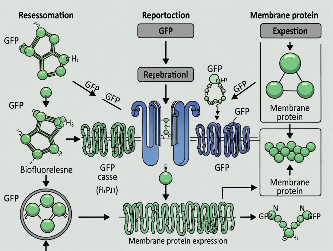
Title: Membrane Protein-GFP Expression Pathway
Within the broader thesis investigating GFP as a reporter for membrane protein expression research, live-cell imaging is a cornerstone technique. It enables the real-time visualization of protein dynamics, trafficking, and localization to specific cellular compartments such as the plasma membrane, endoplasmic reticulum (ER), Golgi apparatus, and mitochondria. This application note details essential microscopy setups and protocols optimized for these purposes, targeting researchers and drug development professionals.
Essential Microscopy Setups: Comparison & Selection
Selecting the correct microscope is critical for balancing resolution, speed, and phototoxicity. The table below summarizes key quantitative parameters for common live-cell imaging modalities.
Table 1: Comparison of Live-Cell Imaging Modalities
| Modality | Typical Resolution (XY) | Acquisition Speed | Phototoxicity Level | Best Suited For |
|---|---|---|---|---|
| Widefield Epifluorescence | ~250-300 nm | Very High | Low-Moderate | High-speed dynamics, membrane trafficking. |
| Spinning Disk Confocal | ~200-250 nm | High | Low | Optical sectioning of organelles, co-localization. |
| Point-Scanning Confocal | ~180-220 nm | Moderate | Moderate-High | High-contrast static localization, fixed cells. |
| TIRF (Total Internal Reflection) | ~100 nm (Axial) | High | Very Low | Plasma membrane-specific events, exocytosis/endocytosis. |
| Super-Resolution (e.g., SIM) | ~100-120 nm | Moderate | Moderate | Sub-organelle structure, protein clusters. |
The Scientist's Toolkit: Research Reagent Solutions
Table 2: Essential Reagents for Live-Cell Imaging of Membrane Proteins
| Reagent / Material | Function & Rationale |
|---|---|
| GFP-Tagged Constructs (e.g., GFP-Farnesyl for PM, GFP-KDEL for ER) | Reporter for localization; genetic fusion allows real-time visualization of protein dynamics. |
| Organelle-Specific Dyes (e.g., MitoTracker Deep Red, ER-Tracker Blue-White DPX) | Counterstains to validate co-localization and define organelle boundaries. |
| Phenol Red-Free Imaging Medium | Eliminates background autofluorescence and maintains physiological pH in CO₂-independent conditions. |
| Environmental Chamber | Maintains cells at 37°C, 5% CO₂, and humidity to ensure viability during extended imaging. |
| Fiducial Markers (e.g., fluorescent beads) | For drift correction during long-term time-lapse experiments. |
| Anti-fade Reagents (e.g., Ascorbic acid for live-cell) | Reduces photobleaching of fluorescent signals, prolonging experiment duration. |
Core Experimental Protocols
Protocol 3.1: TIRF Microscopy for Plasma Membrane Localization
Objective: Visualize the real-time insertion and dynamics of a GFP-tagged membrane protein at the plasma membrane (PM).
Materials:
- Cells expressing GFP-tagged protein of interest (e.g., GFP-GPI for lipid raft localization)
- TIRF microscope with 488nm laser, 60x or 100x high-NA TIRF objective
- Phenol red-free, serum-supplemented medium
- 35mm glass-bottom dish (No. 1.5 coverglass)
Method:
- Cell Preparation: Plate cells 24-48h prior to achieve 50-70% confluency on the day of imaging.
- Microscope Setup:
- Preheat environmental chamber to 37°C for at least 1 hour.
- Mount dish and locate cells using low-intensity transmitted light.
- Illuminate with 488nm laser and adjust the TIRF angle to achieve an evanescent field (typically 100-200nm depth).
- Fine-tune the angle until only the adhesion footprint of the cell is in focus, indicating exclusive PM excitation.
- Image Acquisition:
- Use a sensitive EM-CCD or sCMOS camera.
- Set exposure time to 50-200 ms to minimize blur but maximize signal.
- For time-lapse, acquire images every 0.5-5 seconds depending on process kinetics.
- Keep laser power as low as possible (<5% of maximum) to prevent photobleaching.
- Analysis: Quantify fluorescence intensity at the PM versus cytosolic background using line-scan analysis in software like ImageJ/FIJI.
Protocol 3.2: Spinning Disk Confocal for Organelle Co-localization
Objective: Determine the subcellular localization of a GFP-tagged membrane protein relative to a specific organelle.
Materials:
- Cells expressing GFP-tagged protein and stained with organelle-specific dye (e.g., MitoTracker for mitochondria)
- Spinning disk confocal system with appropriate laser lines and filter sets
- Live-cell imaging medium
Method:
- Staining: Incubate cells with pre-warmed medium containing 50-100 nM MitoTracker Deep Red for 20-30 min at 37°C. Replace with fresh, dye-free medium.
- System Calibration:
- Perform spectral unmixing or sequential acquisition to minimize bleed-through between GFP (emission ~510 nm) and far-red dyes (emission ~665 nm).
- Set pinhole size to achieve optimal optical sectioning (typically 1 Airy Unit).
- Z-stack Acquisition:
- Define a z-stack range covering the entire cell volume (step size: 0.3-0.5 µm).
- Acquire stacks at each time point for dynamic studies.
- Co-localization Analysis:
- Generate orthogonal views and maximum intensity projections.
- Calculate Pearson's or Mander's correlation coefficients using co-localization plugins (e.g., JaCoP in FIJI) on deconvolved images.
Visualizing Experimental Workflows and Pathways
Diagram Title: Workflow for Live-Cell Imaging of GFP-Tagged Proteins
Diagram Title: GFP-Reporter Localization Pathway Logic
Within the broader thesis investigating Green Fluorescent Protein (GFP) as a reporter for membrane protein expression research, this document details quantitative applications of fluorescence intensity. The reliable fusion of GFP to membrane proteins of interest enables the direct, non-destructive monitoring of expression levels in living cells. This forms the cornerstone for quantitative assays, from basic expression optimization in model systems to high-throughput drug screening campaigns targeting membrane proteins, a critical class of drug targets encompassing GPCRs, ion channels, and transporters.
Core Principles and Data Calibration
Fluorescence intensity measured via plate readers or microscopy must be correlated with actual protein concentration. Key control experiments are required to validate the linear relationship between fluorescence and expression level.
Table 1: Essential Calibration Controls and Typical Quantitative Outputs
| Control / Measurement | Purpose | Typical Data Range / Output |
|---|---|---|
| Negative Control | Cells with no GFP vector. Defines background autofluorescence. | 300-1000 RFU (varies by cell type) |
| Positive Control | Cells expressing cytosolic GFP. Sets maximum achievable signal. | 50,000-200,000 RFU |
| Linearity Validation | Serial dilution of expressing cells. Confirms assay dynamic range. | R² > 0.98 for linear fit |
| Fluorescence per Molecule | Quantified using purified GFP or FACS + quantitative Western blot. | ~100-500 RFU/amol (instrument dependent) |
| Z'-Factor (HTS Assay) | Statistical measure of assay robustness and suitability for screening. | Z' > 0.5 is acceptable; >0.7 is excellent |
Experimental Protocols
Protocol 3.1: Quantitative Expression Level Analysis in a Microplate Format
Objective: To quantify the relative membrane protein-GFP fusion expression level under different conditions (e.g., promoters, inducters, host strains).
Materials: See "Research Reagent Solutions" table. Procedure:
- Seed cells: Seed appropriate host cells (e.g., HEK293, Sf9) expressing the membrane protein-GFP construct in a black-walled, clear-bottom 96-well plate. Include negative (no GFP) and positive (cytosolic GFP) controls. Use 3-6 technical replicates.
- Apply treatments: Add experimental variables (e.g., different inducing agents, drug candidates, vehicle controls) in fresh medium.
- Incubate: Incubate under standard growth conditions for the required expression period (e.g., 24-48h).
- Wash and measure: Carefully aspirate medium and wash cells once with 100 µL of pre-warmed, fluorescence-free PBS (pH 7.4). Add 100 µL of PBS to each well.
- Plate reader acquisition: Using a fluorescence microplate reader, measure fluorescence with excitation/emission settings optimal for GFP (e.g., Ex 488 nm, Em 510-530 nm). Use a consistent gain setting.
- Normalize data: Subtract the average negative control RFU from all sample readings. Normalize fluorescence to cell viability (e.g., via parallel MTT assay) or total protein (e.g., via SRB staining) if required.
- Analyze: Calculate mean and standard deviation for replicates. Express data as normalized fluorescence units (NFU) relative to a defined control condition.
Protocol 3.2: High-Throughput Screening (HTS) for Modulators of Membrane Protein Expression or Localization
Objective: To screen a library of compounds for their effect on the expression level or trafficking of a membrane protein-GFP fusion.
Procedure:
- Assay Development & Validation: Optimize Protocol 3.1 for automation. Determine the optimal cell density, incubation time, and DMSO tolerance. Calculate the Z'-factor using high (positive control) and low (negative control or inhibitor) expression controls.
- Library Reformating: Use a liquid handler to transfer compounds from library stocks into the assay plates, maintaining final DMSO concentration ≤0.5%.
- Automated Cell Seeding & Treatment: Use an automated dispenser to seed cells expressing the target protein-GFP fusion directly into compound-containing plates.
- Incubation: Incubate plates in a controlled environment (37°C, 5% CO₂) for the predetermined period.
- Automated Readout: Use a high-throughput plate reader or automated imaging system to measure whole-well fluorescence intensity.
- Primary Hit Identification: Apply a hit threshold, typically defined as fluorescence > 3 standard deviations from the mean of the vehicle control (for enhancers) or < 3 standard deviations (for suppressors).
- Counter-Screening: Confirm hits in a secondary assay that counters for artifacts (e.g., measure fluorescence of a constitutively expressed cytosolic GFP to identify fluorescent compounds or general translation inhibitors).
Visualization: Pathways and Workflows
Title: Quantitative Expression Assay Workflow
Title: HTS Screening Workflow for Expression Modulators
Title: MP-GFP Expression & Reporting Pathway
The Scientist's Toolkit: Research Reagent Solutions
Table 2: Essential Materials for Fluorescence-Based Quantification and Screening
| Item | Function & Rationale |
|---|---|
| Black-Walled, Clear-Bottom Microplates (96/384-well) | Minimizes optical cross-talk between wells; clear bottom allows for optional bright-field/viability imaging. |
| Fluorescence Microplate Reader | Equipped with appropriate filters (Ex 485/20, Em 528/20) for GFP quantification. HTS models enable rapid throughput. |
| Stable Cell Line Expressing MP-GFP | Generates consistent, reproducible expression, critical for screening. Created via lentiviral transduction or clonal selection. |
| Validated GFP-Calibration Beads | Contains a known number of GFP molecules per bead, allowing for absolute quantification and inter-instrument calibration. |
| Automated Liquid Handling System | Enables precise, high-throughput dispensing of cells, compounds, and reagents for screening campaigns. |
| Live-Cell Imaging Compatible PBS | Fluorescence-free buffer for washing and reading to maintain cell viability and minimize background during measurement. |
| Cytotoxicity Assay Kit (e.g., MTT, CellTiter-Glo) | Used in parallel to normalize fluorescence intensity to cell number, distinguishing true expression changes from toxicity. |
| Positive Control (e.g., pEGFP-N1 Vector) | Expresses cytosolic GFP, defining the upper limit of detectable fluorescence in the system. |
| DMSO-Tolerant Assay Medium | Culture medium formulated to maintain cell health at the DMSO concentrations used for compound libraries (typically 0.1-1%). |
Solving the Puzzle: Troubleshooting Common GFP-Membrane Protein Expression Challenges
Within the broader thesis on GFP as a reporter for membrane protein expression research, mislocalization of GFP-fusion proteins presents a significant experimental hurdle. Proper membrane localization—whether plasma membrane, endoplasmic reticulum, or organellar membranes—is often critical for function and a key validation step. This note details common causes, diagnostic protocols, and solutions for when a GFP-fusion protein fails to localize correctly.
Common Causes and Quantitative Data
The table below summarizes frequent causes of mislocalization, their observable effects, and estimated prevalence based on current literature.
Table 1: Primary Causes of Mislocalization for GFP-Membrane Protein Fusions
| Cause Category | Specific Cause | Typical Observation | Estimated Frequency in Failed Experiments* |
|---|---|---|---|
| Fusion Design | GFP Tag Interferes with Targeting Signals | Cytosolic or nuclear fluorescence, diffuse signal. | 25-30% |
| Tag Placement at Wrong Terminus (Blocking signal peptides/STOP-transfer sequences) | Accumulation in ER/Cytosol; lack of surface display. | 20-25% | |
| Protein Health | Misfolding/ Aggregation | Bright puncta (aggresomes), inclusion bodies. | 15-20% |
| Destabilization/ Proteasomal Degradation | Low or absent signal; punctate proteasomes. | 10-15% | |
| Cellular Machinery | Overwhelming Cellular Trafficking (High Expression) | ER retention, accumulation in Golgi. | 10-15% |
| Missing Binding Partner/ Oligomerization Component | Retention in ER or intermediate compartments. | 5-10% |
*Compiled from recent meta-analyses of heterologous expression studies.
Diagnostic Protocol 1: Determining Localization vs. Artifact
Aim: To distinguish true mislocalization from artifacts like protein aggregation or degradation.
Materials & Reagents:
- Cells expressing the GFP-fusion construct.
- Fixative (e.g., 4% paraformaldehyde).
- Permeabilization buffer (e.g., 0.1% Triton X-100).
- Primary antibody against an organelle marker (e.g., Calnexin for ER, GM130 for Golgi).
- Fluorescent secondary antibody (with a spectrum distinct from GFP, e.g., Alexa Fluor 568).
- Nuclear stain (e.g., DAPI or Hoechst).
- Widefield or confocal fluorescence microscope.
Procedure:
- Fixation: 48h post-transfection, fix cells with 4% PFA for 15 min at room temperature (RT).
- Permeabilization & Blocking: Permeabilize with 0.1% Triton X-100 for 10 min, then block with 3% BSA for 1 hr.
- Immunostaining: Incubate with primary antibody against organelle marker (1:500 in blocking buffer) for 1 hr at RT. Wash 3x with PBS. Incubate with secondary antibody (1:1000) for 45 min in the dark.
- Counterstaining & Imaging: Incubate with DAPI (1 µg/mL) for 5 min. Wash and mount. Acquire high-resolution z-stack images using a confocal microscope.
- Analysis: Use image analysis software (e.g., ImageJ/Fiji) to perform colocalization analysis (calculate Pearson's or Manders' coefficients) between the GFP channel and organelle marker channels.
Diagnostic Protocol 2: Assessing Protein Stability and Turnover
Aim: To determine if mislocalization is due to protein instability or rapid degradation.
Materials & Reagents:
- Cells expressing the GFP-fusion construct.
- Cycloheximide (protein translation inhibitor).
- Proteasome inhibitor (e.g., MG132).
- Lysis buffer (RIPA buffer with protease inhibitors).
- SDS-PAGE and Western blot equipment.
- Primary antibodies: anti-GFP, anti-tubulin/actin (loading control).
Procedure:
- Inhibitor Treatment: 24h post-transfection, treat cells with either DMSO (vehicle control), 100 µg/mL cycloheximide, or 10 µM MG132 for 0, 2, 4, and 8 hour time points.
- Cell Lysis & Quantification: Harvest cells at each time point. Lyse in RIPA buffer. Quantify total protein concentration.
- Western Blot: Load equal protein amounts on an SDS-PAGE gel. Transfer to PVDF membrane. Probe with anti-GFP antibody (1:5000) and anti-tubulin (1:10000).
- Analysis: Quantify band intensities. Cycloheximide chase shows degradation rate. MG132 treatment that increases GFP fusion signal implicates proteasomal degradation.
Remediation Protocol: Systematic Optimization of Fusion Design
Aim: To re-engineer the fusion construct to correct for mislocalization.
Procedure:
- Tag Repositioning: If C-terminal tag shows mislocalization, clone GFP to the N-terminus (ensuring the native signal peptide is preserved), and vice versa.
- Linker Insertion: Introduce a flexible linker (e.g., (GGGGS)₃) between the protein of interest and GFP to minimize steric interference.
- Signal Sequence Verification: Use bioinformatics tools (e.g., SignalP, TMHMM) to check for predicted targeting signals. Ensure tag placement does not mask these sequences.
- Truncation Analysis: If the full-length protein mislocalizes, create a series of truncations or domain-specific fusions to identify regions necessary and sufficient for correct targeting.
- Alternative Tags: Consider smaller or more inert tags (e.g., mCherry, HALO tag, or a short epitope tag like FLAG) to compare localization efficiency.
The Scientist's Toolkit: Key Research Reagent Solutions
Table 2: Essential Reagents for Troubleshooting Mislocalization
| Item | Function/Application | Example Product/Catalog Number |
|---|---|---|
| Organelle Marker Antibodies | Co-staining to identify where the fusion protein is accumulating. | Anti-Calnexin (ER), Anti-GM130 (Golgi), Anti-TOMM20 (Mitochondria), Anti-LAMP1 (Lysosomes). |
| Proteostasis Modulators | To probe degradation pathways causing loss of signal. | MG132 (Proteasome inhibitor), Bafilomycin A1 (Lysosome inhibitor), Cycloheximide (Translation inhibitor). |
| Commercial Membrane Protein Expression Kits | Optimized systems for difficult-to-express proteins. | Thermo Fisher Mem-PER Plus Membrane Protein Extraction Kit; Promega HaloTag Membrane Protein Localization Systems. |
| Flexible Linker Peptide Cloning Cassettes | For easy insertion of solubility-enhancing linkers between protein and GFP. | Addgene #101347 (Gly-Ser linker cassette). |
| Live-Cell Imaging Compatible Dyes | To label organelles in live cells expressing GFP fusions. | MitoTracker Deep Red (Mitochondria), ER-Tracker Red (ER), CellMask Deep Red (Plasma Membrane). |
| Validated Positive Control Plasmids | GFP fusions with known, robust membrane localization. | Addgene #15294 (Lck-GFP, PM), #16008 (Sec61β-GFP, ER). |
Visualization of Workflows and Pathways
Diagnostic and Remediation Workflow
Cellular Degradation Pathways for Misfused Proteins
Application Notes
Within a thesis investigating GFP as a reporter for membrane protein expression, a central challenge is managing the inherent cytotoxicity and aggregation of overexpressed target proteins. High-level expression, while desirable for yield, often overwhelms cellular folding and trafficking machinery, leading to endoplasmic reticulum (ER) stress, activation of the unfolded protein response (UPR), and ultimately, reduced cell viability and unreliable fluorescence data. This application note details strategies and protocols to balance expression levels with cell health, ensuring more accurate and reproducible research outcomes.
Key Insights:
- GFP Fluorescence as a Dual Indicator: GFP fusion serves not only to localize the membrane protein but also as a semi-quantitative proxy for expression level and health. A sudden drop in overall fluorescence or the appearance of punctate, aggregated fluorescence signals cytotoxicity and misfolding.
- Inducible Systems are Critical: Using tightly regulated, inducible expression systems (e.g., tetracycline-, cumate-, or temperature-inducible) is non-negotiable. They allow for the separation of growth and production phases, enabling optimization of induction time and inducer concentration.
- Host Engineering Enhances Resilience: Utilizing engineered cell lines (e.g., HEK293T, CHO, or insect cell lines) that overexpress molecular chaperones (like BiP/GRP78) or possess an attenuated UPR can significantly improve tolerance to membrane protein expression.
- Pharmacological Modulation: Small molecules can be employed to transiently modulate cellular pathways. Proteostasis regulators (e.g., chemical chaperones like glycerol or DMSO), ERAD inhibitors, or mild ER stress inducers can sometimes be used to pre-condition cells and enhance folding capacity.
- Temperature Reduction: Lowering the incubation temperature post-transfection/induction (e.g., to 30°C for mammalian cells) slows protein synthesis, allowing the folding machinery to cope more effectively, often reducing aggregation.
Table 1: Impact of Expression Parameters on Cell Health and Yield
| Parameter | Condition A (High Stress) | Condition B (Optimized) | Condition C (Suppressed) | Measured Outcome (vs. Control) |
|---|---|---|---|---|
| Inducer Concentration | 1 µg/mL Doxycycline | 0.1 µg/mL Doxycycline | 0.01 µg/mL Doxycycline | Viability & Expression |
| Cell Viability (48h post-induction) | 45% | 85% | 95% | MTT Assay |
| GFP-Membrane Protein Fluorescence (RFU) | 1,000,000 | 750,000 | 100,000 | Plate Reader |
| % Cells with Aggregates (Punctate GFP) | 65% | 15% | <5% | Microscopy |
| Secretion of ER Stress Marker (CHOP) | 8.5-fold increase | 1.8-fold increase | No change | ELISA |
Table 2: Efficacy of Cytotoxicity Mitigation Strategies
| Strategy | Target System | Key Reagent/Condition | Effect on Viability | Effect on Functional Protein Yield |
|---|---|---|---|---|
| Chaperone Co-expression | HEK293 | Plasmid: pCMV-BiP | +40% | +25% (Binding Assay) |
| Chemical Chaperones | CHO | 2% DMSO (v/v) in media | +35% | +15% (Surface ELISA) |
| Temperature Shift | Sf9 Insect Cells | 28°C post-infection | +50% | +100% (Activity Assay) |
| Proteasome Inhibition | HEK293S | 5 nM Bortezomib (pulse) | +20% | -10% (Note: Increased aggregation risk) |
| UPR Attenuation | Engineered HEK293 | IRE1α kinase inhibitor | +30% | +5% (Folding improved) |
Experimental Protocols
Protocol 1: Titration of Inducer for Optimal Expression
Objective: To determine the inducer concentration that maximizes GFP-tagged membrane protein expression while maintaining >80% cell viability. Materials: Inducible stable cell line, culture media, doxycycline stock (1 mg/mL), 24-well plate, flow cytometer or plate reader. Procedure:
- Seed cells in a 24-well plate at 2.5 x 10^5 cells/mL.
- At 70% confluency, transfert with GFP-tagged membrane protein construct if using a transient system. For stable lines, proceed to step 3.
- Add doxycycline to final concentrations of 0, 0.01, 0.05, 0.1, 0.5, and 1.0 µg/mL in triplicate.
- Incubate for 48 hours at 37°C, 5% CO2.
- Harvest cells: Resuspend 100 µL for viability assay (e.g., Trypan Blue) and analyze the remainder by flow cytometry for GFP median fluorescence intensity (MFI).
- Analysis: Plot MFI and viability (%) against doxycycline concentration. The optimal point is on the plateau of the MFI curve before viability drops below 80%.
Protocol 2: Assessment of Aggregation and ER Stress via Microscopy and qPCR
Objective: To visually identify protein aggregation and molecularly quantify ER stress response activation. Materials: Cells expressing GFP-tagged protein, 4% PFA, Hoechst 33342 stain, RNA extraction kit, cDNA synthesis kit, qPCR primers for BiP (HSPA5) and CHOP (DDIT3). Procedure:
- Imaging: Fix cells with 4% PFA for 15 min. Permeabilize with 0.1% Triton X-100 (optional). Stain nuclei with Hoechst (1 µg/mL) for 5 min. Image using a fluorescence microscope with FITC and DAPI channels.
- Image Analysis: Score cells for diffuse (healthy) vs. punctate (aggregated) GFP signal. Count >200 cells per condition.
- qPCR for ER Stress: In parallel wells, extract total RNA. Synthesize cDNA. Perform qPCR using primers for ER stress markers (BiP, CHOP) and a housekeeping gene (e.g., GAPDH).
- Analysis: Calculate ΔΔCt for BiP and CHOP. A >2-fold increase indicates significant UPR activation correlating with cytotoxic stress.
Protocol 3: Co-expression of Molecular Chaperones
Objective: To improve the soluble yield of a target membrane protein by co-expressing the ER chaperone BiP. Materials: Two plasmids: (1) GFP-tagged membrane protein, (2) Untagged BiP expression vector, transfection reagent. Procedure:
- Seed HEK293T cells in a 6-well plate.
- Prepare two transfection mixes:
- Test: 1 µg target plasmid + 1 µg BiP plasmid.
- Control: 1 µg target plasmid + 1 µg empty vector.
- Transfect according to manufacturer guidelines.
- 48 hours post-transfection, harvest cells.
- Analyze via:
- Western Blot: Non-reducing gel to check for aggregates in the stacking gel.
- Flow Cytometry: Compare GFP MFI and side scatter (SSC, indicator of granularity/complexity).
- Functional Assay: Perform a membrane preparation and ligand binding assay if applicable.
Visualizations
Title: Cytotoxicity Management Workflow for GFP-Reporter Studies
Title: ER Stress & Cytotoxicity Signaling Pathways
The Scientist's Toolkit: Key Research Reagent Solutions
Table 3: Essential Materials for Managing Cytotoxicity
| Reagent / Material | Function / Purpose | Example Product/Catalog Number |
|---|---|---|
| Tetracycline/Doxycycline-Inducible Cell Line | Enables precise, titratable control over GFP-fusion protein expression, separating growth from production. | Flp-In T-REx 293 (Thermo Fisher) |
| Chemical Chaperones (Stock Solutions) | Stabilize protein folding, reduce aggregation in ER. Used in media during expression phase. | 4-Phenylbutyric Acid (PBA), Tauroursodeoxycholic acid (TUDCA) |
| ER-Tracker Dyes | Live-cell stain for visualizing ER morphology and stress in conjunction with GFP signal. | ER-Tracker Red (BODIPY TR Glibenclamide) |
| qPCR Primer Assays for UPR Markers | Quantify transcriptional activation of ER stress pathways (HSPA5/BiP, DDIT3/CHOP, XBP1s). | TaqMan Gene Expression Assays |
| Proteostasis Modulator Library | Small molecule screen to identify compounds that improve functional protein yield. | Selleckchem Proteostasis Library |
| Anti-GFP Nanobody Resin | For one-step purification of GFP-fusion protein; can separate soluble from aggregated material. | GFP-Trap Agarose beads |
| Membrane-Permeant Proteasome Inhibitor | To temporarily inhibit ERAD, allowing assessment of whether degradation is limiting yield. | MG-132 (Z-Leu-Leu-Leu-al) |
| Lipo-friendly Transfection Reagent | For high-efficiency, low-cytotoxicity transfection of adherent cells with GFP-expression plasmids. | Lipofectamine 3000 |
Within the context of a broader thesis on using GFP as a reporter for membrane protein expression, this application note addresses the persistent challenge of weak fluorescence. Efficient membrane protein expression and localization studies rely on a robust fluorescent signal. Weak signals can stem from poor folding of the GFP variant in the challenging oxidizing environment of the endoplasmic reticulum, slow chromophore maturation, or high background noise. This document provides updated protocols and data to optimize these three critical factors: folding, maturation, and signal-to-noise ratio (SNR).
Quantitative Data on GFP Variants and Optimization Strategies
Table 1: Characteristics of GFP Variants for Membrane Protein Fusion
| GFP Variant | Excitation/Emission (nm) | Maturation Half-time (37°C) | Folding Efficiency (Relative %) | Aggregation Propensity | Primary Application Context |
|---|---|---|---|---|---|
| eGFP | 488/509 | ~30 min | 100 (Baseline) | Low | General cytoplasmic |
| sfGFP | 485/510 | ~10 min | ~150 | Very Low | Fused to difficult-to-express MPs |
| Folding Reporter GFP (frGFP) | 400/510 | ~90 min | ~200 (in ER) | Low | ER folding sensor |
| Emerald | 487/509 | ~25 min | ~120 | Low | Brightness & photostability |
| Superfolder GFP (sfGFP) | 485/510 | ~15 min | ~180 | Very Low | Optimal for membrane protein fusions |
| pH-sensitive GFP (pHluorin) | 400/470, 475/509 | ~40 min | ~80 | Medium | Reporting on vesicular pH/ trafficking |
Table 2: Optimization Reagents and Their Quantitative Impact
| Reagent/Condition | Target Process | Typical Concentration | Effect on Fluorescence Signal (Fold Increase) | Effect on SNR |
|---|---|---|---|---|
| 4-Phenylbutyrate (4-PBA) | Chaperone induction, Folding | 1-10 mM | 1.5 - 3.0 | Moderate improvement |
| Trimethylamine N-oxide (TMAO) | Chemical chaperone, Folding | 50-100 mM | 1.2 - 2.0 | Minor improvement |
| Lowered Temperature (30°C) | Folding & Maturation | 30°C vs 37°C | 1.5 - 4.0 (maturation-sensitive variants) | High (reduces background) |
| Proteasome Inhibitor (MG-132) | Reduces degradation | 5-20 µM | 1.3 - 2.5 | Can decrease (increases non-specific signal) |
| N-acetyl Cysteine (NAC) | Redox balance, Maturation | 1-5 mM | 1.2 - 1.8 | Moderate improvement |
| Stable Isotope Labeling (SILAC) with ({}^{15})N, ({}^{13})C | Folding (NMR studies) | Full media replacement | Not directly quantifiable for fluorescence | N/A |
Detailed Experimental Protocols
Protocol 3.1: Evaluating Membrane Protein-GFP Fusion Folding with frGFP
Objective: To assess the folding efficiency of a membrane protein of interest (MPOI) in the endoplasmic reticulum using a folding reporter GFP (frGFP) variant.
Materials:
- Plasmid encoding MPOI-frGFP fusion (frGFP contains redox-sensitive cysteines).
- Appropriate cell line (e.g., HEK293T, CHO).
- Flow cytometer or fluorescence microplate reader.
- 10 mM 4-Phenylbutyrate (4-PBA) stock in PBS.
- 100 mM N-acetyl Cysteine (NAC) stock in water.
- Lysis buffer (e.g., RIPA with protease inhibitors).
Procedure:
- Transfection: Transfect cells with the MPOI-frGFP construct using your preferred method. Include a control plasmid expressing a well-folded secreted protein (e.g., secreted alkaline phosphatase, SEAP)-frGFP fusion as a positive folding control.
- Chemical Treatment: 24 hours post-transfection, split cells into treatment groups:
- Group A: Control (media only).
- Group B: Media + 2 mM 4-PBA.
- Group C: Media + 2 mM NAC.
- Incubate for an additional 24-48 hours at 30°C.
- Sample Preparation: Harvest cells. For total fluorescence, lyse a portion in RIPA buffer. For surface fluorescence (if applicable), keep a portion intact in PBS + 2% FBS.
- Measurement:
- Analyze intact cells by flow cytometry (excitation 400nm, emission 510/20nm bandpass) to measure folded, mature frGFP.
- Measure total protein in lysates by Bradford assay.
- Analysis: Normalize fluorescence intensity (from flow cytometry or plate reader) to total protein concentration. Report values relative to the positive folding control (SEAP-frGFP) set to 100%.
Protocol 3.2: Accelerating Chromophore Maturation for Time-Course Experiments
Objective: To enhance the rate of GFP chromophore maturation to enable faster detection after induction of membrane protein expression.
Materials:
- Cells expressing MPOI-sfGFP (or other variant).
- Fluorescence microplate reader with temperature control.
- 1 M Sodium Azide (NaN₃) stock (CAUTION: Toxic).
- 100 µM Cycloheximide (CHX) stock.
Procedure:
- Induction & Synchronization: Induce expression of the MPOI-GFP construct (e.g., with doxycycline or by changing media for stable lines). Immediately add 10 µg/mL CHX to block new protein synthesis.
- Kinetic Measurement: Transfer cells to a 30°C pre-warmed microplate reader. Immediately begin kinetic reads, measuring fluorescence (ex 485nm, em 510nm) every 5 minutes for 4-8 hours.
- Data Fitting: Plot fluorescence intensity vs. time. Fit the data to a first-order maturation equation:
F(t) = F_max * (1 - e^(-k*t)), wherekis the maturation rate constant. The half-time ist_{1/2} = ln(2)/k. - Optimization Test: Repeat experiment with a parallel set of cells incubated at 30°C (vs. 37°C) from the point of induction. Compare maturation half-times.
Protocol 3.2b: Maturation in Bacterial Membranes
Objective: To optimize fluorescence for membrane proteins expressed in E. coli.
Materials:
- E. coli strain expressing MPOI-GFP (e.g., C41(DE3) or C43(DE3) for toxic proteins).
- Autoinduction media or LB with IPTG.
- 1 M MgSO₄ stock.
- Shaking incubators at 20°C, 30°C, and 37°C.
Procedure:
- Expression Test: Inoculate 5 mL cultures in autoinduction media. Incubate at 20°C, 30°C, and 37°C with shaking for 24, 16, and 6 hours, respectively.
- Measurement: Pellet 1 mL of culture. Resuspend in PBS. Measure OD600 and fluorescence (ex 488nm, em 509nm). Normalize fluorescence to OD600.
- Cofactor Addition: To parallel 30°C cultures, add 2 mM MgSO₄ at induction. Compare final fluorescence/OD600 to untreated controls.
Protocol 3.3: Maximizing Signal-to-Noise Ratio (SNR) in Live-Cell Imaging
Objective: To implement strategies that minimize background autofluorescence and non-specific signal in live-cell imaging of membrane protein-GFP fusions.
Materials:
- Live cells expressing MPOI-GFP.
- Phenol Red-free imaging medium.
- 100 mM Cycloheximide (CHX) stock.
- 50 µM Bafilomycin A1 stock (for lysosomal inhibition control).
- Confocal or widefield fluorescence microscope with environmental chamber.
Procedure:
- Pre-imaging Treatment: 1 hour before imaging, replace culture media with phenol red-free, serum-free imaging medium supplemented with 10 µg/mL CHX to arrest secretion and reduce vesicular background.
- Temperature Control: Maintain sample at 30°C ± 1°C on the microscope stage to promote folding and reduce trafficking.
- Spectral Unmixing Setup:
- Acquire an image of untransfected control cells under identical settings to capture autofluorescence profile.
- Acquire image of MPOI-GFP expressing cells.
- Use software-based spectral unmixing to subtract the autofluorescence component from the GFP channel.
- SNR Calculation:
SNR = (Mean Signal Intensity in ROI - Mean Background Intensity) / Standard Deviation of Background. Report SNR for each optimization condition (e.g., 30°C vs. 37°C, +/- chemical chaperones).
Visualizations
Title: Pathway for GFP Folding & Maturation Optimization
Title: Workflow to Maximize SNR in Live Imaging
The Scientist's Toolkit: Research Reagent Solutions
Table 3: Essential Materials for Fluorescence Optimization
| Item/Category | Example Product/Description | Function in Optimization |
|---|---|---|
| Folding-Enhanced GFP Variants | sfGFP, frGFP plasmids (Addgene #, 54595, 111995) | Intrinsically faster folding, resistant to aggregation, or reporting on ER folding state. |
| Chemical Chaperones | 4-Phenylbutyric Acid (4-PBA), Trimethylamine N-oxide (TMAO) | Stabilize protein native state, reduce aggregation, improve yield of folded protein. |
| Redox Balance Agents | N-acetyl Cysteine (NAC), Reduced Glutathione | Create a more reducing environment in ER, assist disulfide bond formation in GFP chromophore. |
| Proteostasis Modulators | MG-132 (Proteasome Inhibitor), Bafilomycin A1 (V-ATPase inhibitor) | Identify if weak signal is due to degradation; MG-132 blocks proteasomal decay, Bafilomycin blocks lysosomal decay. |
| Temperature-Control Incubators | Refrigerated incubators or shaking incubators with cooling | Enable lower temperature culturing (e.g., 30°C) to slow translation and improve folding/maturation. |
| Phenol Red-Free Media | FluoroBrite DMEM, Leibovitz's L-15 Medium | Eliminates phenol red background fluorescence for live-cell imaging, improving SNR. |
| Spectral Unmixing Software | ZEN (Zeiss), NIS-Elements (Nikon), open-source (ImageJ plugins) | Mathematically separates GFP signal from cellular autofluorescence based on spectral signatures. |
| Validated Control Plasmids | Secreted GFP (e.g., SEAP-GFP), ER-retained GFP (e.g., GFP-KDEL) | Positive controls for folding and localization to benchmark MPOI-GFP performance. |
1. Introduction: GFP in Membrane Protein Expression Research The use of GFP and its spectral variants as reporters for membrane protein localization and expression is a cornerstone of cell biology and drug discovery. A primary confounder in this research is cellular autofluorescence, particularly in stressed, fixed, or drug-treated cells, which can masquerade as true GFP signal at the plasma membrane. Accurate differentiation is critical for quantifying expression levels, studying trafficking, and validating drug effects in high-content screening.
2. Sources of Background Autofluorescence Autofluorescence arises from endogenous fluorophores. Key sources relevant to membrane protein studies include:
- Lipofuscins: Accumulate in lysosomes with cell age or stress.
- NAD(P)H & Flavoproteins: Indicators of metabolic activity.
- Advanced Glycation End-products (AGEs): Present in cultured cells under high glucose.
- Extracellular Matrix Components: e.g., collagen and elastin fibers.
- Drug Compounds & Cell Culture Media: Certain small molecules and phenol red contribute to background.
Table 1: Spectral Profiles of Common Fluorophores vs. Autofluorescence
| Fluorophore | Primary Excitation (nm) | Primary Emission (nm) | Common Interfering Autofluorescence Source |
|---|---|---|---|
| GFP (e.g., EGFP) | 488 | 507-510 | NAD(P)H, Flavoproteins |
| RFP (e.g., mCherry) | 587 | 610 | Lipofuscin, Lysosomal granules |
| YFP (e.g., Venus) | 515 | 528 | Flavoproteins, Drug compounds |
| General Autofluorescence | Broad Spectrum (350-550) | Broad Spectrum (400-600) | Metabolic cofactors, AGEs |
3. Key Experimental Protocols for Artifact Identification
Protocol 3.1: Live-Cell Spectral Unmixing for Membrane GFP Objective: To physically separate the GFP emission signal from overlapping cellular autofluorescence in live cells expressing a membrane-targeted GFP fusion protein. Materials: Confocal microscope with spectral detection or linear unmixing capability; cells expressing membrane protein-GFP; appropriate live-cell imaging medium. Procedure:
- Prepare Control Cells: Seed non-transfected/unlabeled cells of the same type under identical conditions.
- Acquire Reference Spectrum: Image control cells using the 488 nm laser. From a region of interest (e.g., the perinuclear cytoplasm), acquire the complete emission spectrum (e.g., 500-600 nm). Save this as the autofluorescence reference spectrum.
- Acquire Experimental Sample: Image cells expressing your membrane protein-GFP using the same laser and spectral detection settings.
- Perform Linear Unmixing: Using the microscope software, provide the autofluorescence reference spectrum and the known GFP emission spectrum (often a built-in library tool). Execute the linear unmixing algorithm. This computationally separates the contributions of each fluorophore to each pixel.
- Validate: The output will generate two distinct channels: a pure GFP signal channel and an autofluorescence signal channel. Verify membrane-specific localization in the pure GFP channel.
Protocol 3.2: Quenching with TrueBlack Lipofuscin Autofluorescence Quencher Objective: To chemically reduce specific background in fixed-cell preparations for membrane protein imaging. Materials: TrueBlack Lipofuscin Autofluorescence Quencher (Biotium); fixed and immunostained (if applicable) cells; PBS. Procedure:
- Following standard fixation and immunostaining protocols (for GFP or anti-GFP), wash samples 3x in PBS.
- Prepare a 1X solution of TrueBlack quencher in 70% ethanol or PBS as per manufacturer's instructions.
- Incubate the sample in the quencher solution for 30 seconds to 2 minutes. Critical: Optimize time to avoid quenching the true signal.
- Rinse thoroughly with PBS (3x, 5 minutes each).
- Mount and image. Compare quenched and non-quenched samples to assess reduction in broad-spectrum background.
Protocol 3.3: Pharmacologic Modulation of Metabolism for Control Imaging Objective: To modulate cellular metabolic state and its associated autofluorescence for baseline correction. Materials: Live cells in imaging medium; Sodium Azide (10mM stock in PBS) or Rotenone (5mM stock in DMSO); CO₂-independent medium. Procedure:
- Establish Baseline: Image live, non-fluorescent control cells to establish baseline autofluorescence (Ex/Em for GFP filter set).
- Inhibit Metabolism: Add sodium azide (final 10-20mM) or rotenone (final 5-10 µM) to the culture medium. Incubate for 15-30 minutes at 37°C. These inhibitors suppress mitochondrial respiration, reducing NAD(P)H and flavin autofluorescence.
- Re-image: Acquire images of the same field using identical settings. The decrease in signal represents the metabolically-derived autofluorescence component.
- Application: Use this differential to set a more accurate threshold for positive membrane GFP signal in experimental samples.
4. The Scientist's Toolkit: Research Reagent Solutions
Table 2: Essential Reagents for Managing Autofluorescence
| Item | Function & Application |
|---|---|
| TrueBlack Lipofuscin Autofluorescence Quencher | Chemical quenching agent selective for aldehyde-induced autofluorescence and lipofuscin; used post-fixation. |
| MaxBlock Autofluorescence Reducing Reagent Kit | A two-step (photo- and chemical bleaching) system for reducing autofluorescence in tissue and cells pre-immunostaining. |
| CellMask Deep Red Plasma Membrane Stain | A far-red fluorescent stain (Ex/Em ~649/666 nm) to independently visualize plasma membrane architecture, orthogonal to GFP channel. |
| SYTO Red or Orange Dead Cell Stains | Nucleic acid stains for identifying dead/apoptotic cells which exhibit high autofluorescence; allows for their exclusion from analysis. |
| Phenol Red-Free Culture Medium | Eliminates background fluorescence from phenol red, especially critical for live-cell, low-signal imaging. |
| Anti-Fading Mounting Media (e.g., ProLong Diamond) | Contains reagents that slow photobleaching of true fluorophores while often marginally reducing autofluorescence. |
5. Visualization of Key Concepts
Flow: Identifying True Membrane GFP Signal
GFP and Autofluorescence Spectral Overlap
The use of Green Fluorescent Protein (GFP) as a reporter for membrane protein expression has revolutionized the field of cellular biology and drug discovery. By fusing GFP to the intracellular domains of membrane proteins (e.g., GPCRs, ion channels, transporters), researchers can visually quantify total cellular expression via fluorescence microscopy and flow cytometry. However, a critical limitation persists: high total cellular fluorescence does not equate to functional, properly localized protein at the plasma membrane (PM). Much of the expressed protein can remain trapped in the endoplasmic reticulum (ER) due to inefficient folding, assembly, or export, leading to false positives in functional assays. This application note details advanced methodologies to overcome this bottleneck by co-opting specific ER export signals and pharmacological trafficking enhancers to bias the biosynthetic pathway toward the PM, thereby ensuring that GFP-reported fluorescence accurately reflects mature, surface-available protein.
Key Mechanisms & Pathway Diagrams
2.1 ER Export Signaling Pathways for Membrane Proteins The canonical secretory pathway for membrane proteins requires specific motifs to facilitate cargo concentration and loading into COPII-coated vesicles at the ER exit sites (ERES). Native signals are often weak or suboptimal for heterologous expression. Engineering stronger versions of these signals into GFP-tagged constructs can dramatically enhance forward trafficking.
- Di-Lysine Motifs (KKXX): Recognized by the COPI complex; typically a retrieval signal. Its mutation or removal can prevent retrograde transport and enhance surface delivery.
- Di-Acidic Motifs (D/E-X-D/E): Found in channels like the Potassium channel Kir2.1, these interact directly with the Sec24 subunit of COPII.
- Hydrophobic Motifs (e.g., FXF, FF, VXXSL): Common in GPCRs and other multi-pass proteins, often requiring specific escort proteins or chaperones.
- Tetrapeptide Signals (e.g., VXPX): Identified in proteins like the Vesicular Stomatitis Virus G protein (VSV-G), a classic strong export signal.
Diagram: Engineering ER Export for Improved Surface Delivery
2.2 Action of Pharmacological Trafficking Enhancers (Pharmacoperones) These are small molecules that act as molecular scaffolds, binding to misfolded or unstable membrane proteins in the ER, promoting correct folding, masking retention signals, and allowing escape from ER quality control. They are particularly valuable for rescuing disease-associated mutants or proteins with inherently poor trafficking.
Diagram: Mechanism of Pharmacological Trafficking Enhancers
Experimental Protocols
Protocol 1: Evaluating ER Export Signal Efficacy Using GFP-Reported Surface Delivery
Objective: To compare the plasma membrane localization efficiency of a target membrane protein fused with different C-terminal ER export signals.
Materials: See "Scientist's Toolkit" below.
Method:
- Construct Design & Cloning: Clone your gene of interest (GOI), without its native stop codon, into a mammalian expression vector upstream of an in-frame GFP tag. Generate three C-terminal variants:
- V1: GOI-GFP (parental, native signal).
- V2: GOI-GFP-VXPX (adds VSV-G signal).
- V3: GOI-GFP-FCYENE (adds Kir2.1 signal).
- Cell Transfection: Seed HEK293 or HeLa cells in 24-well plates on poly-D-lysine coated coverslips. At 60-70% confluency, transfect each variant (and GFP-only control) using a lipid-based transfection reagent (e.g., Lipofectamine 3000). Include untransfected cells for background.
- Immunostaining (Non-Permeabilized): At 24-48h post-transfection: a. Rinse cells with ice-cold PBS-CM (PBS with Ca2+/Mg2+). b. Block with 3% BSA in PBS-CM for 20 min on ice. c. Incubate with primary antibody against an extracellular epitope of your GOI (1:200 in blocking buffer) for 1h on ice. If no extracellular antibody is available, use an anti-GFP antibody on a parallel, permeabilized set. d. Wash 3x with ice-cold PBS-CM. e. Incubate with Alexa Fluor 647-conjugated secondary antibody (1:500) for 45 min on ice, protected from light. f. Wash 3x and fix with 4% PFA for 15 min on ice. g. Mount slides with antifade medium containing DAPI.
- Image Acquisition & Quantification: Using a confocal microscope, acquire z-stacks under identical settings. Quantify:
- Total Expression: Mean GFP fluorescence intensity per cell (from sum projection).
- Surface Expression: Mean Alexa Fluor 647 fluorescence intensity at the plasma membrane (from a single peripheral plane or line scan analysis).
- Surface Delivery Efficiency: Calculate the ratio of Surface Expression (AF647) to Total Expression (GFP) for at least 50 cells per condition.
Protocol 2: High-Throughput Flow Cytometry Assay for Pharmacoperone Screening
Objective: To quantitatively assess the ability of small molecules to enhance surface delivery of a GFP-tagged, trafficking-deficient membrane protein.
Method:
- Cell Preparation: Seed HEK293 cells stably or transiently expressing your GFP-fused, trafficking-deficient target protein in 96-well plates.
- Compound Treatment: At 6h post-transfection (or at seeding for stable lines), add a library of test compounds (e.g., 10 µM final concentration in 0.1% DMSO). Include controls: DMSO vehicle, known pharmacoperone (positive control), and a protein synthesis inhibitor (e.g., cycloheximide, negative control). Incubate for 16-24h.
- Surface Labeling: Detach cells with gentle enzymatic (e.g., Accutase) or non-enzymatic buffer. Transfer cells to a V-bottom 96-well plate. Pellet cells (300 x g, 5 min). Resuspend in ice-cold staining buffer (PBS, 1% BSA) containing a phycoerythrin (PE)- or APC-conjugated antibody against an extracellular epitope. Incubate on ice for 30 min in the dark.
- Wash & Analysis: Wash cells twice with ice-cold staining buffer. Resuspend in PBS + 1% FBS + propidium iodide (PI, 1 µg/mL) for live/dead discrimination. Analyze immediately on a flow cytometer.
- Gating & Analysis: a. Gate on single, live (PI-negative) cells. b. For the DMSO control, gate the GFP-positive population. c. Apply this gate to all samples. d. For the GFP+ population, measure the median fluorescence intensity (MFI) of the surface stain (PE/APC) and the GFP. e. Rescue Index: Calculate (MFIPEcompound / MFIGFPcompound) / (MFIPEDMSO / MFIGFPDMSO).
Data Presentation
Table 1: Comparative Efficacy of Engineered ER Export Signals on Model GPCR Surface Delivery (Data from representative experiment using the Vasopressin 2 Receptor (V2R)-GFP fusion in HEK293 cells; n=3, mean ± SEM)
| Construct (V2R-GFP + Signal) | Total Fluorescence (GFP MFI, a.u.) | Surface Fluorescence (AF647 MFI, a.u.) | Surface/Total Ratio | Fold-Change vs. Parental |
|---|---|---|---|---|
| Parental (No added signal) | 15,240 ± 1,105 | 2,180 ± 205 | 0.143 ± 0.012 | 1.0 |
| + VSV-G (VXPX) | 14,890 ± 980 | 5,655 ± 320 | 0.380 ± 0.021 | 2.66 |
| + Kir2.1 (FCYENE) | 16,100 ± 1,210 | 4,025 ± 295 | 0.250 ± 0.015 | 1.75 |
| ER-Retained Mutant (L62P) | 18,560 ± 1,450 | 410 ± 85 | 0.022 ± 0.005 | 0.15 |
Table 2: Rescue of Trafficking-Deficient Mutants by Pharmacological Enhancers (Flow cytometry data for V2R-L62P mutant-GFP treated with 10µM compound for 20h; n=4, mean ± SD)
| Treatment Condition | GFP+ Cell Population (%) | Surface MFI (PE) | Total MFI (GFP) | Rescue Index |
|---|---|---|---|---|
| DMSO Vehicle | 32.5 ± 3.1 | 520 ± 45 | 12,300 ± 1,100 | 1.00 |
| Known Pharmacoperone (SR121463B) | 35.1 ± 2.8 | 4,850 ± 320 | 13,050 ± 950 | 9.12 |
| Novel Compound A | 33.8 ± 3.5 | 1,230 ± 110 | 12,900 ± 1,050 | 2.25 |
| Novel Compound B | 31.9 ± 2.9 | 480 ± 65 | 8,750 ± 720* | 1.04 |
| Cycloheximide (50µM) | 5.2 ± 1.1* | 110 ± 30* | 2,100 ± 450* | 1.05 |
*Indicates significant cytotoxicity or protein synthesis inhibition.
The Scientist's Toolkit
| Research Reagent / Material | Function & Application |
|---|---|
| Mammalian Expression Vectors (e.g., pcDNA3.1, pEGFP-N1) | Backbone for cloning GOI-GFP fusions with flexible multiple cloning sites. |
| Site-Directed Mutagenesis Kit | For introducing or removing specific trafficking motifs (e.g., adding VXPX, mutating KKXX). |
| Lipofectamine 3000 | High-efficiency, low-toxicity lipid transfection reagent for delivering plasmid DNA to adherent cell lines. |
| Anti-GFP Nanobody / Chromobody | Live-cell, fluorescently tagged binder for tracking GFP-tagged protein dynamics without fixation. |
| Cell Surface Protein Isolation Kit (Biotinylation-based) | Biochemically isolates PM proteins for western blot validation of surface expression. |
| Conformation-Selective Antibodies | Antibodies that specifically bind the natively folded extracellular domain of your target protein, crucial for non-permeabilized staining. |
| Flow Cytometer with 488nm & 633/640nm lasers | Essential for simultaneous detection of GFP (total protein) and PE/APC (surface label) in high-throughput screening protocols. |
| ER-Tracker Red (BODIPY TR Glibenclamide) | Live-cell stain for visualizing the ER, useful for colocalization analysis with GFP-tagged proteins. |
| Brefeldin A | Fungal metabolite that disrupts ER-to-Golgi transport; used as a negative control in trafficking experiments. |
| Commercial Pharmacoperones (e.g., SR121463B for V2R, IN3 for GnRHR) | Well-characterized positive control compounds for validating trafficking rescue assays. |
Beyond the Green Glow: Validating and Comparing GFP with Other Reporter Technologies
Within the context of membrane protein expression research using Green Fluorescent Protein (GFP) as a reporter, validation of correct localization, surface expression, and function is paramount. GFP fusion proteins allow for real-time visualization, but definitive conclusions require rigorous orthogonal validation. This article details three essential validation controls—co-localization staining, surface biotinylation, and functional assays—providing application notes and protocols to ensure accurate interpretation of GFP-tagged membrane protein data.
Application Notes & Protocols
Co-localization Staining for Subcellular Localization
Application Note: Confirming that a GFP-tagged membrane protein localizes to its intended cellular compartment (e.g., plasma membrane, endoplasmic reticulum, Golgi apparatus) is critical. Co-staining with compartment-specific markers quantitatively validates GFP signal specificity beyond its inherent fluorescence.
Detailed Protocol: Immunofluorescence Co-localization
- Cell Preparation: Seed cells expressing the GFP-tagged membrane protein on poly-D-lysine-coated glass-bottom culture dishes. Fix with 4% paraformaldehyde (PFA) for 15 min at room temperature (RT). Permeabilize with 0.1% Triton X-100 for 10 min if staining internal organelles.
- Blocking: Incubate with blocking buffer (5% normal serum, 1% BSA in PBS) for 1 hour at RT.
- Primary Antibody Incubation: Incubate with a primary antibody against a specific organelle marker (e.g., Na+/K+ ATPase for plasma membrane, Calnexin for ER) diluted in blocking buffer overnight at 4°C.
- Secondary Antibody Incubation: Wash 3x with PBS. Incubate with a spectrally distinct fluorescent secondary antibody (e.g., Alexa Fluor 568 or 647) for 1 hour at RT in the dark.
- Imaging & Analysis: Acquire high-resolution confocal images. Use analysis software (e.g., ImageJ, Coloc2) to calculate Pearson's Correlation Coefficient (PCC) or Mander's Overlap Coefficient (MOC) for multiple cells.
Table 1: Quantitative Co-localization Analysis Results
| GFP-Tagged Protein | Marker Protein (Channel) | Pearson's Coefficient (Mean ± SD) | Mander's Overlap M1 (Mean ± SD) | N (cells) |
|---|---|---|---|---|
| GFP-GPCR A | Na+/K+ ATPase (AF568) | 0.87 ± 0.05 | 0.92 ± 0.03 | 25 |
| GFP-Ion Channel B | Calnexin-ER (AF647) | 0.12 ± 0.08 | 0.15 ± 0.09 | 22 |
| GFP-Transporter C | GM130-Golgi (AF647) | 0.68 ± 0.07 | 0.71 ± 0.06 | 20 |
Diagram 1: Co-localization staining workflow
Surface Biotinylation for Quantifying Plasma Membrane Expression
Application Note: GFP fluorescence intensity alone cannot reliably distinguish between proteins in the plasma membrane (PM) and intracellular pools. Surface biotinylation provides a biochemical method to selectively label, isolate, and quantify the fraction of the GFP-tagged protein present on the cell surface.
Detailed Protocol: Cell Surface Biotinylation and Pull-down
- Labeling: Wash live cells (expressing the GFP-tagged protein) 3x with ice-cold PBS-CM (PBS with 0.1 mM CaCl2, 1 mM MgCl2). Incubate with freshly prepared EZ-Link Sulfo-NHS-SS-Biotin (0.5 mg/mL in PBS-CM) for 30 min on ice with gentle agitation. Quench with 100 mM glycine in PBS-CM for 10 min on ice.
- Lysis: Wash cells 3x with ice-cold TBS. Lyse in RIPA buffer (with protease inhibitors) for 30 min on ice. Clarify lysate by centrifugation at 16,000 x g for 15 min at 4°C.
- Pull-down: Incubate a portion of the lysate (input control) with Laemmli buffer. For the remainder, incubate with pre-washed NeutrAvidin agarose beads for 2 hours at 4°C with end-over-end rotation.
- Wash & Elution: Wash beads 4x with lysis buffer. Elute bound proteins by boiling in 2X Laemmli buffer containing 50 mM DTT (to reduce the disulfide-cleavable biotin linker).
- Analysis: Analyze both Input (total protein) and Eluate (surface protein) fractions by SDS-PAGE and Western blot. Probe for GFP to detect the tagged protein and a control protein (e.g., GAPDH, absent from surface fraction).
Table 2: Surface Biotinylation Quantification (Densitometry)
| GFP-Tagged Protein | Condition | Total Expression (Input, A.U.) | Surface Expression (Pull-down, A.U.) | Surface/Total Ratio (%) |
|---|---|---|---|---|
| GFP-GPCR A | Untreated | 15000 ± 1200 | 11200 ± 950 | 74.7 ± 3.1 |
| GFP-GPCR A | Ligand (1 hr) | 14800 ± 1100 | 5800 ± 600 | 39.2 ± 2.8* |
| GFP-Mutant | Untreated | 8000 ± 700 | 1200 ± 200 | 15.0 ± 1.5* |
(*p < 0.01 vs. Untreated WT)
Diagram 2: Surface biotinylation pull-down logic
Functional Assays for Validating Activity
Application Note: Correct localization does not guarantee functional integrity. Functional assays are required to confirm that the GFP tag does not impair the protein's native activity. The assay is tailored to the protein's function (e.g., ligand binding, ion flux, transporter activity, downstream signaling).
Example Protocol: Calcium Flux Assay for a GFP-Tagged GPCR
- Cell Loading: Harvest cells expressing the GFP-tagged GPCR. Load cells with a calcium-sensitive fluorescent dye (e.g., Fluo-4 AM, 4 µM) in assay buffer for 45 min at 37°C in the dark.
- Baseline Acquisition: Wash and resuspend cells. Using a fluorimeter or plate reader, monitor baseline fluorescence (ex/em ~494/516 nm) for 60 seconds.
- Agonist Stimulation: Add the specific agonist for the GPCR at the desired concentration. Continuously monitor fluorescence for 180+ seconds to capture the calcium transient peak.
- Analysis & Validation: Calculate the peak fluorescence intensity (F) relative to baseline (F0) as ΔF/F0. Compare responses between GFP-tagged and untagged (but expressed) versions of the receptor. Include vector-only controls.
Table 3: Functional Calcium Flux Assay Data
| Cell Line | Agonist | Peak ΔF/F0 (Mean ± SEM) | EC50 (nM) | n (experiments) |
|---|---|---|---|---|
| GFP-GPCR A | Ligand X | 2.8 ± 0.3 | 5.2 ± 0.8 | 6 |
| Untagged GPCR A | Ligand X | 3.1 ± 0.4 | 4.9 ± 1.1 | 6 |
| Vector Control | Ligand X | 0.1 ± 0.05 | N/A | 4 |
The Scientist's Toolkit: Research Reagent Solutions
| Reagent/Material | Function in Validation | Example Product/Catalog |
|---|---|---|
| Organelle-Specific Antibodies | Markers for co-localization (e.g., Na+/K+ ATPase for PM, Calnexin for ER). | Anti-Na+/K+ ATPase α1 (Abcam, ab7671) |
| Spectrally Distinct Secondary Antibodies | Enable multiplex imaging without cross-talk with GFP. | Alexa Fluor 568 anti-mouse (Invitrogen, A-11004) |
| Sulfo-NHS-SS-Biotin | Cell-impermeant, cleavable biotinylation reagent for selective surface protein labeling. | Thermo Scientific, 21331 |
| NeutrAvidin Agarose Beads | High-affinity, low non-specific binding beads for capturing biotinylated proteins. | Thermo Scientific, 29200 |
| Calcium-Sensitive Dyes (Fluo-4, Fura-2) | Report intracellular calcium changes for GPCR or channel functional assays. | Invitrogen, F14201 (Fluo-4 AM) |
| Protease Inhibitor Cocktail | Preserves protein integrity during lysis for biotinylation and biochemical assays. | Roche, cOmplete 4693116001 |
| Poly-D-Lysine | Enhances cell adhesion to glass surfaces for high-resolution imaging. | Sigma-Aldrich, P6407 |
| Laemmli Sample Buffer (2X) | Denatures proteins for SDS-PAGE analysis of input and pull-down samples. | Bio-Rad, 1610737 |
Within the broader thesis on using GFP as a reporter for membrane protein expression research, a central and persistent question is the fidelity of the fluorescent signal. For membrane proteins—often targets in drug development—factors such as incorrect folding, aggregation, failed membrane insertion, and the local chemical environment (e.g., pH, redox state) can decouple fluorescence from functional protein abundance. This application note synthesizes current evidence and provides protocols to empirically determine the GFP-protein correlation in specific experimental systems.
Key Factors Affecting GFP-Protein Correlation
The quantitative relationship between GFP fluorescence and target protein abundance is influenced by multiple variables, summarized below.
Table 1: Factors Influencing GFP Fluorescence to Protein Abundance Correlation
| Factor | Impact on Correlation | Typical Experimental Check |
|---|---|---|
| Protein Maturation Rate | Slow maturation kinetics cause lag, underestimating rapid expression. | Time-course comparing fluorescence to immunoblot. |
| Fusion Design | N- vs. C-terminal fusion can affect folding, activity, & stability. | Compare both fusions & test protein function. |
| Cellular Environment (pH, O₂) | GFP chromophore formation requires O₂; fluorescence is pH-sensitive. | Use ratiometric pH-insensitive variants (e.g., GFPmut3). |
| Protein Stability/Turnover | GFP is very stable; target protein may degrade faster, inflating signal. | Cycloheximide chase followed by immunoblot vs. fluorescence. |
| Misfolding & Aggregation | GFP may fold & fluoresce while target domain is misfolded/inactive. | Solubility fractionation & activity assays. |
| Optical Artifacts | Inner filter effect, photobleaching, quenching. | Ensure linear fluorescence range with cell dilution. |
| Instrument Calibration | Non-standardized detection settings. | Use fluorescent reference standards (e.g., beads). |
Core Validation Protocols
Protocol 1: Direct Correlation via Quantitative Western Blot & Fluorescence
Objective: Establish a calibration curve between fluorescence intensity and absolute protein amount. Materials: Cell lysates expressing the GFP-fusion, purified GFP protein standard, SDS-PAGE system, fluorescent Western blot imager, plate reader.
- Sample Preparation: Generate a series of cell samples expressing the GFP-fusion protein over a range of induction levels or expression times. Prepare lysates under denaturing conditions.
- Absolute Quantification: Create a standard curve using known amounts of purified GFP on the same SDS-PAGE gel. Perform Western blot using anti-GFP antibody.
- Parallel Fluorescence Measurement: Measure the fluorescence of an identical set of live cells or native lysates using a plate reader (ex/~488 nm, em/~510 nm).
- Data Analysis: Plot fluorescence units (y-axis) against protein abundance from the quantitative Western blot (x-axis) for each sample. Calculate the R² value of the linear regression.
Protocol 2: Pulse-Chase Analysis for Stability Decoupling
Objective: Dissect differences in stability between the GFP moiety and the target protein domain.
- Pulse: Treat expressing cells with a short (e.g., 20-min) pulse of labeled methionine/cysteine (e.g., S³⁵-Met/Cys).
- Chase: Replace medium with excess unlabeled amino acids. Collect samples at multiple time points (e.g., 0, 1, 2, 4, 8, 24h).
- Dual Analysis: For each time point:
- Split sample for fluorescence measurement (plate reader).
- Immunoprecipitate the fusion protein using target-protein-specific antibodies.
- Resolve via SDS-PAGE and quantify radioactive signal (phosphorimager) to track target protein decay.
- Comparison: Plot normalized fluorescence vs. normalized radiolabel signal over time. Diverging curves indicate stability mismatch.
Protocol 3: Membrane Protein-Specific Solubility & Activity Check
Objective: Verify that GFP fluorescence correlates with functional, membrane-incorporated protein.
- Membrane Fractionation: Lysc cells gently (e.g., Dounce homogenizer). Perform differential centrifugation to isolate membrane fractions.
- Solubility Assessment: Treat membrane fraction with:
- Mild detergent (e.g., DDM) to solubilize properly folded protein.
- Harsh denaturant (e.g., SDS) to solubilize everything.
- Measure: Quantify GFP fluorescence in each fraction (soluble vs. insoluble). A significant fluorescent signal in the harsh-denaturant-only fraction suggests aggregated fusion protein.
- Functional Assay: Perform a target-specific activity assay (e.g., ligand binding, transport) on the detergent-solubilized fraction. Plot activity vs. fluorescence from the same fraction.
Research Reagent Solutions Toolkit
Table 2: Essential Reagents for Correlation Studies
| Item | Function & Rationale |
|---|---|
| pH-insensitive GFP Variants (e.g., GFPmut3, Superfolder GFP) | Reduces environmental artifact, provides more reliable signal in acidic organelles or extracellular assays. |
| Purified GFP Protein Standard | Enables absolute quantification of GFP-fusion abundance via quantitative Western blot. |
| Fluorescent Calibration Beads | For standardizing flow cytometers and plate readers, enabling cross-experiment comparison. |
| Site-Specific Protease (e.g., TEV, HRV 3C) | To cleave GFP from target protein for separate quantification, checking for independent degradation. |
| Membrane-Permeant & -Impermeant Quenchers | e.g., Trypan Blue or KI. Distinguish surface vs. internalized membrane protein fluorescence. |
| Bicistronic or IRES Constructs | Expresses target protein and free GFP from same mRNA, controlling for translational effects. |
| Crosslinkers (e.g., Formaldehyde, BS³) | To "freeze" protein complexes/complexes before lysis for more accurate native state analysis. |
| Proteasome/Chloroquine Inhibitors | To block degradation pathways, revealing if fluorescence accumulates while target protein is degraded. |
Experimental Workflow & Data Interpretation Pathways
Title: Workflow to Validate GFP-Protein Correlation
Title: Causes & Effects of Poor GFP-Protein Correlation
Within the broader thesis on Green Fluorescent Protein (GFP) as a reporter for membrane protein expression research, a critical comparison with the widely used luciferase system is essential. This application note provides a detailed, quantitative, and methodological comparison of these two fundamental reporter modalities, focusing on their sensitivity, dynamic range, and specific utility for studying membrane-localized targets. The choice of reporter directly impacts the accuracy, throughput, and biological relevance of data in drug discovery and basic research.
Quantitative Comparison: GFP vs. Luciferase
Table 1: Core Performance Characteristics
| Parameter | GFP (e.g., sfGFP, mNeonGreen) | Luciferase (e.g., NanoLuc, Firefly) |
|---|---|---|
| Detection Sensitivity | Moderate-High (pM-nM range for protein) | Extremely High (fM-aM range for enzyme) |
| Dynamic Range | ~3-4 orders of magnitude | ~7-8 orders of magnitude |
| Background Signal | Autofluorescence, direct excitation | Negligible (substrate-dependent chemiluminescence) |
| Temporal Resolution | Real-time, continuous monitoring | Endpoint or real-time (kinetic assays possible) |
| Spatial Resolution | Excellent (subcellular localization) | Poor (lysis required for optimal signal) |
| Direct Suitability for Live-Cell Membrane Targets | Excellent (genetic fusion, live-cell imaging) | Poor (requires lysis, loses spatial data) |
| Assay Time (from sample to read) | Immediate (if expressed) | Minutes post substrate addition |
| Multiplexing Potential | High (with other FPs) | High (with dual substrates/spectral variants) |
Table 2: Suitability for Membrane Protein Research
| Application | GFP Recommendation | Luciferase Recommendation | Rationale |
|---|---|---|---|
| Localization & Trafficking | Ideal | Not Suitable | GFP provides spatial data; luciferase does not. |
| Membrane Protein Expression Level (Bulk) | Good | Ideal (for quantitation) | Luciferase offers superior sensitivity and linear range for quantification in lysates. |
| Real-Time Dynamics in Live Cells | Ideal | Limited | GFP allows continuous monitoring; luciferase kinetics are substrate-limited. |
| High-Throughput Screening (HTS) | Good (for imaging) | Ideal (for plate readers) | Luciferase's low background and high sensitivity are preferred for plate-based HTS. |
| Protein-Protein Interaction at Membrane (e.g., FRET) | Ideal | Not Applicable | GFP variants enable FRET; luciferase systems (e.g., BRET) are an alternative but with lower resolution. |
Experimental Protocols
Protocol 1: Quantifying Membrane Protein Expression Dynamics Using GFP Fusions
Objective: To monitor the expression level and localization of a membrane target protein (e.g., GPCR) in live cells over time.
- Molecular Cloning: Fuse the gene of interest (GOI) in-frame with a bright, monomeric GFP variant (e.g., sfGFP) at the C-terminus (or N-terminus, depending on topology). Use a lentiviral or stable plasmid vector with a selectable marker.
- Cell Culture & Transfection: Seed appropriate cells (e.g., HEK293) in optical-grade multi-well plates or glass-bottom dishes. Transfert with the GFP-GOI construct using a standard method (PEI, lipofection). Include untransfected and GFP-only controls.
- Live-Cell Imaging (Protocol):
- 24-48h post-transfection, replace medium with live-cell imaging medium (phenol-red free, with buffer).
- Use a confocal or high-content microscope with environmental control (37°C, 5% CO₂).
- For Expression Quantification: Capture widefield images using consistent exposure times. Use a nuclear stain (Hoechst) for segmentation.
- For Localization: Capture high-resolution z-stacks. Use a plasma membrane stain (e.g., CellMask Deep Red) for co-localization analysis.
- Image Analysis:
- Segment cells based on nuclear and membrane stains.
- Measure total GFP fluorescence intensity per cell for expression levels.
- Calculate the Pearson's correlation coefficient between GFP and membrane stain channels for localization quantification.
Protocol 2: High-Sensitivity Measurement of Membrane Protein Promoter Activity Using Luciferase
Objective: To quantitatively assess the transcriptional activity of a membrane protein's promoter with high sensitivity.
- Reporter Construct: Clone the putative promoter region of the membrane protein gene upstream of the firefly or NanoLuc luciferase gene in a reporter vector.
- Cell Seeding & Transfection: Seed cells in a 96-well white-walled, clear-bottom assay plate. Co-transfect the promoter-luciferase construct with a Renilla luciferase control plasmid for normalization.
- Stimulation & Lysis: After 24-48h, apply experimental treatments. At assay endpoint, remove medium and lyse cells with 1X Passive Lysis Buffer (for firefly) or simply add Nano-Glo Luciferase Assay Buffer directly to culture medium (for NanoLuc).
- Signal Measurement (Protocol):
- For Firefly/Renilla Dual Assay: Add Firefly Luciferase Assay Reagent to each well, read luminescence. Then, add Stop & Glo Reagent to quench firefly and activate Renilla luciferase, read luminescence again.
- For NanoLuc: Add an equal volume of Nano-Glo Luciferase Assay Reagent, incubate for 3-5 minutes, read luminescence.
- Data Analysis: Normalize firefly or NanoLuc luminescence to the Renilla luminescence signal (for firefly) or to cell viability assay data (e.g., CellTiter-Glo) to account for cell number.
Visualization Diagrams
GFP Reporter Pathway
Luciferase Reporter Pathway
Reporter Selection Workflow
The Scientist's Toolkit
Table 3: Essential Research Reagent Solutions
| Item | Function in Experiment | Key Consideration for Membrane Targets |
|---|---|---|
| Monomeric GFP Variant (sfGFP, mNeonGreen) | Bright, stable fluorescent tag for genetic fusion. Minimizes perturbation of partner protein function and oligomerization. | Essential to use monomeric forms to prevent artifunctional clustering of membrane proteins. |
| NanoLuc Luciferase | Small (19kDa), extremely bright luciferase for high-sensitivity transcriptional reporting. | Superior signal-to-noise for weak promoters. Small size advantageous for some fusion constructs. |
| Plasma Membrane Stain (CellMask, DID) | Fluorescent dye labeling the cell membrane for co-localization analysis with GFP fusions. | Validates correct membrane targeting of the GFP-fused protein of interest. |
| White-Wall/Clear-Bottom Assay Plates | Optimize luminescence signal collection while allowing microscopic visualization. | Enables correlative microscopy and high-sensitivity luminescence reads in the same well. |
| Dual-Luciferase Reporter Assay System | Allows simultaneous measurement of experimental (firefly) and control (Renilla) luciferase activities. | Critical for normalizing transfection efficiency in promoter studies for membrane proteins. |
| Polyethylenimine (PEI) Transfection Reagent | Cost-effective chemical transfection method for plasmid DNA, suitable for many adherent cell lines. | Efficiency can vary for membrane protein expression; optimization required. |
| Live-Cell Imaging Medium (Phenol Red-Free) | Maintains cell health during imaging while minimizing background fluorescence. | Crucial for time-course experiments monitoring membrane protein trafficking. |
| Passive Lysis Buffer (for Firefly Luciferase) | Gentle lysis buffer compatible with dual-luciferase assays, preserving enzymatic activity. | Complete lysis is critical for accurate quantitation of membrane-associated reporter expression. |
Within a thesis focused on GFP as a reporter for membrane protein expression research, the choice of tag is a fundamental decision. This note compares Green Fluorescent Protein (GFP) with small epitope tags (e.g., HA, FLAG), analyzing their distinct trade-offs between enabling live-cell imaging and providing biochemical flexibility for protein characterization.
Comparative Analysis: Key Properties
Table 1: Core Characteristics and Trade-offs
| Property | GFP (and variants e.g., EGFP, mNeonGreen) | Small Epitope Tags (HA, FLAG, Myc, V5) |
|---|---|---|
| Size | ~27 kDa (238 aa) | 1-2 kDa (HA: 9 aa; FLAG: 8 aa) |
| Primary Strength | Direct, intrinsic fluorescence for live-cell imaging, localization, and dynamics. | Minimal perturbation; high accessibility for antibodies in fixed/permeabilized samples. |
| Key Limitation | Large size may perturb folding, trafficking, or function of membrane proteins. | Requires immuno-detection (no live imaging); signal dependent on antibody affinity. |
| Best Applications | Live-cell trafficking, real-time localization, FRET-based interaction studies, flow cytometry (direct). | Immunoprecipitation (IP), Western Blot (WB), immunohistochemistry (IHC), ELISA, studying fragile complexes. |
| Detection Modality | Direct fluorescence (Ex/Em ~488/507 nm for EGFP). | Indirect via high-affinity monoclonal antibodies (anti-HA, anti-FLAG M2). |
| Quantitation | Semi-quantitative; fluorescence intensity can be affected by maturation, pH, O₂. | Highly quantitative in denatured samples (WB); excellent for comparative expression analysis. |
| Typical Construct Design | C-terminal fusion common; N-terminal may affect fluorescence. | Flexible (N- or C-terminal); often used in tandem or with other tags (e.g., HA- tagged GFP). |
Table 2: Performance in Common Membrane Protein Assays
| Assay Type | GFP Suitability | Small Epitope Tag Suitability | Rationale |
|---|---|---|---|
| Live-Cell Confocal Imaging | Excellent | Not Applicable | Intrinsic fluorescence allows real-time tracking of protein localization and movement. |
| Surface Biotinylation + WB | Moderate | Excellent | GFP may obscure epitopes; small tags are more reliably exposed on the extracellular surface. |
| Co-Immunoprecipitation (Co-IP) | Moderate (Native) | Excellent (Denaturing/Native) | GFP can be bulky for native interactions; small tags are less disruptive, with higher-efficiency antibodies. |
| Flow Cytometry (Surface) | Good (Direct) | Excellent (Indirect) | Small tags offer superior signal amplification via secondary antibodies. |
| Protein Purification | Good (GFP-Trap) | Excellent | Anti-FLAG M2 resin allows gentle, high-specificity elution with peptides. |
| Crystallography/Cryo-EM | Poor | Good | Small tags are less likely to interfere with crystal packing or structural analysis. |
Detailed Protocols
Protocol 1: Live Imaging of GFP-Tagged Membrane Protein Trafficking Objective: To visualize the real-time trafficking and plasma membrane localization of a GFP-tagged GPCR. Materials: See "The Scientist's Toolkit" below. Procedure:
- Transfection: Seed HEK293T cells in poly-D-lysine-coated glass-bottom dishes. At 60-70% confluency, transfect with your GPCR-EGFP construct using a lipid-based transfection reagent optimized for minimal cytotoxicity.
- Incubation: Culture cells for 24-48h post-transfection in complete medium at 37°C/5% CO₂ to allow for protein expression and maturation of GFP.
- Live Imaging Prep: Replace medium with pre-warmed, phenol-red-free imaging medium supplemented with 10 mM HEPES.
- Microscopy: Using a confocal microscope with environmental chamber (37°C, 5% CO₂), focus on transfected cells using a 488 nm laser line at low power to minimize photobleaching. Capture time-lapse images (e.g., every 5-10 seconds) to monitor constitutive or ligand-induced trafficking.
- Analysis: Use image analysis software (e.g., ImageJ/Fiji) to quantify fluorescence intensity at the plasma membrane vs. cytosolic compartments over time.
Protocol 2: Sequential Immunoprecipitation and Western Blot of FLAG/HA-Tagged Membrane Protein Complexes Objective: To isolate and detect interaction partners of a dually tagged (FLAG- and HA-) membrane receptor. Materials: See "The Scientist's Toolkit" below. Procedure:
- Sample Preparation: Transfect cells with the dual-tagged construct. 48h post-transfection, lyse cells in 1 mL of gentle lysis buffer (e.g., 1% DDM in TBS with protease inhibitors) for 1h at 4°C with rotation. Clear lysate by centrifugation at 16,000 x g for 15 min.
- First IP (Anti-FLAG): Incubate cleared lysate with 50 µL of pre-washed Anti-FLAG M2 Magnetic Beads for 2h at 4°C.
- Wash: Wash beads 3x with 1 mL of wash buffer (0.1% DDM in TBS).
- Elution (FLAG): Elute the protein complex by incubating beads with 100 µL of 3xFLAG peptide (150 µg/mL) in TBS for 30 min at 4°C. Collect supernatant.
- Second IP (Anti-HA): Dilute the eluate with 900 µL of TBS + 0.1% DDM. Add 25 µL of Anti-HA Agarose Slurry and incubate for 1h at 4°C.
- Final Wash & Elution: Wash HA beads 3x. Elute proteins directly in 2X Laemmli SDS Sample Buffer by boiling for 5 min.
- Analysis: Resolve eluates by SDS-PAGE. Perform Western blot probing sequentially with mouse anti-FLAG (1:2000) and rabbit anti-HA (1:1000) primary antibodies, followed by appropriate fluorescent secondary antibodies to confirm identity and detect co-precipitated partners.
Visualizations
Title: Tag Selection Decision Tree for Membrane Protein Research
Title: Tandem IP Protocol for Protein Complex Isolation
The Scientist's Toolkit
Table 3: Essential Research Reagents & Materials
| Reagent/Material | Primary Function | Key Consideration for Membrane Proteins |
|---|---|---|
| EGFP-N1/CMV Vector | Mammalian expression vector for C-terminal GFP fusions. | Strong CMV promoter drives high expression, which may overwhelm trafficking machinery. |
| pCMV-HA/FLAG Vector | Mammalian expression vectors for N- or C-terminal epitope tagging. | Allows modular cloning; FLAG tag is optimal for surface detection in non-permeabilized cells. |
| Anti-FLAG M2 Magnetic Beads | Immunoprecipitation of FLAG-tagged proteins. | M2 antibody binds Ca²⁺-dependently; use EDTA-free buffers. Ideal for gentle, native elution. |
| Anti-HA Agarose | Immunoprecipitation of HA-tagged proteins. | High affinity; often used in tandem purifications. Elution typically requires denaturation. |
| 3xFLAG Peptide | Competitive elution agent for FLAG IP. | Gentle, specific elution preserves protein complexes for downstream analysis (e.g., 2nd IP). |
| Dynabeads Protein G | Versatile capture beads for antibody-based IP. | Can be conjugated with any anti-epitope antibody, offering flexibility for different tags. |
| n-Dodecyl-β-D-Maltoside (DDM) | Mild, non-ionic detergent for membrane protein solubilization. | Effective at extracting proteins while preserving native complexes; critical for functional IPs. |
| Poly-D-Lysine | Coating agent for cell culture surfaces. | Enhances adherence of cells (e.g., HEK293T) for transfection and imaging, reducing detachment during washes. |
| Phenol-Red Free Medium | Cell culture medium for live-cell fluorescence imaging. | Eliminates background autofluorescence from phenol red, increasing signal-to-noise ratio. |
| HALT Protease Inhibitor Cocktail | Broad-spectrum protease inhibition. | Essential in lysis buffers to prevent degradation of membrane proteins and their partners. |
While green fluorescent protein (GFP) and its variants have been instrumental as reporters for membrane protein localization, trafficking, and expression levels in live cells, they present limitations. Their relatively large size (~27 kDa) can perturb the function and trafficking of sensitive membrane proteins, and their maturation time limits real-time tracking. This application note, framed within the broader thesis of optimizing reporter systems for membrane protein research, assesses the utility of two engineered enzymatic tags—HaloTag and SNAP-tag. These systems offer advantages such as smaller size, rapid covalent labeling with diverse synthetic fluorophores, and capability for pulse-chase and super-resolution imaging, providing powerful alternatives to GFP for advanced membrane protein studies.
Comparative Analysis: GFP vs. HaloTag vs. SNAP-tag
The table below summarizes the core characteristics of these reporter systems for membrane protein applications.
Table 1: Quantitative Comparison of Reporter Systems for Membrane Proteins
| Property | GFP (e.g., EGFP) | HaloTag | SNAP-tag |
|---|---|---|---|
| Size (kDa) | ~27 | ~33 (but see note) | ~20 |
| Labeling Mechanism | Autonomous chromophore | Covalent (chloroalkane) | Covalent (benzylguanine) |
| Labeling Time | N/A (maturation: ~30-90 min) | 15-30 minutes | 15-30 minutes |
| Fluorophore Flexibility | Fixed (genetically encoded) | High (cell-permeable ligands) | High (cell-permeable ligands) |
| Pulse-Chase Feasibility | Poor | Excellent | Excellent |
| FRET Compatibility | Good (with other FPs) | Excellent (with SNAP-tag, etc.) | Excellent (with HaloTag, etc.) |
| Common Applications | Localization, expression | Super-res, PPIs, trafficking | Pulse-chase, dual-color, PPIs |
Note: While HaloTag protein is larger, its minimal functional tag can be as small as 297 amino acids. The key advantage is the small size and versatility of its synthetic ligand.
Key Research Reagent Solutions
Table 2: Essential Toolkit for HaloTag and SNAP-tag Experiments
| Reagent | Function/Description |
|---|---|
| HaloTag Vector | Mammalian expression vector for N- or C-terminal fusion to protein of interest. |
| SNAP-tag Vector | Similar vector system for creating SNAP-tag fusions. |
| HaloTag Ligands (e.g., JF549, TMR) | Cell-permeable, fluorescent chloroalkane ligands for labeling. Various colors available. |
| SNAP-tag Substrates (e.g., SNAP-Cell 647-SiR) | Cell-permeable benzylguanine-fluorophore conjugates for specific labeling. |
| BG- or HaloTag-blocked reagents | For pulse-chase: non-fluorescent substrates to block future labeling of new protein. |
| Live-Cell Imaging Media | Phenol-free medium supplemented for maintaining health during time-course experiments. |
| Membrane Protein Lysis Buffer | Detergent-containing buffer (e.g., DDM, LMNG) for solubilizing tagged proteins for biochemical assays. |
| HaloTag Magnetic Beads | For covalent capture and pull-down of HaloTag fusion protein complexes. |
Protocol 1: Live-Cell Pulse-Chase Labeling of a Membrane Protein Receptor using HaloTag
Objective: To visualize newly synthesized versus existing pools of a G protein-coupled receptor (GPCR) fused to HaloTag.
Materials:
- HEK293T cells expressing GPCR-HaloTag fusion.
- HaloTag JF549 ligand (Pulse ligand).
- HaloTag Block (non-fluorescent chloroalkane ligand).
- Complete growth medium and imaging medium.
Method:
- Pulse Labeling: Incubate cells with 500 nM HaloTag JF549 ligand in complete medium for 15 minutes at 37°C, 5% CO₂.
- Wash: Remove labeling medium. Wash cells 3x with fresh, pre-warmed medium to remove unbound ligand. Incubate in fresh medium for 30 min (chase period) to allow complete covalent binding and washout of free ligand.
- Blocking: Incubate cells with 5 µM HaloTag Block ligand for 30 minutes. This covalently and irreversibly blocks all unlabeled HaloTag molecules from future labeling.
- Chase & Imaging: Return cells to normal growth conditions. Image at defined intervals (e.g., 0, 2, 8, 24h) using a fluorescence microscope with appropriate filter sets. The JF549 signal represents the receptor pool present at the time of the "pulse."
- Optional Secondary Label: To label only the newly synthesized receptor after the block, perform a second label with a spectrally distinct HaloTag ligand (e.g., JF646) at a later time point.
Protocol 2: Proximity Ligation Assay (PLA) for Membrane Protein Dimerization using SNAP-tag and HaloTag
Objective: To detect and visualize close proximity (<40 nm) between two membrane proteins, suggesting direct interaction or dimerization.
Materials:
- Cells co-expressing Protein-A-SNAP-tag and Protein-B-HaloTag.
- Primary antibodies: Anti-SNAP-tag (mouse) and Anti-HaloTag (rabbit).
- Duolink PLA probe oligonucleotide-conjugated secondary antibodies (anti-mouse MINUS, anti-rabbit PLUS).
- Duolink Detection reagents (Ligation, Amplification buffers, fluorescently labeled oligonucleotides).
- Standard fixation, permeabilization, and blocking buffers.
Method:
- Fix and Permeabilize: Fix cells with 4% PFA for 15 min, permeabilize with 0.1% Triton X-100 for 10 min.
- Block and Primary Antibody Incubation: Block with Duolink Blocking Solution. Incubate with mouse anti-SNAP and rabbit anti-HaloTag antibodies diluted in antibody diluent overnight at 4°C.
- PLA Probe Incubation: Wash and incubate with PLA probes (secondary antibodies conjugated to oligonucleotides) for 1h at 37°C.
- Ligation, Amplification, and Detection: Perform ligation of hybridized oligonucleotides to form a circular template. Amplify the circle via rolling-circle amplification using fluorescently labeled nucleotides. Wash.
- Imaging: Image using a fluorescence microscope. Each red fluorescent spot (PLA signal) indicates a single close-proximity event between the SNAP-tag and HaloTag fusion proteins.
Visualizations
Title: Pulse-Chase Labeling Workflow with HaloTag Block
Title: SNAP/HaloTag Proximity Ligation Assay (PLA) Logic
Conclusion
GFP remains an indispensable, versatile tool for illuminating the complex expression and localization dynamics of membrane proteins. This guide has synthesized the journey from foundational concepts through practical application, troubleshooting, and rigorous validation. The key takeaway is that successful implementation requires careful construct design, system-aware methodology, and multi-faceted validation to ensure the fluorescent signal faithfully reports on the biology of interest. Looking forward, the integration of brighter, faster-maturing GFP variants with super-resolution microscopy and automated high-content screening platforms will further empower researchers. For drug development, robust GFP-reporter cell lines are poised to accelerate the discovery of therapeutics targeting G-protein-coupled receptors (GPCRs), ion channels, and transporters by enabling real-time, quantitative analysis of expression, trafficking, and modulation in living systems.