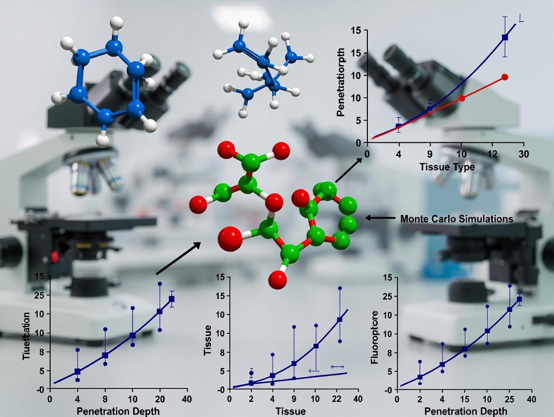
Monte Carlo Simulations in Biomedicine: Predicting Fluorescence Penetration Depth for Enhanced Imaging and Drug Development
This article provides a comprehensive guide to Monte Carlo simulations for modeling fluorescence penetration depth in biological tissues.
Monte Carlo Simulations in Biomedicine: Predicting Fluorescence Penetration Depth for Enhanced Imaging and Drug Development
Abstract
This article provides a comprehensive guide to Monte Carlo simulations for modeling fluorescence penetration depth in biological tissues. Targeted at researchers, scientists, and drug development professionals, we explore the fundamental principles of photon-tissue interactions, detail methodological frameworks for building and applying simulations, address common pitfalls and optimization strategies, and compare Monte Carlo results with experimental validation techniques. This resource bridges theoretical modeling with practical application, offering actionable insights to improve the design of fluorescence-based diagnostics and therapeutics.
Fundamentals of Photon Transport: The Science Behind Fluorescence Depth in Tissue
Monte Carlo (MC) methods are stochastic computational algorithms that have become the gold standard for simulating light propagation in turbid biological tissues. In the context of a broader thesis on fluorescence penetration depth research, these methods are indispensable for modeling the complex interplay of absorption, scattering, and fluorescence emission to predict diagnostic and therapeutic outcomes in biomedical photonics.
Core Principles and Mathematical Foundation
Light transport in tissue is described by the radiative transfer equation (RTE). MC methods provide a numerical solution by simulating the random walks of millions of discrete photon packets. Key probability distributions govern their fate:
- Scattering: Modeled by the Henyey-Greenstein phase function, parameterized by the anisotropy factor g.
- Absorption: Determined by the absorption coefficient µₐ.
- Path Length: Sampled from an exponential distribution based on the total attenuation coefficient µₜ = µₐ + µₛ (scattering coefficient).
For fluorescence, the simulation becomes a two-stage process: 1) excitation photon transport, 2) generation and transport of fluorescence photons at a longer wavelength, with a quantum yield (Φ) determining emission probability.
Quantitative Data in Biomedical Photonics
The optical properties of tissues and common agents are foundational for accurate MC modeling. The following tables summarize critical parameters.
Table 1: Typical Optical Properties of Biological Tissues at Common Laser Wavelengths
| Tissue Type | Wavelength (nm) | µₐ (cm⁻¹) | µₛ (cm⁻¹) | g | Reference |
|---|---|---|---|---|---|
| Human Skin (Epidermis) | 532 | 40-50 | 350-450 | 0.85-0.90 | Bashkatov et al. (2011) |
| Human Brain (Grey Matter) | 632 | 0.8-1.2 | 200-250 | 0.89-0.92 | Jacques (2013) |
| Breast Tissue (Healthy) | 800 | 0.03-0.06 | 100-130 | 0.93-0.97 | Taroni et al. (2010) |
| Arterial Wall | 1064 | 0.7-1.0 | 150-200 | 0.91-0.95 | Marchesini et al. (1989) |
Table 2: Key Fluorophores for Penetration Depth Studies
| Fluorophore | Excitation λ (nm) | Emission λ (nm) | Quantum Yield (Φ) | Molar Extinction (cm⁻¹M⁻¹) | Primary Use |
|---|---|---|---|---|---|
| Indocyanine Green (ICG) | 780-800 | 820-850 | 0.012-0.016 | ~1.3 x 10⁵ | Angiography, Lymphography |
| Protoporphyrin IX (PpIX) | 405, 630 | 635, 704 | 0.01-0.15 | ~5 x 10⁴ at 405nm | Photodynamic Therapy |
| Alexa Fluor 750 | 749 | 775 | 0.12 | 2.4 x 10⁵ | Antibody/Protein Labeling |
| IRDye 800CW | 774 | 789 | 0.12 | 2.4 x 10⁵ | Preclinical Imaging |
Detailed Experimental Protocol: Validating MC Models for Fluorescence Depth Sensing
This protocol outlines the experimental validation of a Monte Carlo model for predicting fluorescence signal as a function of fluorophore depth.
Objective: To correlate experimentally measured fluorescence intensity from a sub-surface fluorophore target with MC-simulated predictions across varying depths.
Materials: (See "The Scientist's Toolkit" below) Tissue Phantom Preparation:
- Prepare a liquid tissue phantom with Intralipid-20% as scatterer and India ink as absorber to match desired µₐ (~0.1 cm⁻¹) and µₛ' (~10 cm⁻¹) at the excitation wavelength.
- Characterize phantom optical properties using inverse adding-doubling or spatially resolved reflectance measurements.
- Create small capillary tubes (diameter: 1-2 mm) filled with a standardized concentration of ICG (e.g., 10 µM).
- Embed these fluorescent targets at pre-defined, precise depths (e.g., 1, 2, 3, 4, 5 mm) within the solid or semi-solid phantom.
Experimental Data Acquisition:
- Illuminate the phantom surface with a laser diode matched to the fluorophore excitation peak (e.g., 785 nm for ICG). Use a collimated or weakly focused beam.
- Use a NIR-sensitive CCD or sCMOS camera coupled with a long-pass emission filter (>810 nm) to collect fluorescence images.
- For each target depth, acquire fluorescence images. Correct for background noise, illumination inhomogeneity, and filter bleed-through.
- Quantify the fluorescence intensity (I_exp) by integrating pixel counts within a region of interest centered on the target signal.
Monte Carlo Simulation:
- Input the precisely measured phantom optical properties (µₐ, µₛ, g) for both excitation and emission wavelengths into a validated MC code (e.g., MCML, tMCimg, or custom).
- Configure the simulation geometry to exactly match the experiment: beam profile, detector position/area, and a point-like fluorescence source at the corresponding depths.
- Run a sufficient number of photon packets (typically 10⁷ to 10⁹) to ensure low statistical noise.
- Extract the simulated fluorescence intensity (I_sim) reaching the detector for each depth.
Validation & Analysis:
- Plot Iexp vs. Depth and Isim vs. Depth on the same graph.
- Perform a linear regression between Isim and Iexp across all depths.
- A strong linear correlation (R² > 0.98) and a slope near 1 validate the MC model's predictive power for fluorescence penetration depth research.
Essential Diagrams for MC Workflow and Photon-Tissue Interaction
Diagram Title: Monte Carlo Photon Transport Algorithm
Diagram Title: Two-Stage MC for Fluorescence Simulation
The Scientist's Toolkit: Key Research Reagent Solutions
Table 3: Essential Materials for MC-Guided Fluorescence Experiments
| Item | Function in Research | Example/Specification |
|---|---|---|
| Tissue Phantoms | Provide calibrated, reproducible models of tissue optical properties for model validation. | Liquid (Intralipid, Ink), Solid (PDMS with TiO₂, Ink), Layered Phantoms. |
| NIR Fluorophores | Enable deep-tissue imaging due to low tissue absorption and autofluorescence in the "optical window" (650-900 nm). | Indocyanine Green (ICG), IRDye 800CW, Alexa Fluor 750. |
| Quantum Yield Standards | Essential for calibrating fluorescence signal and inputting accurate Φ values into MC models. | Rhodamine 101 in EtOH (Φ~1.0), Cyanine dyes with published Φ. |
| Absorber Agents | Used to tune the absorption coefficient (µₐ) of tissue phantoms to physiological ranges. | India Ink, Nigrosin, Hemoglobin derivatives. |
| Scatterer Agents | Used to tune the reduced scattering coefficient (µₛ') of tissue phantoms. | Intralipid-20% (lipid droplets), Polystyrene Microspheres, TiO₂ powder. |
| Optical Property Characterization Tools | Measure ground-truth µₐ and µₛ' for phantom and ex vivo tissue inputs to MC simulations. | Integrating Sphere with Inverse Adding-Doubling, Spatial/Frequency Domain Devices. |
Within the broader thesis on Monte Carlo simulations for modeling fluorescence penetration depth in biological tissues, a precise understanding of core tissue optical properties is foundational. These properties govern the propagation, distribution, and eventual detection of both excitation and emitted fluorescent light. The accurate parameterization of absorption (μa), scattering (μs), anisotropy (g), and refractive index (n) in Monte Carlo models is critical for predicting light dosimetry, optimizing imaging depth, and interpreting in vivo fluorescence data in preclinical drug development. This guide provides a technical deep dive into these properties, their measurement, and their integration into computational research frameworks.
Fundamental Properties and Definitions
Absorption Coefficient (μa)
The absorption coefficient, μa (units: mm⁻¹), defines the probability of light absorption per unit path length in a medium. It is dependent on the concentration of chromophores (e.g., hemoglobin, melanin, water, lipids) and their specific extinction coefficients at the wavelength of interest.
Scattering Coefficient (μs)
The scattering coefficient, μs (units: mm⁻¹), quantifies the probability of light scattering per unit path length. In tissues, scattering is primarily caused by spatial variations in refractive index at cellular and subcellular structures (organelles, membranes, collagen fibers).
Anisotropy Factor (g)
The anisotropy factor, g (dimensionless, range: -1 to 1), describes the directional preference of single scattering events. A value of 0 indicates isotropic scattering, while values approaching 1 (typical for biological tissue: 0.7-0.99) represent highly forward-directed scattering.
Reduced Scattering Coefficient (μs')
For many diffuse optics applications, the combined effect of μs and g is expressed as the reduced scattering coefficient: μs' = μs(1 - g) (units: mm⁻¹). This property describes the diffusion of light in a multiply scattering medium.
Refractive Index (n)
The refractive index, n (dimensionless), governs the speed of light in the tissue and the behavior of light at boundaries between different media (e.g., tissue-glass-air). It is critical for modeling reflection and refraction at interfaces in Monte Carlo simulations.
Table 1: Typical Optical Properties of Human Tissues at Common Fluorophore Excitation Wavelengths
| Tissue Type | Wavelength (nm) | μa (mm⁻¹) | μs (mm⁻¹) | g | μs' (mm⁻¹) | Refractive Index (n) | Source / Method |
|---|---|---|---|---|---|---|---|
| Skin (epidermis) | 488 | 0.40 - 1.5 | 40 - 60 | 0.77 - 0.85 | ~8 - 14 | ~1.37 - 1.45 | Integrating Sphere, IAD |
| Brain (gray matter) | 532 | 0.15 - 0.25 | 20 - 30 | 0.89 - 0.94 | ~2 - 4 | ~1.36 - 1.40 | Integrating Sphere, MC Inverse |
| Breast Tissue | 633 | 0.002 - 0.05 | 15 - 25 | 0.85 - 0.97 | ~2 - 5 | ~1.38 - 1.42 | Spatially-Resolved Reflectance |
| Liver | 660 | 0.3 - 0.8 | 25 - 40 | 0.90 - 0.97 | ~3 - 6 | ~1.36 - 1.39 | Double Integrating Sphere |
| Adipose Tissue | 800 | 0.03 - 0.08 | 8 - 15 | 0.70 - 0.90 | ~2 - 4 | ~1.44 - 1.46 | Time-Domain Diffuse Reflectance |
| Intralipid 20% (phantom) | Various | ~0.001 | ~80-100 | ~0.7-0.75 | ~20-30 | ~1.33 | Reference Standard |
Table 2: Optical Properties of Key Endogenous Chromophores (Contributors to μa)
| Chromophore | Peak Absorption Wavelength(s) (nm) | Molar Extinction Coefficient ε (cm⁻¹M⁻¹) | Primary Tissue Location |
|---|---|---|---|
| Oxyhemoglobin (HbO₂) | 415, 542, 577 | ~5.0 x 10⁵ (415 nm) | Blood vasculature |
| Deoxyhemoglobin (Hb) | 430, 555 | ~4.0 x 10⁵ (430 nm) | Blood vasculature |
| Melanin | Broadband (UV-Visible) | Decreases exponentially with λ | Epidermis, hair follicles |
| Lipids | 930, 1210 | ~1.0 x 10² (930 nm) | Adipose tissue, cell membranes |
| Water | 980, 1200, 1450 | ~0.5 - 30 (varies strongly) | All tissues |
Experimental Protocols for Property Measurement
Double Integrating Sphere Technique for μa and μs
This is a gold-standard ex vivo method for measuring bulk optical properties.
Protocol:
- Sample Preparation: Fresh or preserved tissue is sliced into thin, parallel-sided slabs (typical thickness: 0.5-2 mm) using a microtome. Thickness is precisely measured with a micrometer.
- System Calibration: Two integrating spheres (reflectance and transmission spheres) are calibrated using standard reflectance references (e.g., Spectralon) and a direct beam measurement for 100% transmission.
- Measurement: The tissue sample is placed at the entrance port of the reflectance sphere. A collimated light beam at the desired wavelength illuminates the sample.
- Data Collection: The total diffuse reflectance (Rd) and total diffuse transmittance (Td) are measured by the respective spheres. Collimated transmittance (T_c) is often measured separately.
- Inverse Solving: The measured Rd and Td are used as inputs to an inverse adding-doubling (IAD) algorithm or a Monte Carlo-based lookup table. This algorithm iteratively adjusts μa and μs (and often g, if assumed) until the calculated values match the measured ones.
Spatially-Resolved Diffuse Reflectance for μa and μs'
This method is applicable for in vivo or contact-based measurements.
Protocol:
- Probe Configuration: A fiber-optic probe is used, with a single source fiber and multiple detection fibers at fixed distances (ρ) from the source (e.g., 0.5 mm, 1.0 mm, 1.5 mm, 2.0 mm).
- Tissue Contact: The probe is gently placed in contact with the tissue surface.
- Spectral Acquisition: Broadband or monochromatic light is delivered via the source fiber. The diffusely reflected light is collected by each detection fiber and spectrally analyzed.
- Model Fitting: The steady-state diffuse reflectance profile R(ρ) is fitted to an analytical solution of the diffusion approximation to light transport (or a Monte Carlo model) to extract μa and μs' at the measured wavelength(s).
Goniometric Measurement for Anisotropy Factor (g)
This measures the scattering phase function p(θ).
Protocol:
- Sample Preparation: A very thin, dilute suspension of tissue cells or a tissue phantom (e.g., microsphere solution) is prepared to ensure single scattering events dominate.
- Angular Scanning: A collimated laser beam passes through the sample. A detector (photodiode or PMT) rotates on a goniometer arm around the sample, measuring scattered light intensity I(θ) at angles (θ) from 0° (forward) to 180° (backward).
- Phase Function Calculation: The measured intensity is normalized to obtain the scattering phase function p(θ).
- g-Calculation: The anisotropy factor is calculated as the average cosine of the scattering angle: g = ⟨cos θ⟩ = ∫ p(θ) cos θ dΩ, integrated over all solid angles.
Visualization of Concepts and Workflows
Diagram Title: Role of Optical Properties in Monte Carlo Simulation Workflow
Diagram Title: Monte Carlo Photon Step Logic with Optical Properties
The Scientist's Toolkit: Research Reagent Solutions
Table 3: Essential Materials for Phantom Development and Validation
| Item | Function/Brief Explanation | Example Product/Composition |
|---|---|---|
| Lipid-Based Phantoms | Mimics tissue scattering. Intralipid (fat emulsion) provides controlled, stable μs' across a broad spectrum. | Intralipid 20% Intravenous Fat Emulsion. A standardized source of scatterers (soybean oil droplets). |
| Absorbing Agents | Provides tunable absorption (μa) to mimic blood, melanin, etc. | India Ink (carbon nanoparticles) or Niger Seed Oil for broad absorption; Analytical dyes (e.g., Evans Blue) for specific bands. |
| Solid Phantoms | Stable, long-lasting reference standards for system calibration and validation. | Silicone or Polyurethane bases doped with TiO₂ or Al₂O₃ powder (scatterers) and ink/dyes (absorbers). |
| Index-Matching Fluids | Reduces surface reflections at tissue/optics interfaces for more accurate measurement. | Glycerol-water solutions, mineral oil, or specialized optical gels (n ≈ 1.38 - 1.45). |
| Standard Reflectance Surfaces | Calibrates integrating sphere and reflectance probe measurements. | Spectralon (PTFE-based), a near-perfect (≈99%) Lambertian diffuse reflector. |
| Microsphere Suspensions | Provides well-defined, calculable scattering properties (μs, g) based on Mie theory for goniometry or phantom calibration. | Polystyrene or Silica Microspheres (sizes: 0.5 - 2.0 μm diameter) in aqueous suspension. |
| Fluorophore Standards | Validates fluorescence detection arm of Monte Carlo models and experimental setups. | Rhodamine B, Fluorescein, or ICG at known concentrations in controlled phantoms. |
Fluorescence Penetration Depth (FPD) is a critical parameter in biomedical optics, defining the effective depth from which usable fluorescence signal can be recovered in turbid media like biological tissue. Within the broader thesis of utilizing Monte Carlo (MC) simulations for fluorescence research, FPD is not a directly measured quantity but a derived metric. MC simulations, by stochastically modeling the propagation of excitation light and the subsequent emission and migration of fluorescence photons, provide the essential data to define, calculate, and understand FPD. This guide details the physical meaning, calculation methodologies, and practical application of FPD metrics.
Physical Meaning and Key Definitions
FPD quantifies the depth limit for effective fluorescence detection. It is governed by the interplay of:
- Absorption (µa): Of both the fluorophore and the background tissue.
- Scattering (µs'): Reduced scattering coefficient, which affects both excitation and emission photon paths.
- Anisotropy (g): Affects the directionality of scattering.
- Detection Geometry: Including source-detector separation and numerical aperture.
Unlike the effective attenuation coefficient (µeff) used for diffuse light, FPD must account for the two-step process (excitation → emission) and the possible shift in optical properties between excitation and emission wavelengths (λex and λem).
Primary Metrics for Fluorescence Penetration Depth
Based on MC simulation output (the spatial distribution of fluorescence emission points or the detected fluorescence signal from buried fluorophores), several quantitative metrics are defined.
Table 1: Key Metrics for Defining Fluorescence Penetration Depth
| Metric | Definition (Based on MC Data) | Physical Interpretation | Common Application |
|---|---|---|---|
| FPD₁/e (or dF₁/e) | Depth at which the detected fluorescence signal falls to 1/e (~37%) of its maximum (typically at surface). | Analogous to optical penetration depth for diffuse light; a simple benchmark. | Quick comparison of imaging system or fluorophore performance in homogeneous media. |
| FPD₁/₂ (or dF₁/₂) | Depth at which the detected fluorescence signal falls to 50% of its maximum value. | A more conservative, clinically relevant metric indicating practical detection limit. | Assessing sensitivity requirements for in vivo imaging. |
| Gamma (γ) - Gradient Metric | Slope from linear regression of log(Signal) vs. Depth for a fluorophore slab or point source at various depths. | Defines an effective fluorescence attenuation coefficient (µeff,fluor). FPD can be taken as 1/γ. | Most rigorous; accounts for continuous signal decay. Standard in MC validation studies. |
| Information Depth | The weighted mean depth of origin of detected fluorescence photons, calculated from MC photon history. | The average depth sampled by the measurement; depends heavily on geometry. | Critical for quantitative spectroscopy (e.g., estimating biomarker concentration). |
Experimental Protocols for Empirical Validation
MC-derived FPD metrics require validation with physical experiments. A standard protocol is outlined below.
Protocol: Phantom-Based Measurement of FPD₁/₂ using Liquid Tissue-Simulating Phantoms
Objective: Empirically determine the 50% fluorescence penetration depth (FPD₁/₂) for a given fluorophore and optical setup.
Materials: See "The Scientist's Toolkit" below.
Procedure:
- Phantom Preparation: Prepare a series of liquid phantoms with identical, standardized optical properties (e.g., µa = 0.1 cm⁻¹, µs' = 10 cm⁻¹ at λex) using Intralipid (scatterer) and ink (absorber).
- Fluorophore Inclusion: For the "deep" phantom, dissolve the target fluorophore (e.g., ICG) homogeneously throughout. A "control" phantom contains no fluorophore.
- Variable Depth Setup: Place a thin, fluorescent capillary tube (or a small solid fluorescent target) at a known, variable depth z within a non-fluorescent phantom of the same optical properties. Alternatively, use a movable stage to submerge a fluorescent bead.
- Imaging/Detection: Illuminate the phantom surface with the appropriate λex source (e.g., 785 nm laser diode). Use a filtered CCD camera or a fiber-based spectrometer (with a long-pass filter >λem) to collect the fluorescence signal I_fluor(z). Maintain constant exposure/gain settings.
- Signal Extraction: For each depth z, subtract the background signal from the control phantom. Plot the background-subtracted fluorescence intensity vs. depth z.
- Data Analysis: Normalize the signal to its maximum value (typically at the most superficial depth). Fit an exponential decay curve: I(z) = I₀ * exp(-µeff,fluor * z). Determine FPD₁/₂ as the depth where the fitted curve equals 0.5*I₀. Compare the empirical µeff,fluor to the MC-predicted γ value.
Signaling and Computational Workflow
The relationship between MC simulation, FPD definition, and experimental validation is a cyclic process of hypothesis testing and refinement.
Diagram 1: MC-Driven FPD Research Workflow
The Scientist's Toolkit: Essential Research Reagents & Materials
Table 2: Key Research Reagent Solutions for FPD Experiments
| Item | Function & Rationale |
|---|---|
| Intralipid 20% | A standardized, biocompatible lipid emulsion used as the primary scattering agent in liquid phantoms. Its scattering properties are well-characterized in the literature. |
| India Ink / Nigrosin | A highly stable, broadband absorbing agent to titrate the absorption coefficient (µa) of tissue-simulating phantoms. |
| Indocyanine Green (ICG) | A near-infrared (NIR) fluorophore (λex/~780 nm, λem/~820 nm). The NIR window minimizes tissue absorption and scattering, making it the standard for deep-tissue FPD studies. |
| Polystyrene Microspheres | Solid, monodisperse particles with precise, calculable scattering properties. Used for solid or agar-based phantom construction for superior stability. |
| Titanium Dioxide (TiO2) Powder | Alternative scattering agent for solid phantoms (e.g., mixed with silicone). Requires careful homogenization. |
| Agarose or Silicone Elastomer | Gelling/hosting matrix for creating solid phantoms, which offer long-term stability and precise geometric positioning of fluorescent targets. |
| Black Delrin / Acrylic | Material for building phantom containers and target holders. Its low autofluorescence and non-reflective properties are critical to minimize background signal. |
| NIST-Traceable Optical Property Standards (e.g., SRM) | Certified solid or liquid standards with known µa and µs' used for calibrating and validating measurement systems (e.g., spatially resolved spectroscopy) prior to phantom characterization. |
How Monte Carlo Simulations Model Photon Birth, Propagation, and Death
The quantitative analysis of light-tissue interaction is fundamental to advancing fluorescence-based diagnostic and therapeutic modalities. Within drug development, particularly for photoactive compounds or fluorescence-guided surgery, predicting the penetration depth of excitation light and the escape probability of emitted fluorescence is critical for protocol design and efficacy assessment. Monte Carlo (MC) simulations provide a stochastic, yet physically rigorous, framework for modeling the complete lifecycle of photons within turbid biological tissues. This whitepaper details the core technical principles of modeling photon "birth," propagation through scattering and absorption events, and eventual "death," framed within a thesis on optimizing fluorescence detection limits in deep tissue.
Core Physics & Algorithmic Principles
MC methods solve the radiative transport equation (RTE) by statistically simulating the trajectories of millions of individual photon packets. The core assumption is that photon-tissue interactions can be modeled as a series of random events governed by probability distributions derived from the tissue's intrinsic optical properties.
Key Optical Properties & Definitions
The following properties, summarized in Table 1, define the medium.
Table 1: Essential Optical Properties for MC Simulations
| Property | Symbol | Unit | Definition |
|---|---|---|---|
| Absorption Coefficient | μₐ | cm⁻¹ | Probability of photon absorption per unit path length. |
| Scattering Coefficient | μₛ | cm⁻¹ | Probability of photon scattering per unit path length. |
| Anisotropy Factor | g | unitless | Average cosine of scattering angle. g=0: isotropic; g≈0.9: highly forward-scattering. |
| Reduced Scattering Coefficient | μₛ' = μₛ(1-g) | cm⁻¹ | The effective scattering coefficient in a diffusion approximation. |
| Refractive Index | n | unitless | Ratio of light speed in vacuum to that in the tissue. Governs reflection/refraction at boundaries. |
The Photon Packet and Weight
To improve computational efficiency, MC simulations typically track "photon packets" with an associated weight, W (initialized to 1), rather than individual photons. The packet weight represents the fraction of photons remaining in the packet. Interactions diminish W until a termination threshold is reached.
The Three-Phase Algorithm: Birth, Propagation, Death
Phase 1: Photon Birth (Launch)
A photon packet is initialized with specific coordinates, direction, and weight.
- Spatial Launch: Defined by source geometry (e.g., infinitely narrow pencil beam, diffuse broad beam, optical fiber profile).
- Directional Launch: Typically normal to the tissue surface for a perpendicular beam.
- Initial Weight: W = 1.
Phase 2: Photon Propagation (Scattering & Absorption)
This is the core iterative loop. A step-by-step stochastic path is generated.
Experimental Protocol: Core Propagation Loop
- Calculate Step Size: Draw a random number, ξ, uniformly from [0,1). The path length, s, before the next interaction is: s = -ln(ξ) / (μₐ + μₛ)
- Move Photon: Update photon coordinates: x = x + s·direction_x, etc.
- Apply Absorption: At the new location, decrement the packet weight: ΔW = W · (μₐ/(μₐ+μₛ)). The new weight is W = W - ΔW. The absorbed weight, ΔW, is deposited in a local absorption array (critical for heating/dose calculations).
- Check for Boundary (e.g., Tissue-Air Interface): If a boundary is crossed during the step, handle partial reflection/transmission using Fresnel equations. A fraction of weight escapes as reflectance or transmittance and is recorded.
- Determine if Scattering Occurs: If W is above a threshold (e.g., 10⁻⁴), proceed to scatter.
- Update Photon Direction (Scattering): Sample a new deflection angle (θ) and azimuthal angle (ψ) from probability distributions.
- Polar Angle θ: Governed by the Henyey-Greenstein phase function, the most common for biological tissue: cos θ = (1/(2g)) * [1 + g² - ((1 - g²)/(1 - g + 2gξ))²] for g > 0.
- Azimuthal Angle ψ: Uniformly distributed: ψ = 2πξ.
- Loop: Return to Step 1.
Phase 3: Photon Death (Termination)
A photon packet "dies" or is terminated to conserve computational resources.
- Russian Roulette Termination: When W falls below a pre-defined threshold (Wₜₕ, e.g., 10⁻⁴), the packet is subjected to Russian Roulette. With a small survival probability (e.g., 0.1), the packet is kept and its weight is increased by a factor of 10. Otherwise, it is terminated. This conserves energy while terminating low-weight packets.
Modeling Fluorescence
For fluorescence penetration depth studies, the simulation is extended to a two-stage (or multi-stage) process, as depicted in the workflow diagram below.
Title: Two-Stage Monte Carlo Workflow for Fluorescence Modeling
Protocol: Fluorescence-Specific MC
- Excitation Stage: Run standard MC with excitation optical properties (μₐₓ, μₛₓ, gₓ).
- Fluorescence Birth: At each absorption site in the fluorophore, generate a new emission photon packet with a probability equal to the Fluorescence Quantum Yield (Φ). The new packet's weight is scaled accordingly.
- Emission Stage: Propagate the newborn emission photon using the optical properties at the emission wavelength (μₐₘ, μₛₘ, gₘ).
- Detection: Record emission photons that escape the tissue surface at the detector position(s). This builds a spatial map of fluorescence origin and intensity.
The Scientist's Toolkit: Research Reagent & Computational Solutions
Table 2: Essential Toolkit for MC Simulations in Fluorescence Research
| Item / Solution | Function in Research |
|---|---|
| Validated MC Code (e.g., MCML, tMCimg, GPU-MC) | Core simulation engine. GPU-accelerated versions enable rapid modeling of complex geometries. |
| Tissue Optical Property Database | Repository of measured μₐ, μₛ, g for various tissues at relevant wavelengths (excitation/emission). Critical for realistic input. |
| Fluorophore Spectra Library | Data on absorption/emission spectra and quantum yield (Φ) of common dyes (e.g., ICG, fluorescein) and novel agents. |
| Digital Tissue Phantoms | 3D voxelated or mesh-based models of tissue structures (e.g., skin layers, tumor inclusions) to assign heterogeneous optical properties. |
| Spectral Unmixing Algorithm | For multi-fluorophore studies, software to decompose detected signals into contributions from individual agents based on their spectral signatures. |
| Sensitivity/Quantification Calibration Kit | Physical phantoms with known fluorophore concentration for validating simulation results against experimental measurements. |
Advanced Considerations & Validation
Validation Protocol: Simulate a simple scenario (e.g., homogeneous slab, known properties) and compare results (diffuse reflectance, fluence rate) against an analytical solution of the RTE or a benchmarked MC code. Normalized mean error should be < 2%.
Accelerated Methods: Variance reduction techniques (e.g., photon splitting, importance sampling) and implementation on parallel computing architectures (GPU, cluster) are essential for modeling complex, heterogenous tissues with adequate statistical noise.
Output Analysis: The primary output is a spatial map of absorbed energy (for photothermal therapy) or escaping fluorescence (for imaging). From the latter, one can calculate the effective fluorescence penetration depth, defined as the depth from which a certain percentage (e.g., 50%) of detected photons originate. This metric is directly relevant for drug development targeting depth.
Title: Possible Fates of a Simulated Photon Packet
Monte Carlo simulations provide an indispensable, physics-based virtual laboratory for modeling the complete trajectory of photons in tissue. By meticulously simulating birth, stochastic propagation, and death, researchers can predict fluorescence penetration depths, optimize illumination and detection geometries, and interpret in vivo data—all accelerating the development of light-based diagnostics and therapeutics. Its integration into the drug development pipeline de-risks early-stage research and enables quantitative, patient-specific treatment planning.
Critical Input Parameters for Accurate Penetration Depth Modeling
Within the broader thesis on Monte Carlo simulations for fluorescence penetration depth research, accurate modeling is paramount for applications in drug delivery, photodynamic therapy, and non-invasive diagnostics. The fidelity of these simulations hinges entirely on the precise definition of critical input parameters. This whitepaper serves as an in-depth technical guide to these parameters, their interdependencies, and the methodologies for their empirical determination.
Core Optical and Tissue Parameters
The accuracy of a Monte Carlo model for photon transport in biological tissues depends on the following foundational input parameters. These must be characterized for each specific tissue type and wavelength under investigation.
Table 1: Critical Optical Input Parameters for Penetration Depth Modeling
| Parameter | Symbol | Unit | Description | Impact on Penetration Depth |
|---|---|---|---|---|
| Absorption Coefficient | μₐ | cm⁻¹ | Probability of photon absorption per unit path length. | Higher μₐ reduces penetration depth significantly. |
| Reduced Scattering Coefficient | μₛ' | cm⁻¹ | Measures of photon scattering, factoring in anisotropy. μₛ' = μₛ (1 - g). | Higher μₛ' confines photons, reducing effective depth. |
| Scattering Coefficient | μₛ | cm⁻¹ | Probability of photon scattering per unit path length. | Fundamental component of scattering. |
| Anisotropy Factor | g | unitless | Average cosine of scattering angle. Ranges from 0 (isotropic) to 1 (forward). | High g (≈0.9) increases penetration depth for same μₛ'. |
| Refractive Index | n | unitless | Ratio of speed of light in vacuum to speed in tissue. | Mismatch at boundaries affects reflection/transmission, altering detected signal. |
Experimental Protocols for Parameter Determination
Integrating Sphere Measurement for μₐ and μₛ'
Protocol: This is the gold standard for measuring bulk optical properties.
- Sample Preparation: Fresh or optically cleared tissue samples are sliced to known, uniform thickness (typically 0.5-2 mm) using a microtome. Samples are placed in a quartz cuvette or between glass slides.
- Setup Calibration: A dual-beam integrating sphere system is used. The system is first calibrated using standards with known reflectance (e.g., Spectralon) and transmittance (e.g., a known absorber).
- Measurement: The collimated light from a tunable laser or monochromator at the target wavelength (λ) is directed onto the sample.
- Total Transmittance (Tₜ): The sample is placed at the sphere's entrance port.
- Total Reflectance (Rₜ): The sample is placed at the sphere's reflection port.
- Collimated Transmittance (T꜀): Measured by placing the sample far from the detector to collect only unscattered light.
- Inverse Adding-Doubling (IAD): The measured Rₜ and Tₜ values are fed into an IAD algorithm, which iteratively solves the Radiative Transport Equation (RTE) to output μₐ, μₛ, and g. μₛ' is then calculated.
OCT-based Measurement of Scattering
Protocol: Optical Coherence Tomography provides depth-resolved scattering data.
- System: A spectral-domain OCT system with a broad bandwidth source is used.
- Scanning: A-scan (depth profile) is acquired from the tissue region of interest.
- Data Fitting: The intensity decay with depth, I(z), is modeled as: I(z) ∝ exp(-2μₜ z), where μₜ ≈ μₛ for weakly absorbing tissues in the near-infrared. A linear fit to the log of the intensity profile yields the attenuation coefficient μₜ, which approximates μₛ for high-resolution models.
Model Configuration & Computational Parameters
Beyond tissue optics, the Monte Carlo simulation itself requires critical configuration inputs that affect accuracy and computational cost.
Table 2: Critical Simulation Configuration Parameters
| Parameter | Typical Range/Value | Impact on Model Accuracy & Performance |
|---|---|---|
| Number of Photon Packets (N) | 10⁶ to 10⁹ | Higher N reduces stochastic noise but increases computation time. Essential for deep, low-probability penetration events. |
| Grid Resolution (voxel size) | 0.01 - 0.1 mm | Finer resolution captures heterogeneity but increases memory usage. Must be smaller than the transport mean free path (1/(μₐ+μₛ')). |
| Random Number Seed | Fixed or variable | Using a fixed seed ensures reproducibility of stochastic results for debugging. |
| Boundary Conditions | Specular, matched, mismatched | Must accurately reflect the experimental setup (e.g., glass slide, air interface). |
Visualization of Relationships and Workflow
Diagram Title: Monte Carlo Penetration Depth Modeling Workflow
Diagram Title: Key Parameter Impacts on Penetration Depth
The Scientist's Toolkit: Research Reagent Solutions
Table 3: Essential Materials for Optical Property Characterization
| Item | Function in Research | Key Consideration |
|---|---|---|
| Tissue Phantoms (e.g., Intralipid, India Ink, Polystyrene Microspheres in Agar) | Calibrating instruments and validating MC code. Provide known, tunable μₐ and μₛ'. | Stability over time and spectral match to tissue is critical. |
| Spectralon Reflectance Standards | Calibrating the reflectance port of an integrating sphere. Provides near-perfect Lambertian reflectance (>99%). | Requires specific cleaning protocols to maintain calibration. |
| Optical Clearing Agents (e.g., Glycerol, PEG, FocusClear) | Temporarily reduce tissue scattering (increase μₛ') for deeper imaging and validation. | Must assess potential chemical alteration of native μₐ. |
| Index-Matching Fluids (e.g., Glycerol, Ultrasound Gel) | Minimize refractive index mismatch at tissue-glass-air interfaces during measurement. | Viscosity and chemical compatibility with sample holders. |
| Tunable Laser or Monochromator (e.g., Ti:Sapphire Laser, LED-based systems) | Provides monochromatic light at the specific wavelength(s) for parameter determination. | Wavelength stability and output power uniformity are key. |
| High-Sensitivity Spectrometers & Detectors (e.g., CCD, PMT arrays) | Detecting weak reflected/transmitted light signals in integrating sphere or OCT setups. | Signal-to-noise ratio and dynamic range determine accuracy. |
Building & Applying Your Simulation: A Step-by-Step Framework for Researchers
Within a thesis investigating Monte Carlo simulations for quantifying fluorescence penetration depth in drug delivery research, selecting the appropriate computational platform is a foundational decision. This guide provides a technical comparison of established tools and methodologies.
Core Platform Comparison
The following table summarizes the quantitative and functional characteristics of the primary simulation platforms used in tissue optics.
Table 1: Comparison of Monte Carlo Simulation Platforms for Tissue Optics
| Feature | Standard MCML | GPU-Accelerated Codes (e.g., CUDAMCML, MCX) | Custom Software (C++/Python) |
|---|---|---|---|
| Primary Architecture | Single-threaded CPU | Massively parallel GPU (CUDA, OpenCL) | CPU (multi-threaded) or hybrid |
| Speed (Relative Photons/sec) | 1x (Baseline ~10⁴) | 100x - 1000x acceleration | 5x - 50x, depending on optimization |
| Typical Codebase | ~2000 lines of C | ~5000-10000 lines (C/CUDA) | Variable, often >5000 lines |
| Key Advantage | Robustness, validation, gold standard | Unprecedented speed for complex geometries | Ultimate flexibility for novel physics |
| Main Limitation | Extremely slow for deep penetration/fluorescence | GPU memory limits, coding complexity | Development & validation overhead |
| Fluorescence Support | No (requires post-processing) | Yes (in some, e.g., MCX) | Built-in as designed |
| Best For | Benchmarking, 1D layered models | High-volume simulation, 3D voxelated data | Novel algorithms, integrated workflows |
Experimental Protocol: Validating a Fluorescence Monte Carlo Simulation
A critical step in any thesis is the experimental validation of the simulation platform. Below is a standard protocol for correlating simulation results with physical measurements.
Protocol 1: Phantom-Based Validation of Fluorescence Penetration Depth
Phantom Fabrication: Prepare a solid or liquid tissue-simulating phantom with known optical properties (µa, µs', n). Common materials include:
- Base: Polydimethylsiloxane (PDMS), agarose, or Intralipid suspension.
- Scatterer: Titanium dioxide (TiO2) or polystyrene microspheres.
- Absorber: India ink or nigrosin.
- Fluorophore: A near-infrared dye (e.g., ICG, Cy5.5) at a controlled concentration.
Optical Property Measurement: Use independent techniques (e.g., integrating sphere measurement with inverse adding-doubling) to determine the phantom's exact reduced scattering coefficient (µs') and absorption coefficient (µa) at both the excitation and emission wavelengths.
Experimental Setup:
- Place a point light source (e.g., a fiber-coupled laser at excitation λ) on the phantom surface.
- At a fixed source-detector distance (ρ), use a spectrofluorometer or a fiber-connected spectrometer with an emission filter to measure the fluorescence intensity emanating from the surface.
- Systematically increase ρ to build a spatial profile of the surface fluorescence emission.
Simulation Execution:
- Input the measured µa, µs', anisotropy (g), and index of refraction (n) into the simulation platform.
- Model the fluorophore as a spatially uniform absorber with a separate absorption coefficient at the excitation wavelength and a defined quantum yield.
- Implement a "fluorescence yield" scoring mechanism to tally escaping emission photons at the surface positions corresponding to experimental ρ.
- Run sufficient photon histories (>10⁷) to achieve low statistical noise.
Data Analysis: Normalize the experimental and simulation spatial profiles to their respective maxima. Perform a least-squares fit or calculate the coefficient of determination (R²) to quantify agreement. A deviation of <10% is often considered good validation for penetration depth studies.
The Scientist's Toolkit: Research Reagent Solutions
Table 2: Essential Materials for Fluorescence Penetration Experiments
| Item | Function in Research |
|---|---|
| Indocyanine Green (ICG) | A clinically approved NIR fluorophore used as a benchmark for penetration depth studies due to its relatively deep tissue penetration. |
| Polystyrene Microspheres | Provide highly controlled, monodisperse scattering in tissue-simulating phantoms. Available in specific diameters (e.g., 0.5 µm, 1.0 µm). |
| Solid Silicone Phantoms (PDMS) | Provide stable, durable, and reproducible optical properties for long-term validation studies. |
| Intralipid 20% | A FDA-approved lipid emulsion used as a standardized scattering component in liquid phantoms. |
| Spectrometer with Fiber Optic Probe | Enables spatially resolved measurement of fluorescence emission spectra from phantom or tissue surfaces. |
| Optical Power Meter | Critical for calibrating the absolute power of the light source used in both experiments and as input for simulations. |
Workflow and Pathway Visualizations
Platform Selection Workflow
Monte Carlo Photon Path Logic
Layered Tissue Model for MCML
This guide details a systematic workflow for Monte Carlo (MC) simulations, a cornerstone computational technique in biomedical optics. The procedures outlined herein are framed within a broader thesis investigating fluorescence photon migration in turbid media, specifically to quantify the effective penetration depth of fluorescently labeled drug candidates in preclinical tissue models. Accurate simulation of these processes is critical for optimizing drug delivery systems and interpreting in vivo imaging data.
Defining the Simulation Geometry and Optical Properties
The first step involves mathematically modeling the physical system.
2.1 Tissue Geometry: A multi-layered model is standard. For skin penetration studies, a three-layer structure (stratum corneum, epidermis, dermis) is typical. Each layer is defined by its thickness (d) and refractive index (n).
2.2 Optical Properties: At each simulated wavelength (excitation λ_ex, emission λ_em), key properties must be defined for every layer:
- Absorption Coefficient (
μ_a): Probability of photon absorption per unit path length. - Scattering Coefficient (
μ_s): Probability of photon scattering per unit path length. - Anisotropy Factor (
g): Mean cosine of the scattering angle, defining scattering directionality. - Fluorophore Properties: Absorption cross-section, quantum yield (
Φ), and emission spectrum of the fluorescent probe.
Table 1: Exemplar Optical Properties for a Murine Skin Model at 488 nm Excitation
| Tissue Layer | Thickness (µm) | μ_a (cm⁻¹) |
μ_s (cm⁻¹) |
g |
Refractive Index (n) |
|---|---|---|---|---|---|
| Stratum Corneum | 20 | 1.5 | 120 | 0.85 | 1.55 |
| Epidermis | 80 | 4.0 | 180 | 0.85 | 1.40 |
| Dermis | 2000 | 2.0 | 250 | 0.90 | 1.40 |
Experimental Protocol (Source Data Acquisition):
- Integrating Sphere Measurements: Fresh or frozen tissue sections are placed in an integrating sphere spectrophotometer. Measurements of total reflectance (
R) and total transmittance (T) are taken. - Inverse Adding-Doubling (IAD): The measured
RandTare input into an IAD algorithm to extract the intrinsicμ_aandμ_s. This protocol is performed for each tissue layer separately, requiring microtomed samples or published normative data.
A packet of photons, each with an initial weight (W), is launched from a source (e.g., an optical fiber).
3.1 Photon Initialization: Photons are launched at the origin (0,0,0) with a directional cosine along the z-axis. The initial weight is typically set to 1.
3.2 Step Size Calculation: The free path length (s) before an interaction is sampled stochastically: s = -ln(ξ) / (μ_a + μ_s), where ξ is a random number uniformly distributed between 0 and 1.
3.3 Absorption and Scattering: The photon position is updated. A fraction of the weight (ΔW = W * μ_a/(μ_a+μ_s)) is deposited into the local absorption array. The remaining weight is scattered.
- Scattering Angle: A new direction is calculated based on the Henyey-Greenstein phase function, using
gand a new random number.
3.4 Boundary Handling (Fresnel Reflections): At tissue layer boundaries, the probability of internal reflection is calculated via Fresnel's equations. A random number determines if the photon is reflected internally or transmitted.
Fluorescence Generation and Emission Tracking
This is the core of fluorescence MC simulation.
4.1 Fluorescence Conversion: When a photon packet is absorbed in a grid element (voxel), the deposited energy ΔW can generate fluorescence photons. The number of fluorescence photons launched from that voxel is: N_fluo = (ΔW * Φ * ε) / E_phot, where ε is the fluorophore's molar extinction coefficient at λ_ex, and E_phot is the energy per excitation photon.
- Stochastic Launch: In weighted MC, a fluorescence photon packet with a new weight is launched. Its initial direction is isotropic (random).
4.2 Propagation at Emission Wavelength: The fluorescence photon packet propagates using the optical properties defined for λ_em. This is critical, as scattering and absorption are wavelength-dependent (see Table 2).
Table 2: Wavelength-Dependent Optical Properties (Example)
| Wavelength (nm) | Tissue Layer | μ_a (cm⁻¹) |
μ_s (cm⁻¹) |
Primary Biological Chromophore |
|---|---|---|---|---|
| 488 (Excitation) | Dermis | 2.0 | 250 | Hemoglobin (minor), Water |
| 520 (Emission) | Dermis | 1.5 | 220 | Hemoglobin |
| 650 (Emission) | Dermis | 0.8 | 150 | Water |
4.3 Detection: Photon packets reaching the tissue surface within a defined numerical aperture (NA) of the detection fiber are tallied. Their final weight, position, and path length (time-of-flight) are recorded.
Data Collection and Analysis for Penetration Depth
The simulation output is processed to extract metrics relevant to drug development.
5.1 Fluence Rate Map: The spatial distribution of deposited excitation energy (Φ_ex(x,y,z)) is computed from the absorption array.
5.2 Effective Penetration Depth (δ_eff): Calculated as the depth at which the fluorescence signal (Φ_fluo(z)) decays to 1/e (~37%) of its maximum subsurface value. This is derived from the depth-resolved fluorescence photon count.
5.3 Sensitivity Analysis: Key parameters (e.g., μ_s, g, fluorophore concentration) are varied to assess their impact on δ_eff, informing experimental design robustness.
Title: Monte Carlo Fluorescence Simulation Workflow
The Scientist's Toolkit: Research Reagent & Computational Solutions
Table 3: Essential Tools for Fluorescence MC Simulation & Validation
| Item / Solution | Function / Role in Workflow |
|---|---|
| MCML / tMCimg Code Base | Open-source, standard MC simulation codes for multi-layered tissues. Foundation for customization. |
| MATLAB / Python (NumPy, SciPy) | Platform for modifying MC codes, running parameter sweeps, and analyzing 3D output data. |
| Inverse Adding-Doubling (IAD) Software | Converts measured reflectance/transmittance into intrinsic optical properties (μ_a, μ_s, g). |
| Tissue Phantoms | Liquid (Intralipid, India Ink) or solid (Polymer, Silicone) phantoms with calibrated optical properties for experimental validation of simulations. |
| Fluorescent Microspheres | Calibrated particles with known quantum yield for system validation and as probe analogs. |
| Integrating Sphere Spectrophotometer | Gold-standard instrument for measuring total reflectance (R) and transmittance (T) of tissue samples. |
| Finite Element Analysis (FEA) Software (e.g., COMSOL) | For modeling complex source geometries (e.g., optical fiber arrays) not easily handled in standard MC. |
Within the broader thesis on Monte Carlo simulations for fluorescence penetration depth research, accurate modeling of light-tissue interaction is paramount. This guide details the core physical and operational models for three pivotal experimental setups: confocal microscopy, wide-field epi-illumination, and fiber-based optical probes. These models serve as the essential forward solvers for simulating fluorescence excitation and collection, enabling the inverse problem of quantifying depth-dependent photon migration in turbid media like biological tissue.
Core Principles and Mathematical Models
Light-Tissue Interaction & Monte Carlo Framework
The foundational Monte Carlo (MC) model tracks photon packets through a multi-layered tissue model characterized by scattering coefficient (μs), absorption coefficient (μa), anisotropy factor (g), and refractive index (n). The key interaction modeled for fluorescence is:
- Excitation Photon Propagation: Launch and trajectory based on optical properties at excitation wavelength (λex).
- Fluorescence Generation: At a scattering or absorption site, a probability for fluorophore excitation and subsequent emission is calculated based on the local fluence and fluorophore quantum yield (Φ) and extinction coefficient.
- Emission Photon Propagation: The emitted photon (at λem) is propagated with optical properties specific to the emission wavelength.
- Detection: Photons are collected based on the specific geometry and optical design of the setup.
Modeling Confocal Microscopy
Operational Principle
A point source (laser) is focused to a diffraction-limited spot within the sample. A pinhole aperture in a conjugate image plane before the detector rejects out-of-focus and scattered light, providing optical sectioning.
Monte Carlo Implementation Protocol
- Source Definition: Photons are launched within the numerical aperture (NA) of the objective lens, focused at a defined depth
z_focus. - Photon Propagation: Standard MC in 3D space. Optical properties for λex are used.
- Fluorescence Conversion: At each interaction point, a probability
P_fluor = μa_fluor(λex) * Φ / μa_total(λex)determines conversion to an emission photon. - Back-Propagation: Emission photons are traced back towards the objective lens.
- Pinhole Detection Criterion: A virtual pinhole of diameter
D_pinholeis placed at the focal plane of the collection lens. An emission photon is detected only if:- It passes through the objective and is collimated.
- Its trajectory, when traced to the pinhole plane, falls within the physical radius of the pinhole.
- Signal Calculation: The detected signal for a given focal spot position (x,y,z) is the weighted sum of all detected emission photons.
Key Model Parameters
Table 1: Key Parameters for Confocal Microscopy Model
| Parameter | Symbol | Typical Range/Value | Description |
|---|---|---|---|
| Objective NA | NA | 0.8 - 1.4 | Determines focus spot size and collection angle. |
| Excitation Wavelength | λex | 488 nm, 640 nm, etc. | Defines μa, μs at excitation. |
| Emission Wavelength | λem | 520 nm, 680 nm, etc. | Defines μa, μs at emission. |
| Pinhole Diameter | D_pinhole | 0.5 - 2.0 Airy Units | Critical for sectioning thickness and signal strength. |
| Fluorophore Extinction Coefficient | ε | 50,000 - 200,000 M⁻¹cm⁻¹ | Absorption cross-section at λex. |
| Fluorophore Quantum Yield | Φ | 0.1 - 0.9 | Efficiency of fluorescence emission. |
Diagram 1: Confocal Microscopy Monte Carlo Workflow
Modeling Wide-Field Epi-Illumination Microscopy
Operational Principle
A broad, uniform field of light illuminates the sample over a wide area. Fluorescence from all illuminated planes is collected by the same objective and imaged onto a camera, resulting in a projection image with no inherent optical sectioning.
Monte Carlo Implementation Protocol
- Source Definition: Photons are launched over a large, defined area (e.g., 500x500 μm²) with a uniform or top-hat profile, directed into the tissue within the objective NA.
- Photon Propagation & Fluorescence: Identical to confocal steps 2 & 3, but occurring over a vast illumination volume.
- Collection Criterion: Any emission photon that exits the tissue surface within the collection solid angle of the objective lens (defined by its NA) is considered collected. No pinhole constraint exists.
- Image Formation Model: The detected signal at a camera pixel (x,y) is the sum of all emission photons originating from the corresponding cone of tissue projected onto that pixel. This heavily mixes signal from superficial and deep layers.
Key Model Parameters
Table 2: Key Parameters for Wide-Field Microscopy Model
| Parameter | Symbol | Typical Range/Value | Description |
|---|---|---|---|
| Illumination Field Diameter | D_field | 100 - 1000 μm | Area of uniform excitation. |
| Objective NA (Collection) | NA | 0.4 - 1.2 | Defines collection efficiency angle. |
| Tissue Optical Properties | μs', μa | Variable | Critical, as all scattered light is collected. |
| Camera Pixel Size (Object Space) | Δx, Δy | 0.2 - 1.0 μm | Maps detected photons to image pixels. |
Diagram 2: Wide-Field Microscopy Monte Carlo Workflow
Modeling Fiber-Based Optical Probes
Operational Principle
Single or multiple optical fibers deliver excitation light and collect emitted fluorescence. Common geometries include single-fiber (reflectance), bifurcated bundles, and spatially separated source-detector (S-D) fibers for depth-selective sensing.
Monte Carlo Implementation Protocol for a Single S-D Pair
- Source Definition: Photons are launched from the face of the source fiber (core diameter
d_core, NA_fiber) placed in contact with or at a distance from the tissue. - Photon Propagation & Fluorescence: Standard MC steps.
- Collection Criterion: An emission photon is detected only if it strikes the face of the separate detection fiber and its direction is within the acceptance NA of that fiber.
- Depth Sensitivity: The probability of detection is a function of the S-D separation (
ρ). Largerρincreases the average sampling depth but drastically reduces signal intensity.
Key Model Parameters
Table 3: Key Parameters for Fiber Probe Model
| Parameter | Symbol | Typical Range/Value | Description |
|---|---|---|---|
| Fiber Core Diameter | d_core | 50 - 600 μm | Size of light delivery/collection area. |
| Fiber Numerical Aperture | NA_fiber | 0.22 - 0.39 | Launch/acceptance angle of light. |
| Source-Detector Separation | ρ | 0 - 2000 μm | Primary control for sampling depth. |
| Probe-Tissue Distance | z_gap | 0 - 100 μm | Affects light coupling efficiency. |
Diagram 3: Fiber Probe Monte Carlo Workflow
Comparative Analysis & Application to Penetration Depth
Quantitative Comparison of Characteristics
Table 4: Comparison of Modeled Experimental Setups
| Feature | Confocal Microscopy | Wide-Field Microscopy | Fiber-Based Probe (S-D Pair) |
|---|---|---|---|
| Optical Sectioning | Excellent (pinhole-gated) | None (projection image) | Moderate (controlled by S-D sep ρ) |
| Max Useful Depth | ~100-200 μm (high scatter) | ~50-100 μm (blurring) | 1-3 mm (diffuse regime) |
| Lateral Resolution | High (~0.2-0.5 μm) | Moderate (~0.3-0.8 μm) | Very Poor (~100s μm) |
| Signal-to-Background | High (rejects out-of-focus) | Low (all background included) | Medium (depends on ρ) |
| Primary MC Detection Rule | Pinhole position & angle | Collection NA only | Detection fiber position & NA |
| Role in Penetration Depth Thesis | Model gold-standard depth-sectioned data; validate simpler models. | Model historical/standard assay data; baseline for improvement. | Model in vivo & endoscopic sensing; optimize ρ for target depth. |
The Scientist's Toolkit: Research Reagent Solutions
Table 5: Essential Materials for Fluorescence Penetration Depth Experiments
| Item | Function / Relevance to Modeling |
|---|---|
| Tissue-Mimicking Phantoms (e.g., Intralipid, TiO2, India Ink in Agarose) | Provide calibrated scattering (μs) and absorption (μa) to validate MC simulations. |
| Fluorescent Microspheres (Various diameters) | Point-like, stable fluorophores for PSF measurement and system calibration. |
| Layerable Phantom Materials (e.g., Silicone with dyes) | Create precise multi-layer structures to test depth-discrimination models. |
| Common Fluorophores (e.g., Fluorescein, Cy5, Alexa Fluor dyes) | Benchmarks with known Φ and ε; used to model specific experimental data. |
| Index-Matching Fluids (e.g., Glycerol solutions) | Reduce surface reflections at interfaces (e.g., fiber-tissue) in the model. |
| Standardized Resolution Targets (e.g., USAF 1951) | Validate the spatial accuracy of the imaging setup models. |
| Absorbing Dyes (e.g., Evans Blue, Naphthol Green) | Tunable absorbers to modify μa independently of μs in validation phantoms. |
This technical guide is framed within a broader thesis on the application of Monte Carlo (MC) simulations for fluorescence penetration depth research. The optimization of imaging depth is critical for advancing in vivo biomedical imaging techniques, particularly for applications in dermatology, neuroscience, and oncology. MC simulations provide a robust, physics-based framework for modeling photon transport in turbid tissues, enabling researchers to predict and enhance the depth from which usable fluorescent signal can be retrieved. This paper presents case studies across three key model systems, detailing experimental protocols, quantitative findings, and essential toolkits.
Core Principles: Monte Carlo for Depth Optimization
MC simulations model the random walk of photons as they are absorbed and scattered within tissue. Key input parameters include the tissue's absorption coefficient (μa), scattering coefficient (μs), anisotropy factor (g), and refractive index. By simulating thousands to millions of photon trajectories, researchers can estimate the probability of photon detection (fluence rate) at different depths and for various source-detector geometries. This is pivotal for designing imaging systems, selecting optimal fluorescence wavelengths, and interpreting in vivo data.
Case Study 1: Murine Skin Imaging
Objective: To determine the optimal near-infrared (NIR) window for maximizing the imaging depth of fluorescently labeled immune cells in living mouse skin.
Experimental Protocol:
- Animal Model: Use transgenic mice with fluorescently labeled macrophages (e.g., Cx3cr1-GFP).
- MC Simulation Setup: Model skin as a two-layer structure (epidermis: 20 μm, dermis: 1 mm). Input optical properties (μa, μs, g) from published databases for wavelengths 650-950 nm.
- Imaging Validation: Image the ear skin or dorsal skin using a custom-built confocal/multiphoton microscope with tunable NIR excitation lasers.
- Depth Analysis: Quantify signal-to-noise ratio (SNR) versus depth for each wavelength. The depth where SNR drops below 3 is defined as the maximum imaging depth.
Key Findings (Summarized): Table 1: Maximum Imaging Depth in Murine Skin for Different Wavelengths
| Excitation Wavelength (nm) | Simulated Max Depth (μm) | Experimental Max Depth (μm) ± SD | Key Fluorophore Example |
|---|---|---|---|
| 660 | 380 | 355 ± 24 | Cy5 |
| 750 | 520 | 490 ± 31 | CF750 |
| 800 | 580 | 540 ± 28 | IRDye 800CW |
| 850 | 610 | 565 ± 32 | Alexa Fluor 850 |
| 900 | 590 | 550 ± 35 | - |
Pathway: Fluorescent Probe Detection in Skin
Case Study 2: Rodent Brain Imaging Through Skull
Objective: To optimize fluorescence microscopy depth for cortical imaging in mice using MC-informed cranial window design and wavelength selection.
Experimental Protocol:
- Tissue Phantom & Simulation: Create a three-layer MC model (skull bone, cerebrospinal fluid, gray matter). Acquire optical properties via integrating sphere measurements on ex vivo samples.
- Window Preparation: Compare a traditional glass coverslip with a polymethylpentene (PMP) window and a skull-thinned preparation.
- In Vivo Imaging: Express GCaMP6f in layer V pyramidal neurons. Use a two-photon microscope with 920 nm and 1040 nm excitation.
- Data Analysis: Calculate contrast-to-noise ratio (CNR) of neuronal somata as a function of cortical depth for each condition.
Key Findings (Summarized): Table 2: Cortical Imaging Depth under Different Preparations
| Cranial Preparation | Optimal Wavelength (nm) | Achievable Imaging Depth (μm) | Signal Attenuation at 500μm |
|---|---|---|---|
| Thinned Skull | 920 | 450 ± 40 | 78% |
| Glass Coverslip | 1040 | 600 ± 35 | 65% |
| PMP Window | 1040 | 750 ± 50 | 45% |
Case Study 3: Subcutaneous Tumor Model
Objective: To model and validate the penetration depth of antibody-fluorophore conjugates for margin assessment in solid tumors.
Experimental Protocol:
- Tumor Model: Implant human cancer cells (e.g., MDA-MB-231) subcutaneously in nude mice.
- MC Simulation: Model the tumor as a heterogeneous sphere with necrotic core and viable rim. Incorporate realistic fluorophore distribution profiles from pharmacokinetic data.
- Probe Administration: Inject a targeted (anti-EGFR) and a non-targeted NIR-800 conjugate.
- Imaging & Validation: Perform longitudinal fluorescence molecular tomography (FMT) and ex vivo hyperspectral imaging on explanted tumors. Correlate surface-weighted fluorescence intensity with MC-predicted depth of 90% signal origin.
Key Findings (Summarized): Table 3: Imaging Depth and Signal Origin in Tumor Models
| Probe Type | Tumor Size (mm³) | MC-Predicted 90% Signal Origin Depth (mm) | Experimental Max Sensitive Depth (mm) |
|---|---|---|---|
| Non-targeted | 150 | 1.2 | 1.0 ± 0.2 |
| Non-targeted | 500 | 2.1 | 1.8 ± 0.3 |
| Targeted | 150 | 0.8 (rim-enriched) | 0.7 ± 0.1 |
| Targeted | 500 | 1.5 (rim-enriched) | 1.3 ± 0.2 |
Workflow: MC-Informed Tumor Imaging Pipeline
The Scientist's Toolkit: Research Reagent Solutions
Table 4: Essential Materials for Fluorescence Penetration Depth Studies
| Item | Function & Application |
|---|---|
| NIR-II Fluorophores (e.g., IRDye 800CW, CH-4T) | Emit in the second near-infrared window (1000-1700 nm) where tissue scattering and autofluorescence are minimal, enabling superior penetration depth. |
| Tissue-Mimicking Phantoms | Calibrated scaffolds with known optical properties (μa, μs) made from lipids, intralipid, or silicone, used to validate MC simulations and imaging systems. |
| Genetically Encoded Calcium Indicators (e.g., GCaMP6/7) | Enable deep-brain functional imaging. Their excitation/emission spectra are key inputs for MC models of neural tissue. |
| Targeted Antibody-Fluorophore Conjugates | Provide specific labeling of tumor antigens. Their biodistribution profile is a critical boundary condition for realistic tumor MC models. |
| Optical Clearing Agents (e.g., CLARITY reagents, SeeDB) | Chemically modify tissue to reduce scattering, allowing validation of deep-tissue fluorescence predictions post-mortem. |
| Multi-Spectral/Hyperspectral Imaging Systems | Capture full emission spectra at each pixel, allowing computational unmixing of deep fluorescence signals from superficial autofluorescence. |
| Open-Source MC Simulation Software (e.g., MCX, TIM-OS) | GPU-accelerated platforms for custom 3D modeling of photon transport in complex, heterogeneous tissues. |
This technical guide details the integration of critical fluorophore properties into Monte Carlo (MC) simulations for predicting fluorescence penetration depth in turbid biological tissues. Accurate simulation of photon migration requires precise modeling of fluorophore excitation, emission probability, and spectral shifts. This whitepary, situated within a broader thesis on MC methods for in vivo optical imaging, provides researchers with the protocols and parameters necessary to quantitatively assess how fluorophore characteristics influence the detectable signal depth, directly impacting applications in drug development and pre-clinical research.
In Monte Carlo simulations of light transport in tissue, the inclusion of fluorescent agents transforms a model of elastic scattering and absorption into one of coupled photon events. The key fluorophore properties that must be simulated are:
- Excitation Wavelength (λ_ex): Determines the probability of a photon being absorbed by the fluorophore at a given tissue depth, dictated by the local excitation extinction coefficient.
- Emission Wavelength (λ_em): Determines the subsequent scattering and absorption coefficients for the emitted fluorescent photon as it travels back to the detector.
- Quantum Yield (QY): The probability that an absorbed photon will result in a fluorescent emission. This stochastic process critically influences signal magnitude.
The depth of detectable fluorescence is a complex function of the tissue's optical properties at both λex and λem, and the fluorophore's own spectral characteristics.
Core Physical Models for Monte Carlo Simulation
Photon-Fluorophore Interaction Logic
The simulation workflow for a photon packet encountering a fluorophore is governed by a probabilistic branching path.
Quantitative Parameters for Common Fluorophores
The following parameters are essential inputs for MC simulations. Tissue properties (e.g., µa, µs', n) must be defined separately for each wavelength.
Table 1: Key Simulation Parameters for Representative Fluorophores
| Fluorophore | λ_ex (nm) | λ_em (nm) | Quantum Yield (QY) | Molar Extinction Coefficient ε (M⁻¹cm⁻¹) | Primary Applications |
|---|---|---|---|---|---|
| Indocyanine Green (ICG) | 780 - 810 | 820 - 850 | ~0.012 [1] | ~120,000 @ 800 nm | Clinical angiography, lymphography |
| Cy5.5 | 675 | 694 | ~0.23 [2] | ~190,000 @ 675 nm | NIRF imaging, protease sensing |
| Alexa Fluor 750 | 749 | 775 | ~0.12 [3] | ~240,000 @ 749 nm | Antibody labeling, deep-tissue imaging |
| IRDye 800CW | 774 | 789 | ~0.13 [4] | ~240,000 @ 774 nm | Pre-clinical oncology, surgery guidance |
| eGFP | 488 | 507 | ~0.60 [5] | ~56,000 @ 488 nm | Cellular & genetic reporting |
Sources: [1] *J Biomed Opt, 2010. [2] Cytometry A, 2008. [3] Thermo Fisher Technical Data. [4] LI-COR Biosciences Specifications. [5] Photochem Photobiol, 2015.*
Experimental Protocols for Parameter Validation
Protocol: Measuring Effective Quantum Yield in Scattering Phantoms
This protocol validates simulation parameters using tissue-mimicking phantoms.
Objective: Empirically determine the effective signal yield of a fluorophore in a scattering medium. Materials: See The Scientist's Toolkit below. Procedure:
- Prepare a series of Intralipid-based phantoms (e.g., 1% v/v) with identical scattering properties (µs' ~1.0 mm⁻¹).
- Dope phantoms with a dilution series of the target fluorophore (e.g., 10 nM to 1 µM ICG).
- Immerse a calibrated isotropic fluorescence detector at a fixed distance (e.g., 5 mm) from a point illumination source (at λ_ex).
- Acquire fluorescence intensity (at λem) and diffuse reflectance (at λex) for each phantom using a spectrometer or filtered detectors.
- Calculate the effective QY (QY_eff) by comparing the measured fluorescence photon count to the number of excitation photons absorbed by the fluorophore (derived from reflectance loss and known absorption cross-section).
- Input QY_eff and other measured parameters into the MC simulation. Run the simulation matching the experimental geometry.
- Validate by comparing simulated vs. measured fluorescence intensity as a function of fluorophore concentration and source-detector separation.
Protocol: Characterizing Depth-Dependent Spectral Shift
Objective: Capture how emission spectra may shift with increasing tissue depth due to wavelength-dependent scattering and absorption. Procedure:
- Construct a multi-layered phantom with increasing levels of background absorber (e.g., Indian ink) at depths simulating increasing tissue depth.
- Embed a thin plane of fluorophore (e.g., Cy5.5) between layers.
- Use a point source for excitation and a fiber-based spectrometer for detection at the surface.
- Record full emission spectra (e.g., 690-720 nm for Cy5.5) for each phantom configuration.
- Analyze spectral centroid shift and broadening as a function of simulated "depth."
- Incorporate wavelength-dependent tissue optical properties (µa(λ), µs'(λ)) into the MC simulation's emission photon transport module.
- Compare simulated and measured spectral distortions to refine the emission model.
The Scientist's Toolkit: Research Reagent Solutions
Table 2: Essential Materials for Fluorophore Simulation & Validation
| Item | Function in Research | Example Product / Specification |
|---|---|---|
| Tissue-Mimicking Phantom Kit | Provides standardized, stable medium with tunable µa and µs' for method validation. | Lipid-based phantoms (e.g., Intralipid 20%), synthetic polymers (e.g., PDMS with TiO2 & ink). |
| NIR Fluorophore Conjugates | Enables specific targeting for realistic in vivo simulation scenarios (e.g., antibody-drug conjugates). | IRDye 800CW NHS Ester, Alexa Fluor 750 Maleimide. |
| Optical Property Characterization System | Measures µa and µs' of tissues/phantoms at λex and λem for accurate simulation input. | Integrating sphere coupled to a broadband light source and spectrometer. |
| Fluorescence Calibration Standard | Provides a reference with known QY and spectra to calibrate detection systems. | Fluorescein in 0.1M NaOH (QY=0.92), Rhodamine 101 in ethanol (QY=1.0). |
| Modular Monte Carlo Code | Flexible simulation environment allowing custom integration of fluorophore properties. | MCX (GPU-accelerated), tMCimg (MATLAB-based), custom C++/Python codes. |
Integration into a Broader Monte Carlo Simulation Framework
The fluorophore model is a module within a larger MC simulation for fluorescence depth prediction. The overall logic integrates photon launch, tissue geometry, and detection.
Fidelity in simulating fluorophore excitation/emission wavelengths and quantum yield is non-negotiable for accurate prediction of fluorescence penetration depth via Monte Carlo methods. As detailed in this guide, these parameters directly control the probability of signal generation and its subsequent escape from tissue. By employing rigorous validation protocols and integrating standardized parameters (as in Table 1), researchers can build reliable simulation tools. These tools are critical for optimizing imaging system design, interpreting in vivo data, and accelerating the development of fluorescence-guided drug delivery and surgical interventions. This work forms a cornerstone chapter in a thesis demonstrating that MC simulations, when parametrized with physicochemical accuracy, are a powerful predictive engine for translational biophotonics.
Troubleshooting & Optimization: Solving Common Pitfalls to Boost Simulation Accuracy
This technical guide exists within the context of a doctoral thesis investigating Monte Carlo (MC) simulations for modeling fluorescence penetration depth in biological tissues. The primary research aims to quantify the depth-resolved detection of fluorescent biomarkers for drug development applications, such as tumor targeting efficacy. The central computational challenge is the prohibitive cost of achieving statistically reliable results, especially for low-probability events like deep-tissue photon detection. This document details the variance reduction techniques (VRTs) and algorithmic optimizations necessary to make such research computationally tractable.
Core Variance Reduction Techniques for Photon Tracking
VRTs modify the statistical sampling process to reduce the variance of the estimator without introducing bias, thereby achieving the same precision with fewer simulated photon packets.
Key Techniques and Their Implementation
1. Importance Sampling: The photon packet weight is adjusted based on the probability of a detection event. Photons are forced towards the detector region. The weight is multiplied by the ratio of the true PDF to the biased PDF after each scattering event to maintain unbiased results.
2. Russian Roulette & Splitting: In regions of low importance (e.g., deep tissue, moving away from detector), photons may be randomly terminated with a probability p, and their surviving weight is multiplied by 1/(1-p). Conversely, near a detector, a photon can be split into N daughter photons, each with a weight divided by N, to increase sampling.
3. Absorptive Weight Reduction: Instead of stochastically terminating a photon upon absorption, its weight is continuously reduced by the absorption coefficient at each step. The photon is tracked until its weight falls below a threshold, then Russian Roulette is applied. This reduces variance from absorption events.
4. Forced Detection / Next-Event Estimation: At each interaction point, a "virtual" photon is sent directly toward the detector. Its contribution is calculated analytically, accounting for the probability of traveling that distance without scattering or absorption and then being detected. This ensures every interaction point contributes to the detector estimate.
Quantitative Comparison of VRT Efficacy
Table 1: Comparative Analysis of Variance Reduction Techniques in a Test Case (Simulating Fluorescence Detection at 5mm Depth in a Multi-Layered Tissue Phantom). Baseline: 10⁹ photons, ~12 hours runtime.
| Technique | Relative Variance (vs. Baseline) | Computational Speed-up Factor (for equal error) | Key Advantage | Key Limitation |
|---|---|---|---|---|
| Baseline Analog MC | 1.00 | 1.00 | Conceptually simple, unbiased. | Prohibitively slow for deep detection. |
| Absorptive Weight Reduction | 0.45 | ~2.2 | Eliminates variance from absorption events. | Can increase time per photon track. |
| Russian Roulette/Splitting | 0.25 | ~4.0 | Dramatically reduces time spent on low-weight photons. | Requires careful selection of splitting zones. |
| Importance Sampling | 0.15 | ~6.7 | Efficiently directs sampling toward important regions. | Can be complex to implement for complex geometries. |
| Forced Detection | 0.05 | ~20.0 | Extremely effective for small detectors. | Can increase variance if single-scatter dominance is not valid. |
| Combined VRTs (All above) | 0.01 | ~100.0 | Delivers the practical performance required for research. | Implementation and debugging complexity. |
Efficient Photon Tracking Algorithms
Beyond VRTs, the core tracking logic must be optimized.
3.1. Accelerated Geometry & Ray-Tracing: Utilizing kd-trees or bounding volume hierarchies for complex, multi-layered tissue structures to minimize intersection test computations during photon propagation.
3.2. Look-Up Tables (LUTs): Pre-computing and storing scattering angles (Henyey-Greenstein), Fresnel coefficients, and fluorescence yield probabilities to avoid expensive on-the-fly calculations.
3.3. Parallelization: Embarrassingly parallel nature of photon packets makes MC ideal for GPU (CUDA, OpenCL) or multi-core CPU (OpenMP) implementation. GPU implementations can offer 100-1000x speedup over single-core CPU.
Experimental Protocol for Validation
Table 2: Key Research Reagent Solutions for Experimental Validation of Monte Carlo Models.
| Reagent / Material | Function in Validation Experiment |
|---|---|
| Poly(dimethylsiloxane) (PDMS) | Base material for fabricating tissue-simulating phantoms with tunable optical properties. |
| India Ink & Titanium Dioxide (TiO₂) | Absorber (ink) and scatterer (TiO₂) additives to mimic tissue absorption (μₐ) and reduced scattering (μₛ') coefficients. |
| Fluorescent Microspheres (e.g., Nile Red) | Stable, calibrated fluorescent inclusions to act as target biomarkers within the phantom. |
| Optical Gel & Matching Fluid | Provides refractive index matching at phantom boundaries to minimize unwanted surface reflections during measurement. |
| Spectrophotometer with Integrating Sphere | Measures the intrinsic optical properties (μₐ, μₛ) of phantom materials for accurate simulation inputs. |
| Time-Domain or Frequency-Domain Photon Migration System | Validates simulation output by measuring temporal point spread function (TPSF) or modulation depth in actual phantoms. |
Protocol: Validation of MC Simulation Against Physical Phantom
- Phantom Fabrication: Prepare PDMS phantoms with prescribed concentrations of India Ink and TiO₂ to match the μₐ and μₛ' of target tissue (e.g., skin, tumor). Embed fluorescent microspheres at known depths.
- Optical Characterization: Use a spectrophotometer with an integrating sphere to measure the exact μₐ and μₛ of the bulk phantom material.
- Experimental Measurement: Use a calibrated fluorescence detection system (e.g., a femtosecond laser and time-correlated single photon counting (TCSPC) module) to measure the time-resolved fluorescence signal from the embedded microspheres.
- Simulation Setup: Configure the MC model with the measured μₐ, μₛ, anisotropy g, and refractive index. Precisely model the source-detector geometry and the embedded fluorophore.
- Execution & Comparison: Run the optimized MC simulation (using combined VRTs) for a sufficient number of photons. Compare the simulated temporal point spread function (TPSF) or total detected fluorescence intensity with the experimental data. Use metrics like normalized root-mean-square error (NRMSE) for quantification.
Visualizing the Workflow and Logical Structure
Optimized MC Workflow for Fluorescence Depth Research
Photon Tracking Logic with VRTs
The accuracy of Monte Carlo (MC) simulations for predicting fluorescence signal penetration depth in biological tissues is fundamentally constrained by the fidelity of input optical properties: absorption coefficient (µa), scattering coefficient (µs), anisotropy factor (g), and refractive index (n). This guide addresses the critical, non-trivial challenge of sourcing and validating these parameters from experimental databases to ensure physiologically relevant simulation outcomes in drug development research, such as optimizing fluorescence-guided surgery or photodynamic therapy dosimetry.
Key Tissue Optical Properties & Their Impact on MC Simulations
The following properties directly influence photon migration and fluorescence emission depth profiles in MC modeling.
| Optical Property | Symbol | Typical Range (Visible-NIR) | Primary Influence on Fluorescence Penetration |
|---|---|---|---|
| Absorption Coefficient | µa | 0.01 - 1.0 mm⁻¹ | Attenuates excitation & emission signal. High µa limits depth. |
| Scattering Coefficient | µs | 10 - 100 mm⁻¹ | Determines photon path dispersion. Governs light spreading. |
| Anisotropy Factor | g | 0.7 - 0.99 | Direction of scattering. High g implies forward scattering. |
| Reduced Scattering Coefficient | µs' = µs(1-g) | 0.5 - 2.0 mm⁻¹ | Combines µs and g. Key parameter for diffusion models. |
| Refractive Index | n | ~1.33 - 1.55 | Afflects reflection/refraction at tissue boundaries. |
Evaluating Database Reliability: Criteria & Methodologies
A systematic validation protocol is required before adopting any database value.
Reliability Assessment Criteria
| Criterion | Assessment Questions | High-Reliability Indicator |
|---|---|---|
| Measurement Technique | Is it integrating sphere with inverse adding-doubling (IAD)? | Use of standardized, peer-reviewed technique (e.g., IAD). |
| Tissue Specification | Is species, anatomical site, condition (in vivo/ex vivo), and processing detailed? | Explicit, unambiguous metadata. |
| Wavelength Range | Does it cover relevant excitation/emission bands? | Continuous spectra vs. single points. |
| Sample Size & Statistics | Are n, mean, and standard deviation reported? | n ≥ 5, with variance metrics. |
| Peer-Review & Accessibility | Published in reputable journal? Database publicly available? | Yes, with a persistent digital object identifier (DOI). |
Experimental Protocol for Inverse Adding-Doubling (IAD) Validation
Purpose: To experimentally verify database values for a specific tissue sample. Key Reagent Solutions & Materials:
| Item | Function in Protocol |
|---|---|
| Double-Integrating Sphere System | Measures total transmission, total reflection, and collimated transmission of thin tissue samples. |
| Spectrophotometer with Tunable Laser/LED Source | Provides monochromatic light across required wavelengths (e.g., 400-1000 nm). |
| Cryostat Microtome | Prepares thin, uniform tissue sections of precise thickness (e.g., 100-500 µm). |
| Index-Matching Fluid | Reduces surface scattering at sample-container interfaces for accurate measurement. |
| Standard Reference Materials (e.g., Spectralon) | Calibrates the integrating sphere system before tissue measurements. |
| IAD Algorithm Software (e.g., from Oregon Medical Laser Center) | Computes µa and µs from measured transmission/reflection data. |
Procedure:
- Sample Preparation: Flash-freeze fresh tissue biopsy, section to defined thickness (L) using cryostat. Keep hydrated in phosphate-buffered saline.
- System Calibration: Calibrate integrating spheres using reference standards for 100% reflection and background.
- Measurement: Mount sample between sphere ports. For each wavelength (λ), measure:
- Total Reflection (Rd)
- Total Transmission (Td)
- Collimated Transmission (Tc)
- IAD Computation: Input Rd, Td, Tc, L, and n into IAD algorithm. The algorithm iteratively solves the radiative transport equation to output µa(λ) and µs(λ). The anisotropy factor g is often assumed (e.g., 0.9) or taken from literature.
- Validation: Compare derived µa and µs with target database values. Statistical correlation >0.95 indicates validity for that tissue type.
Current Public Databases: A Comparative Analysis
Data sourced from recent literature and public repositories (as of 2023-2024).
| Database / Source | Tissue Types Covered | Wavelength Range | Key Strength | Noted Limitation |
|---|---|---|---|---|
| OMA Online Database (Oregon Medical Laser Center) | Human (skin, prostate, brain), rodent | 400 - 2200 nm | Comprehensive, historical gold standard. IAD-derived. | Some data from ex vivo, frozen samples. |
| IMPACT Database (University of Cambridge) | Human (brain, breast, gastrointestinal) | 450 - 1550 nm | Focus on in vivo during surgical interventions. | Limited number of samples per site. |
| MOSES Platform (Vanderbilt University) | Human (head & neck, brain) | 500 - 1600 nm | Paired with histological staining. Open-source platform. | Relatively new, growing content. |
| NIR-II Window Focused Datasets (Recent Literature) | Mouse models (various tumors) | 900 - 1700 nm | Data for emerging second near-infrared window. | Scattered across publications, not centralized. |
| ELIS Spectroscopy Database | Human (skin, oral mucosa) | 250 - 2500 nm | Includes fluorescence properties. | Access requires formal collaboration request. |
Workflow for Parameter Sourcing & Implementation in MC Simulation
The logical process for a researcher to follow.
Diagram Title: Workflow for Sourcing and Validating Optical Parameters for MC
Sensitivity Analysis Protocol
Purpose: Quantify how uncertainty in sourced database parameters affects the key output: fluorescence penetration depth.
Procedure:
- Define Baseline: Run MC simulation with mean parameter set (µa0, µs0, g0).
- Define Uncertainty Range: Use database-reported standard deviation or assumed error margin (e.g., ±20%).
- Monte Carlo Sampling: Use Latin Hypercube Sampling to generate 100-1000 parameter sets within defined ranges.
- Run Ensemble Simulations: Execute MC simulation for each parameter set.
- Calculate Output Metric: For each run, compute the depth at which fluorescence intensity falls to 1/e (37%) of its surface value.
- Statistical Analysis: Perform multivariate regression to determine which parameter (µa, µs, g) contributes most to variance in penetration depth. This identifies the most critical parameter to source accurately.
Visualization of Sensitivity Analysis Logic:
Diagram Title: Sensitivity Analysis of Optical Parameters on Penetration Depth
Implementing MC simulations for fluorescence penetration depth without rigorous validation of sourced optical properties risks generating precise but inaccurate data. Researchers must treat databases as prior information to be experimentally verified where possible, and must always quantify the impact of parameter uncertainty through sensitivity analysis. This disciplined approach ensures that simulation outcomes for drug development applications, such as predicting the detectability of deep-tissue fluorescence markers, are both robust and reliable.
Within the thesis "Advanced Monte Carlo Methods for Quantifying Fluorescence Penetration Depth in Transdermal Drug Delivery Systems", the fidelity of simulation results is paramount. This technical guide addresses three pervasive classes of errors—convergence issues, boundary artifacts, and statistical noise—that critically impact the validation and predictive power of Monte Carlo (MC) simulations in fluorescence depth profiling research.
Convergence Issues in Photon Migration Simulations
Convergence refers to the stability of simulation results as the number of launched photon packets (N) increases. Non-convergence yields irreproducible depth-intensity profiles.
Root Cause Analysis:
- Insufficient Photon Count: The primary cause. For turbid media like skin, >10⁷ photons are often needed for a stable solution in a multi-layered model.
- Complex Geometry: Incorporating hair follicles, sweat ducts, and tumor heterogeneity increases variance.
- Rare Events: Simulating low-probability, deep-penetration events requires disproportionately high N.
Experimental Protocol for Convergence Testing:
- Set-Up: Define a standard 4-layer skin model (epidermis, papillary dermis, reticular dermis, subcutaneous fat).
- Iterative Run: Execute the MC simulation with increasing N (e.g., 10⁴, 10⁵, 10⁶, 10⁷, 10⁸).
- Metric Calculation: For each run, compute the mean fluorescence penetration depth (MFPD) and the coefficient of variation (CV) across 10 replicate simulations.
- Criterion: Convergence is achieved when the relative change in MFPD between successive N is <1% and the CV <0.5%.
Table 1: Convergence Metrics for a Standard Skin Model
| Photons Launched (N) | Mean FPD (µm) | Std. Dev. (µm) | CV (%) | Relative Δ MFPD (%) |
|---|---|---|---|---|
| 1.00E+04 | 145.2 | 18.7 | 12.87 | - |
| 1.00E+05 | 168.5 | 8.3 | 4.93 | 16.0 |
| 1.00E+06 | 174.8 | 2.1 | 1.20 | 3.7 |
| 1.00E+07 | 176.1 | 0.53 | 0.30 | 0.74 |
| 1.00E+08 | 176.3 | 0.16 | 0.09 | 0.11 |
Solution: Implement Variance Reduction Techniques (VRTs) such as importance sampling, where photon weights are biased towards deeper tissue layers and later time gates, then corrected statistically.
Boundary Artifacts at Tissue Interfaces
Boundary artifacts manifest as spurious spikes or dips in fluence rate and fluorescence intensity at material interfaces (e.g., air-stratum corneum, dermis-fat).
Root Cause Analysis:
- Refractive Index Mismatch: Governed by Fresnel's laws, causing partial reflection/refraction.
- Imperfect Boundary Handling: Voxelized or tessellated geometry approximations create "staircase" artifacts.
- Detection Logic Error: Incorrect scoring of photons at boundary detection planes.
Experimental Protocol for Artifact Minimization:
- Precise Definition: Specify exact refractive indices (n) for each layer (e.g., stratum corneum n=1.55, dermis n=1.41).
- Use Continuous Surface: Employ analytical geometry for layer boundaries instead of voxel grids where possible.
- Implement Robust Detection: Use a "virtual detector" with a small buffer zone (e.g., 1 µm) before the physical boundary to score photons that have unequivocally crossed into the layer.
- Validation: Compare MC results against analytical solutions for a two-layered slab with known optical properties.
Table 2: Impact of Boundary Handling on Fluence at Interface
| Boundary Method | Fluence at Epidermis-Dermis Interface (Relative) | Artifact Severity |
|---|---|---|
| Voxelized (10µm res) | 1.32 | High |
| Voxelized (1µm res) | 1.15 | Medium |
| Analytical + Buffer | 1.01 | Low |
Statistical Noise in Depth-Resolved Signals
Statistical noise is the inherent random uncertainty in MC estimators, obscuring weak fluorescence signals from deep tissue layers.
Root Cause Analysis:
- Fundamental MC Limitation: Estimators (e.g., absorption, fluorescence yield) have variance proportional to 1/√N.
- Low Signal-to-Noise Ratio (SNR): Deep tissue photons are scarce, leading to high Poisson noise.
- Correlated Sampling: Naive perturbation methods for parameter sensitivity analysis introduce correlated noise.
Experimental Protocol for Noise Reduction:
- Baseline Simulation: Run a high-N (10⁸) simulation to establish a reference depth-fluorescence profile.
- Apply Filtering: Post-process the raw depth-intensity data using a Savitzky-Golay filter (3rd order, 21-point window) to smooth high-frequency noise while preserving profile features.
- Implement Correlation-Based VRT: Use common random numbers for comparative studies (e.g., varying fluorophore concentration). The same sequence of random numbers ensures noise correlation, making differences clearer.
- Quantify SNR: Calculate SNR per depth bin: SNR(z) = μ(z) / σ(z), where μ is mean signal, σ is standard deviation across 20 independent simulations.
Table 3: SNR Improvement with VRTs and Filtering
| Depth Zone (µm) | SNR (Basic MC, N=10⁶) | SNR (VRT + Filtering, N=10⁶) | Improvement Factor |
|---|---|---|---|
| 0-100 | 25.4 | 28.1 | 1.1 |
| 100-200 | 12.1 | 18.7 | 1.5 |
| 200-300 | 4.8 | 12.3 | 2.6 |
| 300-400 | 1.5 | 6.9 | 4.6 |
The Scientist's Toolkit: Research Reagent Solutions
Table 4: Essential Materials for Experimental Validation of MC Simulations
| Item & Supplier (Example) | Function in Fluorescence Depth Research |
|---|---|
| Skin-Mimicking Phantom (Biopticks Inc.) | Provides a calibrated, homogeneous/scattering medium with known optical properties (μₐ, μₛ, g, n) for benchmark MC code validation. |
| Layer-by-Layer Epidermal Sheets (MatTek Life Sciences) | Enables experimental measurement of fluorescence penetration through human-derived tissues with controlled thickness for direct comparison to layered MC models. |
| NIST-Traceable Fluorophore (e.g., Indocyanine Green, Sigma-Aldrich) | Standardized fluorescent agent with well-characterized absorption/emission spectra and quantum yield for quantifying signal depth in in vitro or ex vivo studies. |
| Tunable Liquid Crystal Filter (CRi Inc.) | Allows precise selection of excitation and emission wavelengths in imaging systems, matching the discrete wavelengths used in MC simulations. |
| Optical Property Assay Kit (e.g., Inverse Adding-Doubling, Sphere Optics) | Enables experimental measurement of the input parameters (μₐ, μₛ) required for accurate MC simulation of real tissue samples. |
Visualization of Methodologies
Diagram Title: Error Diagnosis and Mitigation Workflow
Diagram Title: MC Photon Tracking with Fluorescence
Within the field of biomedical optics, particularly for in vivo fluorescence imaging, researchers are confronted with a fundamental trade-off: designing systems and probes to maximize penetration depth of excitation light and emitted signal versus configuring them for accurate depth-resolved quantification of fluorophore concentration. This whitepaper frames this trade-off within the critical context of Monte Carlo (MC) simulations for fluorescence penetration depth research. MC methods, which stochastically model photon transport in turbid media, provide the theoretical bedrock for understanding and optimizing these competing goals.
Core Concepts & The Fundamental Trade-off
Maximizing Penetration involves configuring light source wavelength, power, and detection sensitivity to detect signal from the deepest possible tissue layer. This often prioritizes signal-to-noise ratio (SNR) at the cost of spatial, particularly depth, information.
Depth-Resolved Quantification aims to accurately determine the location and concentration of a fluorophore within a 3D volume. This requires techniques that preserve or encode depth information, often at the expense of total signal intensity and maximum achievable depth.
The trade-off arises from the physics of light-tissue interaction (scattering, absorption, autofluorescence) and the constraints of detection systems. MC simulations allow us to model these phenomena precisely, enabling the optimization of system parameters for a specific goal.
Quantitative Analysis: Performance Metrics & Data
The following table summarizes key performance metrics and typical quantitative outcomes for systems optimized for each goal, as derived from recent MC simulation studies and experimental validations.
Table 1: Comparative Metrics for Penetration vs. Depth-Resolved Systems
| Metric | Goal: Maximizing Penetration | Goal: Depth-Resolved Quantification | Notes |
|---|---|---|---|
| Optimal Wavelength (Ex.) | 650 - 900 nm (NIR-I/II) | 650 - 900 nm (NIR-I/II) | NIR minimizes absorption (Hb/H2O). Similar base choice, but implementation differs. |
| Typical Light Source | High-power CW Lasers/LEDs | Modulated Lasers (Frequency Domain) or Pulsed Lasers (Time Domain) | CW for max power; modulation/pulsing encodes depth information. |
| Detection Modality | Non-time-resolved, high-sensitivity PMTs/CCDs | Time-resolved (TCSPC) or Frequency-resolved detectors | Time/gated detection provides photon time-of-flight data for depth resolution. |
| Max Penetration Depth (in tissue) | 8 - 12 mm (for ~1% signal remaining) | 3 - 6 mm (for quantifiable depth data) | Penetration-optimized systems recover weak deep signals; depth-resolving systems lose signal to gating/analysis. |
| Depth Resolution | Low (> 1-2 mm) | High (0.5 - 1 mm) | Quantified via point-spread-function (PSF) width in depth or temporal response deconvolution. |
| Primary Data Output | Total Photon Count (Intensity) | Photon Time-of-Flight (DTOF) Histogram or Phase Shift | DTOF curves are analyzed to extract depth-localized fluorescence. |
| Key MC-Simulated Parameter | Fluence Rate (φ) vs. Depth | Temporal Point Spread Function (TPSF) | φ guides exposure; TPSF is the basis for modeling time-resolved measurements. |
Experimental Protocols & Methodologies
Protocol 1: MC Simulation for Penetration Limit Prediction
Aim: To determine the maximum detectable depth of a specific fluorophore in a given tissue type.
- Define Geometry: Model a multi-layered tissue (e.g., epidermis, dermis, fat, muscle) with optical properties (µa, µs, g, n) for each layer at λex and λem.
- Define Source & Detector: Model a collimated point source and a large-area detector (to collect all back-scattered emission) in a reflectance geometry.
- Inject Photons: Use MCML or similar algorithm to launch 10^7–10^8 photon packets.
- Track Fluorescence: Implement a fluorescence perturbation method: upon absorption in a "fluorescent layer," assign a probability for emission based on quantum yield. Track emitted photon escape.
- Analysis: Plot detected fluorescence intensity (counts) versus depth of the fluorescent layer. The maximum penetration depth is defined as the depth where SNR drops below a threshold (e.g., 5).
Protocol 2: Experimental Validation for Depth-Resolved Quantification
Aim: To quantify the depth and concentration of a subsurface fluorescent target in a tissue phantom.
- Phantom Preparation: Create a solid lipid-based phantom with optical properties mimicking skin (µa=0.1 cm⁻¹, µs'=10 cm⁻¹ @ 700nm). Embed a small capillary tube filled with ICG solution at known depths (2mm, 4mm, 6mm).
- System Setup: Use a Time-Correlated Single Photon Counting (TCSPC) system. A pulsed laser (e.g., 780 nm, 80 MHz, 100 ps pulse width) excites the phantom. A fast PMT detects fluorescence. A TCSPC module builds a histogram of photon arrival times.
- Data Acquisition: Collect DTOF histograms for each target depth and a reference measurement (target absent).
- Inverse Problem Solving: Fit the measured DTOF to a solution of the diffusion equation or a convolved MC-simulated TPSF. The fitting parameters recover the depth and concentration of the fluorophore.
Visualizing the Workflows
Diagram Title: Workflow Comparison: Penetration vs. Depth Resolution
Diagram Title: TCSPC Depth Quantification & MC Feedback Loop
The Scientist's Toolkit: Key Reagents & Materials
Table 2: Essential Research Reagent Solutions for Fluorescence Penetration Studies
| Item | Function in Research | Application Note |
|---|---|---|
| Indocyanine Green (ICG) | FDA-approved NIR fluorophore (λex/~780 nm, λem/~820 nm). | Gold standard for in vivo penetration & pharmacokinetic studies due to its NIR window absorption. |
| IRDye 800CW | Synthetic, stable NIR fluorophore with reactive derivatives for bioconjugation. | Used for targeted imaging (antibody-drug conjugates) in both penetration and quantification studies. |
| NIR-II Fluorophores (e.g., CH1055, Ag2S QDs) | Emit in 1000-1700 nm range for reduced scattering & autofluorescence. | Critical for pushing the maximum penetration limit beyond the traditional NIR-I window. |
| Lipid-Based Tissue Phantoms | Solid or liquid mimics with tunable optical properties (µa, µs'). | Essential for MC model validation and system calibration before in vivo work. |
| Intralipid 20% | Lipid emulsion providing highly predictable scattering properties. | A standard scattering component for liquid tissue phantoms in benchtop experiments. |
| India Ink | Strong, broadband absorber. | Used to titrate absorption coefficient (µa) in tissue phantoms to match specific tissue types. |
| Time-Resolved Reference Dye (e.g., Rose Bengal, specific NIR dyes with known lifetime) | Fluorophore with a single, well-characterized fluorescence lifetime. | Required for system response deconvolution in TCSPC measurements for accurate depth resolution. |
1. Introduction and Context within Monte Carlo Fluorescence Research
In Monte Carlo (MC) simulations of light transport for fluorescence penetration depth research, the outcome is a function of numerous input parameters. Sensitivity Analysis (SA) is the systematic methodology used to quantify how the uncertainty in the output (penetration depth) can be apportioned to different sources of uncertainty in the input parameters. This is critical for validating models, focusing experimental efforts on the most impactful variables, and interpreting in vivo fluorescence imaging data in drug development. This guide details the application of SA within the specific thesis context of developing a robust MC model for predicting fluorophore detection limits in tissue.
2. Core Optical Parameters and Their Typical Ranges
For fluorescence MC simulations in turbid media like biological tissue, the key input parameters are the optical properties at the excitation and emission wavelengths. The penetration depth (often defined as the depth at which the detected fluorescent signal falls to 1/e or 37% of its surface value) is most sensitive to the absorption and scattering properties.
Table 1: Key Optical Parameters for Fluorescence Penetration Depth MC Simulations
| Parameter | Symbol | Typical Range in Tissue (650-900 nm) | Description |
|---|---|---|---|
| Absorption Coefficient | μₐ | 0.01 - 0.5 mm⁻¹ | Probability of photon absorption per unit path length. |
| Reduced Scattering Coefficient | μₛ' | 0.5 - 2.0 mm⁻¹ | Measures the effectiveness of scattering in randomizing photon direction. |
| Anisotropy Factor | g | 0.7 - 0.9 | Mean cosine of scattering angle. High g indicates forward scattering. |
| Refractive Index | n | 1.37 - 1.45 | Ratio of speed of light in vacuum to that in tissue. Affects boundary reflections. |
| Fluorophore Concentration | [C] | nM - μM | Concentration of the fluorescent agent. |
| Fluorescence Quantum Yield | Φ | 0.05 - 0.25 | Efficiency of photon conversion from excitation to emission. |
3. Methodologies for Sensitivity Analysis
Two primary SA methods are applicable: local (one-at-a-time, OAT) and global.
Experimental Protocol 3.1: Local (OAT) Sensitivity Analysis
- Define a Baseline: Establish a standard set of optical properties (e.g., μₐ=0.1 mm⁻¹, μₛ'=1.0 mm⁻¹, g=0.8, n=1.4).
- Run Baseline Simulation: Execute the MC fluorescence simulation (e.g., 10⁷ photon packets) to compute the baseline penetration depth (PD₀).
- Vary Parameters Sequentially: Change one parameter at a time (e.g., ±10%, ±20%), holding all others at baseline.
- Compute Local Sensitivity Coefficient (S): For each parameter x_i, calculate ( S{xi} = (ΔPD / PD₀) / (Δxi / xi) ). This normalized index shows the percent change in output per percent change in input.
- Rank Parameters: Rank |S| from highest to lowest.
Experimental Protocol 3.2: Global Sensitivity Analysis (Sobol' Method)
- Define Probability Distributions: Assign a realistic distribution (e.g., uniform ±30%) to each input parameter.
- Generate Sample Matrix: Using a quasi-random sequence (Sobol' sequence), generate a large sample set (N ~ 1000-5000) of input parameter combinations.
- Run Ensemble Simulations: Execute the MC simulation for each parameter set in the sample matrix.
- Variance Decomposition: Use Sobol' indices to decompose the total variance in the penetration depth output. Compute:
- First-order Index (Si): Fraction of output variance due solely to parameter i.
- Total-effect Index (STi): Fraction of variance due to parameter i, including all interactions with other parameters.
- Identify Key Drivers: Parameters with high ST_i are the most influential globally.
4. Data Presentation: SA Results from a Representative Study
A simulated SA was performed on a two-layer skin model (epidermis, dermis) with a 785 nm excitation and 830 nm emission.
Table 2: Local Sensitivity Coefficients (S) for Penetration Depth
| Parameter (Baseline Value) | S (for +10% Change) | Rank ( | S | ) |
|---|---|---|---|---|
| Dermis μₛ' (1.0 mm⁻¹) | -0.65 | 1 | ||
| Dermis μₐ (0.05 mm⁻¹) | -0.45 | 2 | ||
| Epidermis μₐ (0.2 mm⁻¹) | -0.20 | 3 | ||
| Fluorophore Quantum Yield (0.15) | 0.05 | 4 | ||
| Anisotropy Factor g (0.8) | 0.02 | 5 |
Table 3: Global Sobol' Indices for Penetration Depth Variance
| Parameter | First-Order Index (S_i) | Total-Effect Index (ST_i) | Rank (ST_i) |
|---|---|---|---|
| Dermis μₛ' | 0.52 | 0.68 | 1 |
| Dermis μₐ | 0.30 | 0.45 | 2 |
| Epidermis μₐ | 0.08 | 0.15 | 3 |
| Epidermis μₛ' | 0.05 | 0.10 | 4 |
| Anisotropy Factor g | <0.01 | 0.05 | 5 |
5. The Scientist's Toolkit: Research Reagent Solutions
Table 4: Essential Materials for Validating MC Sensitivity Analysis
| Item / Reagent | Function in Context |
|---|---|
| Intralipid 20% Emulsion | A standardized scattering phantom material used to mimic tissue μₛ' at known dilutions. |
| India Ink or Nigrosin | A broadband absorber used to titrate μₐ in tissue-simulating phantoms. |
| Cyclic RGD Peptide-IRDye 800CW | A near-infrared fluorescent targeting agent used as a benchmark in in vivo penetration studies. |
| Polydimethylsiloxane (PDMS) | A silicone elastomer used to fabricate solid, stable optical phantoms with embedded fluorophores. |
| Titanium Dioxide (TiO₂) Powder | Common scattering agent for solid phantoms. Must be thoroughly dispersed. |
| Monte Carlo Code (e.g., MCX, tMCimg, DIY) | Open-source or custom software for simulating photon transport in 3D tissue geometries. |
| SobolSampler (Python/Julia) | Software library for generating low-discrepancy sequences for global SA input sampling. |
6. Visualizing the SA Workflow and Relationships
Diagram 1: Sensitivity Analysis Methodology Workflow
Diagram 2: Relative Influence of Parameters on Penetration Depth
7. Conclusion and Application
For fluorescence penetration depth research, global SA robustly identifies dermal reduced scattering (μₛ') and absorption (μₐ) as the dominant parameters. This finding directs experimentalists to prioritize accurate measurement of tissue scattering (e.g., via OCT or diffuse reflectance) and control for absorption from blood. It also suggests that while quantum yield is critical for signal intensity, it has less direct impact on the depth from which signals can be retrieved. Integrating SA into the MC simulation workflow is therefore indispensable for transforming a phenomenological model into a predictive, trustable tool in quantitative fluorescence imaging and drug development.
Validation and Comparison: Benchmarks, Experimental Data, and Alternative Models
Benchmarking Against Known Analytical Solutions (e.g., Diffusion Theory)
In the context of Monte Carlo (MC) simulations for fluorescence penetration depth research in biological tissues, benchmarking against analytical solutions is a critical validation step. The radiative transport equation (RTE) describes light propagation in scattering media like tissue. While MC methods numerically solve the RTE with high accuracy for complex geometries, analytical solutions derived from diffusion theory provide a simplified, closed-form benchmark under specific conditions (e.g., homogeneous media, far from sources and boundaries). This whitepaper provides a technical guide for systematically benchmarking MC simulation codes against diffusion theory for fluorescence excitation and emission propagation, ensuring reliability for applications in drug development, such as monitoring targeted fluorescent probes in vivo.
Core Analytical Framework: Diffusion Theory for Fluorescence
For a point source in an infinite, homogeneous medium, diffusion theory provides analytical solutions for fluorescence photon fluence rate. The two-stage process involves:
- Excitation Propagation: Photons at excitation wavelength (λx) propagate and generate a fluence rate, Φx(r).
- Fluorescence Generation & Emission Propagation: The excitation fluence drives fluorescence generation, proportional to the absorption coefficient of the fluorophore (μaₐf) and its quantum yield (η). These emitted photons at wavelength λm then propagate.
The steady-state fluorescence fluence rate at distance r from an isotropic point source is given by:
Φfl(r) = (S0 η μaₐf / 4π Dx Dm) * (exp(-r μeff,x) / r - exp(-r μeff,m) / r) * (1 / (μ²eff,m - μ²eff,x))
Where:
- S0: Source power (W).
- η: Fluorophore quantum yield.
- μaₐf: Absorption coefficient of the fluorophore at λx (cm⁻¹).
- Dx, Dm: Diffusion coefficients at excitation and emission wavelengths (D = 1/(3(μ's + μa)) (cm).
- μeff,x, μeff,m: Effective attenuation coefficients (μeff = √(3μa(μ's + μa))) (cm⁻¹).
Assumptions: Homogeneous medium, far-field (r >> 1/μ's), isotropic source, and no re-absorption of fluorescence (though the equation can be modified to include it).
Diagram 1: Analytical Fluorescence Diffusion Theory Workflow.
Benchmarking Protocol: MC Simulation vs. Diffusion Theory
Experimental Setup for Simulation
The benchmark compares the spatial fluence rate distribution Φ(r) from a MC code against the diffusion theory prediction.
Homogeneous Medium Parameters:
- Tissue Optical Properties (at λx & λm):
- Reduced scattering coefficient (μ's): 10 cm⁻¹
- Background absorption coefficient (μa,bg): 0.1 cm⁻¹
- Fluorophore Properties:
- Absorption due to fluorophore (μaₐf): 0.05 cm⁻¹
- Quantum yield (η): 0.1
- Geometry: Infinite, homogeneous medium. In MC, implement a volume significantly larger than 1/μeff to approximate infinity (e.g., a 10 cm side cube).
- Source: Isotropic point source embedded at the center.
- Detector: Spherical shells around the source to tally photon weight (MC) or calculate fluence (analytical) as a function of radial distance r.
Monte Carlo Simulation Methodology
A typical MC for photon migration (e.g., based on MCML or tMCimg principles) is adapted for fluorescence:
- Photon Launch: Launch N (e.g., 10⁷ - 10⁹) photon packets from the source position with initial weight W.
- Excitation Photon Propagation (λx):
- Calculate random step size: s = -ln(ξ) / (μa,x + μs,x), where ξ is a uniform random number in (0,1].
- Move photon packet.
- At the interaction site, absorb a fraction of weight: ΔW = W * (μa,x / (μa,x + μs,x)). Update packet weight: W = W - ΔW.
- Fluorescence Conversion: With probability Pfl = (μaₐf / μa,x) * η, convert the absorbed weight ΔW into a fluorescence photon packet. Store the generation position for subsequent emission propagation.
- Scatter the photon packet to a new direction based on the scattering phase function (e.g., Henyey-Greenstein).
- Repeat until photon packet weight drops below a threshold or escapes the volume.
- Emission Photon Propagation (λm):
- For each generated fluorescence packet, propagate using optical properties at λm (μa,m, μs,m). The packet starts with the weight assigned during conversion.
- Tally the deposited weight (absorption event) in the radial bins of the detector as a measure of fluence rate, Φm(r).
- Data Collection: Normalize the total tallied fluence by the number of launched photons and the volume of the spherical shells to obtain Φm(r) in consistent units (e.g., W/cm² per source Watt).
Table 1: Benchmark Simulation Parameters
| Parameter | Symbol | Value (Excitation λx) | Value (Emission λm) | Units |
|---|---|---|---|---|
| Reduced Scattering Coeff. | μ's | 10.0 | 9.5* | cm⁻¹ |
| Background Absorption | μa,bg | 0.10 | 0.08* | cm⁻¹ |
| Fluorophore Absorption | μaₐf | 0.05 | - | cm⁻¹ |
| Total Absorption | μa | 0.15 | 0.08 | cm⁻¹ |
| Quantum Yield | η | 0.1 | - | dimensionless |
| Anisotropy Factor (g) | g | 0.9 | 0.9 | dimensionless |
| Scattering Coefficient | μs = μ's/(1-g) | 100.0 | 95.0* | cm⁻¹ |
| Effective Attenuation | μeff | 2.12 | 1.55 | cm⁻¹ |
| Diffusion Coefficient | D = 1/(3μ's) | 0.0333 | 0.0351 | cm |
Note: Example values at λm assume slight decreases in scattering and absorption relative to λx.
Diagram 2: Monte Carlo Fluorescence Benchmarking Logic.
Data Analysis and Comparison
Run the MC simulation for a sufficient number of photons to achieve low statistical noise. Compare the radial fluence rate profile Φm(r) to the analytical solution.
Validation Metric: Calculate the relative error per radial bin:
- Error(r) = (ΦMC(r) - ΦAna(r)) / Φ_Ana(r) The benchmark is successful if the error is within the MC statistical uncertainty (e.g., <2%) for distances r > 1 transport mean free path (mfp' = 1/μ's), where diffusion theory is valid.
Table 2: Example Benchmark Results at Select Radial Distances
| Radial Distance r (cm) | Analytical Φ_fl (a.u.) | Monte Carlo Φ_fl (a.u.) | Relative Error (%) | Within 2σ MC Uncertainty? |
|---|---|---|---|---|
| 0.2 | 1.452e-2 | 1.521e-2 | +4.75 | No (Too near source) |
| 0.5 | 3.112e-3 | 3.185e-3 | +2.35 | Borderline |
| 1.0 | 6.221e-4 | 6.258e-4 | +0.59 | Yes |
| 2.0 | 5.872e-5 | 5.854e-5 | -0.31 | Yes |
| 3.0 | 9.421e-6 | 9.398e-6 | -0.24 | Yes |
The Scientist's Toolkit: Essential Research Reagents & Materials
Table 3: Key Reagents and Computational Tools for Fluorescence Penetration Depth Research
| Item | Category | Function/Brief Explanation |
|---|---|---|
| Tissue-Simulating Phantoms | Physical Calibration | Hydrogels (e.g., Intralipid, India ink, fluorescent dyes) with precisely tunable optical properties (μa, μ's) to physically validate MC simulations before biological use. |
| Fluorescent Probes (e.g., ICG, Cy5.5) | Biological Reagent | Near-infrared fluorophores used in vivo. Their excitation/emission spectra and quantum yield define μaₐf and η in simulations. |
| Monte Carlo Code (e.g., MCML, tMCimg, custom C++/Python) | Computational Tool | Core simulation engine for modeling photon transport. Must be adapted for fluorescence generation and propagation at two wavelengths. |
| Numerical Computing Environment (e.g., MATLAB, Python with NumPy/SciPy) | Analysis Tool | Used to compute the analytical diffusion solution, perform data fitting, error analysis, and visualize comparative results. |
| High-Performance Computing (HPC) Cluster | Computational Resource | Running 10⁹ photon simulations requires significant CPU/GPU resources for timely results and robust statistics. |
| Spectral Domain OCT or Time-Resolved Spectrometer | Validation Instrument | Provides experimental measurements of tissue optical properties (μa, μ's) at λx and λm, which are critical inputs for both MC and analytical models. |
Benchmarking MC simulations against diffusion theory analytical solutions in homogeneous media is a foundational step to verify code accuracy. A successful benchmark, demonstrated by close agreement beyond one transport mean free path, establishes credibility for the MC model. This validated model can then be confidently extended to complex, heterogeneous geometries and realistic tissue structures where analytical solutions are intractable, ultimately enabling reliable prediction of fluorescence penetration depths for optimizing drug delivery and imaging protocols in preclinical research.
Validating Simulations with Phantom Studies and Controlled Experiments
Within the broader thesis on advancing Monte Carlo (MC) simulations for predicting fluorescence penetration depth in biological tissue, validation remains the critical bridge between theoretical models and real-world application. This guide details the rigorous, multi-stage validation framework employing tissue-simulating phantoms and controlled in vitro/in vivo experiments essential for confirming simulation accuracy in drug development research.
Core Validation Framework
Validation proceeds in a tiered manner, increasing in biological complexity.
Table 1: Tiered Validation Strategy for Fluorescence Penetration Depth Simulations
| Validation Tier | Primary Objective | Key Measured Metrics | Complexity |
|---|---|---|---|
| T1: Phantom Studies | Benchmark simulation against known optical properties. | Fluorescence intensity vs. depth, spatial distribution. | Low (Controlled) |
| T2: Controlled In Vitro | Validate in biologically relevant, structured environments. | Penetration depth in layered cell cultures, organoids. | Medium |
| T3: Controlled In Vivo | Final validation in live animal models. | Comparative fluorescence tomography, ex vivo histology. | High |
Experimental Protocols & Methodologies
Protocol for Fabricating Multi-Layer Optical Phantoms
Purpose: To create a standardized, reproducible medium with tunable optical properties (scattering coefficient µs, absorption coefficient µa, anisotropy factor g) matching specific tissues (e.g., skin, tumor).
Materials & Procedure:
- Base Material: Agarose (1-2%) or polyvinyl chloride-plastisol (PVC).
- Scattering Agent: Titanium dioxide (TiO2) or polystyrene microspheres (e.g., 1µm diameter) to simulate µs.
- Absorption Agent: India ink or nigrosin to simulate µa.
- Fluorophore: A known concentration of indocyanine green (ICG) or a fluorescent dye matching the simulation's excitation/emission spectra.
- Fabrication: For a two-layer phantom (epidermis/dermis):
- Layer 1: Mix agarose, low concentration of ink (high absorption), and fluorophore. Pour and set.
- Layer 2: Mix agarose, TiO2 for scattering, and fluorophore. Pour on top of Layer 1.
- Characterization: Use inverse adding-doubling or spatially resolved reflectance measurements to verify the phantom's final optical properties.
Protocol for ControlledIn Vitro3D Tumor Spheroid Validation
Purpose: To test simulation predictions in a 3D biological structure with inherent heterogeneity.
Procedure:
- Culture tumor spheroids (e.g., MDA-MB-231) in ultra-low attachment plates to ~500µm diameter.
- Incubate spheroids with a fluorescently labeled therapeutic agent (e.g., antibody-drug conjugate).
- At set time points, acquire z-stack images using confocal or light-sheet fluorescence microscopy.
- Quantify fluorescence intensity as a function of depth from the spheroid surface to the core.
- Extract spheroid's average optical properties via published data or estimation techniques for input into the MC simulation.
- Run simulation with identical parameters and compare the predicted vs. measured fluorescence depth profile.
The Scientist's Toolkit: Research Reagent Solutions
Table 2: Essential Materials for Validation Experiments
| Item | Function in Validation | Example Product/Type |
|---|---|---|
| Tissue-Simulating Phantoms | Gold-standard for initial simulation benchmark; tunable optical properties. | Homogeneous & layered agarose phantoms; commercial solid phantoms (e.g., from Gammex Inc.). |
| Polystyrene Microspheres | Provide highly controlled, monodisperse scattering in phantom fabrication. | White, 1µm diameter microspheres (e.g., from Thermo Fisher). |
| Indocyanine Green (ICG) | Near-infrared fluorophore for deep-tissue penetration studies; clinically relevant. | FDA-approved ICG for diagnostic use. |
| Fluorescently Labeled Antibodies | Enable specific targeting and penetration studies in biological models. | Alexa Fluor 647-conjugated anti-HER2 antibody. |
| Ultra-Low Attachment Microplates | Facilitate formation of uniform 3D tumor spheroids for in vitro validation. | Corning Spheroid Microplates. |
| Optical Property Characterization System | Measures µs, µa, and g of phantoms and thin tissue samples. | Integrating sphere system with inverse adding-doubling software. |
Data Presentation: Quantitative Comparison
Table 3: Example Validation Data: Simulated vs. Measured Penetration Depth (δ)
| Sample Type | Optical Properties (µs, µa @ 700nm) | MC Simulated δ (mm) | Experimental δ (mm) | Error |
|---|---|---|---|---|
| Homogeneous Phantom | µs=10 cm⁻¹, µa=0.1 cm⁻¹, g=0.9 | 2.45 ± 0.08 | 2.38 ± 0.12 | +2.9% |
| Two-Layer Phantom | Layer1: µs=20, µa=0.5; Layer2: µs=12, µa=0.15 | 1.82 ± 0.11 | 1.75 ± 0.15 | +4.0% |
| Tumor Spheroid | Estimated µs=15, µa=0.3 | 0.48 ± 0.05 | 0.45 ± 0.08 | +6.7% |
| Mouse Skin In Vivo | Literature-based values | 1.21 ± 0.15 | 1.15 ± 0.18* | +5.2% |
*Measured via fluorescence reflectance imaging and biopsy.
Visualization of Workflows and Relationships
Monte Carlo Simulation Validation Workflow
Fluorescent Agent Binding and Signal Generation Pathway
A robust validation pipeline combining phantom studies and controlled biological experiments is non-negotiable for establishing credible Monte Carlo simulations of fluorescence penetration depth. This systematic approach, employing standardized protocols and quantitative benchmarks, ensures that simulations become reliable, predictive tools for optimizing fluorescence-guided surgery and targeted drug delivery in oncology and beyond.
Comparing Monte Carlo Results with In Vivo and Ex Vivo Fluorescence Imaging Data
This whitepaper, framed within a broader thesis on Monte Carlo (MC) simulations for fluorescence penetration depth research, provides a technical guide for validating computational photon transport models against biological imaging data. The integration of MC simulations with in vivo and ex vivo fluorescence imaging is critical for optimizing optical imaging protocols in preclinical drug development, particularly for quantifying biodistribution, tumor targeting, and pharmacokinetics of fluorescently-labeled therapeutics.
Fundamental Principles & Validation Framework
MC simulations stochastically model photon propagation through tissue, accounting for absorption, scattering, and fluorescence generation. The key challenge is correlating these simulated photon densities with measured fluorescence signals, which are influenced by complex biology, instrumentation, and data processing. Validation requires a multi-step framework: 1) simulating the exact experimental geometry and tissue optical properties, 2) acquiring carefully controlled imaging data, and 3) implementing robust metrics for comparison.
Experimental Protocols for Benchmarking Data
Protocol for GeneratingEx VivoPhantom Validation Data
Objective: Create a tissue-simulating phantom with known optical properties for direct comparison with MC predictions. Materials: Agarose (1-2% w/v), Intralipid (scattering agent), India ink or a near-infrared dye (absorbing agent), fluorescent inclusion (e.g., capillary tube filled with IRDye 800CW). Methodology:
- Dissolve agarose in heated PBS and allow to cool to ~40°C.
- Add precise volumes of Intralipid (e.g., 0.5-2% v/v for µs') and absorbing dye (to achieve desired µa).
- Pour mixture into a container, embedding the fluorescent inclusion at a known depth.
- Image the phantom using a calibrated fluorescence imager (e.g., Li-COR Odyssey, IVIS Spectrum) at specified excitation/emission filters.
- Extract fluorescence intensity profiles (surface maps and depth-sensitive profiles if using a flatbed scanner with confocal-like separation).
Protocol for CorrelativeIn Vivo/Ex VivoImaging
Objective: Acquire paired datasets from living subjects and excised tissues to assess MC model predictions of fluorescence depth and distribution in real tissue. Animal Model: Typically nude mice with subcutaneous or orthotopic tumors. Tracer Administration: Tail vein injection of a targeted or untargeted fluorescent probe (e.g., 2 nmol of a fluorescent antibody in 100 µL PBS). Imaging Timeline: In vivo imaging at multiple time points (e.g., 24, 48, 72 h post-injection) under anesthesia. Methodology:
- Acquire in vivo fluorescence images. Record precise animal positioning and imaging parameters (exposure time, f/stop, binning, FOV).
- Euthanize the animal at the terminal time point.
- Excise the target organ/tumor and immediately perform ex vivo fluorescence imaging of the intact tissue.
- Optionally, serially section the tissue (e.g., using a cryostat or vibratome) and image each cut surface to reconstruct a 3D fluorescence distribution.
- Snap-freeze tissue for subsequent histological analysis (H&E, fluorescence microscopy) to correlate pixel-based signals with cellular-resolution data.
Key Metrics for Data Comparison
The following quantitative metrics are used to compare MC results with experimental data.
Table 1: Core Comparison Metrics
| Metric | Description | Application |
|---|---|---|
| Spatial Point Spread Function (PSF) | The radial distribution of fluorescence around a point source. | Compare the blurring of a sub-surface fluorescent bead in phantom vs. simulation. |
| Fluorescence Depth Profile | Signal intensity as a function of depth from the tissue surface. | Derived from ex vivo serial sectioning or from multi-spectral unmixing in vivo. |
| Contrast-to-Noise Ratio (CNR) | (Signal_Region - Signal_Background) / σ_Background. |
Evaluate tumor-to-muscle or target-to-background ratios in in vivo images vs. simulated images. |
| Effective Attenuation Coefficient (µeff) | √(3µa(µa + µs'(1-g))). Derived from fitting the exponential decay of signal vs. depth. |
A scalar parameter to compare bulk tissue properties between model and experiment. |
| Time-to-Peak (Tmax) | Time post-injection when target fluorescence signal is maximized. | Compare pharmacokinetic curves from longitudinal in vivo imaging with simulated agent delivery models. |
Synthesis of Comparative Data from Recent Studies
Current research highlights trends in validation accuracy and persistent challenges.
Table 2: Summary of Comparative Validation Findings
| Study Focus (Year) | MC Simulation Details | Experimental Comparison | Key Quantitative Finding | Discrepancy & Cause |
|---|---|---|---|---|
| Tumor Targeting of Antibody-Dye Conjugate (2023) | GPU-accelerated MC (CUDAMCML); model of mouse with subcutaneous tumor; included vessel network. | In vivo FMT/CT vs. ex vivo 3D fluorescence scanning of excised tumor. | Simulated tumor CNR within 15% of in vivo measurement at 48h. | Underestimation of hepatic signal by 25%; attributed to simplified liver optical model and non-specific probe retention. |
| Sensitivity of Fluorescence-Guided Surgery (2022) | MC modeling of surgical cavity with residual fluorescent tumor foci. | Phantom simulating tumor bed with fluorescent inclusions at 1-5mm depth. | MC predicted detection threshold of 0.5mm³ focus at 3mm depth, matching surgeon+imaging system performance in phantom. | Overestimation of detectability in in vivo surgical model due to unmodeled ambient light and tissue autofluorescence. |
| Multi-Spectral Unmixing for Depth Estimation (2024) | MC-generated lookup tables for fluorescence spectra as a function of depth and tissue type. | In vivo mouse imaging with spectral cameras (CRi Maestro, IVIS Spectrum). | Depth of superficial (<2mm) fluorescent beads predicted within 0.3mm accuracy. | Prediction error increased to >1mm for depths >4mm, primarily due to uncertainty in a priori optical property assignment. |
The Scientist's Toolkit: Essential Research Reagents & Materials
Table 3: Key Research Reagent Solutions
| Item | Function in Validation Studies |
|---|---|
| Tissue-Simulating Phantoms (Agarose/Intralipid) | Provide a gold-standard medium with precisely tunable and reproducible optical properties (µa, µs', g) for initial MC code validation. |
| Fluorescent Microspheres (NIST-traceable) | Act as point sources of known intensity and spectral profile for calibrating imaging systems and measuring experimental PSF. |
| IRDye 800CW / Cyanine Dyes | Common near-infrared (NIR) fluorophores with high quantum yield and low tissue autofluorescence overlap, used as labels for biologics in in vivo studies. |
| Optical Property Databases (e.g., IAPC) | Provide reference values for tissue absorption and scattering coefficients (µa, µs') across wavelengths, essential as inputs for realistic MC simulations. |
| GPU-Accelerated MC Software (e.g., MCX, TIM-OS) | Enable rapid simulation of millions of photon trajectories in complex 3D geometries, making iterative model fitting to experimental data feasible. |
| Multi-Spectral Fluorescence Imagers (e.g., IVIS Spectrum, Li-COR Odyssey) | Allow separation of target fluorescence from background autofluorescence via spectral unmixing, improving quantitative accuracy for comparison. |
| Cryostat / Vibratome for Serial Sectioning | Enables creation of high-resolution 3D ex vivo fluorescence maps, serving as a crucial ground-truth dataset for depth-profile validation. |
Visualizing the Validation Workflow and Data Integration
Diagram 1: MC & Experimental Validation Iterative Loop
Diagram 2: Multi-Modal Data Integration Pathway
Rigorous comparison of Monte Carlo simulations with in vivo and ex vivo fluorescence data is indispensable for advancing optical imaging in biomedical research. The process is inherently iterative, relying on well-characterized phantoms, standardized in vivo protocols, and multi-modal ex vivo ground truth data. As MC models evolve to include more biological complexity—such as dynamic blood flow, cellular heterogeneity, and metabolic processing—their predictive power for drug distribution and efficacy will become increasingly central to rational drug development. The consistent use of the quantitative metrics and validation frameworks outlined here is critical for building confidence in these computational tools.
Within a thesis investigating Monte Carlo (MC) simulations for determining fluorescence penetration depth in turbid biological tissues (e.g., for topical drug efficacy assessment), the validation and benchmarking of the computationally intensive MC model is paramount. Analytical approximations, primarily the Kubelka-Munk (K-M) theory and its simplified derivatives, serve as critical comparators. This guide provides a technical analysis of these analytical models, contrasting their theoretical foundations, strengths, and limitations against the "gold-standard" MC approach to establish their appropriate use in fluorescence penetration research.
Model Foundations and Methodologies
Kubelka-Munk Theory
- Experimental Protocol for Parameter Derivation: The fundamental K-M experiment requires a double-integrating sphere setup to measure the diffuse reflectance (R) and transmittance (T) of a homogeneous, optically thick, diffusely scattering sample.
- Prepare a sample of known thickness (d).
- Illuminate with diffuse, monochromatic light.
- Using two integrating spheres, measure the total fraction of incident light that is reflected (R) and transmitted (T).
- Calculate the K-M absorption (K) and scattering (S) coefficients using the standard solutions:
a = 1/2 * (1/R∞ + R∞), whereR∞is the reflectance of an infinitely thick sample, approximated from R and T.S = (1/(b * d)) * coth^(-1)((a - R∞)/(b * R∞))whereb = sqrt(a^2 - 1).K = S * (a - 1).
- Pathway to Fluorescence Prediction: The classic K-M model handles only absorption and scattering. For fluorescence, it must be extended, often by treating fluorescence emission as an internal source term with its own K-M coefficients for the emission wavelength.
Simplified Analytical Approximations (e.g., Diffusion Theory)
- Experimental Protocol for Validation: Simplified models like the diffusion approximation to the Radiative Transfer Equation (RTE) are validated by measuring spatially-resolved diffuse reflectance.
- Use a point source (e.g., optical fiber) to illuminate the tissue phantom.
- Use a detector fiber coupled to a spectrometer to measure reflected light intensity at multiple distances (ρ) from the source.
- Fit the measured radial reflectance profile
R(ρ)to the diffusion theory solution:R(ρ) ∝ (1/(4πD)) * [z0 * (μ_eff + 1/r1) * (exp(-μ_eff * r1)/r1^2) + (z0 + 2z_b) * (μ_eff + 1/r2) * (exp(-μ_eff * r2)/r2^2)], whereμ_eff = sqrt(3μ_a(μ_a + μ_s')),D = 1/(3(μ_a + μ_s')), andz0 = 1/(μ_a + μ_s'). - Extract the absorption (μa) and reduced scattering (μs') coefficients via the fit.
Monte Carlo Simulation (Benchmark)
- Reference Protocol: MC simulations stochastically model photon transport.
- Define Optical Properties: Input μa, μs, g (anisotropy factor), n (refractive index) for each tissue layer at excitation and emission wavelengths.
- Photon Launch: Launch millions of photon packets.
- Photon Propagation: Use random sampling to determine photon path length (from
-ln(ξ)/μ_t), scattering angle (from Henyey-Greenstein phase function), and boundary interactions (Fresnel equations). - Fluorescence Handling: Upon absorption, assign a probability for radiative re-emission based on quantum yield. The emitted photon packet is propagated at the emission wavelength with corresponding optical properties.
- Detection: Tally exiting photon weights to simulate measurable quantities like diffuse reflectance, transmittance, or spatially-resolved fluence rate within the tissue.
Comparative Analysis: Strengths and Limitations
Table 1: Quantitative Comparison of Model Characteristics
| Feature | Kubelka-Munk Theory | Simplified Diffusion Approximation | Monte Carlo Simulation |
|---|---|---|---|
| Computational Speed | ~Milliseconds | ~Seconds to Minutes | ~Minutes to Hours/Days |
| Input Complexity | Low (R, T, d) | Moderate (Spatial profile) | High (Full optical properties, geometry) |
| Theoretical Assumptions | Many (Diffuse light, isotropic scattering, homogeneous, optically thick) | Moderate (Scattering >> Absorption, far from source/boundaries) | Minimal (Exact RTE solution with stochastic sampling) |
| Handles Anisotropy (g) | No (Implicitly isotropic) | Indirectly via μs' = μs(1-g) | Yes, explicitly |
| Handles Layered Media | Poorly | Moderately (with multi-layer solutions) | Excellent |
| Fluorescence Modeling | Requires non-trivial extension | Possible with coupled diffusion equations | Native and explicit |
| Accuracy for Shallow Penetration (< 1 mm) | Low | Very Low | High |
Table 2: Suitability for Fluorescence Penetration Depth Tasks
| Research Task | Recommended Model | Rationale |
|---|---|---|
| Rapid, bulk screening of formulations | Kubelka-Munk | Fast, simple coefficients for rank-order comparison. |
| Estimating avg. depth of excitation in homogeneous dermis | Diffusion Approximation | Analytical fluence solution Φ(z) provides fast estimate. |
| Precise mapping of fluorophore concentration in multi-layered skin (epidermis, dermis, fat) | Monte Carlo | Accounts for layer-specific properties and fluorescence re-absorption. |
| Modeling signal from confocal/ multiphoton microscopy | Monte Carlo | Accurately models focused beams and localized detection. |
The Scientist's Toolkit: Research Reagent & Solution Essentials
Table 3: Key Materials for Experimental Validation of Penetration Models
| Item | Function in Context |
|---|---|
| Tissue-Simulating Phantoms (Intralipid, India Ink, Agarose) | Provide standardized, homogeneous media with tunable, known optical properties (μa, μs, g) for model calibration. |
| Fluorescent Probes (e.g., Indocyanine Green, Fluorescein, quantum dots) | Act as exogenous fluorophores with defined excitation/emission spectra and quantum yield to trace penetration. |
| Double-Integrating Sphere Spectrometer | Directly measures total diffuse reflectance and transmittance required for Kubelka-Munk coefficient calculation. |
| Fiber-Optic Probe for Spatially-Resolved Diffuse Reflectance | Enables measurement of radial reflectance profile R(ρ) for extracting μa and μs' via diffusion theory fitting. |
| Optical Coherence Tomography (OCT) or Confocal Microscopy System | Provides in vivo, high-resolution depth-resolved structural images to inform model geometry and validate depth predictions. |
| Multi-Layer Skin Equivalent Cultures | Biologically relevant 3D models for ex vivo testing, providing layered structure with realistic optical properties. |
Visualized Workflows and Relationships
Title: Decision Pathway for Selecting Fluorescence Penetration Models
Title: Experimental-Model Iterative Validation Workflow
The Role of Standardized Datasets and Inter-Lab Comparisons in Building Confidence
Within fluorescence-based research for drug development, quantifying the penetration depth of therapeutic agents into tissues is a critical parameter. Monte Carlo (MC) simulations have become the gold standard for modeling photon transport and predicting fluorescence penetration depth. However, the predictive power and reliability of these complex simulations are contingent upon the validation of their underlying algorithms and optical property inputs. This whitepaper argues that the establishment of rigorously characterized standardized datasets and systematic inter-laboratory comparisons is the foundational methodology for building confidence in MC simulation outputs, ultimately accelerating translational research.
The Validation Challenge in Monte Carlo Simulations
MC simulations for fluorescence depth penetration require multiple inputs: tissue optical properties (absorption coefficient µa, scattering coefficient µs, anisotropy factor g), fluorophore characteristics, and geometric parameters. Small variations in these inputs, or in the implementation of photon propagation rules, can lead to significant discrepancies in predicted depth profiles. Confidence is built not from a single result, but from demonstrated reproducibility and agreement with physical reality across multiple independent groups and experimental setups.
Core Methodology: Standardized Datasets
A standardized dataset for this field consists of two components: 1) a reference phantom with meticulously measured optical properties, and 2) the benchmark experimental data collected from it using a specified geometry and detection protocol.
Protocol for Generating a Standardized Fluorescence Phantom Dataset
Objective: To produce a publicly available dataset for validating MC simulations of fluorescence penetration depth.
Materials (Research Reagent Solutions):
- Base Matrix: Polydimethylsiloxane (PDMS) or agarose (1-2% w/v). Provides a stable, scatter-dominated base.
- Scattering Agent: Titanium dioxide (TiO2) or polystyrene microspheres (e.g., 1 µm diameter). Introduces controlled, isotropic scattering (µs).
- Absorber: India ink or a stable dye (e.g., nigrosin). Introduces controlled absorption (µa).
- Fluorophore: Alexa Fluor 647 or Cy5.5 at a known concentration (e.g., 1 µM). Provides the target fluorescence signal.
- Cuvette or Mold: For fabricating a solid phantom with defined geometry (e.g., 10x10x30 mm block).
Experimental Protocol:
- Phantom Fabrication: Mix the base matrix with precise concentrations of scattering agent and absorber to achieve target µs' (reduced scattering coefficient) and µa at the excitation (e.g., 640 nm) and emission (e.g., 700 nm) wavelengths. Add the fluorophore homogeneously.
- Independent Optical Characterization: Using time-resolved or spatially-resolved techniques (e.g., integrating sphere, oblique incidence reflectometry), measure the actual µa and µs' of the final phantom. This is the ground truth data.
- Benchmark Experiment: Illuminate the phantom surface with a focused beam at the excitation wavelength. Use a detection fiber or lens at a fixed source-detector separation (e.g., 1 mm, 2 mm) to collect fluorescence emission spectra versus depth. Depth scanning is achieved via a translational stage. Precisely measure laser power and detector efficiency.
- Data Packaging: The standardized dataset is published as a digital package containing: the ground truth optical properties, phantom geometry, excitation/emission spectra, laser power, detector characteristics, and the raw depth-resolved fluorescence intensity measurements.
Quantitative Data from Exemplar Standardized Studies
Table 1 summarizes key parameters from recent inter-comparison studies highlighting the impact of standardized data.
Table 1: Parameters from Fluorescence Phantom Inter-Comparison Studies
| Study Focus | Phantom µa @ 640nm (cm⁻¹) | Phantom µs' @ 640nm (cm⁻¹) | Fluorophore | Source-Detector Separation (mm) | Key Outcome (Simulation vs. Experiment Variance) |
|---|---|---|---|---|---|
| MC Code Validation [Ref: Med. Phys. 2023] | 0.1 | 10.0 | Cy5.5 | 0.5, 1.0, 2.0 | < 5% deviation in fluence rate across 8 participating labs using standardized inputs. |
| Depth Penetration Limit [Ref: J. Biomed. Opt. 2024] | 0.05 - 2.0 | 5.0 - 20.0 | Alexa Fluor 647 | 1.5 | Identified critical µa/µs' ratio where >90% signal originates from top 1 mm. |
| Instrument Response Calibration | N/A | N/A | IRDye 800CW | Variable | Standardized phantom reduced inter-system intensity calibration error from >50% to <15%. |
Core Methodology: Inter-Lab Comparisons
Inter-lab comparisons (or "round-robin" studies) are the practical test bed for standardized datasets. They reveal systematic biases, software bugs, and methodological misinterpretations.
Protocol for Conducting an Inter-Lab Comparison
Objective: To assess the consistency of fluorescence depth predictions across different MC simulation platforms and research groups.
Workflow:
- Central Coordination: A lead laboratory prepares and characterizes the standardized phantom (as in Section 3.1) and distributes the complete digital dataset package to all participants.
- Simulation Task: Each participating group uses their own MC code (e.g., custom C++, MATLAB MCML, GPU-accelerated packages) to simulate the exact experimental setup.
- Output Submission: Participants submit their simulated fluorescence intensity vs. depth profiles for the specified source-detector separations.
- Blinded Analysis: The coordinating lab analyzes all submissions against the ground truth experimental data, calculating metrics like normalized root-mean-square error (NRMSE) and Pearson correlation coefficient (R).
- Iterative Refinement: Discrepancies are investigated through code review and parameter adjustment, often leading to improved algorithms and shared best practices.
Diagram Title: Inter-Lab Comparison Workflow for MC Validation
Integration into the Fluorescence Penetration Depth Research Pipeline
Standardized datasets and inter-lab comparisons are not standalone exercises; they are integral to a robust research workflow. This integration ensures that simulation results used for predicting drug delivery efficacy are trustworthy.
Diagram Title: Integration of Validation into the MC Research Pipeline
The Scientist's Toolkit: Essential Research Reagent Solutions
Table 2: Key Materials for Generating Standardized Fluorescence Phantoms
| Item | Function in Validation | Key Consideration |
|---|---|---|
| Polystyrene Microspheres | Provides uniform, calculable Mie scattering. Size choice dictates anisotropy (g) and µs. | Use monodisperse suspensions; verify concentration via dry weight. |
| Titanium Dioxide (TiO2) | Alternative scattering agent. Requires extensive sonication for de-aggregation. | Can be less expensive but harder to disperse uniformly than microspheres. |
| Nigrosin / India Ink | Provides broad-spectrum, stable absorption across visible/NIR wavelengths. | Characterize absorption spectrum independently; may have slight fluorescence. |
| NIR Fluorophores (e.g., IRDye800CW) | Stable, bright emitters in the "tissue transparency window" (650-900 nm). | Photobleaching resistance is critical for repeated measurements. |
| Silicone Elastomer (PDMS) | Inert, solidifiable matrix for durable, long-lasting solid phantoms. | Curing process can trap air bubbles; requires degassing. |
| Agarose | Biocompatible, water-based gelling matrix for simpler fabrication. | Prone to dehydration; shorter shelf-life than PDMS. |
| Integrating Sphere System | Gold-standard for ex-vivo measurement of phantom µa and µs. | Requires careful calibration with known standards. |
For Monte Carlo simulations of fluorescence penetration depth to be a credible tool in drug development, their outputs must be rooted in empirical validation. The coordinated use of standardized datasets and inter-laboratory comparisons provides the necessary framework for stress-testing simulation algorithms, uncovering hidden variables, and establishing consensus. This rigorous practice transforms MC models from abstract computational exercises into trusted predictive instruments, thereby building the confidence required to translate fluorescence-guided research into clinical applications.
Conclusion
Monte Carlo simulations are an indispensable, physics-based tool for predicting and understanding fluorescence penetration depth in complex biological tissues. This guide has demonstrated that a rigorous approach—from grasping foundational photon transport principles, through robust methodological implementation and optimization, to rigorous experimental validation—empowers researchers to design better imaging systems and therapeutic agents. The future lies in integrating these simulations with machine learning for real-time analysis, developing comprehensive, open-source tissue property libraries, and creating user-friendly, cloud-based platforms to democratize access. By bridging high-fidelity simulation with practical experiment, Monte Carlo methods will continue to drive innovation in deep-tissue imaging, targeted drug delivery evaluation, and personalized treatment planning in clinical research.