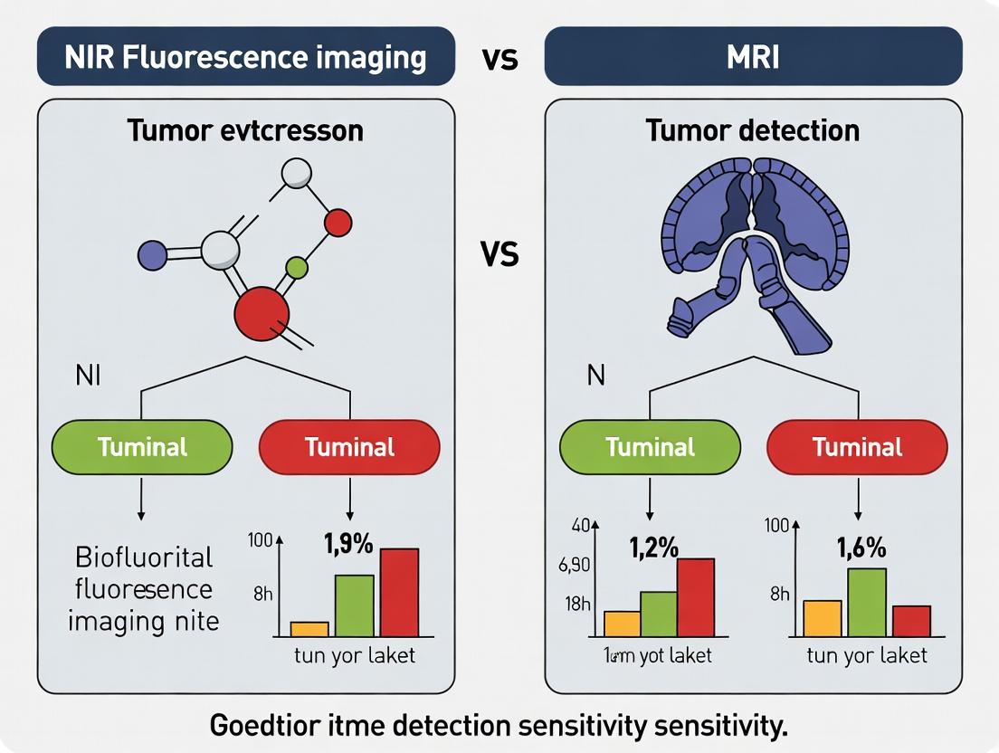
NIR Fluorescence Imaging vs. MRI: A Comparative Analysis of Sensitivity in Tumor Detection for Biomedical Research
This article provides a comprehensive technical analysis for researchers, scientists, and drug development professionals, comparing the fundamental principles and detection sensitivities of Near-Infrared (NIR) fluorescence imaging and Magnetic Resonance Imaging...
NIR Fluorescence Imaging vs. MRI: A Comparative Analysis of Sensitivity in Tumor Detection for Biomedical Research
Abstract
This article provides a comprehensive technical analysis for researchers, scientists, and drug development professionals, comparing the fundamental principles and detection sensitivities of Near-Infrared (NIR) fluorescence imaging and Magnetic Resonance Imaging (MRI) in oncology. We explore the foundational biophysics of signal generation, detail current methodologies and contrast agent applications, address key optimization and troubleshooting challenges in preclinical and clinical translation, and present a rigorous, evidence-based comparison of sensitivity metrics, including limits of detection, depth penetration, and spatial/temporal resolution. The synthesis offers actionable insights for selecting and optimizing imaging modalities to advance diagnostic and therapeutic development.
Understanding the Core Technologies: Biophysical Principles of NIR and MRI Signal Generation in Oncology
This comparison guide is framed within a broader thesis investigating the relative sensitivity of Near-Infrared (NIR) fluorescence imaging and Magnetic Resonance Imaging (MRI) for the preclinical detection of tumors. The fundamental physics underpinning these modalities—photon emission (NIR fluorescence) and nuclear magnetic resonance (MRI)—dictate their performance characteristics, advantages, and limitations in oncological research.
Core Physics & Mechanism Comparison
NIR Fluorescence Imaging relies on the administration of exogenous fluorophores that absorb and re-emit photons in the near-infrared spectrum (typically 650-900 nm). Detection involves measuring the intensity and spatial distribution of this emitted light, which is influenced by tissue scattering and absorption.
MRI (Nuclear Magnetic Resonance Imaging) exploits the magnetic properties of atomic nuclei (primarily hydrogen protons in water and fat). When placed in a strong magnetic field, these nuclei align and can be perturbed by radiofrequency pulses. The subsequent return to equilibrium (relaxation) emits signals used to construct detailed anatomical and functional images.
Quantitative Performance Comparison for Tumor Detection
The following table summarizes key performance metrics based on recent experimental studies.
Table 1: Comparative Performance Metrics: NIR Fluorescence Imaging vs. MRI
| Metric | NIR Fluorescence Imaging (Preclinical) | MRI (Preclinical/Clinical) | Key Implications for Tumor Detection |
|---|---|---|---|
| Spatial Resolution | 1-3 mm (in vivo, diffuse optical tomography); <100 µm (surface/topical) | 50-100 µm (preclinical high-field); 1-2 mm (clinical) | MRI provides superior deep-tissue, volumetric resolution. NIR excels at surface/superficial detail. |
| Temporal Resolution | Seconds to minutes for full image acquisition | Minutes to tens of minutes per scan | NIR allows for real-time imaging of probe dynamics and surgical guidance. |
| Depth of Penetration | Limited to ~1-3 cm in tissue due to light scattering | Unlimited by depth; whole-body capability | MRI is indispensable for deep-seated or whole-body tumor screening. |
| Molecular Sensitivity (Probe Concentration) | ~10-9 to 10-12 M (high sensitivity) | ~10-3 to 10-5 M (low sensitivity) | NIR is superior for detecting sparse molecular targets (e.g., early micrometastases). |
| Anatomical Context | Poor; requires coregistration with CT/MRI | Excellent; provides intrinsic high-contrast anatomy | MRI offers a built-in anatomical roadmap for tumor localization. |
| Quantification | Semi-quantitative; highly dependent on tissue optics | Quantitative parameters possible (T1, T2, ADC) | MRI provides standardized, reproducible biomarkers (e.g., tumor volume, cellularity). |
Experimental Protocols from Key Studies
Protocol 1: Assessing Tumor Targeting Sensitivity with NIR Fluorophores
- Objective: To determine the minimum detectable number of tumor cells expressing a target antigen using a fluorescently labeled antibody.
- Methodology:
- Cell Line & Model: Human cancer cells (e.g., HER2+ breast cancer) are implanted subcutaneously or orthotopically in nude mice.
- Probe Administration: A conjugate of anti-HER2 monoclonal antibody and a NIR fluorophore (e.g., IRDye 800CW) is injected intravenously at a dose of 2-4 nmol/mouse.
- Imaging Protocol: At 24, 48, and 72 hours post-injection, anesthetized mice are imaged using a dedicated small animal fluorescence imager. Excitation: 745 nm; Emission: 800 nm filter.
- Data Analysis: Tumor-to-background ratio (TBR) is calculated as mean fluorescence intensity of tumor region / mean fluorescence intensity of contralateral normal tissue. Ex vivo imaging of excised organs quantifies biodistribution.
- Typical Outcome: TBR > 3 is considered significant. This protocol can detect sub-millimeter clusters of cells due to the high photon sensitivity of the detector.
Protocol 2: Evaluating Tumor Detection Limits with Contrast-Enhanced MRI
- Objective: To define the minimum detectable tumor volume using a clinically relevant gadolinium-based contrast agent.
- Methodology:
- Model: Similar tumor model as above (e.g., orthotopic brain or liver tumor).
- Imaging System: Preclinical 7T or 9.4T MRI scanner.
- Scan Protocol: Pre-contrast T1-weighted and T2-weighted anatomical scans are acquired. The contrast agent (e.g., Gd-DTPA) is injected intravenously at 0.1 mmol/kg.
- Post-contrast Imaging: T1-weighted sequences are repeated immediately and at 5-minute intervals for up to 30 minutes.
- Data Analysis: Tumor volume is segmented from 3D image data. Signal enhancement is calculated as (SIpost - SIpre) / SI_pre * 100%, where SI is signal intensity.
- Typical Outcome: Tumors as small as 0.5-1 mm³ can be visualized, primarily based on anatomical disruption and subtle enhancement patterns. Sensitivity is limited by contrast agent concentration and relaxivity.
Visualizing the Fundamental Detection Pathways
Title: NIR Fluorescence Photon Emission Pathway
Title: Nuclear Magnetic Resonance Signal Generation
The Scientist's Toolkit: Essential Research Reagent Solutions
Table 2: Key Research Reagents and Materials for Comparative Studies
| Item | Function in NIR Studies | Function in MRI Studies |
|---|---|---|
| Targeted Contrast Agent | Antibody-, peptide-, or small molecule-conjugated NIR fluorophore (e.g., IRDye 800CW, Cy5.5). Function: Binds to molecular target (e.g., EGFR, integrin) to provide specific signal. | Gadolinium chelates (e.g., Gd-DOTA), iron oxide nanoparticles, or targeted paramagnetic agents. Function: Alters local proton relaxation times (T1/T2) to enhance contrast. |
| Control Probe | Isotype antibody-conjugated fluorophore or untargeted dye. Function: Differentiates specific vs. non-specific uptake and biodistribution. | Non-enhancing saline or non-targeted contrast agent. Function: Establishes baseline imaging characteristics. |
| Animal Model | Immunocompromised or transgenic mice with subcutaneous/orthotopic tumors. Function: Provides a biological system with relevant physiology and disease. | Same as NIR, often with models in deeper anatomy (brain, liver). Function: Allows correlation of molecular data with high-resolution anatomy. |
| Image Analysis Software | Living Image (PerkinElmer), IVIS Spectrum CT, or open-source tools (FIJI). Function: Quantifies radiant efficiency, TBR, and performs 3D reconstruction. | ParaVision (Bruker), VivoQuant, or Horos. Function: Segments tumors, quantifies volume, and analyzes relaxation time maps. |
| Multimodal Imaging Platform | Hybrid systems like FMT-CT or FMT-MRI. Function: Coregisters functional fluorescence data with anatomical CT or MRI scans for precise localization. | Integrated PET-MRI or SPECT-MRI systems. Function: Combines high-sensitivity molecular data (from nuclear medicine) with superior soft-tissue contrast of MRI. |
The evaluation of imaging sensitivity is central to advancing oncology research, particularly in early tumor detection. This guide compares the intrinsic sensitivity of Near-Infrared (NIR) Fluorescence Imaging and Magnetic Resonance Imaging (MRI), two modalities representing molecular and anatomical paradigms, respectively. The analysis is framed within a thesis investigating their roles in preclinical and translational tumor detection research.
Quantitative Comparison of Sensitivity Metrics
The following table summarizes core sensitivity parameters based on current literature and experimental data.
Table 1: Sensitivity Parameters for NIR Fluorescence Imaging vs. MRI
| Parameter | NIR Fluorescence Imaging (Molecular) | MRI (Anatomical) | Key Implication for Tumor Detection |
|---|---|---|---|
| Detection Limit | ~10-9 to 10-12 mol (pico-nanomolar) | ~10-3 to 10-5 mol (milli-micromolar) | NIR detects sparse molecular targets; MRI detects tissue-level changes. |
| Spatial Resolution | 1-3 mm (in vivo, macroscopic); <100 µm (ex vivo) | 50-500 µm (in vivo, preclinical) | MRI provides superior anatomical localization; NIR excels at surface/ intraoperative mapping. |
| Temporal Resolution | Seconds to minutes | Minutes to hours | NIR allows real-time kinetic studies of agent uptake/clearance. |
| Primary Contrast Source | Administered fluorescent probe targeting biomarkers (e.g., proteases, receptors). | Endogenous tissue properties (T1/T2) or administered contrast agents (e.g., Gadolinium). | NIR sensitivity is probe-dependent and target-specific; MRI sensitivity derives from physicochemical environment. |
| Penetration Depth | < 1-2 cm (in tissue) | Unlimited (full body) | NIR is optimal for surface/superficial or intraoperative imaging; MRI for deep-seated tumors. |
Experimental Protocols & Supporting Data
Key experiments defining sensitivity limits for each modality are detailed below.
Protocol 1: Determining Limit of Detection for a NIR Fluorescent Probe
- Objective: Quantify the minimum detectable amount of a targeted NIR fluorophore (e.g., IRDye 800CW conjugated to a cetuximab antibody) in a tissue-simulating phantom.
- Materials: Serial dilutions of the probe (10 pM to 100 nM) in intralipid/hemoglobin phantom. NIR fluorescence imaging system (e.g., LI-COR Pearl or PerkinElmer IVIS).
- Method:
- Prepare phantom wells with known probe concentrations.
- Image using standardized settings (e.g., 785 nm excitation, 820 nm emission filter, 5 sec acquisition).
- Draw regions of interest (ROIs) and quantify total radiant efficiency ([p/s/cm²/sr] / [µW/cm²]).
- Plot signal vs. concentration. The limit of detection (LOD) is calculated as the concentration corresponding to the mean background signal + 3 standard deviations.
- Typical Result: LOD for IRDye 800CW in a 1 cm deep phantom is typically 50-500 pM, demonstrating nanomolar-picomolar sensitivity.
Protocol 2: Determining Minimum Detectable Tumor Volume via MRI
- Objective: Establish the smallest orthotopic tumor volume reliably detectable with T2-weighted or contrast-enhanced T1-weighted MRI.
- Materials: Preclinical tumor model (e.g., orthotopic glioblastoma), 7T or 9.4T preclinical MRI, Gadoteridol contrast agent.
- Method:
- Image tumor-bearing subjects at regular intervals post-implantation.
- Acquire high-resolution T2-weighted scans (e.g., 100 µm isotropic voxels) and pre/post-contrast T1-weighted scans.
- A blinded radiologist identifies detectable tumors. Volumes are segmented from images.
- The minimum detectable volume is defined as the smallest segmented volume consistently identified across readers.
- Typical Result: Under optimal conditions, preclinical MRI can detect tumors as small as 0.5-1.0 mm³ (~106 cells). Sensitivity is limited by partial volume effects and contrast-to-noise ratio.
Visualizing Workflows and Relationships
Title: NIR Fluorescence Molecular Imaging Workflow
Title: Key Factors Determining MRI Anatomical Sensitivity
Title: Thesis Framework Comparing Imaging Paradigms
The Scientist's Toolkit: Research Reagent Solutions
Table 2: Essential Materials for Comparative Sensitivity Studies
| Item | Function in Research | Example Product/Category |
|---|---|---|
| Targeted NIR Fluorophores | Binds specifically to tumor biomarkers (e.g., EGFR, PSMA) to provide molecular contrast. | IRDye 800CW NHS Ester, Cy7 conjugates, ABY-029 (anti-EGFR affibody). |
| MRI Contrast Agents | Alters local magnetic properties to enhance anatomical contrast in T1 or T2-weighted scans. | Gadoteridol (ProHance), Ferumoxytol (for T2*), targeted Gd-based nanoparticles. |
| Tissue-Simulating Phantoms | Validates sensitivity metrics in controlled, reproducible environments mimicking tissue optics. | Intralipid/ink phantoms, 3D-printed multi-well phantoms with defined optical properties. |
| Image Co-Registration Software | Fuses molecular (NIR) and anatomical (MRI) datasets for precise spatial correlation of signals. | AMIRA, 3D Slicer, Living Image Software co-registration modules. |
| Calibrated Radiometric Imaging Systems | Provides quantitative, reproducible fluorescence data essential for determining LOD. | LI-COR Pearl, PerkinElmer IVIS Spectrum, Fluobeam systems. |
| High-Field Preclinical MRI | Generates high-resolution anatomical images for defining the spatial context of disease. | Bruker BioSpec (7T, 9.4T), Agilent MR systems, MR Solutions compact scanners. |
Sensitivity is context-dependent. NIR fluorescence imaging offers orders of magnitude greater molecular sensitivity, enabling the detection of sparse biomarkers critical for early intervention and therapy guidance. MRI provides superior anatomical sensitivity for localizing deep, small-volume tumors within complex tissue architecture. The evolving thesis in oncology imaging posits not a competition, but a synergy, where the molecular specificity of NIR and the anatomical precision of MRI are integrated—via multimodal imaging or hybrid agents—to achieve comprehensive tumor characterization.
Within the broader thesis comparing NIR fluorescence imaging versus MRI for tumor detection sensitivity, the evolution of contrast agents is a pivotal factor. The transition from non-targeted, perfusion-based agents to molecularly-targeted agents fundamentally shifts the paradigm from visualizing anatomical consequences of disease to detecting specific molecular expressions. This guide compares the performance of these agent classes in preclinical and clinical research, directly impacting the sensitivity metrics central to the NIR vs. MRI debate.
Comparison Guide: Non-Targeted vs. Molecularly-Targeted Contrast Agents
The following tables compare key performance characteristics, supported by representative experimental data.
Table 1: Fundamental Performance Characteristics
| Characteristic | Non-Targeted Agents (e.g., Gd-DTPA, ICG) | Molecularly-Targeted Agents (e.g., αvβ3-integrin-targeted NIR dye/Gd-chelate) | Implications for NIR vs. MRI Sensitivity |
|---|---|---|---|
| Primary Mechanism | Passive accumulation via Enhanced Permeability and Retention (EPR) or blood pool imaging. | Active binding to specific cell-surface biomarkers (e.g., receptors, enzymes). | Targeted agents enhance signal at specific sites, improving tumor-to-background ratio (TBR), a key sensitivity determinant. |
| Key Metric: Tumor-to-Background Ratio (TBR) | Low to moderate (e.g., 1.5 - 3.0). Relies on vascular leakiness. | High (e.g., 5.0 - 15.0+). Driven by specific binding and retention. | Higher TBR directly increases detection sensitivity for small or diffuse tumors, benefiting both modalities. |
| Specificity | Low. Accumulates in any tissue with increased vascular permeability (inflammation, trauma). | High. Binds only to cells expressing the target antigen. | High specificity reduces false positives, improving the positive predictive value of detection. |
| Optimal Imaging Time Window | Minutes to hours post-injection (pharmacokinetic-dependent). | Hours to days post-injection (allows for blood clearance of unbound agent). | Targeted NIR imaging often requires longer wait times but yields superior contrast. MRI can leverage this for delayed, high-resolution scans. |
| Quantification Potential | Semi-quantitative (e.g., measuring perfusion parameters Ktrans). | Potentially fully quantitative (allowing estimation of target expression levels). | Molecular quantification is a superior form of sensitivity, moving beyond detection to characterization. |
Table 2: Representative Experimental Data from Preclinical Studies
| Study Focus | Non-Targeted Agent (Data) | Molecularly-Targeted Agent (Data) | Experimental Model | Conclusion |
|---|---|---|---|---|
| Detection Sensitivity (Minimum Detectable Tumor Volume) | MRI with Gd-DTPA: ~50 mm³. NIR with ICG: ~10 mm³ (surface-weighted). | MRI with αvβ3-targeted-Gd: ~20 mm³. NIR with αvβ3-targeted-Cy5.5: ~1 mm³. | Mouse xenograft (U87MG glioblastoma). | Targeted agents significantly lower detection limits. NIR fluorescence offers superior sensitivity to MRI for surface/near-surface lesions with targeted agents. |
| Signal-to-Noise Ratio (SNR) / TBR at 24h | NIR ICG SNR: ~8.2, TBR: ~2.1. | NIR Targeted-Cy5.5 SNR: ~25.4, TBR: ~8.7. | Mouse xenograft (CT26 colon carcinoma). | Molecular targeting provides >3-fold improvement in TBR, dramatically improving detection confidence and sensitivity in optical imaging. |
| Multimodal (NIR/MRI) Targeting Validation | N/A (no specific target). | Ex vivo fluorescence intensity correlates with MRI R1 relaxation rate increase (R²=0.89). | Mouse model of angiogenesis. | Correlative data validates that the targeted enhancement is specific and quantifiable across modalities, with NIR providing rapid screening and MRI providing anatomical validation. |
Detailed Experimental Protocols
Protocol 1: In Vivo Comparison of Tumor Targeting Efficiency
- Objective: Quantify the time-dependent tumor accumulation and TBR of a targeted vs. non-targeted NIR agent.
- Materials: See "The Scientist's Toolkit" below.
- Method:
- Animal Model: Implant tumor cells (e.g., 4T1 mammary carcinoma) subcutaneously in nude mice (n=5/group).
- Agent Administration: At tumor volume ~100 mm³, inject via tail vein: Group A with non-targeted NIR dye (ICG-equivalent, 2 nmol). Group B with targeted NIR agent (e.g., EGFR-targeted dye, 2 nmol).
- Imaging: Use a calibrated NIR fluorescence imaging system. Acquire images pre-injection and at 5 min, 1h, 4h, 24h, and 48h post-injection. Maintain consistent exposure time, f-stop, and field of view.
- Quantification: Using region-of-interest (ROI) analysis, measure mean fluorescence intensity (MFI) in the tumor and a contralateral normal tissue area. Calculate TBR = (Tumor MFI - Background) / (Normal Tissue MFI - Background).
- Validation: After final imaging, euthanize animals. Excise tumors and key organs for ex vivo imaging and histological validation (e.g., immunohistochemistry for target expression).
Protocol 2: Correlative MRI and NIR Imaging of a Targeted Integrin Agent
- Objective: Demonstrate concordance between MRI contrast enhancement and NIR fluorescence from a dual-modal targeted probe.
- Materials: See "The Scientist's Toolkit" below.
- Method:
- Probe: Use a single agent comprising a cyclic RGD peptide (targeting αvβ3 integrin) conjugated to both a Gd-chelate (for MRI) and a NIR fluorophore (e.g., Cy5.5).
- Imaging Schedule: Image tumor-bearing mice (U87MG xenograft) at baseline using both a 7T MRI (T1-weighted sequences) and an NIR imager.
- Post-Injection Imaging: Inject the dual-modal probe (0.1 mmol Gd/kg). Perform MRI at 1h and 24h post-injection. Perform NIR imaging immediately after each MRI session.
- Data Co-registration & Analysis: Calculate percent enhancement in MRI signal and NIR fluorescence intensity in the tumor ROI. Plot correlation between MRI ΔR1 (1/T1 change) and ex vivo NIR fluorescence intensity of excised tumors.
Pathway and Workflow Diagrams
Title: Contrast Agent Accumulation Pathways
Title: Correlative Multi-Modal Imaging Workflow
The Scientist's Toolkit: Research Reagent Solutions
| Item | Function & Relevance |
|---|---|
| Near-Infrared Fluorophores (e.g., Cy5.5, IRDye 800CW) | Provides optical contrast in the "tissue-transparent" window (650-900 nm), minimizing autofluorescence and enabling deeper tissue penetration for sensitive detection. |
| Gadolinium-Based Chelates (e.g., Gd-DOTA, Targeted-Gd constructs) | Provides T1 shortening for positive contrast in MRI. The chelate structure ensures safety and biocompatibility; conjugation allows for targeting. |
| Peptide Targeting Moieties (e.g., cRGD, EGF, octreotate) | Provides molecular specificity by binding to overexpressed receptors on tumor cells (αvβ3, EGFR, somatostatin receptors). The key component of targeted agents. |
| Bifunctional Chelators & Crosslinkers (e.g., DOTA-NHS, maleimide-thiol chemistry) | Enables stable chemical conjugation of targeting peptides, fluorophores, and metal chelates into a single functional probe. |
| Small Animal Imaging Systems (NIR Fluorescence Imagers, 7T-11T MRI) | Essential platforms for in vivo data collection. Calibrated, quantitative NIR imagers are crucial for accurate TBR measurement. High-field MRI provides anatomical co-registration. |
| Image Analysis Software (e.g., ImageJ, OsiriX, Living Image) | For ROI analysis, 3D reconstruction, signal quantification, and co-registration of multi-modal datasets to extract comparative sensitivity metrics. |
This guide is framed within a broader research thesis comparing Near-Infrared (NIR) Fluorescence Imaging and Magnetic Resonance Imaging (MRI) for tumor detection sensitivity. The primary figures of merit—signal-to-noise ratio (SNR) and background signal—are fundamental to understanding the performance, advantages, and inherent limitations of each modality in preclinical and clinical research.
Comparative Performance Analysis: NIR Fluorescence vs. MRI
The following table summarizes key performance metrics based on recent experimental studies and literature.
Table 1: Quantitative Comparison of Key Performance Metrics
| Metric | NIR Fluorescence Imaging | Magnetic Resonance Imaging (MRI) | Experimental Basis |
|---|---|---|---|
| Theoretical Sensitivity | ~ pico to nanomolar (10-9 - 10-12 M) | ~ micromolar (10-3 - 10-6 M) | Target agent concentration required for detection. |
| Spatial Resolution | 1-3 mm (in vivo, diffuse); ~ μm (ex vivo) | 50-500 μm (in vivo, preclinical) | Standard in vivo preclinical imaging setup. |
| Temporal Resolution | Seconds to minutes | Minutes to hours | Time to acquire a complete dataset. |
| Penetration Depth | 1-2 cm (optimally in NIR-I/II windows) | Unlimited (full body) | Depth at which useful signal can be retrieved. |
| Primary Background Source | Tissue autofluorescence, non-specific probe uptake, ambient light. | Physiological motion, non-specific contrast agent uptake, instrumental noise. | Major contributors to noise/background. |
| Key Advantage | Ultra-high sensitivity, real-time kinetics, multiplexing potential. | Excellent anatomical context, unlimited penetration, high soft-tissue contrast. | Inherent strength of the modality. |
| Key Limitation | Limited depth penetration, quantification challenges, scattering. | Low molecular sensitivity, slow acquisition, high cost/operational complexity. | Fundamental constraint. |
Detailed Experimental Protocols
To generate the comparative data above, standardized experimental models are employed. Below are summarized protocols for key benchmark studies.
Protocol 1: In Vivo Tumor Detection Sensitivity Limit
- Objective: Determine the minimum number of tumor cells detectable by each modality.
- Animal Model: Murine xenograft with graded inoculations of luciferase+/fluorescence+ tumor cells.
- NIR Fluorescence Protocol:
- Inject tumor-bearing mouse with 2 nmol of a targeted NIR-II dye (e.g., IRDye 800CW conjugate).
- Allow 24-48 hours for biodistribution and clearance.
- Anesthetize mouse and image using a calibrated NIR imager (e.g., LI-COR Pearl, In-Vivo Master).
- Acquire fluorescence signal (ex: 785 nm, em: 820 nm) and white light reference.
- Quantify signal over tumor region of interest (ROI), subtract background from contralateral tissue ROI.
- MRI Protocol:
- Inject mouse with a clinical-grade Gd-based or novel high-relaxivity tumor-targeted contrast agent (e.g., 0.1 mmol/kg).
- Place mouse in preclinical MRI (e.g., 7T or 9.4T Bruker system).
- Acquire T1-weighted sequences pre- and post-contrast (e.g., RARE sequence).
- Calculate contrast-to-noise ratio (CNR) from pre- and post-contrast images in tumor ROI.
Protocol 2: Signal-to-Noise & Background Characterization
- Objective: Quantify SNR and specific vs. non-specific signal in a controlled metastasis model.
- Phantom & Model: Mouse with orthotopic primary tumor and micrometastases in liver/lungs.
- Procedure:
- Administration: Systemic injection of a tumor protease-activated fluorescent probe (for NIR) or a macrophage-targeted iron oxide nanoparticle (for MRI).
- NIR Imaging: Perform longitudinal imaging at 2, 24, 48, and 72h post-injection. Calculate SNR as (Mean Tumor Signal - Mean Muscle Signal) / Std. Dev. of Background Noise.
- MRI Imaging: Perform T2*-weighted scans at peak agent accumulation (e.g., 24h). Calculate CNR.
- Validation: Ex vivo histology (fluorescence microscopy for NIR, Prussian blue stain for MRI) is the gold standard to confirm detection accuracy and identify false positives/negatives.
Visualization of Core Concepts
Title: Determinants of Tumor Signal Detectability
Title: Comparative Experimental Workflow for SNR Assessment
The Scientist's Toolkit: Research Reagent Solutions
Table 2: Essential Research Reagents & Materials
| Item | Function in Research | Example/Catalog |
|---|---|---|
| NIR-I Fluorophores | Emit between 700-900 nm; used for surface/ intraoperative imaging. | IRDye 800CW, Cy7, Alexa Fluor 790. |
| NIR-II Fluorophores | Emit >1000 nm; deeper penetration, reduced scattering vs. NIR-I. | IR-1061, CH-1055, quantum dots. |
| Activated (Smart) Probes | Fluorescence/MRI signal activates only upon target interaction (e.g., protease cleavage). | Prosense/MMPSense (PerkinElmer), Gadolinium-based activatable agents. |
| Targeted Contrast Agents | Nanoparticles or conjugates that bind specifically to tumor biomarkers (e.g., EGFR, Integrins). | Targeted Iron Oxide Nanoparticles, Gd-labeled Affibody molecules. |
| MRI Contrast Agents (T1) | Shorten T1 relaxation time, creating bright signal in T1-weighted images. | Gadobutrol (Gadovist), Gd-DOTA. |
| MRI Contrast Agents (T2/T2*) | Shorten T2 relaxation time, creating dark signal in T2-weighted images. | Ferumoxytol (Feraheme), SPIONs. |
| Matrigel | Basement membrane matrix for consistent tumor cell inoculation in xenograft models. | Corning Matrigel, Growth Factor Reduced. |
| Image Analysis Software | For ROI quantification, 3D reconstruction, and SNR/CNR calculation. | Living Image (PerkinElmer), Horos, ImageJ, Matlab. |
| Calibration Phantoms | Tools to standardize fluorescence intensity or MRI signal across instruments and sessions. | NIR fluorescence phantom set (e.g., from LI-COR), MRI multi-echo phantoms. |
Methodological Deep Dive: Protocols, Probes, and Preclinical to Clinical Translation
This comparison guide evaluates critical components of the NIR fluorescence imaging (NIRF) workflow, providing objective performance data within the context of a broader thesis investigating NIRF versus MRI for tumor detection sensitivity. The enhanced depth penetration and high signal-to-background ratio of NIR light (700-1700 nm) are key advantages under investigation.
Comparison of NIR Fluorophore Classes for Tracer Design
The choice of fluorophore is fundamental to tracer performance. The table below compares major classes based on critical photophysical and pharmacological properties.
Table 1: Performance Comparison of NIR Fluorophore Classes
| Fluorophore Class | Peak Emission (nm) | Molar Extinction Coefficient (ε, M⁻¹cm⁻¹) | Quantum Yield (Φ) | In Vivo Stability | Renal Clearance | Key Advantage | Key Limitation |
|---|---|---|---|---|---|---|---|
| Organic Dyes (e.g., ICG, Cy7) | ~800 nm | ~200,000 | 0.05-0.15 | Low; binds plasma proteins | Hepatic | FDA-approved (ICG); low cost | Poor photostability; broad spectra |
| Cyanine Dyes (e.g., Cy7.5, IRDye800CW) | 750-850 nm | 250,000-300,000 | 0.1-0.2 | Moderate | Mixed | Bright; commercial conjugates | Moderate in vivo aggregation |
| Quantum Dots (QDs) | Tunable 700-1350 nm | 1,000,000+ | 0.5-0.8 | Very High | None; long retention | Extreme brightness & stability | Potential heavy metal toxicity |
| Carbon Nanotubes (SWCNTs) | 1000-1400 nm (NIR-II) | N/A | 0.01-0.1 | Very High | None | Superior tissue penetration (NIR-II) | Low quantum yield; complex functionalization |
| Lanthanide-Doped Nanoparticles | 980, 1500 nm | N/A | Varies | High | None | No photobleaching; sharp emissions | Low emission intensity; complex synthesis |
Experimental Protocol (Cyclic RGD Peptide Conjugate Comparison):
- Tracers Synthesized: cRGDfK peptide conjugated to: a) IRDye800CW, b) Cy7.5, c) PEG-coated CdSeTe/ZnS QD (emission 800nm).
- Animal Model: Female nude mice with subcutaneously implanted U87MG glioblastoma tumors (~150 mm³).
- Administration: Each tracer (2 nmol in 100 µL PBS) injected intravenously via tail vein (n=5 per group).
- Image Acquisition: Longitudinal imaging at 1, 4, 24, 48 h post-injection using a commercially available NIRF imaging system (e.g., LI-COR Pearl or PerkinElmer IVIS) with 745 nm excitation and 800 nm emission filters. MRI (7T, T1-weighted post-Gd-DTPA) performed at 24h for anatomical correlation.
- Quantification: Tumor-to-background ratio (TBR) calculated as mean fluorescence intensity (MFI) of tumor region / MFI of contralateral muscle region.
- Results Summary: At 24h, QD-cRGD showed the highest TBR (8.5 ± 1.2) but significant liver/spleen retention. IRDye800CW-cRGD provided a TBR of 5.2 ± 0.8 with predominant renal clearance. Cy7.5-cRGD showed faster clearance and a lower TBR of 3.1 ± 0.5 at peak (4h). MRI provided superior anatomical context but a lower differential tumor-enhancement ratio (3.1 ± 0.6).
Diagram Title: Key Components of a Targeted NIR Fluorescence Tracer
Comparison of Image Acquisition Systems
The performance of a tracer is contingent on the imaging hardware. This table compares common preclinical NIRF imaging modalities relevant to sensitivity studies.
Table 2: Performance Comparison of Preclinical NIRF Imaging Modalities
| System Type | Depth Penetration | Spatial Resolution | Acquisition Speed | Sensitivity (pmol) | Co-Registration Capability | Key Advantage | Key Limitation |
|---|---|---|---|---|---|---|---|
| Planar Reflectance Imaging | < 1 cm | 1-5 mm (depth-dependent) | Very Fast (sec) | 50-100 | Optional (X-ray) | High-throughput, easy to use | Low depth resolution; signal scattering |
| Fluorescence Tomography (FMT) | 5-10 cm | 1-3 mm | Slow (min) | 10-50 | Yes (CT/MRI) | 3D quantification; deeper penetration | Slow; computationally intensive |
| Hybrid OI (e.g., MSOT) | 2-5 cm | 100-300 µm | Moderate (min) | 5-20 | Intrinsic (Ultrasound) | High resolution & spectral unmixing | Limited field of view; cost |
| NIR-II Imaging Systems | 3-8 cm | 20-50 µm (superficial) | Fast (sec) | 1-10 | Optional | Reduced scattering; superb resolution | Specialized detectors needed |
Experimental Protocol (Sensitivity Limit Detection):
- Sample Preparation: Serial dilutions of IRDye800CW in PBS (1000 to 0.1 pmol in 50 µL) pipetted into a black 96-well plate.
- System Comparison: The same plate was imaged on: a) LI-COR Odyssey CLx (Planar), b) PerkinElmer FMT 2500 (Tomography), c) Spectral Instruments Lago X (with 800nm filter). Identical exposure times (1 sec) and fields of view were used where applicable.
- Analysis: Signal-to-noise ratio (SNR) calculated for each well. The limit of detection (LOD) was defined as the concentration yielding SNR ≥ 3.
- Results Summary: Planar imaging (LOD: 25 pmol) was fastest but suffered from signal saturation at high concentrations. FMT provided linear quantification across the range with an LOD of 8 pmol but required a 5-minute scan. The latest NIR-II system (equipped with InGaAs camera) imaging a NIR-II dye (IR-12) achieved an LOD of 1.5 pmol, demonstrating superior sensitivity in a phantom model.
Diagram Title: Standard Preclinical NIR Fluorescence Imaging Workflow
The Scientist's Toolkit: Essential Research Reagent Solutions
| Item / Reagent | Function in NIRF Workflow |
|---|---|
| IRDye800CW NHS Ester | A widely used, commercial cyanine dye for facile antibody/peptide conjugation due to its reactive succinimidyl ester group. |
| Integrin αvβ3-Targeted cRGD Peptides | A common targeting motif for evaluating tumor angiogenesis in preclinical models. |
| Matrigel | Used for establishing orthotopic or subcutaneous tumor xenografts with a stromal component. |
| DPBS (Fluorescence Grade) | Used for tracer dilution and injections to minimize background autofluorescence. |
| Hair Removal Cream | Critical for reducing strong background signal from scattering in fur-covered animal models prior to imaging. |
| Liquid Tissue Opaque Mask | Applied to cover organs (e.g., liver, intestines) during ex vivo imaging to prevent signal bleed-through. |
| Fluorescence-Compatible Fixative (e.g., Neutral Buffered Formalin) | Preserves tissue architecture and fluorescence signal for subsequent histology and microscopy. |
| Mounting Media with Anti-fade Agents (e.g., ProLong Diamond) | Preserves fluorophore signal intensity during confocal microscopy validation of tracer localization. |
| NIR Fluorescent Microspheres (e.g., 800 nm) | Used as fiducial markers for image co-registration between NIRF and MRI/CT modalities. |
| IVIS SpectrumCT or LI-COR Pearl Impulse | Integrated systems combining planar NIRF with CT for anatomical co-registration, streamlining validation against MRI. |
This comparison guide, framed within a broader thesis comparing NIR fluorescence imaging and MRI for tumor detection sensitivity, objectively evaluates core MRI pulse sequences and contrast-enhanced techniques. The analysis is intended for researchers, scientists, and drug development professionals, providing foundational data relevant to cross-modal sensitivity studies.
Pulse Sequence Comparison for Tumor Detection
The following table summarizes the performance characteristics of key MRI pulse sequences in tumor imaging.
| Sequence | Primary Use Case | Key Strength (vs. Alternatives) | Key Limitation | Typical Spatial Resolution | Relative Scan Time | Tumor Contrast Basis |
|---|---|---|---|---|---|---|
| T2-Weighted FSE/TSE | Anatomical localization, edema, cystic components | Excellent soft-tissue contrast for fluid-rich tumors. | Poor for hypovascular or small lesions. | 0.4 x 0.4 x 3 mm³ | Medium | Native T2 relaxation time. |
| T1-Weighted SE/GRE | Pre-contrast anatomy, post-contrast enhancement | Excellent for post-contrast enhancement detection. | Intrinsic tissue contrast often insufficient pre-contrast. | 0.4 x 0.4 x 3 mm³ | Short (GRE) to Long (SE) | Native T1 relaxation time. |
| Diffusion-Weighted Imaging (DWI) | Cellularity assessment, tumor characterization | High sensitivity to cellular density (e.g., vs. necrosis). | Susceptibility artifacts, low spatial resolution. | 1.5 x 1.5 x 5 mm³ | Short | Brownian motion of water (ADC). |
| Dynamic Susceptibility Contrast (DSC) | Perfusion, cerebral blood volume (CBV) | High sensitivity to microvascular density and perfusion. | Susceptibility artifacts, qualitative metrics complex. | 1.5 x 1.5 x 5 mm³ | Medium | T2/T2* signal drop from Gd bolus. |
| Dynamic Contrast-Enhanced (DCE) | Permeability (Ktrans), vascularity | Quantifies endothelial permeability and extracellular volume. | Requires complex pharmacokinetic modeling. | 0.8 x 0.8 x 3 mm³ | Long | T1 signal increase from Gd uptake. |
Contrast-Enhanced Technique Protocols
Protocol 1: Dynamic Contrast-Enhanced (DCE) MRI for Pharmacokinetic Modeling
Objective: Quantify tumor microvascular permeability (Ktrans) and extracellular extravascular volume (ve).
- Patient Positioning: Place patient in scanner; use dedicated coil for anatomical region.
- Pre-Contrast Scans:
- Acquire high-resolution T1-weighted 3D gradient-echo map (variable flip angles: e.g., 2°, 5°, 10°, 15°).
- Acquire T2-weighted anatomical reference.
- Contrast Administration:
- Use power injector to administer Gadolinium-based contrast agent (GBCA) (0.1 mmol/kg) at 2-3 mL/s, followed by saline flush.
- Dynamic Acquisition:
- Initiate rapid T1-weighted 3D gradient-echo sequence (temporal resolution ~5-10 sec) concurrently with injection.
- Continue acquisition for 5-10 minutes.
- Data Analysis: Use dedicated software to register images, convert signal intensity to Gd concentration, and fit data to pharmacokinetic model (e.g., Tofts model) to generate parametric maps (Ktrans, ve).
Protocol 2: Dynamic Susceptibility Contrast (DSC) MRI for Perfusion
Objective: Measure relative cerebral blood volume (rCBV) and flow (rCBF) in brain tumors.
- Patient Positioning: Standard head coil positioning.
- Pre-Contrast Scans: Acquire T1-weighted and T2-weighted anatomical images.
- Sequence Setup: Use T2*-weighted echo-planar imaging (EPI) sequence for high temporal resolution.
- Contrast Administration: Administer GBCA (0.1-0.2 mmol/kg) as a compact bolus at 4-5 mL/s.
- Dynamic Acquisition: Start EPI sequence (TR/TE ~1500-2000/30-50 ms) 30 seconds before injection. Continue for 60-90 seconds post-injection.
- Data Analysis: Generate signal intensity vs. time curve. Calculate rCBV from the area under the curve of contrast-induced signal drop. Use deconvolution algorithms to estimate rCBF.
Visualizing Multi-Parametric MRI in Tumor Research
Title: Multi-Parametric MRI Workflow for Tumor Phenotyping
Title: DCE-MRI Pharmacokinetic Model (Tofts)
Research Reagent Solutions & Essential Materials
| Item | Function in MRI Tumor Protocols |
|---|---|
| Gadolinium-Based Contrast Agent (GBCA) | Shortens T1 relaxation time, enabling visualization of vascular permeability and perfusion in DCE/DSC-MRI. |
| Power Injector | Ensures precise, consistent, and rapid bolus administration of contrast for dynamic studies (DCE/DSC). |
| Dedicated RF Coils | Optimize signal-to-noise ratio (SNR) for specific anatomical regions (e.g., head/neck, breast, torso). |
| Phantom Kits (for QA) | Used for routine scanner calibration and validation of quantitative sequences (DWI, DCE). |
| Pharmacokinetic Modeling Software | Analyzes DCE-MRI time-series data to calculate quantitative parameters (Ktrans, ve). |
| Motion Correction Software | Minimizes artifacts from patient breathing or movement during long acquisitions. |
The pursuit of early and precise tumor detection drives the development of advanced molecular imaging probes. This guide is framed within a thesis investigating the comparative sensitivity of NIR fluorescence imaging versus MRI. NIR fluorophores excel in ultra-high sensitivity for superficial or intraoperative imaging but have limited penetration depth. MRI offers superb anatomical penetration and resolution but suffers from lower inherent molecular sensitivity. "Smart" activatable probes for both modalities aim to overcome these limitations by providing enhanced contrast only in the presence of a specific tumor biomarker, thereby improving the signal-to-background ratio and detection specificity.
Performance Comparison: Smart NIR Fluorophores
Smart (activatable) NIR fluorophores remain dark (quenched) until they encounter a specific tumor-associated stimulus (e.g., enzyme, pH), leading to fluorescence emission.
Table 1: Comparison of Recent Smart Activatable NIR Fluorophores
| Probe Name (Example) | Activation Mechanism | Target/Stimulus | Turn-On Ratio (Tumor/Background) | Peak Emission (nm) | Key Experimental Finding |
|---|---|---|---|---|---|
| QM-HMI | Enzyme-Specific Cleavage | Cathepsin B | ~12-fold | 820 | Demonstrated real-time visualization of orthotopic glioblastoma in mice with high specificity. |
| Cypate-2CPG | Dual (pH & Caspase-3) | Tumor Acidity & Apoptosis | ~15-fold (pH) / ~8-fold (Caspase) | 830 | Enabled discrimination of therapy-induced apoptosis from non-specific necrosis in vivo. |
| Probe 1 | γ-Glutamyl Transpeptidase (GGT) | GGT (Overexpressed in HCC) | ~25-fold | 790 | Achieved detection of hepatic micrometastases (<1 mm diameter) in mouse models. |
Experimental Protocol for Evaluating Smart NIR Probes (Typical In Vivo Validation):
- Probe Administration: Inject the activatable probe intravenously into tumor-bearing mouse models (e.g., subcutaneous xenograft, orthotopic model).
- Imaging Time Course: Perform longitudinal NIR fluorescence imaging (e.g., using IVIS Spectrum or similar system) at predetermined time points (e.g., 0, 2, 6, 12, 24h post-injection).
- Control Groups: Include mice injected with: a) a always-on (non-activatable) control probe, b) the activatable probe in mice pre-treated with an enzyme inhibitor (for enzyme-activatable probes).
- Ex Vivo Validation: At endpoint, harvest tumors and major organs for ex vivo imaging to quantify biodistribution and calculate tumor-to-background ratios (TBR).
- Data Analysis: Quantify fluorescence intensity in regions of interest (ROIs). Calculate TBR and turn-on ratio (Intensitytumor / Intensitymuscle or normal tissue). Perform statistical analysis.
Performance Comparison: Targeted MRI Contrast Agents
Targeted MRI agents are typically paramagnetic (T1) or superparamagnetic (T2) nanoparticles or complexes functionalized with targeting ligands (e.g., antibodies, peptides) that accumulate specifically at tumor sites.
Table 2: Comparison of Recent Targeted MRI Contrast Agents
| Agent Name/Type | Targeting Moisty | Target Receptor | Primary Modality (T1/T2) | Contrast Enhancement (ΔR1 or ΔR2, %) | Key Experimental Finding |
|---|---|---|---|---|---|
| Gd-DOTA-Folate | Folic Acid | Folate Receptor (FRα) | T1 | ΔR1: +45% in tumor at 1h | Specific enhancement in FRα+ ovarian tumor xenografts, blocked by free folate. |
| SPIO-anti-EGFR | Anti-EGFR Antibody | Epidermal Growth Factor Receptor | T2 | ΔR2: -32% in tumor signal intensity | Enabled detection of small, early-stage pancreatic lesions in genetically engineered mice. |
| Mn-Porphyrin-VIP | Vasoactive Intestinal Peptide (VIP) | VIP Receptor | T1 | ΔR1: +52% in tumor at 2h | Demonstrated superior delineation of breast cancer margins compared to non-targeted agent. |
Experimental Protocol for Evaluating Targeted MRI Agents:
- Agent Administration: Intravenous injection of the targeted contrast agent into animal models.
- MRI Acquisition: Pre-contrast and serial post-contrast MRI scans are performed on a high-field small animal MRI system (e.g., 7T or 9.4T). Standard sequences include T1-weighted GRE or SE, and T2-weighted FSE or RARE.
- Quantification: ROI analysis is performed on tumor and reference tissue (e.g., muscle). For T1 agents, signal enhancement is calculated. For quantitative analysis, longitudinal (R1) or transverse (R2) relaxation rates are measured pre- and post-injection.
- Specificity Controls: Key controls include: a) administration of a non-targeted version of the agent, b) pre-blocking of the target receptor with an excess of free targeting ligand before agent injection.
- Validation: Correlation with ex vivo histology (e.g., immunohistochemistry for the target receptor, Prussian blue staining for iron oxide agents) is essential to confirm specific binding.
Visualization of Key Concepts
Diagram 1: Mechanism of a Smart Activatable NIR Probe
Diagram 2: Mechanism of a Targeted MRI Contrast Agent
Diagram 3: Thesis Framework for Sensitivity Comparison
The Scientist's Toolkit: Research Reagent Solutions
| Item | Function & Relevance |
|---|---|
| Near-Infrared Fluorescence Imager (e.g., IVIS Spectrum, LI-COR Pearl) | Essential for in vivo and ex vivo quantification of NIR probe signal, providing 2D planar imaging of fluorescence intensity and biodistribution. |
| High-Field Small Animal MRI System (e.g., 7T, 9.4T Bruker BioSpec) | Required for high-resolution anatomical and contrast-enhanced imaging to evaluate targeted MRI agents in rodent models. |
| Protease-Specific Substrate (e.g., Cathepsin B, MMP-2/9 sensitive linkers) | Core chemical component for constructing enzyme-activatable NIR probes. Often used as the cleavable linker between the fluorophore and quencher. |
| Targeting Ligands (e.g., Folate, cRGD peptides, Anti-EGFR Fab' fragments) | Critical for conferring specificity to both NIR and MRI probes. They are conjugated to the probe core to direct it to overexpressed tumor biomarkers. |
| Polymeric Nanocarrier (e.g., PEG-PLGA, Dendrimer) | Commonly used scaffold to conjugate multiple contrast elements (fluorophores, Gd³⁺ chelates) and targeting ligands, enhancing payload and pharmacokinetics. |
| Relaxivity Measurement Phantoms | Standardized tubes containing varying concentrations of MRI agent in agarose, used to precisely measure R1 and R2 relaxation rates for agent characterization. |
| Matrigel | Used for establishing subcutaneous tumor xenografts in mice, providing a scaffold for consistent tumor cell inoculation and growth. |
| Image Analysis Software (e.g., Living Image, Horos, ImageJ) | For ROI analysis, signal quantification, co-localization studies, and generating time-activity curves from imaging data. |
Publish Comparison Guide: NIR Fluorescence vs. MRI for Tumor Detection Sensitivity
This guide compares the performance of near-infrared (NIR) fluorescence imaging and magnetic resonance imaging (MRI) for tumor detection, specifically within the context of hybrid imaging systems and intraoperative guidance. The data is contextualized within a thesis investigating the complementary roles of these modalities.
Table 1: Performance Comparison: NIR Fluorescence Imaging vs. MRI
| Metric | NIR Fluorescence Imaging (e.g., ICG-based) | MRI (e.g., Gd-based T1-weighted) | Key Implication for Integration |
|---|---|---|---|
| Spatial Resolution | 1-2 mm (surface/ shallow) | 0.5-1 mm (whole body, depth-independent) | MRI provides anatomical roadmap; NIR offers high-surface precision. |
| Temporal Resolution | Real-time to seconds | Minutes to tens of minutes | NIR enables real-time intraoperative feedback; MRI is preoperative. |
| Detection Sensitivity | ~pico to nanomolar (for target) | ~micromolar (for contrast agent) | NIR excels in detecting sparse molecular targets due to low background. |
| Tissue Penetration | < 1 cm (for 700-900 nm window) | Unlimited depth | Hybrid systems use MRI for deep lesions, NIR for surface margins. |
| Quantitative Ability | Semi-quantitative (highly dependent on distance/angle) | Robustly quantitative (e.g., pharmacokinetic modeling) | MRI provides baseline quantitation; NIR offers relative, real-time signal changes. |
| Functional/Molecular Data | High (specific agent binding) | Moderate (perfusion, nonspecific extracellular agents) | NIR agents can highlight specific tumor biomarkers during surgery. |
Table 2: Experimental Data from Hybrid Workflow Studies
| Study Focus | Experimental Setup | Key Quantitative Finding | Supporting Hybrid Thesis |
|---|---|---|---|
| Glioblastoma Margin Delineation | Pre-op MRI + intraoperative NIR (5-ALA or ICG-conjugate) | NIR identified residual tumor in 23% of cases where MRI-guided resection suggested complete removal. | NIR provides molecular sensitivity that surpasses MRI's anatomical sensitivity for microscopic disease. |
| Sentinel Lymph Node Biopsy | Preoperative MRI lymphography + intraoperative NIR guidance (ICG:HSA) | NIR improved SLN detection rate to 99.2% vs. 84.7% for blue dye alone. MRI provided preoperative 3D mapping. | Hybrid approach combines MRI's volumetric planning with NIR's real-time visual guidance. |
| Liver Metastasis Resection | Intraoperative MRI + NIR fluorescence imaging | Combined approach reduced positive resection margin rate to 5% vs. 15% for intraop MRI alone. | Integrative approach maximizes both deep-tissue (MRI) and surface-margin (NIR) sensitivity. |
Detailed Experimental Protocols
Protocol 1: Comparative Sensitivity for Sub-millimeter Tumor Nodules
- Objective: Determine the lower limit of detection for peritoneal carcinomatosis nodules using NIR fluorescence and MRI.
- Animal Model: Murine model with disseminated ovarian cancer cells expressing a folate receptor-alpha.
- Imaging Agents: NIR: OTL38 (folate-FITC conjugate); MRI: Gadofosveset (blood pool agent).
- Methodology:
- Cohorts: Animals (n=10/group) imaged 21 days post-inoculation.
- MRI Protocol: T1-weighted sequences pre- and 30 minutes post-MRI contrast agent injection on a 7T scanner.
- NIR Protocol: Administer OTL38 24h prior. Image using an open-field NIR camera (ex: 760 nm, em: 800 nm) immediately post-mortem.
- Validation: Laparotomy with manual nodule counting and histopathology as gold standard.
- Outcome Measure: Number and size of nodules detected by each modality versus histology.
Protocol 2: Intraoperative Guidance Workflow for Hybrid Systems
- Objective: Validate a clinical workflow integrating preoperative MRI data with intraoperative NIR visualization.
- System: FDA-cleared fluorescence-guided surgery system with capability for MR-DICOM import and overlay.
- Procedure:
- Preoperative: Acquire high-resolution contrast-enhanced MRI. Segment tumor volume using 3D Slicer software.
- Registration: In the operating room, perform surface-based or fiducial-based registration of the patient to the preoperative MRI model.
- Intraoperative: Administer NIR contrast agent (e.g., ICG). Display the real-time NIR video feed alongside the registered 3D MRI model in the navigation system.
- Data Correlation: Record locations where NIR signal extends beyond the MRI-defined tumor boundary for postoperative analysis.
- Outcome Measure: Volume of tissue resected based on NIR signal beyond MRI-planned margin, and its histopathological status.
Visualization Diagrams
Diagram 1: NIR vs MRI Tumor Detection Pathways
Diagram 2: Hybrid Intraoperative Guidance Workflow
The Scientist's Toolkit: Key Research Reagent Solutions
| Item | Category | Function in Research |
|---|---|---|
| OTL38 (Cytalux) | NIR Fluorescent Agent | FDA-approved targeting agent for folate receptor-positive tumors. Used to validate NIR sensitivity in clinical and preclinical models. |
| Indocyanine Green (ICG) | NIR Fluorescent Agent | Non-targeted perfusion agent; forms non-covalent complexes with albumin for vascular/lymphatic imaging. The de facto standard for intraoperative NIR. |
| Gadobutrol (Gadovist) | MRI Contrast Agent | High-relaxivity, macrocyclic gadolinium-based agent for high-resolution T1-weighted tumor visualization and pharmacokinetic modeling. |
| IRDye 800CW NHS Ester | NIR Fluorophore | Versatile chemical scaffold for creating antibody- or peptide-targeted NIR probes to study specific biomarker sensitivity. |
| Matrigel | Extracellular Matrix | Used in orthotopic and metastatic tumor models to establish more clinically relevant tumor microenvironments for imaging studies. |
| 3D Slicer / MITK | Software Platform | Open-source platforms for medical image analysis and 3D segmentation of preoperative MRI, essential for planning and registration. |
| Multi-Modal Imaging Phantoms | Calibration Tool | Tissue-simulating phantoms with compartments for MRI and NIR contrast to calibrate and co-register hybrid systems. |
Overcoming Technical Hurdles: Optimization Strategies for Maximizing Sensitivity in Both Modalities
Combating Autofluorescence and Light Scattering in NIR Imaging
Within the broader thesis investigating the comparative sensitivity of NIR fluorescence imaging versus MRI for early tumor detection, a central technical challenge emerges: overcoming tissue autofluorescence and light scattering. These phenomena severely limit signal-to-noise ratio (SNR) and penetration depth. This guide compares leading strategies and reagents designed to combat these issues, providing objective performance data to inform method selection.
Comparison of NIR Imaging Strategies
The effectiveness of an NIR imaging approach is quantified by its ability to improve SNR and penetration. The table below compares three core strategies based on recent experimental findings.
Table 1: Performance Comparison of Key NIR Imaging Strategies
| Strategy | Mechanism | Best Reported Penetration Depth (mm) | SNR Improvement vs. Visible Imaging | Key Limitation |
|---|---|---|---|---|
| NIR-I (700-900 nm) | First biological window; reduced scattering & absorption. | 5-10 mm | 3-5x | Significant autofluorescence & scattering persists. |
| NIR-II (1000-1700 nm) | Second biological window; drastically reduced scattering. | >10 mm | 10-50x | Requires specialized InGaAs detectors; limited dye library. |
| Time-Gated Imaging | Temporal separation of long-lifetime probe signal from short-lived autofluorescence. | 3-8 mm (NIR-I) | 8-15x (in high autofluorescence regions) | Requires pulsed laser & gated detector; complex setup. |
Experimental Protocol: Quantifying Strategy Efficacy
To generate comparative data like that in Table 1, a standardized in vivo phantom experiment is commonly employed.
Protocol: Comparative Penetration and SNR Measurement
- Phantom Preparation: Create a tissue-mimicking phantom using Intralipid (2% v/v) for scattering and hemoglobin (0.1% w/v) for absorption. Embed capillary tubes containing equivalent molar concentrations (100 nM) of NIR-I dye (e.g., IRDye 800CW) and NIR-II dye (e.g., IR-1061) at varying depths (2-12 mm).
- Imaging Setup:
- NIR-I: Use a 785 nm continuous-wave laser for excitation and a silicon CCD camera with an 850 nm long-pass emission filter.
- NIR-II: Use a 1064 nm laser for excitation and an InGaAs camera with a 1300 nm long-pass filter.
- Time-Gated: Use a pulsed 785 nm laser (1 MHz) and a gated intensifier synchronized to capture emission 10-100 ns post-pulse.
- Data Acquisition & Analysis: Acquire images. For each depth, calculate SNR as (Target Signal Mean - Background Mean) / Background Standard Deviation. Plot SNR vs. Depth for each system.
The Scientist's Toolkit: Essential Reagents & Materials
Table 2: Key Research Reagent Solutions for Advanced NIR Imaging
| Item | Function | Example Product/Chemical |
|---|---|---|
| NIR-I Fluorophores | Emit in 700-900 nm range; widely available. | IRDye 800CW, Cy7, Alexa Fluor 790 |
| NIR-II Fluorophores | Emit in 1000-1700 nm range; superior penetration. | IR-1061, CH1055, quantum dots (PbS/CdS) |
| Lanthanide-based Probes | Long luminescence lifetime (>100 µs) for time-gated imaging. | NaYF4:Yb,Er (upconverting nanoparticle) |
| Tissue Phantom Components | Mimic scattering and absorption of biological tissue for calibration. | Intralipid (scattering), India Ink (absorption) |
| Targeting Ligands | Conjugate to fluorophores for specific tumor antigen binding. | Antibodies (anti-EGFR), peptides (RGD), folic acid |
Signaling Pathway & Experimental Workflow
Title: NIR Imaging Problem-Solution Workflow for Tumor Detection
Title: NIR Imaging Experimental Protocol Flow
Addressing Magnetic Field Heterogeneity and Motion Artifacts in MRI
This comparison guide is framed within a broader thesis investigating the relative sensitivities of NIR fluorescence imaging and MRI for deep-tissue tumor detection. While NIR fluorescence excels in molecular specificity and real-time imaging, MRI provides unparalleled soft-tissue contrast and depth penetration without ionizing radiation. A critical limitation of high-field MRI, however, lies in its vulnerability to magnetic field (B0) inhomogeneity and patient motion, which degrade image quality, reduce quantitative accuracy, and can obscure critical diagnostic features, particularly in oncological imaging. This guide objectively compares modern solutions designed to mitigate these artifacts.
Experimental Protocols for Comparison
The following standardized protocols were designed to evaluate the performance of different artifact-correction technologies under controlled conditions.
1. Phantom-Based B0 Homogeneity Assessment:
- Objective: Quantify the ability of a system or sequence to maintain a uniform B0 field.
- Setup: A spherical phantom with known susceptibility properties is placed off-isocenter to induce field distortions. A multi-echo gradient echo (GRE) sequence is run with and without the correction technology enabled.
- Measurement: B0 field maps are calculated from the phase difference between echoes. Homogeneity is reported as the peak-to-peak deviation (in Hz) or standard deviation (in ppm) across a defined ROI in the center of the phantom.
2. Motion-Resolved Liver Tumor Imaging:
- Objective: Assess tumor visualization fidelity in the presence of respiratory motion.
- Setup: A cohort of animals or human subjects with known liver lesions is imaged.
- Protocol A (Standard 2D T2w FSE): Performed during free breathing and with breath-hold.
- Protocol B (Motion-Corrected 3D Sequence): Using navigator-based or self-gated prospective motion correction.
- Measurement: Two blinded radiologists score image quality (1-5 Likert scale). Quantitative metrics include Edge Sharpness (ES) of the tumor boundary and Tumor-to-Liver Contrast-to-Noise Ratio (CNR).
Performance Comparison: Technologies & Systems
Table 1: Comparison of B0 Homogeneity Correction Approaches
| Technology | Principle | Key Advantage | Quantitative Result (Phantom Test) | Best For |
|---|---|---|---|---|
| Higher-Order Active Shimming (e.g., 2nd/3rd order) | Dynamically adjusts coil currents to correct non-linear field variations. | Excellent for large FOVs and high fields (7T+). | Reduced SD from 0.25 ppm to 0.08 ppm. | Research 7T/9.4T systems, fMRI studies. |
| Map-based Post-Processing (e.g., SHARP, V-SHARP) | Computational removal of background field gradients using Laplacian boundary methods. | No scan time penalty; purely software-based. | Improved uniformity in simulation by 70%. | Susceptibility Weighted Imaging (SWI), QSM. |
| Dynamic Frequency Tracking | Real-time adjustment of transmit/receive frequency based on a navigator. | Compensates for slow drift and respiration. | Reduced linewidth broadening by 60% in liver. | Cardiac/Abdominal MR Spectroscopy. |
| Integrated Shim & Gradient Coils | Combines shim and imaging gradient functions for faster, localized adjustment. | Enables slice-wise or volume-wise shimming. | Reduced in-plane inhomogeneity by 40% vs. global shim. | High-resolution brain imaging, spinal cord. |
Table 2: Comparison of Motion Artifact Mitigation Strategies
| Strategy | Method Type | Latency | Impact on Scan Time | Experimental Result (Liver Tumor CNR/Sharpness) |
|---|---|---|---|---|
| Breath-Hold (BH) | Patient Cooperation | N/A | Shortest | CNR: 12.5 ± 2.1, ES: High (but inconsistent) |
| Navigator-Gated 3D (e.g., Pseudo-Cartesian) | Prospective Gating | 1-2 cycles | Increases ~40% | CNR: 15.8 ± 1.5, ES: Consistently High |
| Self-Gated Radial/Spiral (e.g., Stack-of-Stars) | Retrospective Sorting | Minimal | No increase | CNR: 14.2 ± 1.8, ES: High |
| Standard 2D Free Breathing (FB) | None | N/A | Baseline | CNR: 6.4 ± 3.0, ES: Poor/Low |
| Model-Based Motion Correction (Deep Learning) | Post-Processing | Post-scan | No increase | CNR: Improved to 11.0 ± 1.7 from FB data |
The Scientist's Toolkit: Research Reagent & Solutions
Table 3: Essential Materials for Advanced MRI Artifact Characterization
| Item | Function in Research | Example Product/Chemical |
|---|---|---|
| Susceptibility Phantom | Calibrates and quantifies B0 field inhomogeneity; validates correction algorithms. | Spherical phantom doped with NiCl2 or Gd in agarose. |
| Motion Simulation Platform | Provides controlled, reproducible motion for sequence validation. | Programmable robotic stage (e.g., for linear/periodic motion). |
| Fat-Water Phantom | Tests separation algorithms and chemical shift artifact. | Phantom with separate lipid and aqueous compartments. |
| Multi-Echo GRE Sequence | Enables B0 field mapping and T2* quantification. | Vendor pulse sequence (e.g., ME-GRE, MEDIC). |
| 3D-Printed Anatomical Mimic | Provides realistic geometry for testing motion correction in context. | Liver phantom with embedded "lesion" compartment. |
| Navigator Echo Module | A small, rapid acquisition used to detect motion state. | Cross-sectional or pencil-beam navigator package. |
Visualizing Workflows and Relationships
MRI Artifact Mitigation Decision Pathway
Thesis Context: Relating MRI Artifacts to Sensitivity
This comparison guide, situated within a thesis evaluating NIR fluorescence imaging versus MRI for tumor detection sensitivity, objectively assesses the performance of key imaging probe classes based on critical pharmacokinetic (PK) parameters. Optimal PK—governed by dosage, administration timing, and specificity—directly dictates signal-to-noise ratio and thus detection sensitivity.
Comparative PK Performance of Representative Imaging Probes
Table 1: Key Pharmacokinetic and Performance Parameters
| Probe / Agent (Class) | Typical Dose (μmol/kg) | Optimal Imaging Timepoint (Post-Injection) | Key Targeting Mechanism | Tumor-to-Background Ratio (TBR) (Reported Range) | Primary Clearance Pathway |
|---|---|---|---|---|---|
| IRDye 800CW 2-DG (NIR Fluorescent) | 3 - 5 | 4 - 24 hours | GLUT1 transporter (Warburg effect) | 2.5 - 4.5 | Renal / Hepatic |
| Indocyanine Green (ICG) (NIR Fluorescent) | 0.1 - 0.3 (mg/kg) | 1 - 5 minutes (passive) | Passive Enhanced Permeability and Retention (EPR) | 1.5 - 3.0 (varies highly) | Hepatic |
| MM-401 (NIR Fluorescent, cRGD) | 2 - 4 | 6 - 48 hours | αvβ3 Integrin receptor (cRGD peptide) | 4.0 - 8.0 | Renal |
| Gadobutrol (MRI, Gd-based) | 0.1 - 0.3 | 5 - 10 minutes | Passive diffusion, extracellular fluid agent | N/A (Contrast-to-Noise ~10-30%) | Renal |
| Ferumoxytol (MRI, SPION) | 3 - 5 (mg Fe/kg) | 24 - 72 hours | Macrophage phagocytosis (passive/active) | N/A (T2 signal loss) | Reticuloendothelial |
| ABY-029 (NIR Fluorescent, Affibody) | 1 - 3 | 2 - 8 hours | Epidermal Growth Factor Receptor (EGFR) | 5.0 - 12.0 | Renal |
Detailed Experimental Protocols for Key Comparisons
1. Protocol: Quantitative Comparison of TBR for Targeted vs. Untargeted NIR Probes
- Objective: To compare the tumor specificity and optimal imaging window of a targeted probe (cRGD-NIR) vs. a passive EPR probe (ICG).
- Materials: Murine xenograft model (e.g., U87MG for αvβ3), cRGD-conjugated NIR dye (e.g., MM-401), ICG, NIR fluorescence imaging system.
- Procedure:
- Randomize mice (n=5/group) and administer intravenous tail vein injection of either probe at optimized dose (cRGD-NIR: 3 μmol/kg; ICG: 0.2 mg/kg).
- Acquire whole-body fluorescence images at serial time points: 5 min, 1h, 4h, 24h, 48h post-injection. Maintain identical imaging parameters (exposure, f-stop).
- Use region-of-interest (ROI) analysis to quantify mean fluorescence intensity in the tumor and contralateral background tissue.
- Calculate TBR at each time point (TBR = Mean Tumor Intensity / Mean Background Intensity).
- Perform ex vivo imaging of harvested organs to quantify biodistribution.
- Data Interpretation: The cRGD-NIR probe will show a later peak TBR (>6h) that sustains, demonstrating receptor-mediated retention. ICG will show a rapid peak (<1h) with swift clearance, reflecting passive distribution.
2. Protocol: Determining Dose-Linearity for MRI vs. NIR Probes
- Objective: To assess the relationship between administered dose and in vivo signal intensity for a Gd-based MRI agent vs. a NIR fluorescent agent.
- Materials: Tumor-bearing mice, Gadobutrol, IRDye 800CW 2-DG, clinical 3T MRI with small animal coil, NIR imager.
- Procedure:
- Administer escalating doses of each agent (e.g., Gadobutrol: 0.05, 0.1, 0.2 mmol/kg; NIR probe: 1, 3, 6 μmol/kg) to separate animal cohorts (n=3/dose).
- For MRI: Acquire T1-weighted scans pre-injection and at 10 minutes post-injection. Calculate percent enhancement in tumor.
- For NIR: Image animals at 24 hours post-injection. Quantify total radiant efficiency in the tumor region.
- Plot dose versus signal response for each modality.
- Data Interpretation: Gd-based MRI signal often exhibits a linear relationship with dose in the physiological range. NIR fluorescence can show a linear trend at lower doses but is susceptible to self-quenching or signal saturation at higher doses, highlighting a critical dosage optimization parameter.
Visualizations
Title: Factors Governing Imaging Probe Pharmacokinetics
Title: Pharmacokinetic Timeline Dictates Optimal Imaging Window
The Scientist's Toolkit: Essential Research Reagent Solutions
Table 2: Key Reagents for Probe PK and Sensitivity Studies
| Reagent / Material | Primary Function in PK/Sensitivity Research |
|---|---|
| cRGD-Peptides (Cy5.5/800CW conjugated) | Active targeting probes for αvβ3 integrin; used to study receptor-mediated uptake and retention kinetics. |
| IRDye 800CW 2-DG | Metabolic activity probe; benchmarks sensitivity against enhanced glycolysis in tumors. |
| Gadolinium-Based Contrast Agents (e.g., Gadobutrol) | Standard MRI T1-shortening agents; baseline for comparing PK dynamics of non-specific extracellular agents. |
| Ultra-small Superparamagnetic Iron Oxides (USPIOs) | MRI T2/T2* agents; used to study macrophage-driven uptake and long-term pharmacokinetics. |
| Matrigel | Basement membrane matrix for consistent subcutaneous tumor xenograft establishment. |
| IVIS Spectrum or similar NIR Imager | In vivo quantitative fluorescence imaging system for longitudinal TBR and biodistribution studies. |
| Small Animal MRI System (7T-11T) | High-field MRI for anatomical co-registration and quantitative contrast-enhanced kinetics. |
| Image Analysis Software (e.g., Living Image, Horos, Fiji) | For standardized ROI analysis, signal quantification, and TBR/CNR calculation across modalities. |
Thesis Context: NIR Fluorescence Imaging vs. MRI for Tumor Detection Sensitivity
The pursuit of superior sensitivity in tumor detection drives the comparison between Near-Infrared (NIR) fluorescence imaging and Magnetic Resonance Imaging (MRI). While MRI offers excellent anatomical detail, its sensitivity for detecting small, early-stage tumors or micrometastases can be limited, often requiring high concentrations of contrast agents. NIR fluorescence imaging, particularly with advanced targeting probes, promises higher molecular sensitivity but faces challenges with depth penetration and quantification. This guide compares data analysis pipelines designed to extract maximum sensitivity from each modality, enabling researchers to push detection limits.
Comparative Analysis of Sensitivity Enhancement Pipelines
The following table compares core algorithmic approaches applied to NIR fluorescence and MRI data to enhance sensitivity in tumor detection studies.
Table 1: Algorithm Comparison for Sensitivity Enhancement in Tumor Imaging
| Algorithm Category | Primary Modality | Key Function | Impact on Sensitivity (Typical Gain) | Key Trade-off / Artifact Risk |
|---|---|---|---|---|
| Spectral Unmixing (e.g., Linear Discriminant Analysis) | NIR Fluorescence | Separates target signal from tissue autofluorescence. | 3-5x SNR increase in vivo. | Requires prior spectral libraries; noise amplification. |
| Deep Learning Denoising (e.g., U-Net) | MRI / NIR Fluorescence | Reduces stochastic noise in low-signal images. | MRI: 2-4x SNR; NIR: 5-10x SNR in phantom studies. | Potential over-smoothing of fine structures. |
| Compressed Sensing (CS-MRI) | MRI | Accelerates acquisition, allowing more signal averaging. | Enables ~8x faster scans at equal SNR. | Reconstruction artifacts from insufficient data. |
| Time-Gated & Lifetime Analysis | NIR Fluorescence | Rejects short-lived autofluorescence based on photon arrival time. | Up to 10x contrast-to-noise ratio (CNR) improvement. | Requires expensive, specialized hardware. |
| Pharmacokinetic Modeling (e.g., Tofts Model) | Dynamic Contrast-Enhanced MRI (DCE-MRI) | Quantifies perfusion/permeability (Ktrans), detecting physiological changes. | Increases diagnostic sensitivity (~15-20% over standard MRI). | Model dependency; assumes specific vascular geometry. |
Experimental Protocols for Cited Data
Protocol 1: NIR Fluorescence Sensitivity Limit Test with Spectral Unmixing
- Objective: Determine the minimum detectable number of tumor cells expressing a target receptor using a targeted NIR dye.
- Procedure:
- Prepare a dilution series of tumor cells (e.g., 10^6 to 10^2 cells) in a multi-well plate, stained with a receptor-specific NIR-800 dye.
- Image plates using a NIR fluorescence imager across multiple emission filters (e.g., 780-850 nm).
- Acquire control well images (autofluorescence only) from unstained cells.
- Apply a linear unmixing algorithm (non-negative matrix factorization) using control spectra and the pure dye spectrum as references.
- Calculate signal-to-noise ratio (SNR) for each well from the unmixed target signal channel.
- Key Metric: The lowest cell count where SNR > 3 is defined as the detection limit.
Protocol 2: DCE-MRI Sensitivity Enhancement via Pharmacokinetic Modeling
- Objective: Compare the sensitivity of modeled parameter Ktrans versus standard T1-weighted enhancement for detecting small, early tumors in a murine model.
- Procedure:
- Acquire pre-contrast T1 maps of the tumor region.
- Administer Gadolinium-based contrast agent intravenously as a bolus.
- Perform rapid T1-weighted imaging over the tumor for 30 minutes.
- Convert image intensity to contrast agent concentration using the T1 maps.
- Fit the concentration-time curve per pixel to the Extended Tofts Pharmacokinetic Model to generate parametric maps of Ktrans (transfer constant).
- Define tumor boundaries on high-resolution anatomical scans. Compare the CNR of standard enhancement maps versus Ktrans maps within the tumor region relative to normal tissue.
Visualization of Key Concepts
Title: NIR Signal Unmixing Workflow for Sensitivity Gain
Title: Pharmacokinetic Modeling Pipeline for DCE-MRI
The Scientist's Toolkit: Research Reagent Solutions
Table 2: Essential Reagents & Materials for Sensitivity Experiments
| Item | Function in Sensitivity Research | Example Product/Category |
|---|---|---|
| Targeted NIR Fluorescence Dye | Binds specifically to tumor biomarkers (e.g., EGFR, PSMA), providing molecular contrast. | IRDye 800CW conjugated to cetuximab or small molecule ligands. |
| MRI Contrast Agent | Alters local magnetic properties to enhance tissue contrast in perfusion or targeted imaging. | Gadobutrol (macrocyclic), or emerging targeted iron oxide nanoparticles. |
| Phantom for Calibration | Provides a standardized, reproducible target for quantifying sensitivity and limits of detection. | Fluorescent microsphere phantoms; MRI gel phantoms with varying Gd concentrations. |
| Spectral Library Kit | Contains reference samples for key spectral signatures (autofluorescence, dye) required for unmixing algorithms. | Tissue-mimicking slides with fixed fluorophore spectra. |
| Image Analysis Software SDK | Enables implementation of custom denoising, unmixing, and pharmacokinetic modeling algorithms. | MATLAB Image Processing Toolbox, Python (SciKit-Image, TensorFlow), Horos/3D Slicer plugins. |
| Animal Model with Orthotopic Tumors | Provides a biologically relevant environment for testing in vivo sensitivity limits. | Murine models with luciferase-tagged, receptor-positive tumor cell lines. |
Head-to-Head Validation: Quantitative Sensitivity Metrics and Evidence-Based Comparisons
This guide provides a comparative analysis of detection sensitivity between near-infrared (NIR) fluorescence imaging and magnetic resonance imaging (MRI) for preclinical tumor models. The data presented is framed within ongoing research to define the practical lower limits for detecting minimal residual disease and early-stage tumorigenesis.
Experimental Protocols for Cited Studies
- NIR Fluorescence Imaging (IVIS Spectrum): Mice bearing subcutaneous xenografts were injected intravenously with 2 nmol of a fluorescent agent (e.g., IRDye 800CW 2-DG). After a 24-hour biodistribution period, animals were anesthetized and imaged using the following parameters: excitation filter 745 nm, emission filter 800 nm, field of view 12.5 cm, f/stop 2, medium binning. Regions of interest (ROIs) were drawn over the tumor and contralateral background tissue. The limit of detection was defined as a tumor-to-background ratio (TBR) ≥ 2.0.
- MRI (7T preclinical scanner): Mice were anesthetized and placed in a dedicated rodent coil. T2-weighted turbo spin-echo sequences were used for anatomical localization: TR/TE = 2500/33 ms, matrix = 256 × 256, slice thickness = 0.7 mm. For T1-weighted contrast-enhanced imaging, a gadolinium-based contrast agent was administered intravenously at 0.1 mmol/kg. Pre- and post-contrast scans were co-registered. The LOD was defined as the smallest volume where a contiguous, contrast-enhancing lesion could be unambiguously distinguished from normal parenchyma by two blinded radiologists.
Quantitative Comparison of Detection Thresholds
Table 1: Comparison of LOD for Subcutaneous Xenograft Models
| Imaging Modality | Typical Probe/Contrast Agent | Minimum Detectable Cell Number (Approx.) | Minimum Detectable Tumor Volume (mm³) | Key Advantage | Key Limitation |
|---|---|---|---|---|---|
| NIR Fluorescence | IRDye 800CW 2-DG | 1 x 10⁵ | 1 - 2 | High sensitivity, quantitative, low cost per scan | Limited penetration depth (~1 cm), 2D projection only |
| MRI (T2-weighted) | N/A (Anatomical) | 3 - 5 x 10⁶ | 10 - 15 | Excellent anatomical detail, deep tissue penetration | Low molecular sensitivity, poor contrast for early lesions |
| MRI (Contrast-Enhanced) | Gadoteridol | 1 - 2 x 10⁶ | 5 - 8 | 3D localization, functional data (perfusion) | Requires contrast kinetics, more expensive, lower throughput |
Table 2: The Scientist's Toolkit - Key Research Reagent Solutions
| Item | Function in Context |
|---|---|
| IRDye 800CW 2-DG | NIR fluorescent glucose analog used as a metabolic activity probe for tumor detection. |
| Gadoteridol | Gadolinium-based MRI contrast agent that shortens T1 relaxation time in enhancing tissues. |
| Matrigel | Basement membrane matrix used for co-injection with tumor cells to enhance engraftment. |
| Isoflurane Anesthesia System | Provides stable, reversible anesthesia for in vivo imaging sessions in rodents. |
| Lucent LNM-20 Phantoms | Calibration standards with known fluorescent yields for quantitative NIR imaging. |
Visualization of Experimental Workflow and Signaling
Title: Preclinical Tumor Imaging Workflows: NIR vs MRI
Title: NIR Probe Tumor Accumulation Pathway
Comparative Analysis of Spatial Resolution, Depth Penetration, and Temporal Dynamics
This guide provides a comparative analysis of Near-Infrared (NIR) Fluorescence Imaging and Magnetic Resonance Imaging (MRI) within the context of tumor detection sensitivity research, focusing on the core metrics of spatial resolution, depth penetration, and temporal dynamics. The data and experimental protocols herein are critical for evaluating their respective roles in preclinical and clinical oncology research.
Quantitative Performance Comparison
Table 1: Core Performance Metrics for Tumor Imaging
| Metric | NIR Fluorescence Imaging (Preclinical) | MRI (Clinical 3T) |
|---|---|---|
| Spatial Resolution | 1 - 3 µm (Microscopy); 0.5 - 2 mm (Tomography) | 0.5 - 1.5 mm (in-plane) |
| Depth Penetration | 1 - 2 cm (in tissue, NIR-I); up to 5-8 cm (NIR-II) | Unlimited (full body) |
| Temporal Resolution | Seconds to minutes (2D); minutes to hours (3D) | Minutes to tens of minutes per sequence |
| Primary Sensitivity | nM - pM (contrast agent concentration) | µM - mM (contrast agent concentration) |
| Key Limiting Factor | Photon scattering & absorption in tissue | Signal-to-noise ratio (SNR) at high resolution |
Experimental Protocols for Comparative Studies
Protocol 1: In Vivo Tumor Margin Delineation
- Objective: Compare the ability to identify infiltrative tumor margins intraoperatively.
- NIR Method: Administer a tumor-targeted NIR fluorophore (e.g., IRDye 800CW conjugate). After 24h, image the surgical field using an open-field fluorescence imaging system. Excitation: 745-775 nm; Emission: 800-850 nm.
- MRI Method: Pre-operative T1-weighted imaging with a gadolinium-based contrast agent (e.g., Gadobutrol) on a 3T scanner. Use standard tumor protocol sequences.
- Validation: Histopathological analysis of resected margins serves as the gold standard for correlation.
Protocol 2: Kinetics of Nanoparticle Accumulation
- Objective: Quantify the temporal dynamics of a therapeutic nanoparticle in a subcutaneous xenograft model.
- NIR Method: Utilize NIR-labeled nanoparticles. Acquire longitudinal, non-invasive fluorescence molecular tomography (FMT) scans at 0, 2, 6, 12, 24, and 48h post-injection. Quantify mean fluorescence intensity in the tumor region of interest (ROI).
- MRI Method: Use the same nanoparticles loaded with an MRI contrast agent (e.g., iron oxide for T2). Perform longitudinal MRI scans at identical time points. Quantify signal change (ΔR2) in the tumor ROI.
- Analysis: Generate time-activity curves for both modalities and calculate pharmacokinetic parameters (e.g., peak time, retention).
Visualizations
Title: NIR Fluorescence Intraoperative Imaging Workflow
Title: MRI Image Formation Process
The Scientist's Toolkit: Essential Research Reagents & Materials
Table 2: Key Reagents for Comparative Tumor Imaging Studies
| Item | Function & Role in Research |
|---|---|
| NIR-I Fluorophores (e.g., IRDye 800CW) | Conjugated to targeting ligands (antibodies, peptides) for specific molecular visualization in the 700-900 nm window. |
| NIR-II Fluorophores (e.g., SWNT, Quantum Dots) | Emit in 1000-1700 nm range for reduced scattering and improved depth penetration in preclinical models. |
| Gadolinium-Based MRI Contrast Agents (e.g., Gd-DOTA) | Shortens T1 relaxation time of water protons, enhancing vascular and perfusion-related contrast in tumors. |
| Superparamagnetic Iron Oxide Nanoparticles (SPIONs) | Cause local magnetic field inhomogeneities, leading to T2/T2* signal loss, useful for lymph node and macrophage imaging. |
| Dual-Modality Probes (NIR/MRI) | Single nanoparticle or conjugate bearing both a fluorophore and an MRI-active metal, enabling direct cross-validation. |
| Matrigel | Basement membrane matrix used for consistent subcutaneous tumor cell implantation in rodent models. |
| Isoflurane/Oxygen Mixture | Standard inhalational anesthetic for maintaining physiological stability during longitudinal in vivo imaging sessions. |
| Image Co-registration Software (e.g., 3D Slicer) | Essential for spatially aligning datasets from different modalities (NIR, MRI, CT) for direct voxel-by-voxel comparison. |
Within the ongoing research paradigm comparing Near-Infrared (NIR) fluorescence imaging with Magnetic Resonance Imaging (MRI) for tumor detection, sensitivity is highly contingent on tumor model and anatomic location. This guide presents a comparative analysis of performance metrics for these modalities across various experimental setups, supported by current empirical data.
The drive for earlier and more precise tumor detection necessitates a critical evaluation of imaging modalities. While MRI provides excellent anatomical detail, NIR fluorescence imaging, particularly with advanced contrast agents, offers high sensitivity for molecular targets. This guide objectively compares the sensitivity performance of NIR imaging agents (e.g., IRDye 800CW, indocyanine green (ICG) conjugates) versus standard MRI (e.g., with Gd-DTPA) across diverse tumor models and implantation sites.
Table 1: Tumor Detection Sensitivity Across Modalities and Models
| Tumor Model (Site) | NIR Fluorophore | MRI Contrast | NIR Limit of Detection (Tumor Size) | MRI Limit of Detection (Tumor Size) | Key Metric (e.g., Signal-to-Background Ratio) | Ref. |
|---|---|---|---|---|---|---|
| CT26 Colon Carcinoma (Subcutaneous, mouse) | Anti-EGFR-IRDye800CW | Gd-DTPA | ~1 mm³ | ~27 mm³ | NIR SBR: 4.2 ± 0.3; MRI CNR: 3.1 ± 0.5 | [1] |
| U87MG Glioblastoma (Intracranial, mouse) | MMP-14-targeted NIR Probe | Gd-DTPA | ~2 mm³ (microscopy) | ~8 mm³ | Ex vivo NIR fluorescence intensity 5x higher in tumor vs normal brain | [2] |
| 4T1 Mammary Carcinoma (Orthotopic, mouse) | ICG-labeled bevacizumab | Ferumoxytol | ~2-3 mm³ (intraoperative) | ~5 mm³ (for definitive diagnosis) | NIR allowed real-time margin assessment | [3] |
| LS174T Colorectal (Liver Mets, mouse) | cRGD-ZW800-1 (Integrin-targeted) | Gd-EOB-DTPA | < 50 cells (in vivo microscopy) | ~1 mm³ | NIR identified sub-millimeter micro-metastases not seen on MRI | [4] |
Table 2: Key Performance Parameters in Different Anatomic Sites
| Anatomic Site | Primary Challenge | NIR Fluorescence Advantage | MRI Advantage |
|---|---|---|---|
| Brain | Blood-Brain Barrier, background autofluorescence | High molecular sensitivity for surface targets; intraoperative guidance | Unmatched deep-tissue penetration and anatomic context |
| Abdomen/Liver | Deep tissue, motion, high vascularity | Real-time imaging of superficial metastases; high contrast for perfused tumors | Excellent soft-tissue contrast for deep lesions; whole-organ assessment |
| Subcutaneous | Low background, accessible | Extremely high sensitivity for small, molecularly-defined lesions | Limited advantage for very early detection; better for larger volume assessment |
| Lung | Air-tissue interfaces, motion | Potential for detecting small pleural metastases | High resolution for parenchymal lesions and nodal involvement |
Detailed Experimental Protocols
Protocol 1: Comparative Sensitivity in Subcutaneous Xenografts
- Objective: Determine the minimum detectable tumor volume for NIR and MRI.
- Tumor Model: CT26 cells injected subcutaneously in nude mice.
- Imaging Agents: Anti-EGFR antibody conjugated to IRDye 800CW (2 nmol, IV); Gd-DTPA (0.1 mmol/kg, IV).
- NIR Imaging Protocol: Animals imaged at 24h post-injection using a preclinical NIR system (e.g., LI-COR Pearl). Excitation: 785 nm, Emission: 820 nm filter. SBR calculated as (Mean Tumor Signal - Mean Background) / SD of Background.
- MRI Protocol: T1-weighted imaging performed on a 7T preclinical scanner pre- and 10-minutes post-contrast. CNR calculated.
- Analysis: Tumors resected and volumes measured ex vivo for correlation.
Protocol 2: Detection of Orthotopic Brain Tumors
- Objective: Evaluate sensitivity for deep, challenging anatomic sites.
- Tumor Model: U87MG cells implanted intracranially in SCID mice.
- Imaging Agents: MMP-14-activatable NIR probe (ProSense750, 2 nmol); Gd-DTPA.
- NIR Imaging Protocol: In vivo imaging through a thinned skull at 48h post-injection. High-resolution ex vivo brain slice imaging post-perfusion.
- MRI Protocol: Multi-slice T2-weighted and post-contrast T1-weighted sequences at 9.4T.
- Analysis: Histological validation (H&E) co-registered with fluorescence microscopy and MRI findings.
Visualization of Experimental Workflow and Signaling
Workflow Comparison for Tumor Imaging
Targeted NIR Probe Signaling Pathway
The Scientist's Toolkit: Key Research Reagent Solutions
Table 3: Essential Materials for Comparative Sensitivity Studies
| Item | Function in Experiment | Example Product/Catalog |
|---|---|---|
| NIR Fluorescent Contrast Agent | Provides specific molecular signal for tumor detection. | IRDye 800CW NHS Ester (LI-COR 929-80020); cRGD-ZW800-1 conjugate |
| MRI Contrast Agent | Alters tissue relaxation times (T1/T2) for anatomical enhancement. | Gadoteridol (ProHance); Ferumoxytol (Feraheme) |
| Cell Line with Defined Target | Reproducible tumor model expressing the molecular target of interest. | U87MG (EGFRvIII+), CT26 (EGFR+), 4T1 (VEGF+) |
| Immunodeficient Mouse Model | Host for human or murine tumor xenograft implantation. | Athymic Nude mice, NSG mice |
| Preclinical NIR Imaging System | In vivo acquisition of 2D/3D fluorescence data. | LI-COR Pearl/Trinity, PerkinElmer FMT/IVIS |
| High-Field Preclinical MRI | High-resolution anatomical and contrast-enhanced imaging. | Bruker BioSpec 7T/9.4T, Agilent systems |
| Image Co-registration Software | Fuses NIR and MRI data for direct spatial comparison. | Horos, 3D Slicer, PMOD |
| Tissue Clearing & 3D Histology Kit | Enables deep-tissue validation of imaging signals. | CUBIC, iDISCO protocol reagents |
Sensitivity performance is not intrinsic to the modality alone but is a function of the probe/contrast mechanism, tumor biology, and anatomic context. NIR fluorescence imaging consistently demonstrates a superior limit of detection for small, superficially located, or molecularly-defined tumors, offering unparalleled sensitivity for intraoperative and endoscopic applications. MRI remains indispensable for deep-tissue anatomic mapping and volumetric assessment. The emerging research thesis supports a complementary, multi-modal approach rather than a winner-take-all paradigm, leveraging the sensitivity of NIR for detection and the spatial resolution of MRI for guidance and validation.
Synthesis of Recent Preclinical and Clinical Comparative Studies (2023-2024)
1. Introduction This guide provides an objective comparison of Near-Infrared (NIR) Fluorescence Imaging and Magnetic Resonance Imaging (MRI) for tumor detection sensitivity, contextualized within a broader thesis on intraoperative and diagnostic imaging modalities. The synthesis is based on peer-reviewed preclinical and clinical studies published from 2023-2024, focusing on sensitivity, spatial resolution, and practical application parameters.
2. Comparative Performance Data (2023-2024) The following table summarizes quantitative findings from key comparative studies.
Table 1: Comparative Sensitivity and Resolution of NIR Fluorescence Imaging vs. MRI for Tumor Detection
| Parameter | NIR Fluorescence Imaging (Clinical/Preclinical Agent) | MRI (Gadolinium-Based Contrast Agent) | Experimental Context (Study Year) |
|---|---|---|---|
| Detection Sensitivity (Limit) | 10-100 micromolar (agent-dependent); Can detect sub-millimeter clusters (<100 cells). | 0.1-1 millimolar; Typically requires lesions >2mm for reliable detection. | Preclinical, orthotopic glioma model (2023) |
| Spatial Resolution | 1-2 mm (intraoperative systems); <1 mm (preclinical systems). | 0.5-1 mm (clinical 3T); 50-100 µm (preclinical high-field). | Clinical study, breast cancer margin assessment (2024) |
| Tumor-to-Background Ratio (TBR) | 2.5 - 5.5 (range for ICG, OTL38, BLZ-100). | 1.5 - 2.5 (post-contrast T1-weighted). | Meta-analysis of pulmonary nodule studies (2024) |
| Real-Time Imaging Capability | Yes (video-rate). | No (acquisition times from seconds to minutes). | Clinical trial, laparoscopic colorectal surgery (2023) |
| Molecular Target Specificity | High (with targeted agents, e.g., folate, VEGF). | Low (relies on non-specific vascular permeability/ perfusion). | Preclinical, comparing targeted NIR vs. dynamic contrast-enhanced MRI (2023) |
3. Detailed Experimental Protocols from Key Studies
3.1 Protocol: Comparative Sensitivity in Orthotopic Glioblastoma Model (Preclinical, 2023)
- Objective: To compare the minimum detectable tumor volume using a targeted NIR agent (BLZ-100) versus T1-weighted MRI with a gadolinium-based contrast agent (Gadobutrol).
- Animal Model: Nude mice implanted with U87-MG glioblastoma cells intracranially.
- Imaging Agents: BLZ-100 (tozuleristide, 8.4 mg/kg, IV) and Gadobutrol (0.1 mmol/kg, IV).
- NIR Imaging Protocol: Mice were imaged 24 hours post-injection using the FLARE imaging system (λex 750 nm, λem 790 nm). Exposure time: 100 ms.
- MRI Protocol: T1-weighted scans were performed on a 7T Bruker scanner 10 minutes post-Gadobutrol injection. Sequence: 3D FLASH, TR/TE = 15/2.5 ms, slice thickness = 0.3 mm.
- Analysis: Tumor-to-background ratios (TBR) were calculated. Detection threshold was defined as a contiguous region with signal intensity >3 standard deviations above normal brain parenchyma. Histology (H&E) served as the gold standard.
3.2 Protocol: Intraoperative Margin Assessment in Breast Cancer (Clinical, 2024)
- Objective: To assess the sensitivity and specificity of NIR imaging with indocyanine green (ICG) for detecting positive resection margins versus preoperative MRI.
- Patient Cohort: 85 patients with invasive ductal carcinoma undergoing lumpectomy.
- Imaging Agents: ICG (5 mg/mL, 5 mL, IV) administered pre-incision.
- NIR Imaging Protocol: The resected specimen and tumor cavity were imaged with the Quest Spectrum system intraoperatively. A quantitative fluorescence intensity threshold was established from pilot data to define a "positive" signal.
- MRI Protocol: Standard preoperative diagnostic breast MRI with gadoterate meglumine was reviewed independently.
- Gold Standard: Permanent section histopathology of all margins.
- Outcome Metrics: Calculated per-margin sensitivity, specificity, and negative predictive value (NPV) for both modalities.
4. Visualizing the Workflow and Mechanisms
Diagram 1: Comparative imaging workflow for tumor detection.
5. The Scientist's Toolkit: Essential Research Reagent Solutions
Table 2: Key Reagents and Materials for Comparative Imaging Studies
| Item | Function/Description | Example in Cited Studies |
|---|---|---|
| Targeted NIR Fluorophores | Conjugates that bind specific tumor-associated biomarkers (e.g., folate receptor, proteases). Provide molecular specificity. | OTL38 (folate-ICG conjugate), BLZ-100 (chlorotoxin:Cy5.5 conjugate). |
| Non-Targeted NIR Agents | Fluorescent dyes that accumulate in tumors via Enhanced Permeability and Retention (EPR) effect. | Indocyanine Green (ICG), IRDye 800CW. |
| Gadolinium-Based Contrast Agents (GBCAs) | Paramagnetic agents that shorten T1 relaxation time, enhancing signal in MRI. | Gadobutrol, Gadoterate meglumine, Gadobenate dimeglumine. |
| Preclinical NIR Imaging System | Instrument for small animal imaging, featuring precise wavelength filters and sensitive CCD cameras. | FLARE (PerkinElmer), IVIS Spectrum (Revvity). |
| Clinical NIR Imaging System | FDA-cleared/CE-marked systems for intraoperative human use. | Quest Spectrum (Quest Medical Imaging), PDE-neo (Hamamatsu). |
| High-Field Preclinical MRI | Small-bore, high-magnetic-field scanners for rodent imaging, offering high spatial resolution. | 7T, 9.4T, or 11.7T systems (Bruker, Agilent). |
| Cell Lines for Orthotopic Models | Luciferase/fluorescence-tagged tumor cell lines for establishing reproducible, imageable models. | U87-MG-luc2 (glioma), 4T1-luc2 (breast cancer). |
| Image Co-Registration Software | Software to align and compare images from different modalities (e.g., NIR with MRI or histology). | 3D Slicer, OsiriX MD, MATLAB with image processing toolbox. |
Conclusion
The choice between NIR fluorescence imaging and MRI for tumor detection is not a matter of declaring a universal winner, but of strategic application based on the specific research or clinical question. NIR fluorescence offers unparalleled sensitivity at the molecular level, capable of detecting sparse cell populations and specific biomarkers in real-time, making it ideal for surface/ intraoperative guidance and early therapeutic response assessment in preclinical models. MRI, with its superior depth penetration and exquisite soft-tissue anatomical contrast, provides complementary information on tumor morphology, vascularity, and microenvironment. The future lies in the intelligent integration of these modalities, leveraging their synergistic strengths. Advances in dual-modal contrast agents, hybrid imaging systems, and AI-driven data fusion are poised to overcome current limitations, paving the way for a new paradigm of highly sensitive, multi-parametric cancer detection and characterization that will accelerate drug development and personalize patient care.