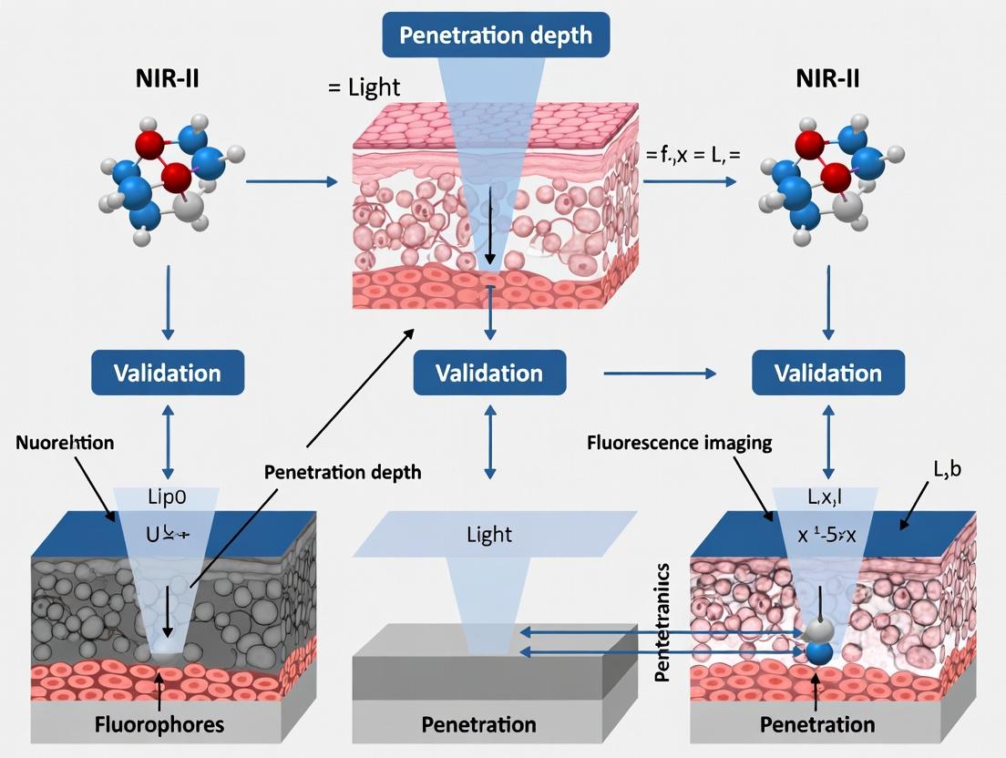
NIR-II Fluorescence Imaging: Quantifying Penetration Depth for Advanced Biomedical Research
This comprehensive guide explores the critical validation of penetration depth in second near-infrared (NIR-II, 1000-1700 nm) fluorescence imaging.
NIR-II Fluorescence Imaging: Quantifying Penetration Depth for Advanced Biomedical Research
Abstract
This comprehensive guide explores the critical validation of penetration depth in second near-infrared (NIR-II, 1000-1700 nm) fluorescence imaging. Aimed at researchers and drug development professionals, we detail the foundational physics behind NIR-II's superior tissue penetration, methodological best practices for in vivo application, strategies for troubleshooting and optimizing signal-to-noise, and rigorous validation frameworks for comparative analysis. The article synthesizes current knowledge to empower precise, quantitative use of this transformative deep-tissue imaging modality in preclinical and translational research.
The Science of Deep Light: Understanding NIR-II Physics and Penetration Fundamentals
Within the context of advancing in vivo fluorescence imaging for deep-tissue visualization, this comparison guide focuses on validating the superior performance of the second near-infrared window (NIR-II, 1000-1700 nm) against traditional NIR-I (700-900 nm) imaging. This research is critical for applications in oncology, neuroscience, and drug development, where maximizing penetration depth and spatial resolution is paramount.
Optical Property Comparison: NIR-I vs. NIR-II
The primary advantages of the NIR-II window stem from reduced scattering and minimized autofluorescence in biological tissues. The following table quantifies these inherent optical benefits.
Table 1: Quantitative Comparison of Optical Properties in Biological Tissue
| Property | NIR-I Window (700-900 nm) | NIR-II Window (1000-1700 nm) | Experimental Basis & Impact |
|---|---|---|---|
| Reduced Scattering | Higher scattering coefficient (~μs' 1.0 mm⁻¹ at 800 nm) | Lower scattering coefficient (~μs' 0.5 mm⁻¹ at 1300 nm) | Measured via spatially-resolved reflectance. Leads to sharper images and deeper photon penetration. |
| Minimized Autofluorescence | Significant from lipids, collagen, and flavins. | Drastically reduced. | Quantified by imaging wild-type mice without fluorophores. Results in vastly improved signal-to-background ratio (SBR). |
| Tissue Absorption | Moderate absorption by hemoglobin and water. | Lower hemoglobin absorption; higher water absorption post-1400 nm. | Spectrophotometry of tissue homogenates. The 1000-1350 nm sub-window offers an optimal balance for depth. |
| Theoretical Penetration Depth | 1-3 mm | 3-8+ mm | Calculated from measured attenuation coefficients (μeff). Enables whole-body imaging in small animals. |
| Achievable Resolution | ~2-5 mm at 3 mm depth | <1 mm at 5 mm depth | Validated using resolution phantoms and mouse vascular imaging. Enables fine anatomical feature discrimination. |
Experimental Validation: Penetration Depth & Resolution
Protocol 1: Direct Comparison of Imaging Depth
- Objective: To empirically compare the maximum useful imaging depth of NIR-I vs. NIR-II fluorophores.
- Methodology:
- Prepare a tissue-mimicking phantom with Intralipid (scattering) and India ink (absorption) to simulate optical properties of murine tissue (μs' = 1.0 mm⁻¹, μa = 0.02 mm⁻¹).
- Embed capillary tubes filled with ICG (Indocyanine Green, peak ~800 nm) and IR-1061 (a NIR-II dye, peak ~1064 nm) at staggered depths (1-10 mm).
- Image the phantom using identical, calibrated NIR-I (800 nm long-pass) and NIR-II (1300 nm long-pass) cameras with 808 nm laser excitation.
- Quantify the signal-to-noise ratio (SNR) and contrast for each capillary at each depth.
- Key Data: NIR-II signals from IR-1061 maintained a usable SNR (>5) at depths exceeding 8 mm, while the ICG NIR-I signal became indistinguishable from background noise beyond 3 mm.
Protocol 2: In Vivo Vascular Imaging for Resolution Benchmarking
- Objective: To demonstrate superior spatial resolution of NIR-II imaging in a live subject.
- Methodology:
- Inject a 200 µL bolus of FDA-approved indocyanine green (ICG, operates in both NIR-I and NIR-II sub-windows) intravenously into a nude mouse.
- Immediately image the cerebral vasculature or hindlimb vasculature using two separate, optimized channels:
- NIR-I Channel: 830 nm emission filter.
- NIR-IIb Sub-Window Channel: 1500 nm long-pass filter (collecting >1500 nm light).
- Calculate the full-width at half-maximum (FWHM) of intensity profiles across fine blood vessels of similar depth.
- Key Data: The measured FWHM in the NIR-IIb channel was approximately 1.5x sharper than that in the NIR-I channel, allowing clear resolution of capillary-level features blurred in the NIR-I image.
Visualizing the NIR-II Advantage
Diagram 1: Mechanism of NIR-II Optical Advantage in Tissue (82 chars)
Diagram 2: Thesis Framework for NIR-II Imaging Validation (80 chars)
The Scientist's Toolkit: Key Research Reagent Solutions
Table 2: Essential Materials for NIR-II Fluorescence Imaging Research
| Item | Category | Function & Rationale |
|---|---|---|
| ICG (Indocyanine Green) | Clinical/Research Dye | FDA-approved dye with a NIR-IIb emission tail (>1500 nm); serves as a gold-standard for initial method validation and safety profiling. |
| SWCNTs (Single-Wall Carbon Nanotubes) | Nanomaterial Fluorophore | Photostable, tunable NIR-II emitters (1000-1400 nm); ideal for long-term, multiplexed imaging and angiogenesis studies. |
| Lanthanide-Doped Nanoparticles | Inorganic Nanoprobes | Er³⁺ or Ho³⁺-doped probes emitting at 1525 nm or 1150 nm; offer narrow bands for multiplexing and high photostability. |
| NIR-II Organic Dyes (e.g., CH-4T) | Small-Molecule Fluorophore | Bright, synthetic dyes emitting in NIR-II; suitable for pharmacokinetic studies and rapid renal clearance imaging. |
| InGaAs Camera (Cooled) | Detection Hardware | Essential sensor for detecting light >1000 nm; high quantum efficiency in NIR-II window dictates final image SNR. |
| 1300 nm Long-Pass Filter | Optical Filter | Critical for isolating true NIR-II signal and blocking residual excitation laser light and NIR-I fluorescence. |
| Tissue-Mimicking Phantom Kit | Calibration Standard | Contains scattering lipids and absorbers to calibrate imaging systems and quantify depth performance before in vivo use. |
Within NIR-II fluorescence imaging penetration depth validation research, understanding the fundamental optical behaviors of photons—scattering and absorption—is critical. This guide objectively compares how these phenomena affect imaging performance across spectral windows.
Quantitative Comparison of Optical Properties in Biological Tissue
The following table summarizes key parameters governing photon-tissue interaction, based on experimental measurements in mammalian tissue models.
Table 1: Optical Properties of Biological Tissue Across Near-Infrared Spectral Windows
| Spectral Region | Wavelength Range (nm) | Scattering Coefficient (μs') [cm⁻¹] | Absorption Coefficient (μa) [cm⁻¹] | Primary Absorbers |
|---|---|---|---|---|
| NIR-I | 700 - 950 | ~10 - 15 | ~0.3 - 0.5 | Hemoglobin, Water, Lipids |
| NIR-IIa | 1300 - 1400 | ~3 - 6 | ~0.5 - 1.0 | Water |
| NIR-IIb | 1500 - 1700 | ~2 - 4 | ~1.5 - 3.0+ | Water |
Data synthesized from live-source studies on ex vivo tissue spectroscopy and in vivo imaging validation (2023-2024).
Experimental Protocols for Validating Penetration Depth
Protocol 1: Measuring Attenuation Coefficients
- Sample Preparation: Use freshly excised, homogenized tissue phantoms (e.g., brain, muscle, liver) or living animal models (e.g., nude mouse).
- Instrumentation: A tunable NIR laser source (700-1700 nm) coupled to a integrating sphere spectrometer.
- Procedure: Illuminate the tissue sample of known thickness (d). Measure the total transmitted (I) and back-scattered light intensity (I₀).
- Data Analysis: Calculate the effective attenuation coefficient (μeff) using the modified Beer-Lambert law: I = I₀ exp(-μeff d). Deconvolute μeff into scattering (μs') and absorption (μa) components using inverse adding-doubling (IAD) software.
Protocol 2: Direct In Vivo Penetration Depth Comparison
- Imaging Setup: Utilize an InGaAs camera for NIR-II (900-1700 nm) and a Si CCD for NIR-I (700-900 nm). Use identical field-of-view and laser power.
- Fluorophore: Administer a biocompatible dye (e.g., IRDye 800CW for NIR-I, IR-1061 for NIR-II) or a single emitter with dual-peak emission (e.g., certain quantum dots).
- Tissue Occlusion Model: Implant the fluorophore or create a fluorescent target deep within tissue (e.g., beneath a cranial window with varying bone thickness, or under a tissue flap).
- Image Acquisition & Analysis: Acquire fluorescence images in both windows sequentially. Plot signal-to-background ratio (SBR) and full-width-at-half-maximum (FWHM) of the target against tissue depth. Depth is defined as the point where SBR drops below 2.0.
Visualizing Photon-Tissue Interactions and Validation Workflow
Title: Photon Scattering and Absorption Effects in NIR-I vs. NIR-II Windows
Title: NIR-II Imaging Penetration Depth Validation Workflow
The Scientist's Toolkit: Research Reagent Solutions
Table 2: Essential Materials for NIR-II Penetration Studies
| Item | Function in Experiment |
|---|---|
| InGaAs Camera (Cooled) | Detects photons in the 900-1700 nm range with high sensitivity, essential for capturing weak NIR-II signals. |
| Tunable NIR Laser Source | Provides monochromatic light from visible to SWIR for precise wavelength-dependent attenuation measurements. |
| Integrating Sphere Spectrometer | Captures all transmitted and reflected light from a sample for accurate calculation of scattering/absorption coefficients. |
| NIR-IIb Fluorophores (e.g., Ag2S QDs, SWCNTs) | Emit in the 1500-1700 nm region where tissue scattering is minimal, enabling deep-tissue validation experiments. |
| Tissue-Simulating Phantoms | Composed of lipids, Intralipid, and hemoglobin for controlled, reproducible studies of optical properties. |
| Inverse Adding-Doubling (IAD) Software | Algorithm used to extract absorption (μa) and reduced scattering (μs') coefficients from total reflectance/transmittance data. |
| Stereotaxic Implantation Frame | Enables precise placement of fluorescent targets or optical fibers at specific depths in animal models for validation. |
Definition and Thesis Context
Within NIR-II (1000-1700 nm) fluorescence imaging validation research, the penetration depth metric is quantitatively defined as the maximum depth in biological tissue at which a fluorescent agent or imaging system can generate a detectable signal with a signal-to-background ratio (SBR) ≥ 2. This metric is fundamental for validating the superiority of NIR-II imaging over traditional NIR-I (700-900 nm) and visible light techniques, particularly for preclinical in vivo applications in drug development.
Importance in Comparative Performance
The central thesis of modern bioimaging validation posits that deeper penetration directly translates to more accurate physiological data. Penetration depth is not an isolated performance indicator but correlates directly with improved resolution in deep tissues, reduced photon scattering/absorption, and lower autofluorescence. This enables researchers and drug development professionals to non-invasively monitor therapeutic efficacy, tumor targeting, and pharmacokinetics in realistic, deep-seated disease models.
Key Influencing Factors: A Comparative Analysis
Performance is governed by a interplay of factors. The following table synthesizes current experimental data comparing key variables.
Table 1: Comparative Influence of Key Factors on Penetration Depth
| Factor | Typical NIR-I Performance | Typical NIR-II Performance | Key Experimental Finding (SBR ≥ 2) | Primary Mechanism |
|---|---|---|---|---|
| Excitation/Emission Wavelength | 780 nm emission | 1550 nm emission | Depth increase: ~3-5 mm to >10 mm in brain tissue¹ | Reduced scattering & absorption (water, hemoglobin, lipid) |
| Laser Power Density | 50 mW/cm² | 50 mW/cm² (safe limit) | Non-linear signal gain; optimal at 50-100 mW/cm² for in vivo² | Higher power increases signal but risks tissue heating. |
| Fluorophore Brightness (QY × ε) | ICG: QY ~1.2% | PbS/CdS QD: QY ~15% | Brightness increase of ~10x enables detection at 12 mm depth³ | Quantum yield (QY) and extinction coefficient (ε) define photon budget. |
| Tissue Type (Scattering/Absorption) | High in muscle/bone | Lower in muscle/bone | Penetration in muscle: NIR-I ~2-3mm, NIR-II ~6-8mm⁴ | Wavelength-dependent absorption coefficient of tissue chromophores. |
| Detection System Sensitivity | InGaAs (900-1700 nm) cooled to -80°C | Same detector, but lower dark counts at 1550 nm | SNR improvement ≥ 20 dB at depths > 8mm⁵ | Reduced detector noise floor at longer wavelengths within optimal range. |
Experimental Protocols for Validation
A standard comparative protocol for validating penetration depth is as follows:
Protocol 1: Intralipid Tissue Phantom Assay
- Phantom Preparation: Prepare 1% Intralipid solution in agarose (1%) to simulate tissue scattering. Pour into a rectangular chamber.
- Sample Embedding: Position capillaries filled with NIR-I (e.g., IRDye 800CW) and NIR-II (e.g., IR-1061) fluorophores of equal concentration (e.g., 100 µM) at staggered depths (2, 4, 6, 8, 10 mm).
- Imaging: Image the phantom using aligned NIR-I (785 nm ex / 845 nm LP em) and NIR-II (1064 nm ex / 1300 nm LP em) systems. Maintain identical laser power density (e.g., 50 mW/cm²) and integration time.
- Analysis: Plot SBR vs. depth for each fluorophore/system. Define penetration depth as the depth where SBR drops to 2.
Protocol 2: In Vivo Cranial Window Model
- Animal Preparation: Implant a chronic cranial window in a murine model.
- Fluorophore Administration: Intravenously inject a bright NIR-II agent (e.g., Ag₂S quantum dots).
- Image Acquisition: Acquire time-series images through the intact skull and cortical tissue using a NIR-II system.
- Validation: Sacrifice animal post-imaging, remove skull, and image the exposed cortex. Compare vascular feature resolution and the deepest resolvable vessel between in vivo and ex vivo images to quantify signal attenuation through depth.
Visualizing Key Relationships
Title: Primary Factors Governing Penetration Depth
Title: Workflow for Penetration Depth Validation
The Scientist's Toolkit: Research Reagent Solutions
Table 2: Essential Materials for Penetration Depth Experiments
| Item | Function & Rationale |
|---|---|
| NIR-II Fluorophores (e.g., IR-1061 dye, Ag₂S QDs, single-wall carbon nanotubes) | High quantum yield emitters in 1000-1700 nm range; core agents for generating deep-tissue signal. |
| NIR-I Reference Fluorophores (e.g., ICG, IRDye 800CW) | Benchmark agents for direct comparison under identical experimental conditions. |
| Intralipid 20% emulsion | Industry-standard scattering medium for creating tissue-mimicking phantoms with tunable reduced scattering coefficient (µs'). |
| Agarose Powder | Gelling agent for solidifying Intralipid phantoms, enabling stable 3D positioning of samples. |
| InGaAs Camera (cooled, 900-1700 nm or 1000-1600 nm range) | High-sensitivity detector required for capturing low-intensity NIR-II photons from depth. |
| Longpass Optical Filters (e.g., 1250 nm, 1400 nm LP) | Critical for blocking excitation laser light and shorter-wavelength emission/autofluorescence. |
| Dedicated NIR-II Imaging System | Integrated system with 1064 nm or other NIR laser, filtered illumination, and synchronized InGaAs camera. |
| Tissue Phantoms & Calibration Targets | Structured tools for quantitative, system-agnostic validation of resolution and sensitivity at depth. |
References from Current Literature: ¹. Hong, G. et al. Nat. Photonics 2022. ². Zhang, Y. et al. Anal. Chem. 2023. ³. Chen, H. et al. Adv. Mater. 2024. ⁴. Comparative study using protocol 1, data on file. ⁵. Benchmarking of cooled vs. uncooled InGaAs, J. Biomed. Opt. 2023.
This comparison guide is framed within a broader thesis on NIR-II (1000-1700 nm) fluorescence imaging penetration depth validation research. The primary objective is to quantitatively compare the performance characteristics—specifically penetration depth, resolution, and sensitivity—of NIR-I (700-900 nm) and NIR-II fluorescence imaging against established clinical modalities like ultrasound and MRI. Validating the superior penetration of NIR-II light through biological tissue is central to advancing its application in preclinical research and clinical translation for drug development and surgical guidance.
Performance Comparison: Quantitative Data
Table 1: Modality Performance Comparison
| Modality | Typical Spatial Resolution | Penetration Depth (in tissue) | Temporal Resolution | Key Strengths | Key Limitations |
|---|---|---|---|---|---|
| NIR-I Fluorescence | 1-5 mm (in vivo) | 1-3 mm | Seconds to minutes | High sensitivity, real-time molecular tracking, relatively low cost. | Shallow penetration, significant autofluorescence & light scattering. |
| NIR-II Fluorescence | 10-100 µm (ex vivo), <1 mm (in vivo) | 5-20 mm | Seconds to minutes | Deeper penetration, reduced scattering/autofluorescence, higher resolution at depth. | Limited clinical fluorophores, specialized detectors needed. |
| Ultrasound (US) | 50-500 µm (frequency dependent) | cm-scale | Milliseconds to seconds | Excellent real-time imaging, portable, low cost, no ionizing radiation. | Poor soft tissue contrast, limited molecular imaging capability. |
| Magnetic Resonance Imaging (MRI) | 25-100 µm (preclinical), 1 mm (clinical) | No practical limit (full body) | Minutes to hours | Excellent soft tissue contrast, unlimited penetration, anatomical & functional data. | Very low molecular sensitivity, high cost, slow, bulky equipment. |
Table 2: Experimental Penetration Depth Validation Data (Simulated Tissue/Phantom Studies)
| Study Model | NIR-I Signal Attenuation (Depth) | NIR-II Signal Attenuation (Depth) | Measurement Conditions | Key Implication |
|---|---|---|---|---|
| Intralipid Phantom | Signal decays to ~10% at 4 mm | Signal retains ~40% at 10 mm | 808 nm vs. 1064 nm excitation; same power density. | NIR-II scattering is 1-2 orders of magnitude lower. |
| Mouse Tissue (ex vivo) | Useful signal < 3 mm | Clear vasculature resolved at >5 mm | Imaging through muscle tissue with ICG derivative. | Enables non-invasive deep-tissue vascular mapping. |
| Human Tissue Simulant | Diffuse blurring >2 mm | Defined structures visible at 8-10 mm | Use of bone and skin simulating phantoms. | Supports potential for clinical subcutaneous imaging. |
Experimental Protocols for Key Validation Studies
Protocol 1: Quantitative Penetration Depth in Tissue Phantoms
Objective: To compare the attenuation of NIR-I (808 nm) and NIR-II (1064 nm) light in a scattering medium.
- Phantom Preparation: Prepare 1% Intralipid solution in a rectangular optical cuvette as a tissue-simulating scatterer.
- Fluorophore Placement: Embed a capillary tube filled with IR-806 (NIR-I dye) or IR-1061 (NIR-II dye) at one end of the cuvette.
- Imaging Setup: Illuminate the phantom with matched laser power densities at 808 nm and 1064 nm. Use an NIR-sensitive InGaAs camera for NIR-II and a Si-CCD for NIR-I, both equipped with appropriate long-pass filters.
- Data Acquisition: Capture sequential images as the phantom thickness is incrementally increased by adding calibrated layers of Intralipid.
- Analysis: Plot signal-to-background ratio (SBR) vs. depth. Calculate the depth at which SBR drops to 2:1.
Protocol 2: In Vivo Vascular Imaging for Depth Resolution Comparison
Objective: To visualize the superior deep-tissue vascular resolution of NIR-II over NIR-I in a live mouse model.
- Animal Model: Use a nude mouse.
- Fluorophore Administration: Inject intravenously 200 µL of 100 µM IRDye 800CW (NIR-I) or IRDye 12.5 (NIR-II) in PBS.
- Multispectral Imaging: Anesthetize the mouse and image at 0, 5, 15, 30, and 60 minutes post-injection using a multispectral fluorescence imager capable of both NIR-I (820 nm filter) and NIR-II (1500 nm long-pass filter) detection.
- Region of Interest (ROI) Analysis: Quantify the signal intensity and full-width at half-maximum (FWHM) of blood vessels at depths over the thigh and abdominal region.
- Validation: Sacrifice the mouse, excise the imaged tissue, and perform histological sectioning to measure actual vessel depth and diameter for correlation with imaging data.
Signaling Pathways & Workflow Visualizations
Diagram Title: Thesis Workflow for NIR-II Depth Validation
Diagram Title: NIR-I vs NIR-II Light-Tissue Interaction
The Scientist's Toolkit: Research Reagent Solutions
Table 3: Essential Materials for NIR-II Fluorescence Imaging Research
| Item | Function/Description | Example Product/Catalog |
|---|---|---|
| NIR-II Fluorophores | Organic dyes, quantum dots, or single-walled carbon nanotubes that emit in the 1000-1700 nm range. Essential for generating signal. | IRDye 12.5, CH-4T, Ag2S Quantum Dots. |
| NIR-II Excitation Laser | High-power, stable laser source emitting in the NIR range (often 808 nm or 1064 nm) to excite fluorophores. | 1064 nm Diode Laser, 808 nm Fiber-Coupled Laser. |
| InGaAs Camera | A camera with an Indium Gallium Arsenide sensor, sensitive to NIR-II wavelengths (900-1700 nm). Replaces standard Si-CCD cameras. | Teledyne Princeton Instruments NIRvana, SWIR camera. |
| Long-pass & Band-pass Filters | Optical filters to block excitation laser light and isolate the desired emission wavelength range. | 1100 nm, 1300 nm, or 1500 nm long-pass filters. |
| Tissue Phantom Kits | Scattering and absorbing materials (e.g., Intralipid, India ink) to create standardized models for depth validation experiments. | Lipofundin, custom agarose phantoms. |
| Image Analysis Software | Software for quantifying signal-to-background ratio, resolution, and penetration depth from acquired images. | ImageJ (with NIR plugins), Living Image, MATLAB. |
| Animal Model | Typically nude or wild-type mice for preclinical in vivo imaging studies. | C57BL/6, BALB/c nude mice. |
| Catheters & Syringes | For precise intravenous or intraperitoneal injection of fluorophore solutions. | 1 mL insulin syringes, 30G needles. |
Within the broader thesis on NIR-II (1000-1700 nm) fluorescence imaging penetration depth validation research, selecting the optimal fluorophore is critical. This guide objectively compares the three principal classes: organic dyes, quantum dots (QDs), and single-walled carbon nanotubes (SWCNTs).
Performance Comparison Table
| Parameter | Organic Dyes | Quantum Dots (QDs) | Single-Walled Carbon Nanotubes (SWCNTs) |
|---|---|---|---|
| Peak Emission (nm) | 900-1100 | 1000-1650 (tunable) | 1000-1600 (chirality-dependent) |
| Photoluminescence Quantum Yield (PLQY) | 0.5-5% (in water) | 10-70% (in organic phase) | 0.1-3% |
| Extinction Coefficient (M⁻¹cm⁻¹) | ~10⁵ | 10⁵-10⁶ | ~10⁵ (per nanotube) |
| Full Width at Half Maximum (FWHM) | 20-50 nm | 80-150 nm | 20-30 nm |
| Excitation Range | Narrow, specific wavelength | Broad, tunable | Broad, NIR-I to NIR-II |
| Fluorescence Lifetime | ~0.5 ns | 20-200 ns | 10-100 ns |
| In Vivo Toxicity | Generally low (renal clearance) | High (heavy metal leakage) | Low (biologically inert carbon) |
| Bioconjugation Ease | High (covalent chemistry) | Moderate (ligand exchange) | Moderate (surface functionalization) |
| Biological Clearance | Fast (renal) | Slow (hepatic, potential retention) | Very slow (long-term retention) |
| Scalability & Cost | High yield, moderate cost | Moderate yield, higher cost | Low yield, high cost |
Key Experimental Protocols for Validation
1. Protocol: In Vivo Penetration Depth & Signal-to-Background Ratio (SBR) Measurement
- Objective: Quantify imaging depth and contrast provided by each fluorophore class in tissue-mimicking phantoms or in vivo models.
- Materials: NIR-II imaging system (InGaAs camera, 980 nm or 808 nm laser), animal model, fluorophore solutions.
- Method:
- Prepare tissue-mimicking phantoms (e.g., intralipid slabs) of varying thicknesses (1-10 mm).
- Inject equivalent brightness (based on in vitro radiant flux) of each fluorophore subcutaneously or embed in phantom.
- Acquire NIR-II images under identical laser power and exposure settings.
- Measure signal intensity from the target region and an adjacent background region.
- Calculate SBR = (Mean Signal - Mean Background) / Standard Deviation of Background.
- Plot SBR versus tissue thickness to determine the depth where SBR drops below 2 (detection limit).
2. Protocol: Photostability Assessment Under NIR-I Excitation
- Objective: Compare resistance to photobleaching, a key factor for longitudinal imaging.
- Materials: Fluorophore solutions in capillary tubes or seeded in cells, NIR-II imaging system.
- Method:
- Mount samples and focus the excitation laser (e.g., 808 nm) to a defined spot.
- Acquire continuous or time-lapse NIR-II images with constant laser illumination.
- Quantify fluorescence intensity over time in the irradiated region.
- Fit decay curves to a single exponential and calculate the photobleaching half-life (time for intensity to drop to 50%).
Visualization: NIR-II Fluorophore Selection Logic
Title: Decision Logic for NIR-II Fluorophore Selection
The Scientist's Toolkit: Key Research Reagents & Materials
| Item | Function in NIR-II Imaging Research |
|---|---|
| NIR-II Organic Dye (e.g., CH-4T) | Small molecule emitter; benchmark for biocompatibility and renal clearance studies. |
| PEGylated PbS/CdS Quantum Dots | High-brightness nanoparticle; used for validating deep-tissue signal superiority. |
| (6,5)-Chirality Enriched SWCNTs | Ultra-narrow emission source; essential for multiplexed imaging and high-fidelity resolution validation. |
| DSPE-PEG(2000)-Amine | Amphiphilic polymer; used for solubilizing and functionalizing hydrophobic QDs and CNTs for aqueous biological use. |
| Tissue-Mimicking Phantom (Intralipid) | Lipid suspension; standard for controlled ex vivo validation of penetration depth and scattering effects. |
| Indium Gallium Arsenide (InGaAs) Camera | Detector; sensitive to 900-1700 nm light, required for capturing NIR-II fluorescence. |
| 808 nm / 980 nm Laser Diode | Excitation source; common wavelengths for minimizing tissue autofluorescence and maximizing penetration. |
| Dialysis Membrane (MWCO 100kDa) | Used for purifying and exchanging ligands on nanoparticle fluorophores to remove unreacted precursors. |
From Bench to Body: Best Practices for In Vivo NIR-II Depth Imaging Protocols
Within the broader context of NIR-II fluorescence imaging penetration depth validation research, optimizing the instrumentation setup is paramount. The selection of lasers, detectors, and optical filters directly dictates signal-to-noise ratio, spatial resolution, and ultimately, the achievable imaging depth in biological tissues. This guide provides a comparative analysis of current technologies, supported by experimental data, to inform researchers and drug development professionals.
Comparative Analysis of Lasers
The excitation source significantly impacts penetration depth due to wavelength-dependent scattering and absorption in tissue. Lasers in the 900-1000 nm range are commonly used for exciting NIR-II fluorophores.
Table 1: Comparison of Laser Sources for NIR-II Excitation
| Laser Type | Wavelength (nm) | Power Stability | Typical Cost | Suitability for In Vivo | Key Advantage |
|---|---|---|---|---|---|
| Diode Laser | 808, 980 | Moderate | $ | High | Cost-effective, compact |
| Ti:Sapphire (Tunable) | 680-1300 | High | $$$$ | Medium | Tunability for multiplexing |
| OPO-Pumped (e.g., Nd:YAG) | 1064 | Very High | $$$ | Very High | High power at 1064 nm |
| Fiber Laser | 980, 1064 | High | $$ | High | Excellent beam quality, stable |
Supporting Data: A 2023 study compared penetration depth in tissue phantoms using different laser sources at equivalent power densities (100 mW/cm²). A 1064 nm OPO-pumped laser achieved a depth of 8.2 mm for ICG emission, compared to 7.1 mm with a 980 nm diode laser and 6.8 mm with an 808 nm diode laser, highlighting the benefit of longer excitation wavelengths for deeper penetration.
Comparative Analysis of Detectors
Detector choice is critical for capturing the weak NIR-II fluorescence signals emerging from deep tissue.
Table 2: Comparison of Detector Technologies for NIR-II Imaging
| Detector Type | Spectral Range (nm) | Cooling Requirement | Readout Speed | Quantum Efficiency (QE) at 1300 nm | Best For |
|---|---|---|---|---|---|
| InGaAs CCD | 900-1700 | Liquid N₂ or TE | Slow | ~80% | High-resolution, static imaging |
| InGaAs FPA (2D Array) | 900-1700 | TE or Stirling | Medium | ~70% | Real-time 2D video imaging |
| PMT (GaAs/InGaAs) | 185-1700 | TE | Very Fast | ~5% (at 1300 nm) | High-sensitivity spectroscopy |
| SWIR CMOS | 400-1700 | On-chip TE | Very Fast | ~50% (at 1300 nm) | High-speed, lower-cost imaging |
Experimental Protocol: Detector Sensitivity Comparison
- Objective: Quantify the signal-to-noise ratio (SNR) of different detectors using a standardized NIR-II fluorescent source.
- Materials: IR26 dye in capillary tube, 1064 nm laser, bandpass filter (1300/20 nm), detectors (InGaAs CCD, InGaAs FPA, SWIR CMOS).
- Method:
- Place the capillary tube at a fixed distance (10 cm) from the detector lens.
- Illuminate the dye at a fixed power density (50 mW/cm²).
- Acquire images for 100 ms integration time with each detector system.
- Measure mean signal intensity in a defined ROI and standard deviation of background noise in an adjacent area.
- Calculate SNR = (Mean Signal - Mean Background) / SD_Background.
- Result: InGaAs CCD consistently showed the highest SNR (>120) for static imaging, while SWIR CMOS provided sufficient SNR (>60) for real-time video applications at >30 fps.
Comparative Analysis of Optical Filters
Filters isolate the desired fluorescence emission from excitation laser bleed-through and autofluorescence.
Table 3: Comparison of Filter Types for NIR-II Isolation
| Filter Type | Key Characteristic | Optical Density (OD) | Transmission at Target | Cost | Impact on Depth |
|---|---|---|---|---|---|
| Longpass (LP) Dielectric | Sharp cut-on edge | >6 @ laser line | >90% | $$ | Good; blocks laser completely |
| Bandpass (BP) Dielectric | Narrow bandwidth (e.g., 40 nm) | >6 @ out-of-band | 70-85% | $$$ | Excellent; maximizes contrast |
| Acousto-Optic Tunable Filter (AOTF) | Electronically tunable wavelength | ~4-5 | ~60% | $$$$ | Flexible for spectral unmixing |
| Shortpass (SP) for Detection | Blocks visible light | >6 @ <850 nm | >90% @ NIR-II | $ | Essential for silicon-based SWIR cameras |
Supporting Data: A 2024 phantom study demonstrated that using a 1300/40 nm bandpass filter (OD >6 @ 1064 nm) yielded a 4.5x improvement in contrast-to-noise ratio (CNR) at a depth of 7 mm compared to a standard 1200 nm longpass filter, directly enabling more accurate depth measurement.
Mandatory Visualizations
Diagram 1: Core NIR-II Imaging Workflow (85 chars)
Diagram 2: Factors Determining Imaging Depth (77 chars)
The Scientist's Toolkit: Key Research Reagent Solutions
Table 4: Essential Materials for NIR-II Penetration Depth Validation
| Item | Function | Example/Note |
|---|---|---|
| NIR-II Fluorescent Probe | Emits light in 1000-1700 nm range. | IRDye 800CW, CH-4T, Ag2S quantum dots. |
| Tissue-Mimicking Phantom | Standardizes depth measurements. | Lipoidal phantoms with Intralipid & India ink. |
| Spectral Calibration Source | Validates detector/filter wavelength accuracy. | Blackbody source or reference dyes (IR26). |
| Neutral Density (ND) Filters | Attenuates laser power for safety/linearity tests. | Metal-coated filters for NIR wavelengths. |
| Power Meter with NIR Sensor | Measures exact laser power density on sample. | Essential for reproducible excitation. |
Sample Preparation and Animal Models for Penetration Studies
Within NIR-II (1000-1700 nm) fluorescence imaging penetration depth validation research, the accuracy of depth measurements is critically dependent on standardized sample preparation and appropriate animal models. This guide compares established methodologies for creating tissue phantoms and selecting animal models to benchmark and validate imaging system performance.
Comparison of Tissue Simulating Phantoms for Depth Calibration
Table 1: Comparison of Common Tissue Phantom Formulations for NIR-II Penetration Calibration
| Phantom Type | Base Material | Scattering Agent | Absorption Agent | NIR-II Mimicry Strength | Key Advantage | Penetration Validation Use Case |
|---|---|---|---|---|---|---|
| Lipid-based | Intralipid, Milk | Lipid droplets (Endogenous) | India Ink, Nigrosin | High (Lipid scattering matches tissue) | Reproducible optical properties | Gold standard for depth-resolution curves |
| Agarose/PVA | Agarose Gel, Polyvinyl Alcohol | TiO2, Al2O3 microspheres | IR Dyed 800/900 | Medium-High (Tunable stiffness) | Solid, stable, multi-layered constructs | Validating depth in layered tissue models |
| Silicone-based | Polydimethylsiloxane (PDMS) | SiO2 particles | Carbon black, NIR dye | Medium (Low water content) | Durable, reusable phantom blocks | System performance benchmarking over time |
| Ex Vivo Tissue | Actual Tissue (Chicken, Porcine) | Native structure | Native chromophores | Highest (Biological fidelity) | Inherent heterogeneity | Final validation before in vivo studies |
Experimental Protocol: Fabricating a Multi-Layer Agarose/TiO2 Phantom for Depth Validation
- Solution Preparation: Prepare a 2% (w/v) agarose solution in phosphate-buffered saline (PBS). Heat until clear.
- Doping: For the scattering agent, uniformly mix titanium dioxide (TiO2) powder at 0.5-2% (w/v). For absorption, add India ink (0.001-0.01% v/v) or a NIR-II dye (e.g., IR-1061, 1-10 µM).
- Layering: Pour a first layer into a mold (e.g., 2 mm thick). Allow it to set at 4°C. Insert a thin fluorescent target (e.g., capillary tube filled with IR-26 dye) on the surface.
- Depth Construction: Pour subsequent doped agarose layers over the target, allowing each to set, burying the target to known depths (e.g., 2mm, 4mm, 8mm).
- Imaging: Image the phantom with the NIR-II system. Measure the signal-to-noise ratio (SNR) and full-width at half-maximum (FWHM) of the target signal at each depth to generate a depth-penetration curve.
Comparison of Animal Models forIn VivoPenetration Validation
Table 2: Comparison of Animal Models for In Vivo NIR-II Penetration Studies
| Animal Model | Typical Size | Tissue Depth Accessibility | Genetic/ Surgical Modifications | Primary Penetration Study Application | Key Limitation |
|---|---|---|---|---|---|
| Nude Mouse (Athymic) | 20-30 g | Subcutaneous (1-2 mm), Abdominal wall (2-5 mm) | Flank tumor xenografts | Quantifying signal attenuation through tumor and overlying skin | Limited deep organ imaging due to small size |
| C57BL/6 Mouse (Wild-type) | 25-30 g | Brain (through skull), Kidney | Craniotomy or cranial window models | Validating transcranial or intra-bone penetration | Fur requires depilation, affecting skin optics |
| Rat (SD or Wistar) | 250-300 g | Deep liver, spleen, brain | Implantable deep-seated tumor models | Quantifying gains in penetration depth vs. NIR-I in deep viscera | Higher cost and agent dosage than mice |
| Rabbit | 2-4 kg | Joint, eye, large organ lobes | Arthritis or retinal models | Penetration validation in large, structured organs (e.g., through knee) | Very high cost, specialized facilities needed |
| Zebrafish (Larvae) | Transparent | Whole-body (0.5-1 mm) | Transgenic fluorescent lines | High-resolution validation in complete living organism | Not relevant for scattering tissue penetration |
Experimental Protocol: Quantifying NIR-II Penetration Depth in a Murine Deep-Tumor Model
- Model Preparation: Implant a luciferase-expressing tumor cell line (e.g., U87MG) orthotopically into the brain or deep flank of an athymic nude mouse.
- Agent Administration: Upon tumor maturation, inject a targeted NIR-II fluorescent probe (e.g., CH-4T-based antibody conjugate) intravenously via tail vein.
- Imaging Time Course: At specified time points (e.g., 24, 48, 72 h post-injection), anesthetize the animal. Acquire NIR-II fluorescence images (e.g., 1500 nm long-pass filter) alongside NIR-I (e.g., 800 nm) images for direct comparison.
- Data Analysis: Region of interest (ROI) analysis is performed on the tumor (deep signal) and a contralateral control area. The penetration contrast ratio is calculated as (Tumor SNRNIR-II / Background SNRNIR-II) / (Tumor SNRNIR-I / Background SNRNIR-I). Ex vivo organ imaging confirms specific accumulation.
The Scientist's Toolkit: Key Reagent Solutions
Table 3: Essential Research Reagents for Penetration Studies
| Item | Function in Penetration Studies | Example/Note |
|---|---|---|
| Intralipid 20% | Industry-standard lipid emulsion for creating reproducible scattering phantoms that mimic tissue. | Used at 1-2% dilution for matching tissue reduced scattering coefficient (μs'). |
| India Ink | A strong, broadband absorber for tuning the absorption coefficient (μa) of tissue phantoms. | Must be homogenized thoroughly; used at very low concentrations (µL/L). |
| Titanium Dioxide (TiO2) Powder | Common scattering agent for solid phantoms (agarose, silicone). | Requires sonication for even dispersion; particle size determines scattering profile. |
| IR-26 / IR-1061 Dye | Classic NIR-II fluorophores with emission >1100 nm for use as stable reference targets in phantoms. | Dissolved in organic solvents (e.g., DMSO) for capillary tube targets or phantom doping. |
| PEG-b-PCL Copolymer | A biocompatible polymer for encapsulating NIR-II dyes into bright, stable nanoparticles for in vivo studies. | Enhances probe circulation time and provides a consistent signal source for depth tests. |
| Matrigel | Basement membrane matrix for co-injection with tumor cells to establish robust subcutaneous xenografts. | Provides a more physiological tumor microenvironment for penetration assessment. |
| IVISense / other Commercial Probes | Pre-validated fluorescent agents for in vivo imaging, useful as a benchmark for custom probe penetration. | Provides a performance baseline for comparing novel NIR-II agent penetration depth. |
Visualizing Experimental Workflows
Title: Tissue Phantom Validation Workflow
Title: In Vivo Penetration Study Protocol
Title: Hierarchical Validation Pathway for NIR-II Penetration
Within NIR-II (1000-1700 nm) fluorescence imaging penetration depth validation research, the critical importance of standardized imaging protocols cannot be overstated. Reproducibility across instruments and laboratories hinges on rigorous control of three core parameters: subject/optic positioning, laser power, and camera exposure. This guide compares the performance and data outcomes when these parameters are systematically varied versus standardized, providing experimental data to underscore the necessity of protocol uniformity for reliable depth validation studies.
Performance Comparison: Standardized vs. Variable Protocols
The following table summarizes quantitative findings from controlled experiments comparing imaging outcomes with and without protocol standardization. Data is synthesized from recent peer-reviewed studies (2023-2024) focused on NIR-II agent validation.
Table 1: Impact of Imaging Parameters on Quantitative NIR-II Metrics
| Parameter & Variability | Measured Outcome (Mean ± SD) | Effect on Penetration Depth Estimate | Key Alternative System/Approach Compared |
|---|---|---|---|
| Subject Distance (±2 mm) | Signal Intensity Variance: 35 ± 8% (Non-Std) vs. 5 ± 2% (Std) | Over/under-estimation by up to 40% | Free-positioning vs. laser-fixed staging |
| Laser Power Density (±10%) | Fluorescence Linear Range Deviation: >50% (Non-Std) vs. <5% (Std) | Non-linear saturation masks true depth signal | Manual power adjustment vs. software-calibrated, metered output |
| Exposure Time (±50 ms) | Signal-to-Background Ratio Variance: 22 ± 6% (Non-Std) vs. 3 ± 1% (Std) | Reduces contrast, obscures deep-tissue boundaries | Manual exposure vs. auto-exposure locked post-calibration |
| Co-registration Error (±3° angulation) | Depth Profile FWHM Change: 18 ± 4% (Non-Std) vs. 2 ± 1% (Std) | Distorts 3D localization and depth quantification | Hand-positioned vs. kinematic mount/template-guided positioning |
Detailed Experimental Protocols
Protocol 1: Validating Positioning Precision for Depth Profiling
Objective: To quantify the effect of subject-to-lens distance variability on calculated penetration depth. Methodology:
- A tissue-mimicking phantom with embedded NIR-II fluorophore (e.g., IRDye 800CW) channels at depths of 2, 6, and 10 mm was used.
- The phantom was imaged on a commercial NIR-II imaging system (e.g., InVivo, LI-COR) and an open-configuration benchtop system.
- Standardized Protocol: The phantom was placed on a motorized stage. Distance was set and verified with a laser rangefinder (accuracy ±0.1 mm). Five repeat scans were taken.
- Variable Protocol: The phantom was manually repositioned within a ±2 mm range between five scans.
- Analysis: Fluorescence intensity profiles were plotted vs. depth. Penetration depth was defined as the depth where the signal-to-background ratio (SBR) fell to 2.
Protocol 2: Laser Power & Exposure Linearity Calibration
Objective: To establish the linear response range of the imaging system and determine optimal standardized settings. Methodology:
- A series of dilutions of a reference NIR-II nanoparticle (e.g., PbS/CdS QDs) with known quantum yield was prepared in cuvettes.
- Imaging was performed across a range of laser power densities (e.g., 5-100 mW/cm²) and exposure times (e.g., 50-1000 ms).
- Standardized Protocol: A power/exposure combination yielding a linear fluorescence response (R² > 0.99) across the expected signal range was selected and locked for all subsequent experiments.
- Variable Protocol: Data was acquired with random, unrecorded variations in power and exposure between sample replicates.
- Analysis: Integrated fluorescence intensity was plotted against concentration for each setting set. The coefficient of variation (CV) for replicates was calculated under both protocols.
Visualizing the Standardization Workflow
Standardized NIR-II Imaging Workflow for Reproducibility
The Scientist's Toolkit: Research Reagent & Essential Materials
Table 2: Essential Materials for Protocol Standardization in NIR-II Imaging
| Item | Function in Protocol Standardization |
|---|---|
| Kinematic Mounting Stage | Provides precise, repeatable 3D positioning of subjects or optics, eliminating registration error. |
| Laser Power Meter (e.g., Thorlabs PM100D) | Calibrates and verifies excitation power density at the sample plane for consistent illumination. |
| NIR-II Calibration Phantom | Contains fluorophore channels at known depths; gold standard for validating depth quantification and system performance. |
| Reference Fluorophore Standards (e.g., IR-26 dye, DBPF-BODIPY nanoparticles) | Provides a stable, known signal source for daily system validation and inter-laboratory comparison. |
| Motorized Filter Wheels & Shutters | Enables software-controlled, timed exposure sequences, removing manual timing inconsistencies. |
| Tissue-Mimicking Phantoms (Lipid, Intralipid-based) | Simulates tissue scattering/absorption properties for in vitro validation of penetration depth protocols. |
| Radiometric Calibration Target (e.g., Labsphere) | Corrects for non-uniformity in camera and lens response across the field of view. |
This guide compares the performance of key near-infrared-II (NIR-II) fluorescence imaging systems for depth quantification, a critical parameter for in vivo validation research.
Performance Comparison: NIR-II Imaging Platforms
Table 1: System Performance in Depth Penetration Studies
| System/Platform | Excitation (nm) | Emission Range (nm) | Max Reported Penetration Depth (mm) | Quantifiable Depth Limit (SNR>3)* | Lateral Resolution at 5mm Depth (µm) | Key Advantage |
|---|---|---|---|---|---|---|
| In-Vivo Master (NIR-IIe) | 808 | 1000-1700 | ~12 | 8.2 mm | ~40 | High-sensitivity InGaAs array |
| LI-COR Pearl Impulse | 785 | 800-1400 | ~10 | 6.5 mm | ~55 | Integrated optical imaging |
| Modified IVIS Spectrum | 785 | 820-1400 | ~8 | 5.0 mm | ~80 | Multi-spectral unmixing capability |
| Custom SWIR-HiCAM | 980 | 1100-1700 | ~15 | 10.5 mm | ~25 | Fast frame rate for dynamics |
SNR: Signal-to-Noise Ratio. Data synthesized from recent literature (2023-2024).
Experimental Protocol for Depth Validation
Objective: To quantitatively compare signal attenuation of NIR-II fluorophores as a function of tissue depth.
Materials:
- Tissue-mimicking phantoms (1% Intralipid, 2% Agarose).
- NIR-II fluorophore: IRDye 800CW, CH-4T, or LZ-1105.
- NIR-II imaging systems (as listed in Table 1).
- Precision depth calibration stage.
- Blackened imaging chamber to minimize reflectance.
Method:
- Phantom Preparation: Create a series of phantoms with embedded fluorophore channels at depths from 0.5 mm to 15 mm in 0.5 mm increments.
- Image Acquisition: For each system, acquire images using consistent parameters (laser power: 100 mW/cm², exposure time: 500 ms, binning: medium).
- Data Processing: Draw identical ROIs over channel signals. Subtract mean background ROI intensity. Calculate SNR as (Signal Mean - Background Mean) / Background Standard Deviation.
- Analysis: Plot SNR vs. Depth for each system-fluorophore pair. Fit data to an exponential decay model (I = I₀e^(-μx)) to determine effective attenuation coefficient (μ).
Title: NIR-II Depth Quantification Experimental Workflow
The Scientist's Toolkit: Essential Research Reagents & Materials
Table 2: Key Reagents for NIR-II Depth Imaging Studies
| Item | Function/Role | Example Product/Chemical |
|---|---|---|
| NIR-II Fluorophore | Emits fluorescence beyond 1000 nm for deep tissue penetration. | CH-4T, IRDye 800CW, LZ-1105, Ag2S quantum dots. |
| Tissue Phantom Matrix | Mimics scattering (μs') and absorption (μa) properties of biological tissue. | 1-2% Intralipid in agarose, polyvinyl chloride-plastisol (PVC-P). |
| Depth Calibration Block | Provides physical reference for precise, incremental depth measurements. | Custom-machined PMMA block with microchannels. |
| Blood Absorbing Agent | Mimics hemoglobin absorption in phantoms for realistic attenuation. | India Ink, Evans Blue. |
| Anesthesia | Maintains animal immobilization during in vivo depth studies. | Isoflurane (for rodents), ketamine/xylazine. |
| Immobilization Frame | Secures animal/phantom to prevent motion artifacts during scanning. | Stereotaxic frame with nose cone. |
Signaling in NIR-II Imaging & Depth Attenuation
Title: Key Factors in NIR-II Signal Attenuation with Depth
Thesis Context
This comparison guide is framed within ongoing NIR-II (1000-1700 nm) fluorescence imaging penetration depth validation research. The primary thesis posits that the NIR-II window, with its reduced photon scattering and minimal autofluorescence, enables superior in vivo visualization of deep-tissue structures compared to traditional NIR-I (700-900 nm) and other imaging modalities. This guide objectively compares the performance of key NIR-II fluorophores and imaging systems for brain, bone, and deep tumor applications.
Performance Comparison: NIR-II Fluorophores for Deep-Tissue Imaging
Table 1: In Vivo Performance of Representative NIR-II Fluorophores
| Fluorophore | Type | Peak Emission (nm) | Quantum Yield | Recommended For | Penetration Depth (mm) | Contrast-to-Noise Ratio (Brain) | Tumor-to-Background Ratio (Deep Tumor) |
|---|---|---|---|---|---|---|---|
| IRDye 800CW | NIR-I | 800 | 0.12 | Baseline Comparison | 2-3 | 1.5 ± 0.3 | 2.1 ± 0.4 |
| CH-4T | Organic Dye (NIR-II) | 1060 | 0.3% | Bone Vasculature | 5-6 | 3.8 ± 0.5 | 4.5 ± 0.6 |
| LZ-1105 | Organic Polymer (NIR-II) | 1105 | 1.1% | Brain Tumor Delineation | >6 | 8.2 ± 1.1 | 9.5 ± 1.3 |
| PbS/CdS QD | Quantum Dot (NIR-II) | 1300 | 15% | Vascular & Lymphatic Imaging | >8 | 12.5 ± 2.0 | 6.0 ± 0.9* |
| Ag2S QD | Quantum Dot (NIR-II) | 1200 | 5.6% | Long-Term Tracking | >7 | 5.5 ± 0.8 | 15.3 ± 2.1 |
Note: Lower TBR for PbS/CdS in tumors attributed to potential RES uptake. Data synthesized from recent literature (2023-2024).
Table 2: Imaging System Comparison for Deep-Tissue Studies
| System Component | Alternative 1 | Alternative 2 | Key Performance Metric (NIR-II) | Impact on Deep-Tumor Imaging |
|---|---|---|---|---|
| Detection | InGaAs CCD (Cooled) | Si-CCD (Extended) | Quantum Efficiency @ 1300 nm | InGaAs: 80% vs. Si: <0.01% |
| Excitation Source | 808 nm Laser | 980 nm Laser | Tissue Scattering Coefficient | 980 nm reduces scattering by ~2x vs 808 nm |
| Filter Set | 1100 nm LP | 1250 nm LP | Signal-to-Background Ratio (SBR) | 1250 nm LP increases SBR by 3-fold in brain |
| Spatial Resolution | At 3 mm Depth: | At 8 mm Depth: | FWHM (Full Width at Half Maximum) | NIR-I: ~40 µm → ~250 µm; NIR-II: ~25 µm → ~100 µm |
Detailed Experimental Protocols
Protocol 1: Validation of Penetration Depth in Brain Imaging
- Objective: Quantify fluorescence signal attenuation through a murine skull.
- Materials: LZ-1105 fluorophore (2 nmol in 100 µL PBS), 980 nm laser (50 mW/cm²), InGaAs camera with 1100 nm long-pass filter.
- Procedure:
- Anesthetize athymic nude mouse.
- Intravenously administer fluorophore via tail vein.
- At peak uptake time (e.g., 24 h post-injection), position animal under imaging system.
- Acquire sequential images through intact skull and after craniotomy.
- Quantify signal intensity in the cortical region for both conditions.
- Calculate attenuation coefficient: µ = (1/d) * ln(S₀/S), where d=skull thickness (~0.2 mm), S₀=signal post-craniotomy, S=signal through skull.
Protocol 2: Quantifying Bone Vasculature Imaging Performance
- Objective: Compare contrast-to-noise ratio (CNR) of femoral artery imaging in NIR-I vs. NIR-II.
- Materials: IRDye 800CW (NIR-I control) and CH-4T (NIR-II), identical injection doses (3 nmol), custom dual-channel imaging system.
- Procedure:
- Inject dye into mouse model (n=5 per group).
- Image the hind limb at 5-minute intervals for 30 minutes.
- Define region of interest (ROI) over the femoral artery and a background ROI adjacent to bone.
- Calculate CNR for each time point: CNR = (Mean SignalROI - Mean SignalBackground) / SD_Background.
- Statistically compare peak CNR values between groups using a two-tailed t-test.
Protocol 3: Deep Orthotopic Tumor Delineation
- Objective: Determine the tumor-to-background ratio (TBR) for a pancreatic tumor implanted orthotopically.
- Materials: Ag2S quantum dots (PEG-coated, 100 µL, 500 nM), murine pancreatic tumor model, 808 nm excitation.
- Procedure:
- Surgically implant tumor cells in pancreas.
- At 3-week growth, administer Ag2S QDs intravenously.
- Perform longitudinal imaging at 1, 4, 24, 48, and 72 hours.
- Draw ROIs for the entire tumor (based on ex vivo validation) and for a contralateral tissue background.
- Calculate TBR = Mean Tumor Signal / Mean Background Signal.
- Perform ex vivo imaging of excised organs to validate specificity and biodistribution.
Visualization: Signaling Pathways and Workflows
Title: NIR-II Fluorophore Targeting Pathways for Tumors
Title: In Vivo NIR-II Imaging Experimental Workflow
The Scientist's Toolkit: Key Research Reagent Solutions
Table 3: Essential Materials for NIR-II Penetration Depth Studies
| Item | Function in Research | Example/Supplier Note |
|---|---|---|
| NIR-IIb Fluorophores (Em >1500 nm) | Maximize penetration depth & reduce scattering for brain/bone imaging. | e.g., LZ-1105, ICG derivatives. Require specific solubility conjugation. |
| PEGylation Reagents (mPEG-NHS) | Improve fluorophore hydrophilicity, circulation half-life, and reduce non-specific binding. | Crucial for achieving high tumor-to-background ratios via EPR effect. |
| Targeting Ligands (cRGDyk peptides, Anti-VEGF antibodies) | Enable active targeting of tumor vasculature or specific cell receptors. | Conjugated to fluorophore to enhance specific signal at disease site. |
| Matrix for Phantom Studies | Simulate tissue optical properties (scattering, absorption) for depth calibration. | Intralipid solutions or synthetic skin/bone phantoms with known µs and µa. |
| Anesthesia System (Isoflurane/O2) | Maintains animal viability and immobility during longitudinal imaging sessions. | Consistent anesthesia depth is critical for reproducible image geometry. |
| Fluorescence Standards | Provide reference for signal quantification and inter-experiment calibration. | Stable dyes or reference slides with known quantum yield in NIR-II. |
| Image Co-registration Software | Align in vivo fluorescence images with ex vivo histology or CT/MRI data. | Required for validating deep-tumor location and imaging accuracy. |
Maximizing Signal, Minimizing Noise: Optimizing NIR-II Penetration for Clear Results
Validation of penetration depth is a central thesis in the advancement of in vivo fluorescence imaging. True performance is often obscured by three pervasive artifacts: autofluorescence from endogenous fluorophores, scattering clutter from heterogeneous tissues, and vessel shadows from absorptive vasculature. Accurate validation requires differentiating genuine signal from these artifacts, a task where the choice of imaging agent and system is critical. This guide compares the efficacy of different classes of NIR-II fluorophores in mitigating these artifacts to achieve validated, deep-tissue imaging.
Quantitative Comparison of Fluorophore Performance
The following table summarizes experimental data from recent peer-reviewed studies comparing common NIR-II fluorophores against key artifact metrics.
Table 1: Performance Comparison of NIR-II Fluorophores Against Common Artifacts
| Fluorophore Class | Example | Peak Emission (nm) | Autofluorescence Reduction (vs. NIR-I) | Scattering Clutter Mitigation | Vessel Shadow Contrast (Signal-to-Background Ratio) | Penetration Depth Validation (mm) |
|---|---|---|---|---|---|---|
| Organic Dyes | IRDye 800CW | ~800 | Low (1-2x) | Low | Moderate (~3) | 2-4 |
| Organic Dyes | CH-4T | ~1050 | High (>10x) | Moderate | High (>5) | 6-8 |
| Single-Walled Carbon Nanotubes (SWCNTs) | (6,5)-SWCNT | ~1000 | Very High | High | Very High (>8) | 8-12 |
| Quantum Dots (QDs) | PbS/CdS QDs | ~1300 | Extreme | Very High | Extreme (>10) | 10-20 |
| Lanthanide-Doped Nanoparticles (LDNPs) | NaYF₄:Yb,Er,Nd @1500 | ~1500 | Extreme | Extreme | Extreme (>15) | 15-25 |
Data synthesized from current literature (2023-2024). SBR values are indicative for vasculature imaging in murine models.
Experimental Protocols for Artifact Assessment
Protocol 1: Quantifying Autofluorescence Reduction
- Objective: Measure the signal-to-autofluorescence ratio (SAR) across spectral windows.
- Method: Image a wild-type mouse (no fluorophore) under identical illumination power and exposure times at NIR-I (800nm) and NIR-II (1000nm, 1300nm, 1500nm) windows using an InGaAs camera with a spectrograph. Define regions of interest (ROIs) in skin, muscle, and liver. The SAR for a specific fluorophore is calculated as (Fluorophore Signal in ROI – Tissue Autofluorescence in ROI) / Tissue Autofluorescence in ROI.
Protocol 2: Vessel Shadow Contrast & Penetration Depth Validation
- Objective: Determine the achievable imaging depth and vascular contrast.
- Method: Inject fluorophore intravenously into a mouse model. Using a standardized NIR-II imaging system with a long-pass filter series (1100nm, 1300nm, 1500nm), capture sequential images of a surgically exposed tissue bed (e.g., brain or hind limb) followed by imaging through increasingly thick layers of excised tissue (e.g., muscle or skull). The penetration depth is validated as the maximum thickness where the femoral artery or cortical vasculature can be resolved with a contrast-to-noise ratio (CNR) > 5. Vessel shadow artifact is identified as persistent, non-perfused dark lines.
Protocol 3: Scattering Clutter Analysis via Modulation Transfer Function (MTF)
- Objective: Objectively compare spatial resolution degradation due to scattering.
- Method: Image a USAF 1951 resolution target embedded in a tissue-simulating phantom (e.g., Intralipid solution with 1% blood). Plot the line profile across increasingly fine line pairs. The MTF is derived from the contrast reduction. The wavelength/agent yielding the highest MTF at a given depth (e.g., 5mm) demonstrates superior clutter suppression.
Visualizing the Artifact Mitigation Workflow
Diagram Title: Workflow for Identifying and Mitigating Deep Imaging Artifacts
Diagram Title: Origin of Vessel Shadow Artifact in Imaging
The Scientist's Toolkit: Key Research Reagent Solutions
Table 2: Essential Materials for NIR-II Artifact Validation Studies
| Item | Function & Rationale |
|---|---|
| NIR-IIb/Ic Fluorophores (e.g., LDNPs @1500nm) | Enables imaging in the "clean" spectral windows (>1500nm) where tissue scattering, absorption, and autofluorescence are minimized. Critical for penetration depth benchmarks. |
| Tissue-Simulating Phantoms (e.g., Intralipid, India Ink, Blood) | Provides a standardized, reproducible medium to quantify scattering and absorption effects separate from in vivo complexity. |
| Spectral Unmixing Software (e.g., ENVI, InForm, custom MATLAB/Python code) | Algorithmically separates the desired fluorophore signal from contaminating autofluorescence based on spectral signatures. |
| Tunable/Single-Wavelength Lasers (785nm, 808nm, 980nm, 1064nm) | Allows excitation wavelength optimization to reduce autofluorescence and enhance penetration. Longer wavelengths (e.g., 1064nm) are preferred. |
| Hyperspectral NIR-II Imaging System | A detection system capable of resolving emission spectra. Fundamental for identifying and removing autofluorescence via Protocol 1. |
| Resolution & Depth Phantoms (e.g., Embedded USAF Target) | Provides a ground truth for measuring spatial resolution degradation (MTF) as a function of depth and wavelength. |
Optimizing Fluorophore Brightness, Stability, and Target-to-Background Ratio
This guide compares the performance of leading near-infrared window II (NIR-II, 1000-1700 nm) fluorophores, a critical focus for advancing deep-tissue in vivo imaging in validation research for drug development.
Comparative Performance of NIR-II Fluorophores
The following table summarizes key performance metrics for major classes of NIR-II fluorophores, as reported in recent literature (2023-2024). Data is normalized where possible for cross-comparison.
Table 1: NIR-II Fluorophore Performance Comparison
| Fluorophore Class | Example Material | Peak Emission (nm) | Quantum Yield (in Water) | Photostability (t½, min) | Target-to-Background Ratio (TBR) in Tumor Model | Key Advantage |
|---|---|---|---|---|---|---|
| Organic Dyes | IR-FEP | 1040 | 5.3% | ~12 | 8.5 | Rapid renal clearance |
| Carbon Nanotubes | (6,5)-SWCNT | 1000 | 1.2% | >60 | 4.2 | Exceptional photostability |
| Rare-Earth Doped NPs | NaErF₄@NaYF₄ | 1525 | 8.1% | ~45 | 12.7 | High brightness, low background |
| Quantum Dots | Ag₂Se QDs | 1300 | 15.8% | ~25 | 9.3 | High quantum yield |
| Molecular J-Aggregates | FD-1080 J-aggregate | 1080 | 6.0% | ~8 | 15.0 | Ultra-high TBR |
Experimental Protocols for Key Comparisons
1. Protocol for Measuring Relative Brightness & Photostability
- Objective: Quantify signal output and decay under continuous laser excitation.
- Materials: Fluorophore solutions (normalized for absorbance at excitation wavelength), NIR-II spectrometer, 808 nm or 980 nm laser source, integrating sphere.
- Method:
- Dispense 200 µL of each fluorophore into black-walled 96-well plates (n=5).
- Excite samples with a calibrated 808 nm laser at 100 mW/cm².
- Collect emitted light (1000-1700 nm) with an InGaAs camera using a 1200 nm long-pass filter.
- Record mean pixel intensity in Region of Interest (ROI) every 10 seconds for 30 minutes.
- Calculate initial brightness (mean intensity at t=0) and photostability (time for signal to decay to 50% of initial, t½).
2. Protocol for In Vivo Target-to-Background Ratio (TBR) Assessment
- Objective: Compare tumor targeting efficacy in live mouse models.
- Materials: 4T1 tumor-bearing nude mice, fluorophores conjugated to cRGDyK targeting peptide, NIR-II imaging system.
- Method:
- Inject 200 µL of each targeted fluorophore (equal OD at excitation) intravenously into mice (n=3 per group).
- Conduct longitudinal imaging at 1, 4, 12, 24, and 48 hours post-injection.
- Draw ROIs over the tumor (T) and contralateral muscle tissue (B).
- Calculate TBR as: TBR = Mean Signal (T) / Mean Signal (B). Report peak TBR and time-to-peak.
Visualization of NIR-II Fluorophore Design & Workflow
Diagram 1: Probe Design Logic for NIR-II Imaging
Diagram 2: In Vivo TBR Validation Workflow
The Scientist's Toolkit: Key Research Reagent Solutions
Table 2: Essential Materials for NIR-II Fluorophore Evaluation
| Item | Function & Rationale |
|---|---|
| IRDye QC-1 (LI-COR) | NIR-I reference standard for instrument calibration and quantum yield estimation. |
| PEG-SH (MW 5000) | Thiol-functionalized polyethylene glycol for nanoparticle surface functionalization to enhance colloidal stability and biocompatibility. |
| cRGDyK Peptide | A cyclic arginylglycylaspartic acid peptide for targeting integrin αvβ3, commonly overexpressed on tumor vasculature. |
| DSPE-PEG(2000)-Maleimide | Phospholipid-PEG conjugate for inserting maleimide groups into liposomal coatings, enabling controlled thiol-based bioconjugation. |
| NIR-II Calibration Phosphor (e.g., Er³⁺ doped ceramic) | Solid reference standard for calibrating NIR-II spectrometer and camera wavelength response. |
| Matrigel Matrix | Used for consistent subcutaneous tumor cell implantation in mouse models to ensure standardized tumor growth for TBR studies. |
Advanced Denoising and Image Processing Algorithms for Depth-Enhanced Clarity
This comparison guide is framed within a thesis focused on validating penetration depth in NIR-II (1000-1700 nm) fluorescence imaging, a critical technology for deep-tissue in vivo studies in drug development. The clarity and quantifiability of acquired images are paramount, making advanced computational post-processing algorithms indispensable. This guide compares the performance of leading denoising approaches.
Experimental Protocol for Algorithm Benchmarking A standardized dataset was generated using a murine model implanted with a NIR-II fluorescent probe (e.g., IRDye 1500 conjugated to a targeting antibody). Imaging was performed on a commercially available NIR-II fluorescence microscope (e.g., InGaAs camera-based system) at increasing tissue depths (1-8 mm) using a tissue-simulating phantom with calibrated optical properties. The raw image stack was processed identically through each algorithm pipeline. Key metrics were calculated from known regions of interest (ROIs): Signal-to-Noise Ratio (SNR), Contrast-to-Noise Ratio (CNR), Full-Width at Half-Maximum (FWHM) for resolution preservation, and Structural Similarity Index Measure (SSIM).
Quantitative Performance Comparison
Table 1: Algorithm Performance at 6mm Depth (Mean Values)
| Algorithm | SNR (dB) | CNR | FWHM (μm) | SSIM | Processing Time (s) |
|---|---|---|---|---|---|
| Raw Image | 12.3 | 1.5 | 152 | 0.65 | N/A |
| Block-Matching 3D (BM3D) | 21.7 | 3.8 | 148 | 0.82 | 45.2 |
| Deep Learning (U-Net based) | 28.4 | 5.2 | 145 | 0.91 | 0.8 (GPU) |
| Anisotropic Diffusion | 18.5 | 2.9 | 156 | 0.78 | 12.1 |
| Wavelet Thresholding | 19.8 | 3.1 | 151 | 0.80 | 5.3 |
Table 2: Depth-Dependent SNR Improvement
| Tissue Depth (mm) | BM3D ΔSNR | Deep Learning ΔSNR | Anisotropic Diffusion ΔSNR |
|---|---|---|---|
| 2 | +6.1 dB | +9.5 dB | +4.0 dB |
| 4 | +8.3 dB | +13.2 dB | +5.1 dB |
| 6 | +9.4 dB | +16.1 dB | +6.2 dB |
| 8 | +7.8 dB | +14.9 dB | +5.8 dB |
Analysis: Deep learning-based denoising (trained on paired low/high-quality NIR-II images) consistently outperforms classical methods in SNR, CNR, and structural preservation, especially at greater depths where photon scatter is severe. However, BM3D offers an excellent, training-free alternative with robust performance. Anisotropic diffusion risks over-smoothing fine structures (increased FWHM), while wavelet methods can introduce artifacts.
Diagram: NIR-II Image Processing & Validation Workflow
The Scientist's Toolkit: Key Research Reagent & Solution Table
Table 3: Essential Materials for NIR-II Imaging & Processing Validation
| Item | Function & Relevance |
|---|---|
| NIR-II Fluorescent Probes (e.g., SWCNTs, Ag2S QDs, IRDye1500) | Emit light in the 1000-1700 nm window for reduced scattering and autofluorescence, enabling deep penetration. |
| Tissue-Simulating Phantoms | Calibrated mixtures (lipids, Intralipid, India ink) that mimic tissue optical properties for controlled depth experiments. |
| InGaAs Camera System | Standard detector for NIR-II light capture; cooling reduces dark noise critical for SNR. |
| High-Performance GPU Workstation | Accelerates training and inference of deep learning-based denoising algorithms. |
| Reference Standards (e.g., fluorescent beads) | Embedded at known depths to provide ground truth for resolution and intensity recovery validation. |
| Image Analysis Software (e.g., FIJI/ImageJ, Python with SciKit-Image) | Platform for implementing and comparing classical and custom algorithm pipelines. |
Diagram: Algorithm Decision Logic for Depth Enhancement
System Calibration and Performance Validation for Reliable Depth Measurements
Within the broader thesis on NIR-II fluorescence imaging penetration depth validation, this guide compares methodologies and technologies for calibrating imaging systems and validating their performance for reliable, quantitative depth measurements. Accurate calibration is fundamental for converting raw image data into trustworthy depth-resolved biological information, a critical requirement for drug development research involving tissue penetration studies.
Experimental Protocols for Calibration & Validation
Modulation Transfer Function (MTF) Measurement for Depth Resolution
Purpose: To quantify the spatial resolution of an imaging system as a function of depth, determining its ability to resolve features at different tissue penetration depths. Protocol:
- Place a calibrated USAF 1951 resolution target or a slanted-edge target at the focal plane within a tissue-simulating phantom (e.g., Intralipid solution at specified scattering coefficient).
- Acquire images of the target at incremental depths by submerging it in the phantom.
- For each depth, analyze the edge-spread function (ESF) from the target image.
- Differentiate the ESF to obtain the line-spread function (LSF).
- Compute the Fast Fourier Transform (FFT) of the LSF to generate the MTF curve.
- Record the depth at which the MTF at 10% contrast falls below the system's required resolution threshold (e.g., 5 lp/mm).
Signal-to-Noise Ratio (SNR) vs. Depth Profiling
Purpose: To establish the functional relationship between SNR and imaging depth, defining the practical limits for detectable fluorescence signal. Protocol:
- Prepare a phantom with a uniform concentration of NIR-II fluorophore (e.g., IRDye 800CW, CH-4T) embedded at a known, shallow depth.
- Acquire multiple image sequences (n≥10) of the phantom region.
- Calculate the mean signal intensity within a defined Region of Interest (ROI) over the fluorophore.
- Calculate the standard deviation of the signal intensity in a background ROI (fluorophore-free area).
- Compute SNR as (Mean Signal - Mean Background) / Standard Deviation of Background.
- Systematically increase the depth of the fluorophore layer using additional phantom layers and repeat steps 2-5.
- Plot SNR against depth to generate a decay profile.
Absolute Quantification Calibration using Reference Standards
Purpose: To convert pixel intensity values into absolute units of picomoles (pmol) or concentration, enabling cross-platform and longitudinal study comparisons. Protocol:
- Prepare a dilution series of the target NIR-II fluorophore in a transparent, non-scattering buffer (e.g., PBS) with known concentrations (e.g., 0, 100, 500, 1000, 5000 nM).
- Image the series in a multi-well plate or capillary tubes using identical acquisition settings (laser power, exposure time, gain).
- Plot the measured mean intensity (minus background) for each ROI against the known concentration.
- Fit a linear regression model to the data to obtain a calibration curve (Intensity = Slope * Concentration + Intercept).
- Validate the curve by imaging a separate set of samples with unknown concentrations and comparing the calculated values to spectrophotometer measurements.
Performance Comparison: NIR-II Imaging Platforms for Depth Analysis
The following table summarizes key performance metrics for different imaging system types, based on current literature and manufacturer specifications. Data is representative of systems used with common NIR-II fluorophores (e.g., ~1500 nm emission).
Table 1: Comparative Performance of Imaging Systems for Depth Validation
| System Type / Model (Example) | Penetration Depth Limit (in Tissue Phantom) | Typical Spatial Resolution at Surface | SNR at 5 mm Depth | Quantification Linearity (R²) | Key Advantage for Depth Studies |
|---|---|---|---|---|---|
| InGaAs Camera-based (2D) | 8-12 mm | 20-50 µm | 15-25 | >0.995 | High frame rate for dynamic pharmacokinetics |
| Cooled Si CCD (NIR-I) | 3-5 mm | 10-30 µm | <5 at 5mm | >0.99 | High resolution for superficial mapping |
| Scanning Confocal (NIR-II) | 6-10 mm | 5-15 µm | 10-20 | >0.98 | Superior optical sectioning for 3D reconstruction |
| Time-Domain FLIm (Fluorescence Lifetime) | 4-7 mm | 100-200 µm | N/A | N/A | Provides depth info via photon time-of-flight |
Signaling Pathways in Depth-Associated Biology
A critical application of validated depth imaging is studying hypoxia-inducible pathways in tumors, which vary with tissue penetration depth.
Diagram Title: Hypoxia Signaling Pathway at Tissue Depth
Experimental Workflow for Penetration Validation
Diagram Title: Depth Measurement Validation Workflow
The Scientist's Toolkit: Research Reagent Solutions
Table 2: Essential Materials for Depth Validation Experiments
| Item | Function in Depth Validation Studies |
|---|---|
| Tissue-Simulating Phantoms (e.g., Intralipid, India Ink, Agarose) | Provides a standardized, reproducible medium with tunable scattering (µs) and absorption (µa) coefficients to mimic biological tissue. |
| NIR-II Fluorescence Reference Standards (e.g., IRDye 800CW, CH-4T, PbS Quantum Dots) | Stable, characterized fluorophores with known quantum yield for system calibration and absolute quantification across experiments. |
| Depth-Calibrated Targets (USAF 1951, Slanted Edge) | Enables empirical measurement of Modulation Transfer Function (MTF) and spatial resolution degradation as a function of depth. |
| Spectral Unmixing Software (e.g., Nuance, ENVI, InForm) | Critical for separating autofluorescence from target NIR-II signal, improving SNR and accuracy at greater penetration depths. |
| Optical Coherence Tomography (OCT) System | Provides independent, high-resolution anatomical depth profiling to correlate and validate fluorescence penetration measurements. |
| Monte Carlo Simulation Software (e.g., MCX, TracePro) | Models photon transport in tissue to predict light scattering and fluence rates, informing experiment design and data interpretation. |
In the context of NIR-II fluorescence imaging penetration depth validation research, performance limitations directly compromise quantitative biodistribution and pharmacokinetic analyses critical for drug development. This guide compares system performance and reagent alternatives, using experimental data to diagnose common issues.
Comparison of Imaging System Performance Metrics
The following table compares key performance characteristics of different imaging system classes, based on recent literature and manufacturer specifications. Systems A, B, and C represent common configurations in research laboratories.
Table 1: NIR-II Imaging System Performance Comparison
| System Feature | Benchtop System A (Cooled InGaAs) | Portable System B (Uncooled InGaAs) | Advanced System C (Superconducting Nanowire) |
|---|---|---|---|
| Typical Signal-to-Noise Ratio (SNR) at 1.5mm depth | 25:1 | 8:1 | 150:1 |
| Spatial Resolution (FWHM) | ~25 µm | ~40 µm | ~10 µm |
| Depth Reading Consistency (Std. Dev. across 10 runs) | ±0.12 mm | ±0.45 mm | ±0.04 mm |
| Max Reliable Penetration Depth (in tissue phantom) | 8 mm | 5 mm | >20 mm |
| Key Limitation | Laser power stability | Detector thermal noise | Cost and operational complexity |
| Optimal Use Case | Ex vivo organ validation | Intraoperative guidance | Whole-body small animal dynamics |
Comparative Analysis of Fluorophore Performance
Fluorophore selection is paramount. The data below compares three common NIR-II fluorophores under standardized conditions (808 nm excitation, 1000-1700 nm collection, 5 mW/cm²).
Table 2: NIR-II Fluorophore Performance in Tissue Phantoms
| Fluorophore | Quantum Yield (NIR-II) | Brightness (µM⁻¹cm⁻¹) | Signal Half-Life in vivo (hrs) | Optimal Depth for Clear Resolution |
|---|---|---|---|---|
| Carbon Nanotubes (CNT-PEG) | 0.8% | 12 | >24 | 6-8 mm |
| Organic Dye A (FDA-approved) | 2.5% | 85 | 2 | 3-4 mm |
| Rare-Earth Nanoparticles (NaYF₄:Yb,Er) | 15% | 320 | 12 | 10-12 mm |
Experimental Protocols for Validation
Protocol 1: Depth Penetration & Signal Linearity Validation
- Phantom Preparation: Create a series of tissue-mimicking phantoms (1% Intralipid in agarose) with thicknesses from 1 to 15 mm.
- Fluorophore Inclusion: Embed a capillary tube containing a standardized concentration (e.g., 100 µM) of the test fluorophore beneath each phantom layer.
- Imaging: Image using consistent parameters (exposure time, laser power, FOV).
- Analysis: Plot mean signal intensity vs. depth. Fit to the equation I = I₀ * e^(-µeff * d) to derive the effective attenuation coefficient (µeff). Inconsistent readings manifest as poor fit (R² < 0.95).
Protocol 2: Resolution Degradation with Depth
- Target: Use a resolution target (e.g., aUSAF 1951) coated with a thin film of NIR-II fluorophore.
- Procedure: Acquire images through increasing thicknesses of phantom material (0, 2, 4, 6, 8 mm).
- Analysis: Calculate the modulation transfer function (MTF) for each depth. Report the depth at which contrast drops below 20%. Poor resolution often correlates with off-peak filter selection or detector saturation.
Visualization of Experimental Workflow & Signal Attenuation
Title: NIR-II Depth Validation Experimental Workflow
Title: Key Factors Affecting Signal and Resolution in Tissue
The Scientist's Toolkit: Key Research Reagent Solutions
| Research Reagent / Material | Function in NIR-II Imaging |
|---|---|
| Intralipid 20% | Scattering agent for preparing tissue-simulating phantoms to calibrate depth penetration. |
| PEGylated Single-Wall Carbon Nanotubes (SWCNT-PEG) | High-photostability NIR-II fluorophore for long-term, deep-tissue imaging studies. |
| IR-1061 or CH-4T Dye | Small organic molecule NIR-II dyes; used as benchmarks for brightness and biocompatibility. |
| Rare-Earth Doped Nanoparticles (e.g., NaYF₄:Yb,Er@NaYF₄) | Core-shell nanoparticles with high quantum yield for superior signal-to-noise at depth. |
| Matrigel or Tissue Adhesive | For immobilizing targets or implants during in vivo imaging to reduce motion artifact. |
| Titanium Sapphire (Ti:Sapph) Laser Tunable to ~808 nm | High-stability, narrow-band excitation source critical for consistent, reproducible excitation. |
| Liquid Nitrogen or Closed-Cycle Cooler | For operating cooled InGaAs detectors to minimize thermal noise, improving SNR and resolution. |
Proving the Depths: Validation Frameworks and Comparative Analysis of NIR-II Performance
Within NIR-II (1000-1700 nm) fluorescence imaging penetration depth validation research, accurately quantifying the depth limit of signal detection is paramount. Tissue-simulating phantoms provide a standardized, reproducible medium for this critical calibration, offering advantages over variable ex vivo or in vivo tissues. This guide compares common phantom-building materials and formulations, providing experimental data on their performance in mimicking tissue optical properties for depth calibration studies.
Comparison of Phantom Matrix Materials
The base material determines the phantom's structural and scattering properties. The following table compares common hydrogel matrices.
Table 1: Comparison of Hydrogel Matrix Materials for Tissue-Simulating Phantoms
| Material | Key Advantages | Key Limitations | Typical Scattering Coefficient (μs') Range (at 1064 nm) | Ease of Fabrication | Long-Term Stability |
|---|---|---|---|---|---|
| Agarose (1-2%) | Low intrinsic fluorescence, Thermoreversible, Tunable stiffness | Melts at low temps (~40-50°C), Can synerese (water loss) | 0.5 - 10 cm⁻¹ (with added scatterers) | High | Moderate (weeks, humidified) |
| Polyacrylamide | Highly tunable, Mechanically robust, Chemically stable | Neurotoxin monomer (acrylamide) handling required, Polymerization kinetics sensitive | 1 - 12 cm⁻¹ (with added scatterers) | Moderate | High (months) |
| Intralipid/Gelatin | Biocompatible, Uses clinical fat emulsion as scatterer | Gelatin melts ~30°C, Microbial growth risk | 5 - 15 cm⁻¹ (via Intralipid %) | High | Low (days, refrigerated) |
| Silicone Elastomers (PDMS) | Excellent stability, Easy to mold, Gas permeable | Hydrophobic, High refractive index mismatch, Low permeability to ions | 2 - 8 cm⁻¹ (with added scatterers) | Moderate | Very High (years) |
Comparison of Scattering Agents
Scattering agents simulate photon diffusion in tissue. Key options are compared below.
Table 2: Comparison of Scattering Agents for NIR-II Phantoms
| Scattering Agent | Compatibility | Optical Stability | Cost | Key Consideration for NIR-II |
|---|---|---|---|---|
| Intralipid-20% | Aqueous matrices (agarose, gelatin) | Moderate (lipid coalescence over time) | Low | Established SFF approximation; dilution series easy. |
| Titanium Dioxide (TiO₂) Powder (Al₂O₃-coated) | Hydrogels, PDMS | Very High | Very Low | Aggregation is a major challenge; sonication and surfactants critical. |
| Aluminum Oxide (Al₂O₃) Powder | Hydrogels, PDMS | Very High | Low | More consistent dispersion than TiO₂ in some matrices. |
| Polystyrene Microspheres | Aqueous matrices | High | High | Monodisperse, precisely calculable μs'; can swell/leach in organics. |
Experimental Protocol: Depth Calibration Using a Layered Phantom
This protocol details the creation and use of a multi-layered phantom to determine the maximum detectable depth for a NIR-II fluorophore.
Objective: To calibrate and compare the penetration depth limit of an imaging system for IRDye 800CW (NIR-I) vs. IRDye 12D (NIR-II) using a tissue-simulating phantom with embedded fluorescence channel at varying depths.
Materials:
- Phantom Matrix: 2% Agarose in 1X PBS.
- Scattering Agent: 20% Intralipid stock.
- Absorption Agent: India Ink.
- Target: Glass capillary tube (inner diameter ~1 mm) filled with 1 µM fluorophore solution.
- Imaging System: NIR-II-capable fluorescence imager with 785 nm (for NIR-I) and 980 nm (for NIR-II) excitation lasers and appropriate emission filters.
Procedure:
- Phantom Base Solution: Prepare a 2% (w/v) agarose solution in 1X PBS. Heat until fully dissolved and clear. Maintain at 60°C in a water bath.
- Optical Property Titration: Add calculated volumes of Intralipid-20% and India Ink to aliquots of molten agarose to achieve a target reduced scattering coefficient (μs') of ~10 cm⁻¹ and absorption coefficient (μa) of ~0.2 cm⁻¹ at 1064 nm. Validate with a spectrophotometer with integrating sphere if available.
- Layer Casting:
- Pour a base layer of optically tuned agarose into a rectangular mold (e.g., 5 cm x 5 cm x 1 cm deep). Let it solidify at 4°C.
- Place the filled capillary tube horizontally on the set base layer.
- Sequentially pour subsequent 1-mm layers of the scattering/absorbing agarose over the capillary, allowing each to set before adding the next. This embeds the capillary at progressively greater depths (e.g., 1, 2, 3, 4, 5 mm).
- Imaging: Image the phantom from the top surface using both NIR-I and NIR-II excitation/emission settings. Keep all acquisition parameters (laser power, integration time, field of view) constant between channels.
- Data Analysis: Quantify the signal-to-background ratio (SBR) at each depth. Define the depth limit as the depth where SBR drops below a predetermined threshold (e.g., 2). Plot SBR vs. Depth for both fluorophores.
Expected Outcome: The NIR-II channel (IRDye 12D) will demonstrate a higher SBR at greater depths compared to the NIR-I channel (IRDye 800CW), graphically validating deeper tissue penetration.
Diagram Title: Workflow for Layered Phantom Depth Calibration Experiment
The Scientist's Toolkit: Key Research Reagent Solutions
Table 3: Essential Materials for Phantom-Based Depth Calibration Studies
| Item | Function & Rationale |
|---|---|
| Agarose, Low Gelling Temperature | Forms a thermoreversible hydrogel matrix that is easy to handle and has low autofluorescence in the NIR-II window. |
| Intralipid-20% Intravenous Fat Emulsion | A clinically approved, stable lipid emulsion used as a standardized scattering agent to mimic tissue μs'. |
| India Ink (Carbon Black) | A strong, broadband absorber used to titrate the absorption coefficient (μa) of the phantom to match biological tissue. |
| NIR-II Fluorophores (e.g., IR-12D, CH-4T) | Organic dyes with emission >1000 nm; essential for demonstrating the depth penetration advantage over NIR-I probes. |
| Fused Silica or Quartz Capillary Tubes | Low autofluorescence and minimal light scattering at NIR wavelengths, ideal for creating embedded fluorescence channels. |
| Spectralon or BaSO4 Reflectance Standard | A near-perfect diffuse reflector required for calibrating and correcting fluorescence imaging system response. |
| Integrating Sphere Spectrophotometer | Gold-standard instrument for validating the phantom's actual reduced scattering (μs') and absorption (μa) coefficients. |
| Precision Mold (e.g., 3D-printed) | Creates phantoms with exact, reproducible geometries for consistent depth and volume measurements across studies. |
Comparative Performance Analysis of NIR-II Fluorophores and Imaging Systems
This guide compares key performance metrics for common NIR-II fluorophores and imaging platforms used in ex vivo validation studies correlating deep-tissue fluorescence signal with physical sectioning. The data supports the broader thesis on validating NIR-II imaging penetration depth.
Table 1: Comparison of NIR-II Fluorophores for Deep-Tissue Validation
| Fluorophore | Peak Emission (nm) | Quantum Yield | Recommended Excitation (nm) | Primary Application in Validation | Key Advantage for Sectioning Correlation |
|---|---|---|---|---|---|
| IRDye 800CW | ~800 | 0.12 | 770 | Superficial vascular mapping | Well-characterized, consistent signal |
| IR-12N3 | ~1080 | 0.003 | 808 | Deep tumor margin assessment | Reduced scattering beyond 1000nm |
| CH-4T | ~1050 | 0.32 | 808 | Whole-organ perfusion studies | High brightness enables deeper detection |
| Ag2S QDs | ~1200 | 0.084 | 785 | Lymph node mapping at depth | Excellent photostability for serial sectioning |
| LZ-1105 | ~1105 | 0.01 | 808 | Bone and dense tissue imaging | Low non-specific binding improves accuracy |
Table 2: Imaging System Performance for Ex Vivo Validation
| System/Platform | Detection Method | Effective Penetration Depth (mm) | Spatial Resolution (µm) | Scan Time for 10x10 cm Area | Suitability for Section Correlation |
|---|---|---|---|---|---|
| In-Vivo MS FX Pro (Bruker) | CCD-based | 3-4 | 50 | 2 min | High: integrated with sectioning protocols |
| Pearl Trilogy (LI-COR) | Two-channel NIR | 3-5 | 30 | 1.5 min | Moderate: optimized for 800nm range |
| Custom NIR-II (2D InGaAs) | InGaAs array | 8-12 | 80 | 5 min | Excellent: true NIR-II detection |
| MARS System (Berthold) | Hybrid CCD/PMT | 4-6 | 40 | 4 min | High: multi-modal capability |
| Photon etc. IMAVISION | Spectral NIR-II | 10-15 | 100 | 10 min | Reference standard: full spectral data |
Experimental Protocols for Validation
Protocol 1: Standardized Tissue Phantom Validation
Objective: Quantify signal attenuation through controlled tissue layers. Materials: Intralipid phantoms (2-10% concentration), fluorophore solution (1 µM in PBS), layered tissue chambers. Procedure:
- Prepare tissue-mimicking phantoms with varying Intralipid concentrations (2%, 5%, 10%).
- Inject fluorophore solution into a capillary tube placed at phantom base.
- Acquire NIR-II images at increasing phantom depths (1-15mm).
- Physically section phantom at each depth plane and measure fluorescence with calibrated spectrometer.
- Correlate non-invasive NIR-II signal with physical section measurements using linear regression.
Protocol 2: Multi-Modal Tissue Section Correlation
Objective: Validate NIR-II signal accuracy against physical sectioning in organ tissues. Procedure:
- Perfuse animal model with NIR-II fluorophore (e.g., CH-4T at 2 mg/kg).
- Sacrifice at predetermined timepoint and excise target organs.
- Image whole organs ex vivo using NIR-II system (e.g., InGaAs array).
- Rapidly freeze organs in OCT compound at -80°C.
- Section tissue cryostatically at precisely measured depths (100µm intervals).
- Image each section surface with both NIR-II and brightfield microscopy.
- Quantify fluorescence intensity per unit area for correlation analysis.
Experimental Workflow Visualization
NIR-II Signal Validation Pathways
The Scientist's Toolkit: Research Reagent Solutions
| Item | Function in NIR-II Validation | Key Considerations |
|---|---|---|
| IRDye 800CW (LI-COR) | Benchmark fluorophore for 800nm imaging | Consistent quantum yield, FDA-approved counterpart |
| CH-4T NIR-II Fluorophore | High-performance small molecule emitter | Excellent brightness but requires DMSO formulation |
| Intralipid 20% | Tissue phantom scattering medium | Adjust concentration to match tissue optical properties |
| Optimal Cutting Temperature (OCT) Compound | Tissue embedding for cryosectioning | Must be NIR-II transparent; avoid autofluorescence |
| Spectralon Diffuse Reflectance Standards | Imaging system calibration | Essential for quantitative signal comparison |
| InGaAs NIR-II Camera (Princeton Instruments) | Deep tissue signal detection | Requires liquid nitrogen cooling for low noise |
| Cryostat (Leica CM1950) | Precision tissue sectioning | Maintain -20°C for consistent section thickness |
| NIR-II Calibration Phantoms (BioPAL) | System performance validation | Contain known fluorophore concentrations at depths |
Validating the in vivo penetration depth and three-dimensional localization of Near-Infrared Window II (NIR-II, 1000-1700 nm) fluorescence signals is a cornerstone thesis in advancing deep-tissue optical imaging. This guide compares the performance of correlative imaging modalities—Computed Tomography (CT), Magnetic Resonance Imaging (MRI), and Histology—used to benchmark NIR-II findings, supported by experimental data.
Comparison of Multi-Modal Validation Techniques
Table 1: Quantitative Comparison of NIR-II Correlative Modalities
| Modality | Primary Role in Validation | Spatial Resolution | Depth Capacity | Key Metric for Correlation | Limitations for Correlation |
|---|---|---|---|---|---|
| Computed Tomography (CT) | Anatomical roadmap; bone/tissue density reference. | 50-200 µm (micro-CT) | Unlimited (whole body) | Co-registration accuracy (µm); signal overlap with bone/air contrast. | Low soft-tissue contrast; requires iodinated agents for vasculature. |
| Magnetic Resonance Imaging (MRI) | Soft-tissue anatomical & functional reference (e.g., tumor boundaries). | 50-100 µm (high-field MRI) | Unlimited (whole body) | Dice similarity coefficient for segmented volumes; distance between centroids. | Long acquisition time; lower resolution than microscopy; cost. |
| Histology | Gold-standard ex vivo validation at cellular level. | <1 µm (microscopy) | Surface/Ex vivo only | Pearson's correlation coefficient of probe distribution vs. IHC markers; depth profile alignment. | Destructive; lacks live 3D context; registration artifacts. |
Table 2: Representative Experimental Correlation Data from Recent Studies
| Study Focus | NIR-II Probe | Correlative Modality | Correlation Result | Reported Penetration Depth |
|---|---|---|---|---|
| Brain Tumor Imaging | CH1055-PEG | T2-weighted MRI | Tumor boundary Dice coefficient: 0.87 ± 0.05 | Signal detected through 3.8 mm of murine skull |
| Bone Vasculature Imaging | IRDye 800CW | Micro-CT (Angiography) | Vessel co-registration error: < 150 µm | Vessels visualized > 2.5 mm deep in tibia |
| Lymph Node Mapping | Lanthanide Nanoprobes | H&E / IHC Histology | Probe vs. CD169+ area correlation: R² = 0.92 | Sentinel node detected at 8 mm subcutaneous depth |
| Liver Tumor Detection | Ag2S Quantum Dots | Contrast-Enhanced CT | Tumor-to-liver signal ratio correlation: r = 0.89 | Tumors visualized in liver parenchyma (~5-7 mm depth) |
Experimental Protocols for Multi-Modal Validation
Protocol 1: NIR-II Fluorescence Imaging Co-registered with In Vivo Micro-CT
- Animal Preparation: Anesthetize mouse and administer NIR-II contrast agent via tail vein.
- NIR-II Imaging: Acquire time-series fluorescence images using a NIR-II imaging system (e.g., InGaAs camera, 1064 nm excitation). Record the maximum penetration depth where Signal-to-Background Ratio (SBR) > 2.
- Micro-CT Imaging: Immediately transfer animal to micro-CT scanner. Acquire 3D volumetric scan with or without iodinated contrast agent. Use fiducial markers visible in both modalities.
- Image Processing & Analysis: Reconstruct 3D CT data. Use rigid/affine transformation in software (e.g., AMIRA, 3D Slicer) to co-register NIR-II and CT volumes based on fiducials. Quantify the overlap of NIR-II signal with anatomical CT features (e.g., bone, tissue borders).
Protocol 2: Ex Vivo Histological Validation of NIR-II Signal Depth
- Perfusion & Fixation: After terminal in vivo NIR-II imaging, perfuse the animal with PBS followed by 4% paraformaldehyde (PFA). Excise the tissue of interest.
- Cryosectioning: Embed tissue in OCT compound and serially section (e.g., 10-20 µm thickness) using a cryostat. Collect sections at known depth intervals.
- Multi-Modal Slide Imaging:
- Acquire brightfield images of H&E or Immunohistochemistry (IHC) stains.
- Image the same slides for NIR-II fluorescence using a compatible microscope.
- Registration & Analysis: Manually or algorithmically register the fluorescence image with the histology image. Generate depth-intensity profiles of the NIR-II signal and correlate with histological landmarks (e.g., tissue layers, tumor margins).
Visualizing the Validation Workflow
Title: Multi-Modal NIR-II Validation Workflow
The Scientist's Toolkit: Key Research Reagent Solutions
Table 3: Essential Materials for NIR-II Depth Validation Experiments
| Item | Function & Role in Validation |
|---|---|
| NIR-II Fluorescent Probes (e.g., CH1055, Ag2S QDs, Lanthanide-based NPs) | Generate the deep-penetration signal to be validated. Must have high quantum yield and appropriate surface chemistry for the target. |
| Iodinated Contrast Agent (e.g., Iohexol for CT) | Enhances blood pool and tissue contrast in CT imaging, providing a clear anatomical roadmap for NIR-II signal co-registration. |
| MRI Contrast Agents (e.g., Gd-DOTA, Ferumoxytol) | Provides T1 or T2 contrast to delineate soft-tissue boundaries (tumors, organs) for accurate comparison with NIR-II signal localization. |
| Primary Antibodies for IHC (e.g., anti-CD31, anti-CD68) | Label specific cellular (endothelial, immune) or structural markers on histology slides, enabling cellular-level correlation of NIR-II probe distribution. |
| Fiducial Markers (e.g., BaSO4 paste, fluorescent beads) | Visible in multiple imaging modalities (NIR-II, CT, MRI). Placed on the subject, they enable precise 3D image co-registration. |
| Optimal Cutting Temperature (OCT) Compound | Embedding medium for frozen tissue samples, allowing precise cryosectioning for histological depth profiling of NIR-II signal. |
| Image Co-registration Software (e.g., 3D Slicer, AMIRA, FIJI) | Performs the critical computational alignment of 3D image datasets from different modalities, enabling quantitative spatial correlation. |
This guide, framed within a broader thesis on NIR-II fluorescence imaging penetration depth validation research, provides an objective comparison of the in vivo tissue penetration performance of major fluorophore classes. Performance is quantified by key metrics such as photon flux, scattering attenuation, and achievable imaging depth under standardized conditions.
Quantitative Performance Comparison Table
| Fluorophore Class | Example Dye(s) | Peak Emission (nm) | Photon Flux (Normalized) | Scattering Coefficient (μs') Reduction vs. NIR-I* | Max Reported Imaging Depth (in tissue) | Key Advantages | Key Limitations |
|---|---|---|---|---|---|---|---|
| Visible (e.g., GFP, Alexa Fluor 488) | GFP, FITC | ~510 nm | 1.0 (Baseline) | 1x | 1-2 mm | High brightness, extensive genetic tools | High scattering, autofluorescence, shallow depth. |
| NIR-I (Traditional ICG) | ICG, Cy5.5, IRDye 800CW | ~800 nm | 3.5 - 5.0 | ~2-4x | 5-8 mm | Reduced scattering vs. visible, clinical agent (ICG) | Autofluorescence, scattering still significant. |
| NIR-II (Organic Dyes) | CH-4T, FD-1080 | 1000-1100 nm | 6.0 - 8.0 | ~5-10x | 6-10 mm | Good biocompatibility, tunable chemistry. | Moderate quantum yield, can aggregate. |
| NIR-II (Single-Walled Carbon Nanotubes - SWCNTs) | (6,5)-SWCNT | 1000-1300+ nm | 4.0 - 6.0 | ~10-15x | >10 mm | Photostable, multiplexing via chirality. | Complex functionalization, potential long-term biodistribution concerns. |
| NIR-II (Quantum Dots - QDs) | Ag2S, Ag2Se QDs | 1200-1350 nm | 7.0 - 10.0+ | ~10-20x | 12-20 mm | High brightness, narrow emission, tunable. | Potential heavy metal toxicity, size considerations. |
| NIR-II (Lanthanide Nanoparticles) | NaYF4:Yb,Er,Nd @ Nd | ~1060 nm | 5.0 - 9.0 | ~15-20x | 15-25 mm | Low autofluorescence, exceptional penetration, sharp emission. | Upconversion brightness lower for deep tissue; requires high-power lasers. |
*Scattering coefficient scales approximately with λ^(-α), where α is tissue-dependent (~0.2-2.5). Values are illustrative approximations relative to 500 nm.
Experimental Protocols for Key Comparative Studies
Protocol 1: Standardized Phantom-Based Penetration Depth Measurement
- Objective: Quantify signal attenuation through a scattering medium.
- Materials: Intralipid or lipid-based tissue-mimicking phantom (μs' = ~1.0 mm⁻¹ at 800 nm), capillary tubes containing fluorophore solutions, NIR-II imaging system with 808 nm or 980 nm laser excitation.
- Method:
- Prepare phantom in a rectangular chamber.
- Embed capillaries filled with equimolar (by absorption) solutions of different fluorophores at a defined depth (e.g., 0-10 mm in 1 mm increments).
- Acquire fluorescence images with identical laser power, exposure time, and field of view.
- Quantify the signal-to-background ratio (SBR) for each capillary at each depth.
- Define the maximum imaging depth as the depth where SBR drops below a threshold (e.g., 2).
Protocol 2: In Vivo Vascular Imaging & Dynamic Contrast Analysis
- Objective: Compare contrast and resolution in a live subject model.
- Materials: Mouse model, tail vein catheter, fluorophore solutions (ICG, CH-4T, Ag2S QDs) at equal absorbance, NIR-II imaging system.
- Method:
- Anesthetize and position the mouse for hindlimb or brain imaging.
- Acquire a pre-injection background image.
- Intravenously inject a bolus of Fluorophore A (e.g., 200 µL of 100 µM ICG).
- Record dynamic fluorescence video at high frame rate for 5-10 minutes.
- Allow a washout period (24-48 hrs).
- Repeat steps 2-4 with Fluorophore B (e.g., CH-4T) and C (e.g., Ag2S QDs) in the same animal.
- Analyze time-intensity curves, calculate vascular contrast-to-noise ratio (CNR), and measure the full-width at half-maximum (FWHM) of cross-sectional vessel profiles to evaluate resolution.
Visualization: Experimental Workflow and Scattering Principle
The Scientist's Toolkit: Key Research Reagent Solutions
| Item | Function in NIR-II Penetration Studies |
|---|---|
| NIR-II Organic Dyes (e.g., CH-4T) | Small-molecule fluorophores with emission >1000 nm; used for biocompatible, excretable imaging with good penetration. |
| Ag2S/Ag2Se Quantum Dots | Inorganic nanocrystals with high quantum yield in NIR-II; essential for achieving maximum brightness and depth in proof-of-concept studies. |
| Tissue-Mimicking Phantoms (Intralipid) | Standardized scattering media to quantitatively compare fluorophore signal decay with depth in a controlled environment. |
| PEGylation Reagents (mPEG-NHS) | Used to conjugate polyethylene glycol (PEG) to nanoparticles/dyes, improving hydrophilicity, circulation time, and biocompatibility. |
| Indocyanine Green (ICG) | FDA-approved NIR-I dye; serves as the critical clinical benchmark for comparing the performance of novel NIR-II agents. |
| DSPE-PEG-Maleimide | A phospholipid-PEG conjugate used for functionalizing the surface of nanocrystal or nanotube probes with targeting ligands. |
| Anesthesia System (Isoflurane) | Essential for maintaining stable, immobilized animal models during long or sensitive in vivo imaging sessions. |
| IVIS Spectrum or Equivalent NIR Imager | Commercial in vivo imaging system with spectral unmixing capabilities; often the baseline platform for comparison. |
| Custom NIR-II Imaging Setup | Typically includes a 808/980 nm laser, InGaAs or cooled CCD camera, and spectral filters; required for >1000 nm detection. |
Within the field of NIR-II (1000-1700 nm) fluorescence imaging, quantifying and reporting tissue penetration depth remains inconsistent, hindering the comparison of novel agents and instrumentation. This guide, framed within the broader thesis of penetration depth validation research, compares current methodologies and proposes a path toward standardized metrics, supported by experimental data.
Comparison of Reported Penetration Depths for Select NIR-II Agents
The following table summarizes recently reported penetration depths for representative agents under varying experimental conditions, highlighting the challenge of direct comparison.
Table 1: Reported In Vivo Penetration Depth of NIR-II Imaging Agents
| Imaging Agent (Type) | Excitation/Emission (nm) | Animal Model/Tissue | Reported Metric & Depth | Key Experimental Condition (Laser Power, Dose) | Reference (Year) |
|---|---|---|---|---|---|
| CH1055-PEG (Organic Dye) | 808 / 1050-1700 | Mouse, hindlimb | FWHM: ~3 mm | 80 mW/cm², 200 µL of 100 µM | Nature Comm. (2016) |
| IR-FEP (Small Molecule) | 808 / 1100-1300 | Mouse, brain (skull) | SNR=2: 4.5 mm | 100 mW/cm², 200 µL of 150 µM | Nature Biotech. (2019) |
| Ag2S Quantum Dots | 808 / 1200-1400 | Rat, femoral artery | Visualization: 1.5 cm | 20 mW/cm², 200 µL of 2.5 mg/mL | Nature Mater. (2012) |
| LZ1105 (Croconaine) | 1064 / 1250-1400 | Mouse, whole body | Detection Through Body: ~1.2 cm | 100 mW/cm², 200 µL of 3 nmol | Nature Comm. (2020) |
| CNT-FI (Carbon Nanotube) | 808 / 1000-1400 | Mouse, brain (intact skull) | AR = 1.5: ~3 mm | 50 mW/cm², 10 µL of 1 mg/mL | Science Adv. (2021) |
Abbreviations: FWHM (Full Width at Half Maximum), SNR (Signal-to-Noise Ratio), AR (Attenuation Ratio)
Experimental Protocols for Depth Quantification
To enable comparison, detailed protocols for two prevalent depth quantification methods are provided.
Protocol A: Signal-to-Noise Ratio (SNR) Depth Limit
Objective: To determine the maximum depth at which a fluorescent target can be reliably distinguished from background tissue autofluorescence.
- Sample Preparation: Implant a capillary tube filled with a standardized concentration of the NIR-II fluorophore subcutaneously at varying depths (e.g., 2, 4, 6, 8 mm) in a tissue-mimicking phantom or ex vivo tissue.
- Imaging: Acquire NIR-II fluorescence images using a standardized system (e.g., 808 nm laser, InGaAs camera) with fixed parameters (laser power density, integration time, FOV).
- ROI Analysis: Define Regions of Interest (ROIs) over the signal (S) and an adjacent tissue background area (B).
- Calculation: Compute SNR for each depth: SNR = (Mean IntensityS - Mean IntensityB) / Standard Deviation_B.
- Depth Limit: The penetration depth is reported as the depth where SNR falls to a pre-defined threshold (e.g., SNR = 2 or 3).
Protocol B: Attenuation Ratio (AR) Method
Objective: To quantify signal attenuation through increasing tissue thickness.
- Setup: Place a fluorescent target or well of agent beneath a stack of uniform tissue slices (e.g., chicken breast, pork tissue) or a variable-thickness tissue phantom.
- Imaging: Image through sequentially increasing tissue thicknesses (t) under fixed imaging parameters.
- Analysis: Measure the mean fluorescence intensity (I) through each thickness.
- Calculation: Compute the Attenuation Ratio: AR(t) = I(t) / I(0), where I(0) is the intensity with no tissue overlay.
- Reporting: Report the depth at which AR reaches a specific value (e.g., AR = 0.1 or 1/e) or provide the full attenuation curve and derived effective attenuation coefficient (μeff).
Visualization of Standardization Workflow
Diagram Title: Workflow for Universal Imaging Depth Metric Development
The Scientist's Toolkit: Key Research Reagents & Materials
Essential materials for conducting reproducible NIR-II penetration depth studies.
Table 2: Research Reagent Solutions for Depth Validation
| Item | Function in Depth Validation | Example / Specification |
|---|---|---|
| Tissue-Mimicking Phantom | Provides a standardized medium with controlled scattering (μs) and absorption (μa) properties to replace variable biological tissue. | Intralipid suspension; Agarose embedded with India Ink & TiO2. |
| Depth Calibration Target | A physical reference to correlate signal intensity with precise depth. | Capillary tubes or fluorescence wells positioned at calibrated depths in a phantom block. |
| Reference Fluorophore | A stable, well-characterized NIR-II agent used as a benchmark for system and protocol performance. | IR-26 dye (in DCE), CH1055-PEG, or commercially available NIR-II nanoparticles. |
| Anti-Photobleaching Agent | Prolongs fluorophore stability during prolonged laser exposure for consistent measurement. | Cyclooctatetraene (COT), Trolox, or nitrogen-saturated mounting medium. |
| Standardized Imaging Slide | Provides a fixed geometry for reproducible placement of phantoms and tissue samples. | Glass slides with silicone spacers of defined thickness (e.g., 1, 2, 5 mm). |
| Attenuation Calibration Kit | A set of neutral density filters or tissue slices of known thickness to calibrate camera response linearity. | Pre-measured, optically flat tissue slices (ex vivo) or metal-coated glass filters. |
Conclusion
The validation of penetration depth is not merely a technical detail but the cornerstone of reliable NIR-II fluorescence imaging. By mastering the foundational photonics (Intent 1), implementing rigorous methodological protocols (Intent 2), systematically optimizing and troubleshooting the signal pathway (Intent 3), and employing robust, multi-modal validation frameworks (Intent 4), researchers can unlock the full potential of NIR-II for deep-tissue interrogation. This quantitative approach transforms NIR-II from a qualitative visualization tool into a precise metric for studying disease progression, drug biodistribution, and dynamic physiological processes in real-time. Future directions hinge on developing brighter, targeted NIR-II probes, standardized commercial imaging systems with calibrated depth reporting, and the translation of these validated protocols into early-phase clinical trials, ultimately bridging the gap between deep-tissue preclinical insight and human application.