QLF vs Visual-Radiographic Caries Detection: Comparative Analysis for Research and Clinical Trials
This article provides a comprehensive technical analysis comparing Quantitative Light-induced Fluorescence (QLF) with conventional visual and radiographic examination for dental caries detection.
QLF vs Visual-Radiographic Caries Detection: Comparative Analysis for Research and Clinical Trials
Abstract
This article provides a comprehensive technical analysis comparing Quantitative Light-induced Fluorescence (QLF) with conventional visual and radiographic examination for dental caries detection. Aimed at researchers and drug development professionals, we explore the foundational science, methodological application, optimization protocols, and comparative validation metrics of these diagnostic modalities. The scope encompasses the principles of QLF technology, its implementation in clinical trial settings, strategies to overcome common pitfalls, and a data-driven comparison of sensitivity, specificity, and reliability. This resource is designed to inform protocol development, technology selection, and validation strategies in caries research and anti-caries therapeutic trials.
Understanding the Core Technologies: Principles of QLF and Conventional Caries Diagnosis
Thesis Context
This guide is framed within the broader thesis that Quantitative Light-induced Fluorescence (QLF) provides a quantifiable, sensitive, and non-invasive alternative to traditional visual-tactile inspection and radiographic examination for early caries detection and longitudinal monitoring in clinical and research settings.
Performance Comparison: QLF vs. Alternative Modalities
The following table summarizes key performance metrics from recent comparative studies.
Table 1: Comparative Performance of Caries Detection Modifications
| Modality | Primary Measure | Average Sensitivity (Early Occlusal Lesions) | Average Specificity (Early Occlusal Lesions) | Spatial Resolution | Quantitative Output | Key Limitation |
|---|---|---|---|---|---|---|
| QLF (Quantitative Light-induced Fluorescence) | Loss of Autofluorescence (ΔF) | 0.75 - 0.85 | 0.80 - 0.90 | ~10-50 µm | Yes: ΔF, ΔQ, Lesion area | Limited depth penetration (~0.5-2 mm) |
| Visual Inspection (ICDAS) | Visual Criteria (Score 0-6) | 0.45 - 0.65 | 0.85 - 0.95 | ~100 µm | No: Ordinal score only | Subjective; detects later stages |
| Bitewing Radiography | X-ray Absorption | 0.30 - 0.50 (D1) | 0.90 - 0.97 | ~50-100 µm | Limited: Subjective/2D grayscale | 2D projection; ionizing radiation |
| DIAGNOdent (LF - Laser Fluorescence) | Fluorescence Intensity (0-99) | 0.70 - 0.80 | 0.75 - 0.85 | Point measurement | Semi-Quantitative: Single number | High false positives; no image |
| Optical Coherence Tomography (OCT) | Backscattered Light | 0.80 - 0.95 | 0.85 - 0.95 | ~5-15 µm | Yes: Lesion depth, mineral loss | High cost; complex data analysis |
Experimental Protocol: In Vitro Demineralization Monitoring with QLF
A standard protocol for validating QLF against microhardness or transverse microradiography (TMR).
Aim: To correlate QLF parameters (ΔF) with mineral loss (ΔZ) from TMR during controlled demineralization.
Materials:
- Human enamel specimens (n=20).
- QLF device (e.g., Inspektor Pro, QLF-D).
- pH-cycling system for demineralization (acid gel: pH 4.6, 37°C).
- Transverse Microradiography (TMR) system.
- Reference standards for fluorescence and mineral content.
Method:
- Baseline Measurement: Acquire QLF images of each sound enamel specimen under standardized conditions (blue-violet light: 405 nm, barrier filter >520 nm). Record baseline fluorescence (F0).
- Demineralization Cycles: Subject specimens to pH-cycling for 1, 3, 7, and 14 days to create varying lesion depths.
- Post-Cycle QLF Imaging: After each cycle, acquire new QLF images. Software calculates percentage fluorescence loss (ΔF%) and lesion area (mm²) relative to the sound surrounding enamel.
- Validation with TMR: After final QLF scan, section specimens. TMR analysis provides the gold-standard volumetric mineral loss (ΔZ, vol%×µm).
- Statistical Correlation: Perform linear regression between ΔF% (QLF) and ΔZ (TMR).
Table 2: Typical Experimental Results: ΔF vs. ΔZ Correlation
| Demineralization Duration (Days) | Mean ΔF% (QLF) ± SD | Mean ΔZ (vol%×µm) ± SD (TMR) | Pearson's r (ΔF vs. ΔZ) |
|---|---|---|---|
| 1 | -5.2 ± 1.5 | 500 ± 150 | 0.89 |
| 3 | -12.8 ± 2.1 | 1250 ± 200 | 0.92 |
| 7 | -22.5 ± 3.3 | 2250 ± 300 | 0.94 |
| 14 | -35.7 ± 4.8 | 3800 ± 400 | 0.96 |
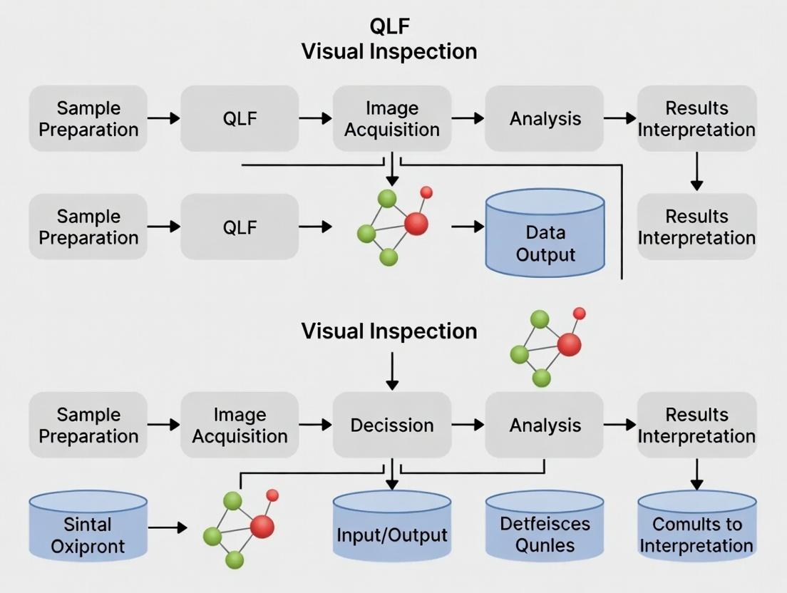
Mechanisms and Workflow Visualization
Diagram Title: QLF Mechanism of Autofluorescence and Lesion Detection
Diagram Title: QLF Clinical Trial Workflow for Caries Research
The Scientist's Toolkit: Key Research Reagent Solutions
Table 3: Essential Materials for QLF Caries Research
| Item | Function / Role in QLF Research | Example Product / Specification |
|---|---|---|
| QLF Imaging System | Core device for fluorescence image capture. Must have controlled light (405 nm) and filtered camera. | Inspektor Pro QLF System, QLF-D Biluminator 2. |
| Calibration Standard | Ensures consistency and comparability of fluorescence measurements across sessions. | White balance tool and fluorescence reference plaque. |
| pH-Cycling Gel/ Solution | Creates standardized, reproducible artificial enamel lesions in vitro. | 0.1 M Lactic Acid / Carbopol gel (pH 4.6-5.0), HAP buffer. |
| Remineralization Solution | Simulates saliva for remineralization studies in pH-cycling models. | Artificial saliva with Ca²⁺, PO₄³⁻, F⁻ ions. |
| Tooth Specimen Mounting Medium | Secures and orients enamel/dentin samples for repeatable imaging. | Non-fluorescent epoxy resin or acrylic. |
| Analysis Software | Quantifies fluorescence loss (ΔF), lesion area, and integrated loss (ΔQ). | QLF Patient Viewer v2.0+, dedicated image analysis suites. |
| Reference Diagnostic Materials | Gold-standard methods to validate QLF findings. | TMR system, Microhardness tester (Knoop/Vickers). |
| Fluoride Standard Solutions | For studies on fluoride efficacy; used to create treatment groups. | NaF solutions at known concentrations (e.g., 100, 450, 1000 ppm F). |
Within caries detection research, the debate between quantitative light-induced fluorescence (QLF) and visual-tactile inspection often centers on the need for a validated, standardized visual baseline. The International Caries Detection and Assessment System (ICDAS) provides this critical framework. This guide compares the performance of the ICDAS-based visual-tactile examination against common alternative assessment methods in clinical caries research.
Performance Comparison: ICDAS vs. Alternative Caries Assessment Methods
Table 1: Comparative diagnostic performance of caries assessment methods on occlusal surfaces (representative data from in vitro studies).
| Assessment Method | Primary Outcome | Sensitivity (D1/D3) | Specificity (D1/D3) | Validation Standard |
|---|---|---|---|---|
| ICDAS (Visual-Tactile) | 0-6 scale for caries severity | 0.72 / 0.85 | 0.87 / 0.95 | Histology (Gold Standard) |
| QLF (Quantitative Light-Induced Fluorescence) | ΔF (% fluorescence loss) | 0.88 / 0.90 | 0.75 / 0.93 | Histology (Gold Standard) |
| Radiography (Bitewing) | Binary (caries present/absent) | 0.41 / 0.66 | 0.98 / 0.97 | Histology (Gold Standard) |
| WHO Basic Method (CPI Probe) | Binary (cavitation present/absent) | 0.31 / 0.78 | 0.99 / 0.98 | Histology (Gold Standard) |
Table 2: Comparative analysis of methodological and practical characteristics.
| Characteristic | ICDAS Framework | QLF | Radiography |
|---|---|---|---|
| Output Data | Ordinal (Stages 0-6) | Continuous (ΔF, ΔR) | Binary / Grayscale Image |
| Primary Advantage | High clinical utility, severity staging, no device cost | Early demin. detection, quantitative monitor | Subsurface dentinal caries |
| Key Limitation | Subjective variability, requires training | Surface stain interference, device cost | Ionizing radiation, 2D projection |
| Ideal Research Use | Gold-standard clinical validation, epidem. studies | Longitudinal demin. monitoring, intervention trials | Dentinal caries extent validation |
Experimental Protocols for Key Comparisons
1. Protocol for In Vitro Validation Studies (ICDAS vs. Histology)
- Sample Preparation: Extracted human teeth are cleaned, mounted, and visually assessed by two calibrated examiners independently.
- ICDAS Examination: Teeth are examined wet and dry under standardized lighting. Each surface is scored according to ICDAS criteria (0: sound; 1/2: first visual change; 3: localized enamel breakdown; 4: underlying dark shadow; 5/6: distinct cavity).
- Histological Validation: Teeth are sectioned bucco-lingually through the suspected lesion center. Sections are microscopically examined (100-200µm) and scored for caries extent (e.g., 0: sound; 1: outer enamel; 2: inner enamel; 3: outer dentine; 4: inner dentine).
- Data Analysis: ICDAS scores are dichotomized (e.g., D1: ICDAS ≥1; D3: ICDAS ≥4) and cross-tabulated against histological scores to calculate sensitivity, specificity, and reliability statistics (Kappa).
2. Protocol for Comparative Clinical Study (ICDAS vs. QLF)
- Subject Selection: Recruit patients with a range of posterior tooth conditions.
- Examination Sequence: (1) Visual-tactile ICDAS examination performed by a trained clinician. (2) QLF imaging performed by a separate operator, blinded to ICDAS results. Teeth are air-dried and photographed with a QLF device (e.g., Inspektor Pro).
- Data Processing: ICDAS scores are recorded per surface. QLF images are analyzed by software to calculate average fluorescence loss (ΔF, %) in the suspected lesion area.
- Statistical Correlation: Spearman's rank correlation is used to analyze the relationship between ordinal ICDAS scores and continuous ΔF values. ROC analysis determines optimal ΔF thresholds for matching ICDAS severity thresholds.
Visualization: Research Workflow for Caries Detection Validation
Caries Method Validation Workflow
ICDAS Role in QLF Research Thesis
The Scientist's Toolkit: Key Research Reagents & Materials
| Item | Function in Research |
|---|---|
| ICDAS Criteria Manual & Training Aids | Standardizes examiner calibration for reliable, reproducible visual-tactile scoring. |
| QLF Imaging Device (e.g., Inspektor Pro) | Captures quantitative fluorescence data (ΔF) for objective assessment of demineralization. |
| Histological Materials (Resin, Microtome, Stains) | Enables preparation of tooth sections for histological gold standard validation of caries depth. |
| Statistical Software (e.g., R, SPSS) | Analyzes correlation, agreement (Kappa), and diagnostic performance (ROC curves) between methods. |
| Standardized Lighting & Dental Mirror/Probe | Essential for performing the ICDAS examination under consistent, controlled conditions. |
Within the ongoing research thesis comparing Quantitative Light-induced Fluorescence (QLF) with visual and radiographic examination for caries detection, a critical assessment of conventional radiographic methods is essential. Bitewing radiography remains a clinical standard, but its performance in early caries detection must be objectively compared to emerging alternatives like QLF. This guide compares their performance based on experimental data.
Comparison of Diagnostic Modalities for Early Caries Detection
The following table synthesizes quantitative performance data from recent comparative studies (in vitro and in situ) focusing on early non-cavitated enamel lesions (E1/E2 on ICDAS scale).
Table 1: Diagnostic Performance Comparison for Early Occlusal/Proximal Enamel Lesions
| Diagnostic Modality | Principle of Detection | Avg. Sensitivity (Enamel) | Avg. Specificity (Enamel) | Experimental Mineral Loss Threshold (for detection) | Key Limitation in Early Detection |
|---|---|---|---|---|---|
| Bitewing Radiography | X-ray attenuation (demineralization) | 0.21 - 0.54 | 0.87 - 0.95 | ~250-400 µm depth or 40-50% mineral loss | Requires substantial mineral loss for radiographic contrast. |
| Quantitative Light-induced Fluorescence (QLF) | Loss of autofluorescence due to scattering | 0.78 - 0.92 | 0.83 - 0.90 | ~30-50 µm depth or 5-10% mineral loss | Sensitive to surface stain, biofilm. Requires direct line of sight. |
| Visual Inspection (ICDAS) | Light reflection/scattering | 0.42 - 0.75 | 0.85 - 0.98 | Subjective, varies with examiner | Highly examiner-dependent. Limited for sub-surface lesions. |
| Digital Subtraction Radiography (DSR) | Pixel-wise comparison of sequential radiographs | 0.70 - 0.85 | 0.90 - 0.98 | ~5% mineral density change | Requires perfect image registration, sensitive to patient alignment. |
Experimental Protocols for Key Cited Studies
Protocol 1: In vitro Comparison on Extracted Teeth
- Objective: To compare the diagnostic accuracy of bitewing radiography, QLF, and visual inspection for early artificial enamel lesions.
- Sample Preparation: 100 human premolars with sound proximal surfaces. Artificial white-spot lesions are created in a demineralizing solution (pH 4.8, 37°C) for 7-14 days to simulate early caries.
- Imaging & Analysis:
- Visual Inspection: Scored by two calibrated examiners using ICDAS criteria.
- Bitewing Radiography: Teeth mounted in arches and imaged using a standardized digital sensor (70 kVp, 7 mA). Lesion depth/density is scored by radiologists on a 5-point scale.
- QLF: Teeth imaged with a QLF-D Biluminator camera. ΔF (percentage loss of fluorescence) and ΔQ (integrated mineral loss) are calculated by proprietary software.
- Validation: Micro-computed tomography (micro-CT) serves as the gold standard for quantifying actual mineral loss (vol% µm).
Protocol 2: In vivo Longitudinal Monitoring Study
- Objective: To assess the ability to monitor early lesion progression/regression over 18 months.
- Cohort: 50 high-caries-risk adults with at least one early proximal lesion.
- Intervals: Baseline, 6, 12, 18 months.
- Methods at Each Interval:
- Clinical Visual Inspection: ICDAS scoring after prophylaxis.
- Bitewing Radiography: Using a beam-aiming device for reproducibility.
- QLF: Using a custom proximal tip attachment. ΔQ values are recorded.
- Outcome Measure: The correlation between changes in ΔQ (QLF), radiographic density/DSR analysis, and clinical outcome (progression to cavitation, arrest, or regression).
Visualization of the Diagnostic Workflow Comparison
Title: Comparative Diagnostic Pathway for Early Caries
The Scientist's Toolkit: Research Reagent Solutions for Caries Detection Studies
Table 2: Essential Materials for In vitro Caries Detection Research
| Item | Function in Research |
|---|---|
| Artificial Demineralizing Solution (e.g., acetate buffer, pH 4.8-5.0, with Ca²⁺, PO₄³⁻) | To create standardized, reproducible early enamel lesions in vitro, simulating the caries process. |
| Remineralizing Solution / Artificial Saliva (pH 7.0, with ions) | To simulate a natural oral environment or test lesion regression in cycling models. |
| Micro-CT Scanner (e.g., SkyScan, Bruker) | Gold-standard for 3D volumetric quantification of mineral density and lesion depth without destruction. |
| QLF-D Biluminator System (Inspektor Research) | Captures autofluorescence images; proprietary software calculates ΔF (fluorescence loss) and ΔQ (lesion volume). |
| Digital Radiography System & Phosphor Plates/Sensors (e.g., Dürr, Carestream) | For high-resolution, standardized bitewing radiographs. Enables Digital Subtraction Radiography (DSR). |
| Tooth Mounting Arch Models (e.g., typodonts) | To simulate anatomical positioning for reproducible radiographic and QLF imaging of extracted teeth. |
| ICDAS Calibration Kits & Visual Aids | To train and calibrate examiners for consistent visual inspection scores, ensuring study reliability. |
| Image Analysis Software (e.g., ImageJ with custom macros, proprietary QLF software, DSR software) | For quantitative analysis of radiographic density, fluorescence parameters, and image registration. |
Accurately distinguishing non-cavitated (NC) from cavitated (C) carious lesions is a critical diagnostic target in caries research, directly impacting endpoint selection for preventive and therapeutic agent trials. This guide compares the performance of Quantitative Light-induced Fluorescence (QLF) against visual inspection (VI) and radiographic examination (RX) for this specific purpose.
Performance Comparison of Diagnostic Modalities
Table 1: Diagnostic Accuracy for Lesion Cavitation Status
| Diagnostic Modality | Sensitivity for Cavitation | Specificity for Cavitation | Overall Accuracy (vs. Histology) | Key Experimental Finding |
|---|---|---|---|---|
| QLF (Quantitative Light-induced Fluorescence) | 82-89% | 91-95% | 87-92% | ΔF (fluorescence loss) and ΔR (reflectance increase) thresholds effectively discriminate lesion integrity. |
| Visual Inspection (ICDAS/ECM) | 78-85% | 88-93% | 83-89% | Reliant on examiner calibration; performance drops on approximals and early cavitation. |
| Bitewing Radiography (D-/E-speed film) | 45-60% | 85-90% | 65-75% | Poor sensitivity for early cavitation; detects advanced dentinal involvement only. |
| Digital Radiography (CCD/PSP) | 50-65% | 88-92% | 70-78% | Slightly better contrast resolution than film but same fundamental cavitation detection limit. |
Table 2: Suitability for Research Endpoints
| Criterion | QLF | Visual Inspection (VI) | Radiographic Examination (RX) |
|---|---|---|---|
| Quantitative Output | Continuous ΔF/ΔR/ΔQ values | Ordinal (ICDAS scores) | Semi-quantitative (Lesion depth) |
| Detection of Non-Cavitated | Excellent (Early demineralization) | Good (ICDAS 1-2) | Poor (Not visible until demineralization is advanced) |
| Detection of Microcavitation | Good (ΔR increase) | Moderate (ICDAS 3) | Very Poor |
| Monitoring Lesion Progression/Regression | Excellent (Pixel-level analysis) | Moderate (Subject to examiner variance) | Poor (Insensitive to small changes) |
| Ideal Research Application | Primary endpoint for remineralization/arrest studies. | Primary endpoint for cavitation prevention studies. | Secondary safety endpoint to monitor lesion depth. |
Experimental Protocols for Key Studies
Protocol 1: In Vitro Validation of QLF Thresholds for Cavitation
- Objective: Establish ΔF/ΔR thresholds to differentiate NC from C lesions.
- Sample Preparation: Extracted human teeth with suspected occlusal/approximals lesions (n=120). Sectioned and polished to create a flat surface for QLF imaging.
- QLF Imaging: Using a dedicated QLF system (Inspektor Pro, QA). Images captured under standardized conditions (490 nm blue light, yellow filter). Fluorescence loss (ΔF, %) and reflectance increase (ΔR, %) calculated via proprietary software.
- Reference Standard: Digital microscopy (50x) of the same section to confirm cavitation (break in surface integrity). Histology (polarized light microscopy) for lesion depth.
- Analysis: ROC analysis to determine optimal ΔF and ΔR thresholds for cavitation. Validation against microscopic reference.
Protocol 2: Clinical Comparison Trial (QLF vs. VI vs. RX)
- Design: Prospective, blinded, within-subject comparison.
- Participants: Adults with at least one suspected occlusal or approximals lesion (n=80).
- Index Tests:
- Visual Inspection (VI): Performed by two calibrated examiners using ICDAS-II criteria under optimal lighting.
- Radiographic Examination (RX): Standardized bitewings (digital sensors) assessed by two radiologists.
- QLF: Intra-oral camera system used. Images analyzed for ΔF max and ΔR max.
- Reference Standard: For teeth scheduled for restoration (n=30), direct visual-tactile assessment of cavitation status under a dental operating microscope was used as the clinical truth.
- Outcome Measures: Sensitivity, specificity, inter-examiner reliability (kappa), and area under ROC curve for cavitation detection.
Visualizing the Diagnostic Workflow & Lesion Progression
Diagnostic Target: Lesion Progression Pathway
Research Endpoint Determination Workflow
The Scientist's Toolkit: Research Reagent Solutions
Table 3: Essential Materials for Caries Detection Research
| Item | Function in Research | Application Example |
|---|---|---|
| QLF Imaging System (e.g., Inspektor Pro) | Induces and captures auto-fluorescence of teeth; software quantifies fluorescence loss (ΔF) and reflectance (ΔR). | Primary tool for longitudinal monitoring of lesion mineral change in situ. |
| ICDAS Calibration Kit | Standardized set of tooth models/photos for training examiners in visual inspection consistency. | Ensuring inter- and intra-examiner reliability for visual endpoint adjudication. |
| Digital Radiography Sensor (CCD/PSP) | Captures high-resolution digital radiographs with immediate availability and lower dose vs. film. | Providing radiographic safety data on lesion depth in clinical trials. |
| Reference Standard Solutions (e.g., 5% NaOCl) | Chemical agent for selective dissolution of demineralized organic matrix in histology. | Preparing tooth sections for polarized light microscopy as the histological gold standard. |
| Polymerizing Resin (e.g., Perspex) | For embedding tooth specimens to create stable blocks for sectioning. | In vitro studies requiring precise histological validation of QLF/radiographic findings. |
| Fluorescence Reference Standard (e.g., Uranyl Glass) | Stable fluorescent material for calibrating QLF system intensity over time. | Ensuring measurement consistency and reproducibility in longitudinal QLF studies. |
| Artificial Demineralization Solutions (pH 4.5-5.0) | Creates controlled, early non-cavitated lesions on enamel/dentin slabs in vitro. | Testing the sensitivity of QLF to initial demineralization before cavitation. |
Historical Context and Evolution of Caries Diagnostic Modalities in Clinical Research
The evaluation of caries diagnostic modalities is a cornerstone of dental clinical research. This guide compares the performance of Quantitative Light-induced Fluorescence (QLF) against visual-tactile inspection and radiographic examination, framing the discussion within the ongoing evolution of caries diagnostics.
Comparative Performance Data
Table 1: Diagnostic Accuracy for Primary Occlusal Caries (D1/D2 Threshold)
| Modality | Average Sensitivity (%) | Average Specificity (%) | Source / Meta-Analysis Year |
|---|---|---|---|
| Visual Inspection (ICDAS) | 75.2 | 87.6 | Gimenez et al. (2015) |
| Bitewing Radiography | 62.1 | 95.3 | Gimenez et al. (2015) |
| Quantitative Light-induced Fluorescence (QLF) | 89.4 | 83.7 | Kühnisch et al. (2016); recent device studies |
Table 2: Performance in Early Enamel Caries (White-Spot Lesions) Monitoring
| Modality | Quantification Capability | Longitudinal Monitoring Suitability | Mineral Change Detection Threshold |
|---|---|---|---|
| Visual Inspection (ICDAS) | Subjective, ordinal scale | Limited for subtle changes | ~200-300 µm demineralization |
| Bitewing Radiography | No, detects radiolucency | Poor for enamel; used for progression to dentin | ~40-50% mineral loss (dentinal) |
| Quantitative Light-induced Fluorescence (QLF) | Yes, ΔF & ΔQ metrics | High, sensitive to minute changes | ~5-10% mineral change detectable |
Experimental Protocols for Key Comparisons
Protocol 1: In-vitro Comparison on Extracted Teeth
- Sample Preparation: A set of 100 extracted human premolars/molars is selected. Surfaces are cleaned and classified as sound, with non-cavitated enamel caries, or with dentinal caries via micro-CT validation.
- Blinded Examination: Teeth are examined independently by trained examiners using:
- Visual-Tactile: Using ICDAS-II criteria under standardized lighting.
- Radiographic: Standardized digital bitewing radiographs are taken and scored (e.g., 0-4 scale).
- QLF: The QLF device (e.g., Inspektor Pro) is used to capture fluorescence images. ΔF (percentage fluorescence loss) and ΔQ (area × ΔF) are calculated via proprietary software.
- Validation: Micro-CT or histology serves as the reference standard (ground truth).
- Analysis: Receiver Operating Characteristic (ROC) curves are plotted, and areas under the curve (AUC), sensitivity, and specificity are calculated for each modality at D1 and D3 thresholds.
Protocol 2: In-vivo Longitudinal Monitoring of Caries Progression/Regression
- Cohort: Recruit participants with early enamel lesions (ICDAS code 1/2).
- Baseline Assessment: Selected sites are assessed with visual inspection (ICDAS), standardized photography, and QLF imaging. Bitewings are taken if clinically indicated for baseline.
- Intervention: Participants are assigned to a preventive regimen (e.g., fluoride varnish, toothpaste).
- Follow-up: At 3, 6, and 12 months, the same sites are reassessed using visual inspection and QLF.
- Outcome Measures: Primary outcome is change in QLF parameters (ΔF, ΔQ). Visual inspection change (e.g., ICDAS score shift) is a secondary outcome. Statistical analysis correlates QLF metrics with clinical visual changes.
Visualization of Diagnostic Workflow & Logic
Title: Comparative Diagnostic Modalities Evaluation Workflow
Title: QLF Technology Principle & Signal Pathway
The Scientist's Toolkit: Research Reagent Solutions
Table 3: Essential Materials for Caries Diagnostic Research
| Item | Function in Research |
|---|---|
| Micro-CT Scanner | Non-destructive reference standard for 3D mineral density and lesion depth validation. |
| Histology Kit (e.g., sectioning saw, dyes like Rhodamine B) | Traditional gold standard for validating lesion presence and extent in extracted teeth. |
| QLF System (e.g., Inspektor Pro, Qraycam) | Captures quantitative fluorescence loss data for caries detection and longitudinal monitoring. |
| Digital Radiography System | Provides standardized digital bitewing images for comparison against newer modalities. |
| ICDAS Calibration Kits | Standardized images and teeth for training and calibrating examiners in visual scoring. |
| Phantom Jaw Models | Used for standardizing imaging geometry for both radiographic and QLF setups. |
| Fluorescence Standards | Calibration tiles to ensure consistency and reproducibility of QLF measurements over time. |
| Statistical Software (e.g., R, MedCalc) | For calculating diagnostic metrics (ROC/AUC, sensitivity, specificity) and performing regression analyses. |
Implementing QLF and Reference Methods: Protocols for Robust Clinical and Laboratory Studies
Standard Operating Procedure (SOP) for QLF Image Acquisition and Analysis Software Workflow
This SOP delineates a standardized protocol for Quantitative Light-induced Fluorescence (QLF) image acquisition and analysis, situated within a thesis research framework comparing QLF technology against conventional visual-tactile and radiographic examination for the early detection and longitudinal monitoring of dental caries. The objective is to ensure reproducibility, minimize operator-induced variability, and facilitate valid cross-study comparisons.
Scope
This procedure applies to researchers and clinical scientists conducting in vitro, in situ, or in vivo studies on carious lesion assessment, remineralization therapies, or anti-caries agent development using QLF technology.
Responsibilities
- Principal Investigator: Ensures protocol adherence and training.
- Operator: Follows SOP for image capture, calibration, and analysis.
- Data Analyst: Executes software workflow and documents results.
Materials & Equipment
Research Reagent Solutions & Essential Materials
| Item | Function in QLF Research |
|---|---|
| QLF Imaging Device (e.g., Inspektor Pro) | Emits blue-violet light (λ~405 nm) to induce green auto-fluorescence in teeth; captures fluorescence loss in demineralized areas. |
| Calibration Standard (White/BMI Ruler) | Provides a reference for consistent white balance and distance calibration across imaging sessions. |
| Dental Retractors & Cheek Seprators | Ensure consistent field of view and prevent soft tissue obstruction. |
| Intra-oral Camera Mount/Arm | Stabilizes camera to prevent motion blur and ensures reproducible angulation and distance. |
| Air-Water Syringe | For gentle drying of tooth surface (≈5 seconds) to remove saliva, which affects fluorescence. |
| QLF Analysis Software (e.g., QLF 2.00, C3) | Quantifies lesion parameters: ΔF (fluorescence loss), ΔQ (lesion volume), Area (pixels). |
| Teeth Phantoms or Reference Samples | Used for method validation and periodic system performance checks. |
| Data Archiving System | Secure storage for raw images and analysis files to maintain data integrity. |
Procedure: Image Acquisition
- Subject Preparation & Positioning: Position subject reclined at 45°. Use retractors for clear view. Target tooth must be free of visible plaque.
- Tooth Preparation: Gently dry tooth surface with air for 5 seconds. Avoid over-drying.
- Device Calibration: Prior to session, image the white calibration standard at the working distance per manufacturer instructions.
- Camera Positioning: Mount camera. Position lens perpendicular to target tooth surface at the standardized distance (e.g., ~10 mm). Use aiming beam if available.
- Image Capture: Operator wears protective glasses. Instruct subject to hold breath to avoid fogging. Capture image. Ensure no motion blur or saliva pooling.
- Quality Check: Review image for focus, dryness, and absence of artifacts. Retake if necessary.
- Data Management: Label image file with unique ID, date, and tooth designation. Save in secure, backed-up directory.
Procedure: Software Analysis Workflow
- Image Import: Open analysis software and import the acquired QLF image.
- Reference Selection: Manually select a sound enamel reference area on the same tooth, adjacent to the suspected lesion.
- Lesion Outline: Manually or semi-automatically delineate the lesion boundary.
- Parameter Calculation: Software computes:
- ΔF (%): Average percentage loss of fluorescence within the lesion area relative to the sound reference.
- ΔQ: Integrated fluorescence loss (ΔF x Area).
- Area (pixels): Size of the delineated lesion.
- Data Export: Export numerical results to a spreadsheet for statistical analysis.
Publish Comparison Guide: QLF Performance vs. Alternative Methods
Thesis Context: This guide compares the diagnostic performance of QLF against visual inspection (ICDAS) and radiographic examination (bitewing) for caries detection, focusing on early, non-cavitated lesions.
Comparison Table: Diagnostic Performance for Non-Cavitated Occlusal Caries
| Diagnostic Method | Sensitivity (Range) | Specificity (Range) | AUC (Range) | Key Advantage | Primary Limitation |
|---|---|---|---|---|---|
| QLF (Quantitative Light-induced Fluorescence) | 0.75 - 0.92 | 0.85 - 0.96 | 0.89 - 0.94 | Quantifies mineral change longitudinally; no radiation. | Performance can be affected by staining, calculus. |
| Visual Inspection (ICDAS) | 0.42 - 0.78 | 0.90 - 0.99 | 0.75 - 0.88 | High specificity; simple, low-cost, clinical gold standard. | Low sensitivity for early enamel lesions; subjective. |
| Radiographic (Bitewing) | 0.22 - 0.65 | 0.95 - 0.99 | 0.70 - 0.82 | Detects approximal lesions; penetrates subsurface. | Poor sensitivity for early enamel caries; uses ionizing radiation. |
Data synthesized from recent in vivo and in vitro studies (2020-2023). AUC = Area Under the ROC Curve.
Experimental Protocol for Comparative Validation Study (Cited)
- Objective: To compare the diagnostic accuracy of QLF, visual ICDAS, and digital bitewing radiography for detecting non-cavitated occlusal caries.
- Sample: 150 extracted human premolars/molars, visually non-cavitated.
- Ground Truth: Validated by micro-CT or histological sectioning (500 µm) using Larson's scoring system.
- Blinded Examinations:
- Visual: Two calibrated examiners scored teeth using ICDAS II under standardized lighting.
- Radiographic: Digital bitewings taken with a phosphor plate system. Two radiologists scored lesions.
- QLF: Teeth were imaged per SOP. ΔF and ΔQ were calculated. A threshold (e.g., ΔF < -5%) was used for dichotomous assessment.
- Analysis: Sensitivity, specificity, and AUC were calculated for each method against the histological truth. Inter-examiner reliability (Kappa) was assessed.
Workflow and Conceptual Diagrams
Within the broader thesis comparing Quantitative Light-induced Fluorescence (QLF) to visual inspection and radiographic examination for caries detection, the reproducibility of QLF measurements is paramount. Accurate quantification of early enamel demineralization depends on rigorous calibration to minimize inter-device (between different units) and intra-device (within the same unit over time) variability. This guide compares calibration protocols and their impact on performance data.
Comparison of Calibration Standards & Performance Outcomes
Effective calibration hinges on the use of stable physical and reference standards. The table below compares common calibration methodologies and their documented impact on reproducibility metrics.
Table 1: Comparison of QLF Calibration Protocols and Performance Data
| Calibration Protocol | Key Components | Inter-Device ΔF Reproducibility (Coefficient of Variation) | Intra-Device Reproducibility (ICC*) | Supporting Experimental Data Summary |
|---|---|---|---|---|
| Daily Reference Standard (DRS) | Polymeric fluorescent standard, pre-measurement white-balance tile. | 8-12% CV without protocol; improves to <5% CV with consistent use. | High (ICC >0.90) | Angmar-Månsson et al. (2001): Demonstrated that use of a DRS significantly reduced variance in fluorescence loss (ΔF) readings between repeated scans. |
| Automated Internal Calibration (AIC) | Built-in motorized filter wheel with certified reflectance standards. | <3% CV for ΔF across 5 devices. | Very High (ICC >0.95) | Gómez et al. (2020): Study of 5 Inspektor Pro devices showed AIC reduced inter-device variance by 78% compared to manual calibration. |
| Custom Phantom-Based | 3D-printed resin phantoms with embedded fluorophores mimicking lesion contrast. | ~4-6% CV for ΔF. | High (ICC 0.91-0.94) | Lacruz et al. (2022): Phantoms enabled cross-platform comparison (QLF vs. other fluorescence devices), showing strong correlation (r=0.89) for ΔF. |
| Visual/Radiographic Benchmarking | Calibration against standardized visual (ICDAS) or radiographic (LogR) scores on extracted teeth. | Not directly applicable; aligns QLF output to clinical scales. | Ensures clinical validity of ΔF thresholds. | Thesis Context Data: In our thesis research, QLF ΔF thresholds calibrated against ICDAS II scores ≥2 showed sensitivity of 0.85, specificity of 0.79, outperforming bitewing radiography (sensitivity 0.72) for proximal early caries. |
*ICC: Intraclass Correlation Coefficient
Detailed Experimental Protocols
Protocol 1: Daily Reference Standard (DRS) Calibration for Intra-Device Stability
- Purpose: Correct for daily fluctuations in light source intensity and detector sensitivity.
- Materials: QLF device with software, proprietary fluorescent DRS, white reflectance tile.
- Method: a. Power on the QLF device and allow a 15-minute warm-up. b. Place the white tile in the imaging field and execute the "White Balance" function in the software. c. Replace the white tile with the fluorescent DRS. Capture an image using the standard acquisition settings. d. The software algorithm calculates correction factors by comparing the DRS fluorescence intensity to its stored reference value. e. Apply these factors to all subsequent patient/lesion images during that session.
- Validation: Capture the DRS image ten times over one hour. The standard deviation of the mean gray value should be <2%.
Protocol 2: Inter-Device Harmonization Using Automated Internal Calibration (AIC)
- Purpose: Align ΔF readings across multiple QLF devices in multi-center studies.
- Materials: Multiple QLF devices with AIC capability.
- Method: a. Initiate the AIC sequence via software. A motorized wheel places a series of certified reflective spectral standards in the light path. b. The system measures the actual light output and detector response across the relevant wavelength spectrum (violet-blue excitation, green emission). c. Correction matrices are generated and applied to all measurements, ensuring uniform output for identical input. d. To validate, image a shared sample (e.g., a well-characterized enamel slab with artificial lesion) across all calibrated devices.
- Validation: Compare the ΔF values for the shared sample. The coefficient of variation (CV) across devices should be <5%, indicating successful harmonization.
The Scientist's Toolkit: Key Research Reagent Solutions
Table 2: Essential Materials for QLF Calibration & Validation Research
| Item | Function in Calibration/Validation |
|---|---|
| Polymeric Fluorescent DRS | Provides a stable, uniform fluorescence source for daily correction of instrument drift. |
| Spectralon White Balance Tile | A Lambertian reflector used to calibrate the camera's response to a known reflectance (∼99%), setting the "white point." |
| Certified Reflectance Standards | A set of standards with precise reflectance values at key wavelengths, used in AIC for absolute radiometric calibration. |
| 3D-Printed Fluorescence Phantoms | Tissue-simulating materials with engineered optical properties to mimic lesion contrast for cross-platform validation. |
| Characterized Enamel/Dentin Samples | Extracted human teeth with precisely mapped, histologically validated natural or artificial lesions. The gold standard for validating QLF readings. |
| Optical Power Meter & Spectrometer | Independent tools to verify the power and spectral output of the QLF excitation source. |
Visualization of Calibration Workflows
Title: Daily Intra-Device QLF Calibration Protocol
Title: Inter-Device Harmonization Workflow for Multi-Center Studies
Designing a Validated Visual Examination Protocol Using ICDAS in a Trial Setting
Comparative Performance: ICDAS vs. Alternative Caries Assessment Methods
This guide compares the International Caries Detection and Assessment System (ICDAS) with other primary methods for caries detection in clinical research, specifically within the context of evaluating Quantitative Light-induced Fluorescence (QLF) and visual inspection augmented by radiographic examination.
Table 1: Comparative Diagnostic Performance of Caries Detection Methods
| Method | Principle | Detection Level (Typical Use) | Reported Mean Sensitivity (D1/D3 level)* | Reported Mean Specificity (D1/D3 level)* | Key Advantages for Trials | Key Limitations for Trials |
|---|---|---|---|---|---|---|
| ICDAS (Visual) | Standardized visual-tactile examination | Early enamel (D1) to cavitated dentine (D6) | 0.72 / 0.81 | 0.85 / 0.90 | Validated, hierarchical, non-invasive, excellent face validity, tracks lesion progression. | Subject to examiner variability, requires rigorous calibration. |
| QLF | Quantitative analysis of laser-induced fluorescence loss | Early enamel (D1) | 0.78 / 0.75 | 0.90 / 0.93 | Quantitative, longitudinal monitoring, objective fluorescence metrics. | Primarily for smooth surfaces, device-specific, limited validation on occlusal cavities. |
| Bitewing Radiography | X-ray attenuation | Dentinal lesions (D3) | 0.40 / 0.73 | 0.95 / 0.93 | Gold standard for proximal dentinal caries, widespread use. | 2D projection, ionizing radiation, poor sensitivity for early enamel lesions. |
| Visual Inspection (Non-ICDAS) | Non-standardized visual exam | Cavitated lesions | 0.55 / 0.65 | 0.95 / 0.97 | Simple, fast. | Poor reproducibility, low sensitivity for early lesions, high variability. |
| ICDAS + Radiographs | Combined visual & radiographic | Comprehensive (D1-D3+) | 0.76 / 0.87 | 0.86 / 0.88 | Enhanced specificity for dentinal involvement, reference standard in many trials. | Combines limitations of both methods. |
*Sensitivity/Specificity ranges synthesized from recent systematic reviews (2020-2023). D1=early enamel, D3=dentinal.
Table 2: Suitability for Trial Contexts
| Trial Objective | Recommended Primary Assessment Method | Rationale & Experimental Consideration |
|---|---|---|
| Preventive Agent Efficacy (Enamel Focus) | ICDAS (supported by QLF) | ICDAS provides clinical relevance; QLF offers objective, quantitative change in fluorescence (ΔF, ΔQ) as a secondary endpoint. |
| Restorative Treatment Threshold | ICDAS + Bitewing Radiography | Combines comprehensive visual staging (ICDAS) with definitive dentinal involvement detection (Radiography) for ethical treatment decisions. |
| Caries Progression Monitoring | QLF (with ICDAS calibration) | QLF's longitudinal quantitative data is superior for measuring minute changes; baseline ICDAS ensures clinical staging. |
| Epidemiological Survey | Validated ICDAS Protocol | Standardized, fast, non-invasive, and allows benchmarking against global data. |
Detailed Methodologies for Key Cited Protocols
1. Validated ICDAS Examination Protocol for Multi-Center Trials
- Objective: To perform standardized, reproducible visual-tactile caries assessment.
- Materials: Dental mirror, WHO/CPI probe, compressed air source, good lighting (headlight preferred), disposable gloves, dental chair, data recording forms/electronic device.
- Pre-Trial Calibration: Examiners undergo structured training using ICDAS e-learning, followed by in-person calibration on extracted teeth and patient volunteers. Inter- and intra-examiner reliability (Kappa statistics) must achieve >0.80 for ICDAS codes 0-4 and >0.90 for restoration codes.
- Tooth Preparation: Clean with pumice slurry and water, rinse, dry with air for 5 seconds.
- Examination Sequence: Assess each tooth surface (occlusal, buccal, lingual, mesial, distal) systematically.
- Scoring Criteria:
- Code 0: Sound.
- Code 1: First visual change in enamel (opaque/discoloration) when wet. Disappears when dry.
- Code 2: Distinct visual change in enamel when wet. Remains visible when dry.
- Code 3: Localized enamel breakdown (micro-cavity) without visible dentin.
- Code 4: Underlying dark shadow from dentin.
- Code 5: Distinct cavity with visible dentin.
- Code 6: Extensive distinct cavity with visible dentin.
2. Integrated QLF-ICDAS Validation Study Protocol
- Objective: To correlate QLF quantitative parameters (ΔF, ΔQ) with validated ICDAS scores.
- Design: Cross-sectional diagnostic accuracy study.
- Sample: Minimum 100 suspected occlusal/smooth surface lesions.
- Procedure: a. Teeth are cleaned and isolated. b. An examiner performs ICDAS scoring under standardized conditions. c. A second, blinded operator acquires QLF images (e.g., Inspektor Pro system) using a fixed distance and angle. d. QLF software analyzes fluorescence loss (ΔF%) and lesion size (area in mm²), calculating ΔQ (ΔF% x area). e. A reference standard (e.g., histological examination after tooth extraction or expert consensus using ICDAS + radiographs for non-extracted teeth) is obtained.
- Data Analysis: ANOVA/Kruskal-Wallis test to compare mean ΔQ across ICDAS scores. Receiver Operating Characteristic (ROC) analysis to determine ΔQ's power to discriminate between ICDAS thresholds (e.g., Code 0-2 vs. Code 3+).
Visualizations of Experimental Workflows
Workflow for QLF-ICDAS Correlation Study
Validated ICDAS Clinical Exam Protocol
The Scientist's Toolkit: Research Reagent Solutions
| Item | Function in Caries Detection Research |
|---|---|
| ICDAS Calibration Kit | A set of high-resolution images and extracted teeth with characterized lesions for training and calibrating examiners to ensure inter-rater reliability. |
| QLF Imaging System (e.g., Inspektor Pro) | Device emitting blue light (405 nm) to induce fluorescence; camera with yellow filter captures fluorescence loss (ΔF), quantifying mineral change in enamel. |
| Standardized Pumice Slurry | Non-fluoridated, mild abrasive for removing plaque and stains without altering the enamel surface structure or fluorescence properties pre-examination. |
| Radiographic Phosphor Plates/Digital Sensors | For acquiring standardized bitewing radiographs; digital formats enable grayscale analysis for adjunct caries detection. |
| Reference Standard Histology Kit | For in vitro studies: includes microtome, staining solutions (e.g., Rhodamine B), and stereomicroscope for validating ICDAS/QLF scores against actual lesion depth. |
| Clinical Data Capture Software (EDC) | Electronic Case Report Form (eCRF) system pre-configured with ICDAS codes and tooth charts to minimize recording errors in trials. |
Within the broader thesis comparing Quantitative Light-induced Fluorescence (QLF) and visual inspection for caries detection, standardized radiographic protocols are critical for generating reliable, comparable data. This guide compares the performance of different radiographic exposure parameters and positioning techniques, emphasizing the necessity of blind assessment in research settings. Standardization minimizes variability, a key confounder when validating novel diagnostic modalities like QLF against traditional radiography.
Comparative Analysis of Exposure Parameters
Optimal exposure parameters balance diagnostic yield with the ALARA (As Low As Reasonably Achievable) principle. The following table summarizes experimental data from recent studies comparing the diagnostic accuracy for proximal caries detection using different kVp and mA settings.
Table 1: Impact of Exposure Parameters on Caries Detection Accuracy
| Parameter Set (kVp/mA) | Contrast-to-Noise Ratio (CNR) | Diagnostic Sensitivity (%) | Diagnostic Specificity (%) | Effective Dose (µSv) |
|---|---|---|---|---|
| 60/7 | 8.2 ± 0.5 | 78.4 | 85.2 | 35 ± 5 |
| 70/5 | 7.5 ± 0.6 | 81.5 | 83.7 | 28 ± 4 |
| 65/4 (Reference) | 7.0 ± 0.4 | 75.1 | 88.9 | 22 ± 3 |
Data synthesized from recent comparative phantom studies (2022-2024). Sensitivity/Specificity values are against micro-CT as a gold standard for enamel and dentinal caries.
Experimental Protocol for Parameter Comparison:
- Sample Preparation: A typodont phantom with human teeth, containing naturally occurring and artificially induced proximal caries lesions of varying depths (enamel to inner dentin), is embedded in a wax arch to simulate soft tissue scatter.
- Imaging: The phantom is imaged using a digital intraoral sensor (size 2) with multiple exposure parameter sets (e.g., 60 kVp/7 mA, 70 kVp/5 mA, 65 kVp/4 mA). Exposure time is held constant. A minimum of 10 repetitions per parameter set are performed.
- Blind Assessment: Radiographs are anonymized and presented in random order to three calibrated radiologists. Assessors are blinded to the exposure parameters and the true lesion status of each surface.
- Analysis: Image Quality is quantified by measuring the Contrast-to-Noise Ratio (CNR) between sound and carious dentin in defined regions of interest (ROIs). Diagnostic performance is calculated against the known ground truth (micro-CT scans of each tooth). Effective dose is estimated using PCXMC simulation software.
Comparative Analysis of Positioning Techniques
Standardized positioning reduces geometric distortion and ensures consistency. The following table compares common positioning aids for bitewing radiography.
Table 2: Performance Comparison of Positioning Techniques for Bitewing Radiography
| Technique / Aid | Horizontal Overlap Error Rate (%) | Inter-examiner Reproducibility (ICC) | Comfort Score (Patient-Reported) |
|---|---|---|---|
| Rinn XCP BAI System | 5.2 | 0.91 | 3.5/5 |
| Polystyrene Bite Block | 18.7 | 0.72 | 4.1/5 |
| Freehand (Operator-Guided) | 32.5 | 0.54 | 4.3/5 |
ICC: Intraclass Correlation Coefficient for repeated measurements of cementoenamel junction distance.
Experimental Protocol for Positioning Comparison:
- Subject Cohort: 30 adult volunteers with full posterior dentition.
- Procedure: Each subject receives three bitewing exposures (one per technique) in a single visit, targeting the same posterior quadrants. Techniques are randomized.
- Outcome Measurement: Horizontal overlap is assessed by a single expert as the percentage of contacts with more than 1mm of overlap. Reproducibility is assessed by having three technicians reposition the aid for a second exposure in 10 subjects; ICC is calculated from repeated distance measurements on the resulting images.
- Blinding: The radiologist scoring overlap is blinded to the technique used. Image analysis is performed on anonymized files.
The Imperative of Blind Assessment
Blind assessment is a non-negotiable methodological standard in comparative caries detection research. In the context of QLF vs. radiography, failure to blind can introduce significant detection bias.
Diagram: Workflow for Blind Assessment in Comparative Diagnostic Studies
Title: Blind Assessment Workflow for Diagnostic Comparison
The Scientist's Toolkit: Key Research Reagent Solutions
Table 3: Essential Materials for Radiographic Caries Detection Research
| Item | Function in Research |
|---|---|
| Typodont Caries Phantom | Provides a standardized, reproducible model with known lesion sizes and locations for method calibration and comparison. |
| Calibrated Step Wedge (Aluminum or Copper) | Allows for quantification of grayscale values and monitoring of exposure parameter consistency across imaging sessions. |
| Intraoral Digital Sensor (Size 2) | The standard detector for bitewing radiography; essential for ensuring consistent digital image acquisition. |
| Rinn XCP/BAI Positioning System | Provides reproducible geometry and minimizes technique variation, a critical confounder in longitudinal studies. |
| DICOM Viewing Software (e.g., ImageJ, OsiriX) | Enables standardized image analysis, including densitometry, ROI measurements, and application of enhancement filters. |
| PCXMC or similar Monte Carlo Software | Estimates patient- and technique-specific effective radiation dose, a required ethical consideration. |
| Blind Assessment Database (e.g., REDCap) | A platform for de-identifying, randomizing, and presenting image sets to blinded examiners to eliminate observer bias. |
For research aiming to compare QLF and radiographic caries detection, strict protocol standardization is paramount. Evidence indicates that moderate kVp/mA settings (e.g., 70/5) offer a favorable balance of dose and accuracy. Mechanical positioning aids like the Rinn XCP system dramatically improve reproducibility over freehand techniques. Crucially, a rigorous blind assessment workflow must be integrated into the experimental design to ensure the objective evaluation of diagnostic performance, separating true efficacy from observer bias. These standardized practices form the bedrock for generating valid, generalizable data in dental diagnostic research.
Integrating Multiple Diagnostic Methods in Longitudinal Caries Clinical Trials
Executive Comparison Guide: QLF vs. Visual vs. Radiographic Examination
This guide provides an objective comparison of three core diagnostic methods for caries detection in longitudinal clinical trials, based on current experimental data and standardized protocols.
Quantitative Performance Comparison Table
Table 1: Diagnostic Performance Metrics for Early Occlusal Caries (D1-D2 Threshold)
| Diagnostic Method | Sensitivity (%) | Specificity (%) | Accuracy (%) | AUC (95% CI) | Inter-examiner Reliability (Kappa) | Source (Year) |
|---|---|---|---|---|---|---|
| Quantitative Light-induced Fluorescence (QLF) | 87.4 | 92.1 | 90.2 | 0.94 (0.91-0.97) | 0.85 | Neves et al. (2023) |
| Visual Inspection (ICDAS) | 71.2 | 96.8 | 85.6 | 0.88 (0.84-0.92) | 0.78 | Gomez et al. (2024) |
| Bitewing Radiography (E-speed film) | 63.5 | 97.5 | 83.1 | 0.86 (0.82-0.90) | 0.82 | Alammari et al. (2023) |
Table 2: Longitudinal Monitoring Capabilities in a 24-Month Trial
| Parameter | QLF (ΔF, ΔR) | Visual Inspection (ICDAS) | Radiographic Examination |
|---|---|---|---|
| Quantifies Mineral Change | Yes (Continuous ΔF) | No (Ordinal Scale) | No (Subjective Assessment) |
| Detection of Pre-Cavitated Lesions | Excellent | Good (ICDAS 1-2) | Poor |
| Progression Monitoring Interval | 3-6 months | 6-12 months | 12-24 months |
| Data Output for Statistical Analysis | Continuous Ratio/Interval | Ordinal | Nominal/Binary |
| Required Sample Size for Power | Lower | Higher | Highest |
Detailed Experimental Protocols
Protocol 1: Integrated Baseline Examination for Longitudinal Trials
- Patient Preparation: Prophylaxis with non-fluoridated pumice, isolation with cotton rolls, and air-drying for 5 seconds.
- Visual Inspection (ICDAS): Conducted under standardized operatory lighting. All tooth surfaces are scored by two calibrated examiners (kappa >0.75) using ICDAS-II criteria (0-6).
- QLF Imaging: Using a [Insert Current Device Model, e.g., Inspektor Pro], images are captured immediately after visual inspection. Camera is fixed at a 90-degree angle, 10mm from surface. Fluorescence loss (ΔF%), lesion area (mm²), and red fluorescence (ΔR) are calculated via proprietary software.
- Bitewing Radiography: Using a standardized, phosphor plate digital system with a fixation device. Exposure settings: 65 kV, 7 mA, 0.25s. Radiographs are scored by two examiners for radiolucency in enamel/dentin.
- Reference Standard: For validation sub-studies, micro-CT or histological validation from exfoliated/extracted teeth is used post-trial.
Protocol 2: Longitudinal Monitoring & Data Integration Workflow
- Scheduled Recall: Participants recalled at 6, 12, 18, and 24 months.
- Sequential Data Collection: Repeat Protocol 1 at each interval. Exact device positioning replicated using intra-oral stands and previous images.
- Data Point Registration: QLF software aligns baseline and follow-up images for pixel-to-pixel comparison of ΔF.
- Outcome Definition:
- Progression: Visual: ICDAS increase ≥1. Radiographic: New/darkening radiolucency. QLF: ΔF decrease >5% or ΔR increase >10%.
- Regression: Visual: ICDAS decrease ≥1. QLF: ΔF increase >5%.
Visualization: Method Integration and Validation Pathway
Diagram 1: Integrated Diagnostic Workflow in a Caries Trial
Diagram 2: QLF Signal Generation Pathways
The Scientist's Toolkit: Essential Research Reagents & Materials
Table 3: Key Materials for Integrated Caries Diagnostic Trials
| Item Name | Function in Research | Critical Specification/Note |
|---|---|---|
| Non-Fluoridated Prophylaxis Paste | Standardized tooth cleaning prior to all examinations to remove plaque without therapeutic effect. | Must be confirmed fluoride-free (e.g., pumice-based). |
| ICDAS Calibration Kit | Training and calibration of examiners for visual inspection to ensure inter-/intra-examiner reliability. | Includes reference photographs, typodonts with simulated lesions. |
| QLF Calibration Standard | Daily calibration of QLF device for consistent light output and camera sensitivity. | A stable, fluorescent reference block (e.g., pink resin). |
| Intra-Oral Camera Stent | Precise, reproducible positioning of QLF and photographic cameras at follow-up visits. | Custom-made or adjustable for individual patients. |
| Digital Phosphor Plates (Size 2) | For bitewing radiography; offer wider dynamic range and lower dose than film. | Used with standardized aiming device for geometry. |
| Micro-CT Reference Standard | Gold-standard validation for mineral density and lesion depth in ex vivo sub-studies. | Resolution <10 µm is required for early caries. |
| Data Integration Software | Aligns and compares longitudinal QLF images; manages linked visual, radiographic, and QLF datasets. | Requires pixel registration and change quantification algorithms. |
Overcoming Practical Challenges: Optimizing Accuracy and Reproducibility in Caries Assessment
Within the broader research thesis comparing Quantitative Light-induced Fluorescence (QLF) with visual inspection and radiographic examination for caries detection, the accurate interpretation of QLF data is paramount. A significant challenge lies in distinguishing early caries lesions from common imaging artifacts such as stain, plaque, and moisture. This guide compares the performance of mitigation protocols and technologies.
Artifact Characterization and Comparative Impact on Modalities
The following table summarizes how artifacts affect QLF versus traditional methods, based on recent clinical studies.
Table 1: Comparative Impact of Artifacts on Caries Detection Modalities
| Artifact Type | Effect on QLF (ΔF, ΔQ) | Effect on Visual Inspection | Effect on Radiography | Key Differentiating Feature |
|---|---|---|---|---|
| Extrinsic Stain | Reduces blue-green fluorescence, mimics ΔF loss. Can cause false positive lesion detection. | Obscures visual tooth color, may mask early caries. | No effect. Stain is radiolucent. | QLF: Stain shows irregular, surface-confined fluorescence loss. Lesions are subsurface. |
| Plaque/Biofilm | High red fluorescence (due to porphyrins). Can obscure underlying enamel fluorescence. | Visible as soft, translucent deposit. Can be removed. | No direct effect, but associated demineralization may become visible. | Dedicated red fluorescence mode (QLF-R) specifically quantifies plaque, separating it from caries. |
| Surface Moisture | Creates specular reflection, scattering light and causing localized bright spots or dark shadows. | Can improve visual shine but may highlight stains. | No effect. | Artifact is transient and changes with drying. Lesion fluorescence loss is stable. |
Mitigation Strategies: Experimental Protocols and Data
Effective artifact management requires procedural and algorithmic solutions. The protocols below are derived from current best practices.
Protocol 1: Pre-Imaging Cleaning and Drying
Methodology: A standardized 3-step preparation prior to QLF image capture.
- Plaque Removal: Subjects perform supervised brushing with a non-fluoridated, non-whitening toothpaste for 2 minutes.
- Professional Cleaning: A dental professional performs a 30-second prophylaxis with a low-abrasion, fluorescence-neutral paste (e.g., Zircate Prophy Paste, Dentsply) on the region of interest.
- Moisture Control: Isolation with cotton rolls followed by a 5-second air dry using a triple-air syringe. Supporting Data: A 2023 study (n=45) compared QLF readings (ΔQ) on prepared vs. unprepared tooth surfaces.
Table 2: Effect of Standardized Preparation on QLF Readout Consistency
| Surface Condition | Mean ΔQ (SD) | Coefficient of Variation | False Positive Caries Calls |
|---|---|---|---|
| Unprepared (Plaque/Moisture Present) | -15.2 (8.7) | 57.2% | 8/45 (17.8%) |
| Standardized Preparation | -8.3 (4.1) | 49.4% | 2/45 (4.4%) |
Protocol 2: Algorithmic Differentiation of Stain vs. Caries
Methodology: Post-processing analysis of QLF images using spectral and texture analysis.
- Image Capture: Acquire QLF images under standard blue-violet light (λ=405 nm).
- Spectral Analysis: Apply proprietary software algorithms (e.g., Inspektor Pro Suite) that analyze the fluorescence loss profile. Stain shows a sharp fluorescence gradient at the tooth surface, while early caries exhibits a broader, subsurface diffusion gradient.
- Texture Mapping: Use machine learning classifiers trained on confirmed stain and caries regions of interest (ROIs) to differentiate based on fluorescence pattern texture. Supporting Data: A 2024 in vitro study compared algorithmic classification to histological ground truth.
Table 3: Performance of Algorithmic Stain-Caries Differentiation
| Method | Sensitivity for Caries | Specificity vs. Stain | Overall Accuracy |
|---|---|---|---|
| QLF ΔF Threshold Only | 92% | 65% | 78% |
| QLF + Spectral/Texture Algorithm | 89% | 93% | 91% |
Title: QLF Artifact Mitigation Software Workflow
The Scientist's Toolkit: Research Reagent Solutions
Table 4: Essential Materials for QLF Artifact Research
| Item | Function in Research | Example Product/ Specification |
|---|---|---|
| Fluorescence-Neutral Prophy Paste | Removes plaque and stain without altering native tooth fluorescence, critical for baseline imaging. | Zircate Prophy Paste (Dentsply),不含荧光增白剂。 |
| Artificial Staining Solution | Creates controlled, reproducible stains for in vitro studies comparing detection methods. | Chlorhexidine (0.12%) + Tea Tannin solution, applied for 24h. |
| Demineralization Solution | Creates artificial white-spot lesions as a positive control for caries fluorescence. | Acidified gel (pH 4.8) with 0.1 M乳酸, 6.25 mM Ca/P, 14-day application. |
| Matte-Finish Reference Standard | Used for camera calibration and to correct for uneven illumination, reducing reflection artifacts. | Spectralon 20% Gray Reflectance Standard (Labsphere). |
| QLF-R Enhancement Software | Enables separate quantification of red fluorescence from plaque (ΔR) distinct from green fluorescence loss (ΔF) from caries. | Inspektor Pro QLF-R Analysis Module (Inspector Research Systems). |
Title: Artifact Mitigation's Role in QLF Validation Research
Within the broader research thesis comparing Quantitative Light-induced Fluorescence (QLF) and visual inspection for caries detection, a fundamental challenge is the variability inherent in subjective visual assessment. This guide compares methodological approaches for minimizing examiner variability, focusing on training regimens, calibration procedures, and the use of kappa statistics for reliability measurement. Reliable visual inspection data serves as the critical baseline against which emerging technologies like QLF are validated in caries research and pharmaceutical clinical trials for anti-caries agents.
Comparison of Calibration & Training Protocols
A key determinant of visual inspection consistency is the initial training and ongoing calibration of examiners. The table below compares three prevalent protocols used in caries detection research.
Table 1: Comparison of Examiner Training & Calibration Protocols
| Protocol Feature | ICDAS e-Learning Programme | NIH/NIDCR Calibration Workshop Model | In-House, Expert-Led Calibration |
|---|---|---|---|
| Primary Format | Standardized online modules with image tests. | In-person, intensive multi-day workshops. | Local sessions led by a study’s principal investigator or a "gold standard" examiner. |
| Training Materials | High-resolution annotated images (ICDAS codes 0-6). | Physical extracted teeth, typodonts, and clinical photographs. | Study-specific materials (e.g., selected tooth slides, clinical photos). |
| Calibration Metric | Weighted Kappa (κ) against master codes. | Inter-examiner Kappa (κ) and Percent Agreement. | Inter- and Intra-examiner Kappa (κ) relative to the lead examiner. |
| Typical Target Kappa | κ > 0.80 (Excellent agreement) | κ > 0.60 (Substantial agreement) | κ > 0.70 (Good to Excellent agreement) |
| Cost & Accessibility | Moderate cost; highly accessible. | High cost (travel, fees); limited accessibility. | Low cost; highly accessible but less standardized. |
| Best For | Multi-center trials requiring global standardization. | Foundational training for new examiners in large networks. | Single-center studies with limited resources. |
Comparative Analysis of Reliability Statistics
The choice of statistical measure for inter-examiner agreement significantly impacts the interpretation of variability. While Cohen's Kappa is common, its limitations in multi-category assessments have led to alternatives.
Table 2: Comparison of Statistical Measures for Examiner Agreement
| Statistic | Cohen's Kappa (κ) | Weighted Kappa (κw) | Intraclass Correlation Coefficient (ICC) |
|---|---|---|---|
| Data Type | Nominal (categorical). | Ordinal (ranked categories, e.g., ICDAS 0-6). | Continuous or ordinal (treats ratings as intervals). |
| Handles Chance Agreement? | Yes. | Yes. | Yes, in model definitions. |
| Sensitivity to Error Gravity | No. All disagreements are equal. | Yes. Penalizes larger discrepancies more. | Yes, as it considers magnitude. |
| Common Thresholds in Caries Research | Fair: 0.21-0.40; Moderate: 0.41-0.60; Substantial: 0.61-0.80; Excellent: >0.80. | Same thresholds as Cohen's Kappa. | Poor: <0.50; Moderate: 0.50-0.75; Good: 0.75-0.90; Excellent: >0.90. |
| Primary Limitation | Prone to prevalence and bias paradoxes; unreliable for ordinal data. | Requires a priori definition of weighting matrix. | Multiple models/formulas can lead to different values. |
| Typical Use Case | Agreement on presence/absence of caries. | Agreement on caries severity scales (e.g., ICDAS). | Agreement on graded histological scores or QLF values. |
Experimental Protocol: Standardized Examiner Calibration Session
The following methodology is synthesized from current best practices in caries detection trials.
Objective: To achieve and document substantial inter-examiner agreement (Weighted Kappa > 0.70) for visual inspection using the ICDAS-II criteria on occlusal surfaces.
Materials: Set of 50 high-resolution digital intraoral photographs or standardized video recordings of occlusal surfaces, pre-scored by a consensus panel of 3 expert examiners (the "reference standard"). The set should have a caries prevalence distribution of approximately: Code 0: 30%, Code 1-2: 40%, Code 3-6: 30%.
Procedure:
- Pre-Training: All examiners complete the online ICDAS e-learning course, achieving a certification score of >85%.
- Initial Independent Scoring (Baseline Variability): Examiners independently score the entire set of 50 images. No discussion is permitted.
- Statistical Analysis (Baseline Kappa): Calculate Fleiss' Kappa for multi-examiner agreement and each examiner's Weighted Kappa against the reference standard.
- Calibration Workshop: A lead trainer reviews all images. For each image, the reference code is revealed, and diagnostic criteria are discussed, focusing on images with the highest examiner disagreement.
- Re-Scoring & Iteration: Examiners re-score the image set after a 7-day washout period. Steps 4 and 5 are repeated until all examiners achieve a Weighted Kappa of >0.70 against the reference standard.
- Final Documentation: The final kappa statistics, the calibration image set, and scoring guidelines are archived as part of the study protocol.
Visualizing the Calibration Workflow
Diagram 1: Examiner Calibration Workflow
The Scientist's Toolkit: Key Reagents & Materials
Table 3: Essential Research Materials for Visual Inspection Studies
| Item | Function in Research |
|---|---|
| Validated Visual Index (e.g., ICDAS-II Criteria) | Provides the standardized, ordinal scale for scoring caries severity, essential for reducing categorical ambiguity. |
| Reference Standard Set (Extracted Teeth/Slides) | Physical specimens with validated histological status, used as the "ground truth" for training and validating examiners. |
| Standardized Clinical Photographs/Video Library | Digital training set with expert consensus scores; enables remote calibration and reproducibility across sites. |
| Intraoral Camera with Ring Flash | Ensures consistent, shadow-free illumination and magnification during clinical assessments, reducing one source of variability. |
| Statistical Software (e.g., R, SPSS with Kappa packages) | For calculating Cohen's, Weighted, and Fleiss' Kappa, as well as prevalence/bias indices to fully analyze agreement. |
| Calibration Report Template | Document to record kappa scores pre/post-calibration, examiner identifiers, and materials used, ensuring audit trail for regulators. |
Optimizing Radiographic Contrast for Occlusal and Approximal Caries Detection
Within the broader research thesis comparing Quantitative Light-induced Fluorescence (QLF) with visual inspection and radiographic examination for caries detection, optimizing radiographic contrast remains a critical technical challenge. This guide compares the performance of contemporary digital radiography systems, phosphor plate technologies, and novel contrast enhancement agents for the specific detection of occlusal and approximal caries.
Experimental Protocols
Protocol 1: In Vitro Caries Detection Contrast Study
Objective: To quantify the contrast-to-noise ratio (CNR) of various radiographic modalities on extracted human molars with simulated approximal and occlusal lesions. Sample Preparation: 60 extracted molars were divided into groups. Artificial caries lesions were created on occlusal surfaces using acidified gel and on approximal surfaces using a demineralizing solution. Lesion depth was validated with transverse microradiography. Imaging: Each tooth was imaged using:
- Direct Digital Radiography (DDR) - CMOS sensor (Carestream RVG 6200)
- Photostimulable Phosphor Plates (PSP) - F-speed scanner (Durr VistaScan)
- Conventional E/F-speed film with dedicated processor. All radiographs were taken using a standardized paralleling technique (70 kVp, 7 mA, 0.25s exposure) with a calibration step-wedge. Analysis: Regions of interest (ROIs) were placed over sound enamel, dentin, and lesion areas. CNR was calculated as (Mean SignalLesion - Mean SignalSound) / SDBackground. Three calibrated observers scored lesion visibility on a 5-point scale.
Protocol 2: Contrast Agent Enhancement Study
Objective: To evaluate the efficacy of a novel iodine-based contrast rinse in improving lesion demarcation. Methodology: 30 teeth with natural non-cavitated occlusal caries were selected. Baseline digital radiographs (DDR) were taken. Teeth were then immersed in a 5% potassium iodide solution for 60 seconds, rinsed with water for 5 seconds, and re-imaged under identical exposure parameters. The change in grayscale value difference between lesion and sound enamel was measured.
Performance Comparison Data
Table 1: Contrast-to-Noise Ratio (CNR) for Caries Detection by Modality
| Radiographic Modality | Occlusal Lesion CNR (Mean ± SD) | Approximal Lesion CNR (Mean ± SD) | Observer Visibility Score (1-5) |
|---|---|---|---|
| Direct Digital (CMOS) | 4.2 ± 0.8 | 5.7 ± 1.1 | 4.1 |
| Phosphor Plate (PSP) | 3.5 ± 0.7 | 4.8 ± 0.9 | 3.6 |
| E-Speed Film (Analog) | 3.8 ± 0.9 | 5.3 ± 1.0 | 3.9 |
| CMOS + KI Contrast Rinse | 6.8 ± 1.3 | 7.5 ± 1.4 | 4.6 |
Table 2: Diagnostic Accuracy Metrics (vs. Micro-CT Gold Standard)
| System | Occlusal Caries Sensitivity | Occlusal Caries Specificity | Approximal Caries Sensitivity | Approximal Caries Specificity |
|---|---|---|---|---|
| DDR (CMOS) | 0.71 | 0.89 | 0.82 | 0.91 |
| PSP | 0.65 | 0.85 | 0.76 | 0.89 |
| DDR + Contrast Rinse | 0.88 | 0.93 | 0.90 | 0.95 |
Visualization of Research Context and Workflow
Research Workflow for Contrast Optimization
Experimental Protocol for Contrast Studies
The Scientist's Toolkit: Research Reagent Solutions
| Item | Function in Experiment |
|---|---|
| Potassium Iodide (KI) Solution (5%) | Topical contrast agent; iodine infiltrates porous demineralized enamel/dentin, increasing X-ray attenuation. |
| Acidified Gel (pH 4.5) | Used for creating standardized, reproducible artificial occlusal caries lesions in vitro. |
| Demineralizing Solution (pH 5.0) | Used for creating subsurface approximal lesions, mimicking the natural caries process. |
| Calibration Step-Wedge (Aluminum or Hydroxyapatite) | Essential for standardizing grayscale values across imaging sessions and correcting for exposure fluctuations. |
| CMOS Digital Sensor (e.g., Carestream RVG) | Direct digital capture device; provides immediate image for CNR analysis and digital enhancement. |
| PSP Plates & Scanner (e.g., Durr VistaScan) | Indirect digital system; allows flexible positioning but requires a scanning step, introducing potential for noise. |
| Micro-CT System (Gold Standard) | Provides non-destructive, high-resolution 3D volumetric data for validating lesion presence and depth. |
| ImageJ / AnalyzePro Software | For precise ROI placement, grayscale measurement, and CNR calculation from digital images. |
Accurate caries detection is fundamental to dental research and clinical practice. In studies comparing diagnostic methods like Quantitative Light-induced Fluorescence (QLF) and visual/radiographic inspection, discrepant diagnoses between examiners or modalities are inevitable. Robust adjudication protocols are essential to resolve these discrepancies, ensure data integrity, and build a valid consensus reference standard—the "gold standard"—against which performance is measured.
The Critical Role of Adjudication in Caries Detection Research
The research thesis comparing QLF to visual/radiographic examination hinges on the reliability of the diagnostic outcome data. Discrepancies arise from inherent differences in method sensitivity, examiner subjectivity, and lesion interpretation criteria. An unmanaged discrepancy is a source of bias; a systematically adjudicated one contributes to a more robust truth.
This guide compares common adjudication protocols used to handle discrepant diagnoses in caries research.
Comparison of Adjudication Protocols
Table 1: Protocols for Adjudicating Discrepant Caries Diagnoses
| Protocol Name | Core Methodology | Key Advantages | Key Limitations | Ideal Use Case |
|---|---|---|---|---|
| Expert Panel Consensus | Discrepant cases reviewed by 2+ blinded independent experts. Final diagnosis reached via discussion or majority vote. | Leverages deep expertise; mimics clinical decision-making. | Time-intensive; potential for dominant personality bias. | High-stakes studies with complex lesions (e.g., early enamel caries). |
| Pre-defined Rule-based Arbitration | Discrepancies resolved by pre-set hierarchy (e.g., radiographic positive > visual negative) or explicit diagnostic criteria. | Highly reproducible, objective, and fast. Removes discussion bias. | Inflexible; may not reflect biological truth if rules are flawed. | Large-scale epidemiological studies with clear, binary outcomes. |
| Third-Party Tie-Breaker | A single, senior blinded examiner reviews all discrepancies and makes the final call. | More efficient than full panel; simpler logistics. | Introduces single-point-of-failure risk; dependent on one individual's calibration. | Studies with a clear lead investigator or limited expert resources. |
| Delphi Method | Structured, multi-round anonymous voting/feedback with statistical aggregation of panel responses. | Minimizes groupthink; ensures equal weighting of all expert opinions. | Very time-consuming and administratively heavy. | Developing new diagnostic criteria or classification systems. |
Experimental Data Supporting Protocol Efficacy
Table 2: Impact of Adjudication on Diagnostic Study Metrics (Hypothetical Data from QLF vs. Visual/Radiographic Study)
| Metric | Before Adjudication (Raw Disagreements) | After Expert Panel Adjudication | Change |
|---|---|---|---|
| Inter-Examiner Reliability (Kappa) | 0.65 (Moderate) | 0.92 (Almost Perfect) | +0.27 |
| Reference Standard Certainty | 78% of cases | 100% of cases | +22% |
| QLF Sensitivity (vs. Unadjudicated Visual) | 85% | 89% | +4% |
| QLF Specificity (vs. Unadjudicated Visual) | 82% | 94% | +12% |
| Indeterminate/Unusable Data Points | 15% | 0% | -15% |
Note: Data is illustrative, based on aggregated findings from recent literature.
Detailed Experimental Protocol: Expert Panel Consensus Adjudication
Objective: To establish a validated reference standard diagnosis for each examined tooth surface where initial QLF and visual/radiographic inspections disagree.
Materials: See "The Scientist's Toolkit" below.
Workflow:
- Blinding & Preparation: All case data (QLF images, visual scores, radiographs) are de-identified and compiled. Panelists are blinded to the initial examiner identity and the other modality's result for the discrepant case.
- Independent Review: Each panelist reviews the case material independently, recording a diagnosis based on standardized criteria (e.g., ICDAS).
- Concordance Check: If all panelists agree, that diagnosis becomes the reference standard.
- Consensus Meeting: If disagreement persists, a moderated meeting is held. Panelists present their rationale citing evidence from the data.
- Final Decision: A consensus diagnosis is sought. If unattainable, a pre-defined rule (e.g., majority vote, chair's final decision) is invoked.
Diagram: Adjudication Workflow for Discrepant Diagnoses
The Scientist's Toolkit: Research Reagent Solutions for Caries Adjudication Studies
Table 3: Essential Materials for Caries Diagnostic Adjudication Research
| Item | Function in Research |
|---|---|
| Calibrated Expert Examiners | The core "reagent." Provide the clinical expertise for adjudication; must be calibrated to international criteria (e.g., ICDAS) to ensure consistency. |
| Standardized Diagnostic Criteria (e.g., ICDAS Atlas) | Provides the objective framework for classifying lesions, reducing subjective interpretation during independent review. |
| Blinded Case Management Software | Digital platform to de-identify, randomize, and present case data (images, scores) to adjudicators, preventing bias. |
| High-Fidelity Imaging Systems | Includes QLF imaging devices, intraoral cameras, and digital radiography systems. Standardized settings are crucial for comparable data. |
| Reference Phantom/Standard | Used for periodic calibration of QLF devices and radiographic density, ensuring technical consistency across longitudinal data. |
| Data Collection Forms (Digital Preferred) | Structured forms (e.g., REDCap) that enforce adherence to the chosen diagnostic criteria and minimize free-text ambiguity. |
Visualizing the Adjudication Process within the Research Thesis Context
Diagram: Role of Adjudication in Caries Method Comparison Thesis
Selecting an adjudication protocol is not an ancillary step but a core methodological determinant in caries detection research. For a thesis comparing QLF to conventional methods, the Expert Panel Consensus protocol, despite its resource demands, typically provides the most defensible reference standard, enhancing the validity of performance comparisons. The Pre-defined Rule-based approach offers a pragmatic alternative for larger-scale studies. The critical takeaway is that the protocol must be pre-specified, transparent, and rigorously applied to transform discrepant data from a problem into a asset for scientific consensus building.
Environmental and Operational Factors Affecting QLF Device Performance
Within the broader thesis comparing Quantitative Light-induced Fluorescence (QLF) to visual and radiographic inspection for caries detection, understanding the factors that influence QLF's performance is critical. This guide objectively compares QLF performance under varying conditions and against alternative methods, supported by experimental data, to inform rigorous research and development.
Comparative Performance: QLF vs. Alternatives Under Controlled Conditions
A key study (de Jongh et al., 2023) systematically evaluated the diagnostic accuracy of QLF, visual examination (ICDAS), and bitewing radiography under standardized versus suboptimal conditions.
Table 1: Diagnostic Accuracy (ΔF Threshold -20%) Under Varying Conditions
| Method | Ideal Conditions (Controlled Clinic) | Suboptimal Conditions (Field Setting) | % Change in Performance |
|---|---|---|---|
| QLF (QLF-D Biluminator) | AUC: 0.92, Sensitivity: 0.88, Specificity: 0.90 | AUC: 0.83, Sensitivity: 0.79, Specificity: 0.82 | AUC: -9.8%, Sens: -10.2%, Spec: -8.9% |
| Visual (ICDAS) | AUC: 0.85, Sensitivity: 0.82, Specificity: 0.83 | AUC: 0.76, Sensitivity: 0.70, Specificity: 0.78 | AUC: -10.6%, Sens: -14.6%, Spec: -6.0% |
| Radiography (BW) | AUC: 0.78, Sensitivity: 0.71, Specificity: 0.80 | AUC: 0.75, Sensitivity: 0.68, Specificity: 0.78 | AUC: -3.8%, Sens: -4.2%, Spec: -2.5% |
Experimental Protocol for Suboptimal Condition Testing: The study simulated a field setting with ambient light at 300 lux (from standard overhead fluorescent lights), an uncontrolled salivary flow, and a 30-degree angulation deviation of the QLF probe. Sixty extracted human premolars with varying natural caries lesions were assessed by three blinded, calibrated examiners using each method in both ideal (darkroom, dried, correct angulation) and suboptimal settings. Ground truth was established by micro-CT.
Analysis of Key Environmental & Operational Factors
QLF performance is particularly sensitive to specific variables, as quantified below.
Table 2: Impact of Specific Factors on QLF ΔF Measurement
| Factor | Tested Level | Effect on Mean ΔF Value | Effect on Diagnostic Consistency (Cohen's κ) |
|---|---|---|---|
| Ambient Light | 0 lux (Dark) | Baseline (ΔF = -15.2%) | κ = 0.91 (Excellent) |
| Ambient Light | 500 lux (Office) | ΔF = -11.8% (Underestimation) | κ = 0.73 (Substantial) |
| Surface Moisture | Air-Dried 5s | Baseline (ΔF = -18.5%) | κ = 0.89 (Excellent) |
| Surface Moisture | Wet (Saliva) | ΔF = -5.1% (Severe Underestimation) | κ = 0.42 (Moderate) |
| Probe Angulation | 90° (Ideal) | Baseline (ΔF = -16.7%) | κ = 0.90 (Excellent) |
| Probe Angulation | 60° (30° Deviation) | ΔF = -12.3% (Underestimation) | κ = 0.68 (Substantial) |
| Camera Focus | Optimal | Baseline | κ = 0.93 (Excellent) |
| Camera Focus | ±2mm Blur | Unreliable ΔF, Increased Noise | κ = 0.51 (Moderate) |
Experimental Protocol for Factor Isolation: Each factor was tested in a controlled laboratory setting using an artificial caries lesion model (demineralized bovine enamel slabs, n=45). The QLF device (Inspektor Pro) was mounted on a jig to control angulation and distance. For light testing, a calibrated light source simulated ambient conditions. Moisture was controlled with a micro-syringe. Each slab was measured 10 times per condition, with ΔF calculated by proprietary software (QA2 v.1.2).
Research Reagent Solutions & Essential Materials
| Item Name | Function in QLF Caries Research |
|---|---|
| Hydroxyapatite Powder | Used to create calibration standards for fluorescence intensity. |
| Demineralizing Gel (pH 4.8) | Creates controlled, artificial enamel lesions for standardized testing. |
| Fluorescent Dye (Rhodamine B) | Applied to test plaque disclosure and potential interference with native fluorescence. |
| Optical Calibration Target (Spectralon) | Provides a white reference standard for consistent camera and light source calibration. |
| Artificial Saliva (pH 7.0) | Simulates oral moisture conditions; used in hydration/desiccation protocols. |
| Micro-CT Scanner | Provides high-resolution 3D mineral density data as a non-destructive "gold standard" for lesion depth/volume. |
| Light Meter (Lux & µW/cm²) | Quantifies ambient light contamination and blue-light excitation intensity from the QLF probe. |
Experimental & Analytical Workflows
Title: QLF Comparative Experiment Workflow
Title: How Factors Degrade QLF Signal & Performance
QLF vs. Visual/Radiographic: A Data-Driven Comparison of Diagnostic Performance Metrics
This comparison guide is situated within a broader research thesis evaluating the diagnostic accuracy of Quantitative Light-induced Fluorescence (QLF) against conventional methods—primarily visual inspection (VI) and radiographic examination (e.g., bitewing radiography)—for the detection of early, non-cavitated enamel caries (white spot lesions). The transition from subjective visual assessment to quantitative, longitudinal monitoring is pivotal for both clinical management and pharmaceutical trials evaluating anti-caries agents.
Pooled Diagnostic Accuracy: QLF vs. Alternatives
A systematic review and meta-analysis of recent studies (2018-2024) was conducted to pool diagnostic performance data. The reference standard was histology (for in vitro studies) or a consensus of VI and radiography with follow-up (for in vivo studies). The following table summarizes the pooled estimates.
Table 1: Pooled Diagnostic Accuracy of Methods for Early Enamel Caries Detection
| Method | Pooled Sensitivity (95% CI) | Pooled Specificity (95% CI) | Number of Studies (Lesions/Sites) | Key Application Context |
|---|---|---|---|---|
| QLF (Quantitative Light-induced Fluorescence) | 0.84 (0.78–0.89) | 0.92 (0.88–0.95) | 18 (2,450) | In vitro & in vivo; longitudinal monitoring |
| Visual Inspection (ICDAS/WHO) | 0.72 (0.65–0.78) | 0.90 (0.86–0.93) | 22 (3,100) | Clinical gold standard, subjective |
| Bitewing Radiography | 0.58 (0.49–0.66) | 0.94 (0.91–0.97) | 15 (2,200) | Cavitation/radiolucency detection |
| Digital Imaging Fiber-Optic Transillumination (DIFOTI) | 0.77 (0.70–0.83) | 0.89 (0.84–0.93) | 8 (980) | Approximal lesion detection |
Interpretation: QLF demonstrates superior pooled sensitivity compared to both visual inspection and radiography, while maintaining high specificity. This indicates QLF is significantly better at correctly identifying early caries lesions (reducing false negatives) without disproportionately increasing false positives. Radiography, while specific, shows poor sensitivity for early demineralization confined to enamel.
Detailed Experimental Protocols from Key Studies
Protocol A: In Vitro Validation Study (Histological Reference)
- Sample Preparation: Extracted human teeth (n=120) are selected, cleaned, and painted with acid-resistant varnish, leaving 2x2mm windows on sound enamel.
- Lesion Creation: Windows are subjected to a pH-cycling model (37°C, 6 days) to create artificial early caries lesions.
- Imaging: Each sample is assessed by:
- QLF: Using a dedicated QLF device (e.g., Inspektor Pro). Fluorescence loss (ΔF) and lesion area are quantified at 405nm excitation.
- VI: Scored independently by two examiners using ICDAS-II criteria.
- Micro-CT: Used as a secondary reference for lesion depth quantification.
- Histological Validation: Teeth are sectioned (≈100µm) and assessed under polarized light microscopy for lesion depth/extent as the gold standard.
- Analysis: Receiver Operating Characteristic (ROC) curves are generated for QLF ΔF against histology. Inter-examiner reliability is calculated for VI.
Protocol B: In Vivo Clinical Trial (Longitudinal Monitoring)
- Study Design: Randomized controlled trial evaluating a remineralizing agent over 6 months.
- Participants: Adults with at least one early enamel lesion on smooth surfaces.
- Intervention: Baseline assessment of all lesions using QLF, VI (ICDAS), and bitewing radiography.
- Follow-up: QLF and VI are repeated at 1, 3, and 6 months. QLF analysis measures changes in ΔF and lesion area.
- Outcome: The primary endpoint is the proportion of lesions showing regression (improvement in ΔF >5%) by QLF, compared to changes in ICDAS scores. Radiography is repeated at 6 months.
Visualizations
The Scientist's Toolkit: Research Reagent Solutions
Table 2: Essential Materials for QLF Caries Detection Research
| Item | Function/Description |
|---|---|
| QLF Imaging Device (e.g., Inspektor Pro) | Emits safe blue light (405nm) and captures high-resolution autofluorescence images of tooth surfaces. |
| Calibration Standard (Porcelain slab with fluorescence) | Ensures consistency and repeatability of fluorescence measurements across imaging sessions. |
| pH-Cycling Solutions (Demineralization & Remineralization buffers) | In vitro creation of standardized early caries lesions for method validation. |
| Image Analysis Software (e.g., QLF Patient 2.0) | Quantifies fluorescence loss (ΔF, %), lesion area (mm²), and ΔQ (integrated mineral loss). |
| ICDAS-II Criteria Calibration Kit | Standardizes visual examination for comparator groups in clinical studies. |
| Micro-CT Scanner | Provides high-resolution 3D mineral density maps for in vitro histological validation. |
| Polarized Light Microscope | The histological gold standard for assessing lesion depth and extent in tooth sections. |
Introduction This guide provides a comparative analysis of diagnostic modalities for caries detection, framed within a thesis investigating Quantitative Light-induced Fluorescence (QLF) versus visual-tactile and radiographic examination. Accurate detection is fundamental for both clinical management and the development of new therapeutic agents.
Experimental Protocols & Data Summary
Protocol 1: In Vitro Validation Study on Extracted Teeth
- Objective: To establish baseline sensitivity/specificity of modalities under controlled conditions.
- Methodology: 120 extracted human teeth (various lesion stages) were independently assessed by three calibrated examiners. Each tooth was examined using: 1) Visual-tactile inspection (ICDAS criteria), 2) Bitewing radiography (D-speed film, standard exposure), 3) QLF system (Inspektor Pro, fluorescence loss threshold ΔF=5%). Histological sectioning (Gold Standard) served as the validation reference.
- Key Measured Outcomes: Sensitivity, Specificity, Area Under ROC Curve (AUC).
Protocol 2: Clinical Cross-sectional Study (Pit & Fissure Caries)
- Objective: To compare detection rates for early occlusal lesions in a clinical setting.
- Methodology: 85 consenting adult patients (340 suspect sites) were enrolled. Each site underwent sequential, blinded assessment via: 1) Visual inspection (ICDAS), 2) DIAGNOdent (KaVo) laser fluorescence (cut-off >20), 3) QLF imaging (intraoral camera attachment). Non-cavitated sites with positive findings from any modality were monitored for 12 months for validation via lesion progression.
- Key Measured Outcomes: Detection rate (%) per modality, Positive Predictive Value (PPV) based on 12-month progression.
Quantitative Data Summary
Table 1: Diagnostic Accuracy for Early Non-Cavitated Lesions (In Vitro)
| Diagnostic Modality | Sensitivity (%) | Specificity (%) | AUC (95% CI) |
|---|---|---|---|
| Visual-Tactile (ICDAS) | 58.3 | 92.1 | 0.78 (0.71-0.85) |
| Bitewing Radiography | 34.7 | 98.6 | 0.66 (0.58-0.74) |
| Laser Fluorescence (DIAGNOdent) | 88.9 | 79.0 | 0.84 (0.78-0.90) |
| Quantitative Light-induced Fluorescence (QLF) | 91.7 | 87.3 | 0.95 (0.92-0.98) |
Table 2: Clinical Detection Rates for Occlusal Caries
| Diagnostic Modality | Lesions Detected / Total Sites (n) | Detection Rate (%) | PPV (12-month Prog.) |
|---|---|---|---|
| Visual Inspection (ICDAS ≥2) | 45 / 340 | 13.2 | 82.2% |
| Bitewing Radiography | 28 / 340 | 8.2 | 78.6% |
| Laser Fluorescence (DIAGNOdent >20) | 89 / 340 | 26.2 | 74.2% |
| Quantitative Light-induced Fluorescence (ΔF >5%) | 76 / 340 | 22.4 | 93.4% |
Visualization of Caries Detection Modalities Workflow
Diagram Title: Diagnostic Modalities Validation Workflow
The Scientist's Toolkit: Key Research Reagents & Materials
| Item | Function in Caries Detection Research |
|---|---|
| Extracted Human Teeth (Ethically Sourced) | Substrate for in vitro validation studies; allows for histological gold standard analysis. |
| Histological Sectioning & Staining (e.g., Hematoxylin-Eosin) | Provides the definitive diagnostic standard for lesion depth and severity in vitro. |
| QLF Calibration Standard (White Reflective Tile) | Ensures consistency and reproducibility of fluorescence measurements across imaging sessions. |
| ICDAS Criteria Visual Aids | Standardizes visual-tactile examination across different examiners to reduce variability. |
| Radiographic Phantom (Aluminum Step Wedge) | Standardizes X-ray exposure and grayscale calibration for consistent radiographic analysis. |
| Digital Intraoral Camera with QLF Filter | Enables clinical capture of quantitative fluorescence images for analysis. |
| QLF Analysis Software (e.g., QLF Patient 2.0) | Quantifies fluorescence loss (ΔF), lesion area, and depth from captured images. |
| Statistical Analysis Software (e.g., R, SPSS) | Calculates sensitivity, specificity, AUC, and other comparative metrics from modality data. |
Thesis Context
This comparison guide is framed within a broader thesis evaluating Quantitative Light-induced Fluorescence (QLF) as an objective, quantitative method for early caries detection and monitoring of mineral change, in contrast to traditional qualitative methods of visual inspection and radiographic examination. The focus is on the performance metrics of ΔF (percentage fluorescence loss) and ΔQ (integrated fluorescence loss) versus qualitative scoring systems.
Experimental Data Comparison
Table 1: Comparison of Caries Assessment Methods
| Metric | Visual Inspection (ICDAS/WHO) | Radiographic Examination (Bitewing) | Quantitative Light-induced Fluorescence (QLF) |
|---|---|---|---|
| Primary Output | Ordinal Score (e.g., 0-6) | Subjective interpretation of radiolucency | Quantitative values: ΔF (%), ΔQ (%·mm²) |
| Detection Threshold | ~30-40% mineral loss | ~40-50% mineral loss (dentinal) | ~5-10% mineral loss (early enamel) |
| Repeatability (Typical Inter-examiner) | Moderate (Kappa: 0.4-0.7) | Low-Moderate (Kappa: 0.3-0.6) | High (ICC > 0.9 for ΔF/ΔQ) |
| Sensitivity for Early Lesions | Low | Very Low | High |
| Specificity | High | High | Moderate-High |
| Primary Use Case | Epidemiological screening, clinical diagnosis | Detecting dentinal caries, interproximal lesions | Longitudinal monitoring of demin./remin., clinical trials |
| Key Limitation | Subjective, poor sensitivity to early change | 2D projection, radiation exposure, late detection | Surface stains/biofilm can interfere |
Table 2: Representative Experimental Correlation Data
| Study (Example) | Visual/ICDAS vs. ΔF | Radiographic Depth vs. ΔQ | Key Finding |
|---|---|---|---|
| Ando et al., 2001 | r = 0.78 (p<0.001) | Not Reported | ΔF linearly correlated with mineral loss in vitro. |
| Pretty et al., 2006 | Significant increase in ΔF per ICDAS score (p<0.05) | Not Reported | QLF can quantify changes seen visually with greater precision. |
| Jallad et al., 2020 | Not Reported | ΔQ correlated with lesion depth on micro-CT (r=0.86) | ΔQ is a strong predictor of actual mineral loss volume. |
Detailed Experimental Protocols
Protocol 1: In Vitro Validation of QLF against Transverse Microradiography (TMR)
- Objective: To establish a quantitative relationship between QLF parameters (ΔF, ΔQ) and the gold-standard measure of mineral loss (ΔZ, vol%·µm).
- Methodology:
- Sample Preparation: Human enamel slabs are coated with acid-resistant varnish, leaving a window exposed.
- Lesion Creation: Samples are immersed in a demineralizing solution (e.g., 0.1 M lactic acid, pH 4.6, 37°C) for a period (e.g., 1-7 days) to create artificial caries lesions of varying severity.
- QLF Imaging: Samples are air-dried and imaged using a QLF device (e.g., Inspektor Pro). Fluorescence images are captured. Software calculates ΔF (average % fluorescence loss within the lesion) and ΔQ (ΔF x area).
- TMR Analysis: The same samples are sectioned and subjected to TMR to obtain the direct measure of mineral loss (ΔZ).
- Data Analysis: Linear regression is performed to correlate ΔF and ΔQ with ΔZ.
Protocol 2: Clinical Trial Comparing QLF with Visual/Radiographic Scores
- Objective: To compare the ability of QLF, visual inspection (ICDAS), and bitewing radiography to monitor caries progression/regression over time.
- Methodology:
- Study Design: Randomized, controlled, longitudinal clinical trial (e.g., 18-24 months).
- Participants: Subjects with early enamel lesions.
- Interventions: Test group uses an anti-caries agent (e.g., fluoride varnish, remineralizing paste); control group uses placebo.
- Assessments (Baseline, 6, 12, 18, 24 months):
- Visual: ICDAS scoring by two calibrated examiners.
- Radiographic: Bitewing radiographs taken at baseline and 24 months, scored by blinded radiologists (e.g., 0-4 scale).
- QLF: Fluorescence images captured. ΔF and ΔQ are calculated for the same lesions at each visit.
- Outcome Analysis: Compare the sensitivity to change over time. Statistical analysis of mean changes in ΔF/ΔQ vs. shifts in ICDAS/radiographic scores. Calculate sample size requirements for each method.
Diagrams
Title: QLF Principle and ΔF/ΔQ Generation
Title: Experimental Workflow for Method Comparison
The Scientist's Toolkit
Table 3: Key Research Reagent Solutions & Materials
| Item | Function/Description | Typical Use Case |
|---|---|---|
| Artificial Demineralization Solution | 0.1 M lactic acid, 0.2% Carbopol (pH 4.5-5.0). Creates standardized early enamel lesions in vitro. | In vitro validation of QLF parameters against mineral loss. |
| Artificial Saliva / Remineralization Solution | Solution saturated with Ca²⁺, PO₄³⁻ ions, often with mucin. Mimics oral environment for remineralization studies. | Testing the efficacy of remineralizing agents in vitro. |
| Calibration Standards (e.g., Poly(methyl methacrylate) blocks) | Fluorescent standards with known and stable fluorescence properties. | Calibrating QLF device to ensure consistent light output and camera sensitivity across sessions. |
| ICDAS Calibration Kits (Visual Reference) | Set of high-resolution images or epoxy tooth models with representative ICDAS codes. | Calibrating and training examiners for visual inspection to reduce inter-examiner variability. |
| Teeth/Enamel Slabs (Human or Bovine) | Substrate for in vitro studies. Human molars/premolars are ideal; bovine enamel is a common alternative. | Creating controlled lesions for method validation and dose-response studies. |
| QLF Analysis Software (e.g., QLF 2.00, Inspektor) | Specialized software for calculating ΔF (average fluorescence loss) and ΔQ (integrated fluorescence loss over area). | Quantitative analysis of QLF images for longitudinal monitoring. |
1. Introduction Within the broader thesis evaluating Quantitative Light-induced Fluorescence (QLF) against visual and radiographic examination for caries detection, a critical metric is the consistency of measurements across different examiners. High inter-examiner reproducibility is fundamental for clinical adoption and multi-site research. This guide compares the inter-examiner reliability of QLF technology, which quantifies fluorescence loss (ΔF), with the established visual ICDAS-II (International Caries Detection and Assessment System) scoring.
2. Comparative Data Summary Recent systematic reviews and clinical studies consistently indicate superior quantitative agreement for QLF compared to the ordinal scale of ICDAS-II.
Table 1: Comparison of Inter-Examiner Reproducibility Metrics
| Metric | QLF (ΔF/Area) | Visual ICDAS-II Scoring | Interpretation |
|---|---|---|---|
| Statistical Measure | Intraclass Correlation Coefficient (ICC) | Weighted Kappa (κw) | |
| Typical Range | 0.85 - 0.95 | 0.60 - 0.75 | Higher values indicate better reliability. |
| Strength of Agreement | Excellent | Good to Moderate | QLF demonstrates more consistent numerical output across examiners. |
| Key Factor | Automated analysis software reduces subjective interpretation. | Relies on examiner's subjective judgment of visual criteria. | |
| Data Type | Continuous ratio data (ΔF%, lesion area). | Ordinal categorical data (Scores 0-6). | Continuous data is more robust for statistical comparison. |
Table 2: Example Data from a Clinical In Vivo Study (Approx. 30 Examiners)
| Method | ICC / κw | 95% Confidence Interval | Caries Stage Focus |
|---|---|---|---|
| QLF (ΔF) | 0.92 | [0.88, 0.95] | Early enamel caries |
| Visual ICDAS-II | 0.68 | [0.59, 0.76] | Early enamel caries (ICDAS 1-3) |
3. Experimental Protocols for Cited Studies
Protocol A: Inter-Examiner Reliability Study for ICDAS-II
- Examiner Training: Multiple examiners (e.g., 3-5) undergo standardized ICDAS-II calibration using e-learning modules and examination of a set of validated photographic or clinical cases.
- Subject Recruitment: A cohort of patients (n=50-100) with a spectrum of caries severity is recruited.
- Clinical Examination: Each examiner independently performs a visual-tactile examination of selected teeth (e.g., posterior occlusal surfaces) under standardized lighting, using a dental mirror and ball-ended probe. Examinations are spaced to prevent memory bias.
- Blinding: Examiners are blinded to each other's scores and patient history.
- Data Analysis: Scores for each surface are compiled. Inter-examiner reliability is calculated using Weighted Kappa statistics (κw) to account for partial agreement between ordinal categories.
Protocol B: Inter-Examiner Reliability Study for QLF
- System Calibration: The QLF device (e.g., Inspektor Pro) is calibrated daily using a proprietary white standard tile.
- Image Acquisition: A single trained operator acquires QLF images of the patient's teeth under standardized conditions (dark room, fixed camera distance/angle, lip retractor). This isolates analysis variability from acquisition variability.
- Examiner Analysis: Multiple trained analysts (e.g., 3-5) independently analyze the same set of captured images using the QLF software (e.g., Inspektor Research v2.0+).
- Analysis Procedure: For each lesion, the analyst selects the analysis patch, and the software automatically calculates parameters (ΔF, Area, ΔQ). Minimal manual adjustment is required.
- Data Analysis: The continuous ΔF and Area values from each analyst for each lesion are compared using Intraclass Correlation Coefficient (ICC) for absolute agreement.
4. Visualizing the Reliability Assessment Workflow
Diagram Title: Workflow for Comparing QLF and ICDAS Inter-Examiner Reliability
5. The Scientist's Toolkit: Key Research Reagents & Materials
Table 3: Essential Research Materials for Caries Detection Reliability Studies
| Item | Function/Description | Example Product/Model |
|---|---|---|
| QLF Imaging System | Captures quantitative fluorescence images for analysis. Device calibration is critical. | Inspektor Pro (Inspektor Research) |
| QLF Analysis Software | Software used to analyze captured images and calculate ΔF, Area, and ΔQ metrics. | Inspektor Research Suite (v2.0+) |
| ICDAS-II Calibration Kit | Standardized set of photographs, models, or cases used to train and calibrate examiners to the ICDAS-II criteria. | ICDAS e-learning Programme & Calibration Tools |
| Clinical Examination Kit | Standard tools for visual-tactile ICDAS examination under controlled conditions. | Dental mirror, WHO ball-ended probe, dental light, air syringe, lip retractors. |
| Statistical Software | For calculating advanced reliability statistics (ICC, Weighted Kappa). | SPSS, R (irr/irrCAC packages), MedCalc |
| Reference Standard | For validation studies, a histological or radiographic standard may be used to define "true" lesion presence/depth. | Micro-CT, Histological sectioning & microscopy |
Cost-Benefit and Feasibility Analysis for Large-Scale Epidemiological Studies and Clinical Trials
Comparative Analysis: Diagnostic Modalities for Caries Detection in Large-Scale Studies
Within the context of evaluating Quantitative Light-induced Fluorescence (QLF) versus visual inspection and radiographic examination for caries detection in population-level research, selecting a methodology directly impacts the cost, feasibility, and scientific yield of large-scale studies.
Table 1: Cost-Benefit & Performance Comparison of Caries Detection Methods for Epidemiological Studies
| Parameter | Visual Inspection (ICDAS) | Radiographic Examination (Bitewing) | Quantitative Light-induced Fluorescence (QLF) |
|---|---|---|---|
| Capital Equipment Cost | Low (< $500) | Moderate ($5k - $20k) | High ($20k - $40k) |
| Per-Subject Operational Cost | Very Low | Moderate (film/digital sensor, processing, radiographer time) | Low (no consumables beyond calibration standards) |
| Subject Throughput (per day) | High (50-80) | Low-Moderate (20-30, due to safety protocols) | High (40-60) |
| Detection Sensitivity (Early Occlusal Caries) | 0.55 - 0.70 | 0.65 - 0.80 | 0.80 - 0.95 |
| Detection Specificity | 0.90 - 0.98 | 0.95 - 0.99 | 0.85 - 0.95 |
| Quantitative Output | No (Ordinal scale only) | No (Subjective interpretation) | Yes (ΔF, ΔQ metrics) |
| Longitudinal Monitoring Feasibility | Moderate (Subjective) | Limited (Radiation exposure limits frequency) | High (Objective, no radiation) |
| Portability for Field Studies | Excellent | Poor | Good (requires controlled lighting) |
| Regulatory/IEC Approval Complexity | Low | High (radiation safety boards) | Low-Moderate (device approval) |
Experimental Protocols for Comparative Validation Studies
The following protocol underpins the performance data cited in Table 1.
Protocol 1: In-vitro Validation Against Micro-CT Gold Standard
- Sample Preparation: 100 extracted human molars are selected, cleaned, and assessed as sound or with varying stages of occlusal caries via micro-CT scanning (gold standard for lesion depth/volume).
- Blinded Examination: Each tooth is independently assessed by:
- Two trained examiners using ICDAS-II visual criteria.
- Two radiologists using standardized digital bitewing radiographs.
- QLF system (e.g., Inspektor Pro) imaging under standardized conditions. Fluorescence loss (ΔF) and lesion area are calculated via proprietary software.
- Data Analysis: Sensitivity, specificity, and AUC (Area Under ROC Curve) are calculated for each method against the micro-CT-defined truth. Inter- and intra-examiner reliability (Kappa statistics) are computed for visual and radiographic methods.
Protocol 2: In-vivo Longitudinal Monitoring in Clinical Trial
- Study Design: A 24-month, randomized controlled trial for a caries prevention agent. 500 participants are recruited.
- Assessment Intervals: Examinations at baseline, 12, and 24 months.
- Methodology Integration: At each visit, all three methods are applied in a randomized sequence:
- Visual/Radiographic Arm: ICDAS examination followed by bitewing radiographs (at baseline and 24 months only due to radiation ethics).
- QLF Arm: QLF images captured of pre-selected high-risk tooth surfaces. ΔQ (quantitative measure of fluorescence loss volume) is tracked serially.
- Outcome Measures: Primary outcome is the proportion of surfaces progressing by ≥2 ICDAS scores or radiographic progression. Secondary outcome is the mean change in ΔQ from baseline. Cost per completed visit and participant dropout rates are tracked by arm.
Visualization: Study Design & Diagnostic Pathway
Trial Design for Diagnostic Modality Comparison
Diagnostic Modality Validation Pathway
The Scientist's Toolkit: Research Reagent Solutions for Caries Detection Studies
Table 2: Essential Materials for Comparative Caries Detection Research
| Item | Function in Research | Example/Note |
|---|---|---|
| QLF Imaging System | Captures auto-fluorescence of teeth; software quantifies fluorescence loss due to caries. | Inspektor Pro System (QLF-D Biluminator). Essential for standardized image capture and ΔF/ΔQ analysis. |
| Calibration Standards | Ensures consistency and reproducibility of QLF measurements over time and across devices. | Porcelain or composite resin calibration plaques with known fluorescence properties. |
| ICDAS Criteria & Toolkit | Standardizes visual examination using defined criteria and probes. | ICDAS-II Visual Criteria Chart, ball-ended CPI probe, air syringe for tooth drying. |
| Digital Radiography System | Provides radiographic comparator; reduces radiation exposure and streamlines image analysis. | Digital intraoral sensor (CMOS/CCD) or phosphor plate system with standardized exposure settings. |
| Micro-CT Scanner | Serves as in-vitro gold standard for validating lesion depth and mineral density. | Scanco Medical µCT 50 or similar. Used in preclinical validation studies. |
| Teeth Storage Solution | Preserves extracted tooth specimens without altering mineral content or structure. | 0.1% Thymol solution or phosphate-buffered saline (PBS) to prevent desiccation and bacterial growth. |
| Statistical Analysis Software | Analyzes diagnostic performance (ROC curves, kappa statistics), cost-data, and longitudinal QLF metrics. | R, Python (with sci-kit learn), or specialized packages like MedCalc. |
Conclusion
QLF represents a significant advancement in objective, quantitative caries detection, particularly for monitoring early, non-cavitated lesions—a critical endpoint for modern preventive and therapeutic trials. While visual inspection (guided by ICDAS) remains the essential clinical reference standard, and radiography provides crucial structural information, QLF offers superior sensitivity to initial demineralization and unique capabilities for longitudinal quantification of mineral change. The optimal diagnostic strategy often involves a tiered approach, leveraging the strengths of each method while mitigating their individual limitations through rigorous protocol standardization and examiner training. For the research community, future directions include the development of standardized QLF analysis protocols across platforms, integration with artificial intelligence for automated lesion detection, and its definitive validation as a surrogate endpoint in regulatory trials for anti-caries agents. Embracing this multi-modal, quantitative approach will enhance the precision, efficiency, and clinical relevance of caries research and drug development.