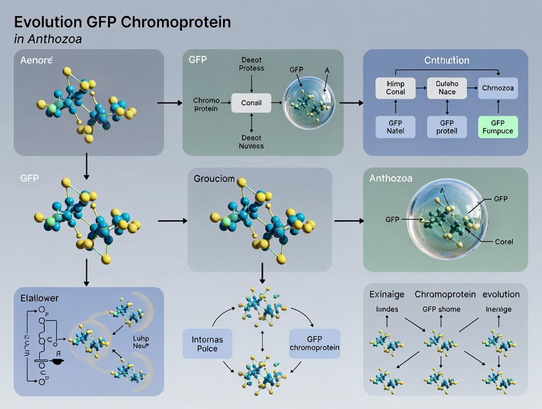
The Evolution of Anthozoan GFP Chromoproteins: From Coral Reefs to Biomedical Innovation
This article provides a comprehensive analysis of GFP (Green Fluorescent Protein) chromoprotein evolution within Anthozoa (corals and anemones), exploring its foundational biology, methodological applications in research, troubleshooting for experimental use,...
The Evolution of Anthozoan GFP Chromoproteins: From Coral Reefs to Biomedical Innovation
Abstract
This article provides a comprehensive analysis of GFP (Green Fluorescent Protein) chromoprotein evolution within Anthozoa (corals and anemones), exploring its foundational biology, methodological applications in research, troubleshooting for experimental use, and comparative validation. We detail the molecular diversification of these proteins from their initial discovery in reef-building corals, highlighting their unique non-fluorescent, brightly colored properties. The article outlines critical protocols for chromoprotein expression, maturation, and tagging in model systems, addresses common challenges in stability and quantification, and offers a comparative framework against traditional fluorescent proteins. Targeted at researchers, scientists, and drug development professionals, this review synthesizes current knowledge to bridge evolutionary insights with practical applications in biosensing, optogenetics, and high-throughput screening.
Unveiling Anthozoan Chromoproteins: Evolutionary Origins and Structural Diversity
The study of green fluorescent protein (GFP)-like proteins in Anthozoa (corals and anemones) has revealed a remarkable evolutionary diversification. Within this broader thesis on GFP chromoprotein evolution in Anthozoa research, a critical distinction exists between canonical fluorescent proteins (FPs) and their non-fluorescent counterparts, known as chromoproteins (CPs). This whitepaper provides a technical guide to defining CPs, elucidating their unique structural and functional characteristics that distinguish them from classical FPs, and discusses their emerging applications in biomedical research.
Core Characteristics & Comparative Analysis
Chromoproteins are a class of GFP-like proteins that possess a mature chromophore identical or similar to that of fluorescent proteins but exhibit negligible fluorescence due to ultra-fast non-radiative decay processes. Their primary characteristic is strong visible-light absorption, imparting intense coloration.
The table below summarizes the key quantitative distinctions between canonical FPs and CPs.
Table 1: Quantitative Comparison of Key Characteristics: Fluorescent Proteins vs. Chromoproteins
| Characteristic | Fluorescent Proteins (FPs) | Chromoproteins (CPs) |
|---|---|---|
| Primary Photophysical Property | High Fluorescence Quantum Yield (QY) | Very Low/Zero QY; High Molar Extinction Coefficient (ε) |
| Typical Quantum Yield Range | 0.1 - 0.8 (10-80%) | < 0.001 - 0.01 (<0.1-1%) |
| Typical ε (M⁻¹cm⁻¹) | ~50,000 - 100,000 | Often > 50,000 (e.g., 50,000 - 95,000) |
| Dominant Energy Dissipation | Radiative Decay (Photon Emission) | Non-radiative Decay (Internal Conversion, Vibrational Relaxation) |
| Mature Chromophore Structure | GFP-type: p-hydroxybenzylidene-imidazolinone. DsRed-type: (Z)-2-{2-[(E)-4-(imidazol-5-yl)buta-1,3-dien-1-yl]-5-(4-hydroxybenzylidene)-4-oxoimidazolidin-1-yl}ethanoate. | Often identical or highly similar to FPs (e.g., Kaede-type, DsRed-type). Key modifications (e.g., cis/trans isomerization, extended conjugation) alter energy states. |
| Common Applications | Intracellular tagging, gene expression reporting, FRET, protein trafficking. | Colorimetric reporters, in vivo labeling without fluorescence bleed-through, acceptors in FRET, photoswitching precursors. |
| Representative Examples | GFP, EGFP, YFP, mCherry, DsRed. | asCP, aeCP597, cjBlue, amilCP, Rtms5. |
Structural & Mechanistic Basis for Non-Fluorescence
The defining feature of CPs is the efficient quenching of fluorescence. Mechanisms identified through structural biology and spectroscopy include:
- Cis-Trans Isomerization of the Chromophore: In proteins like asCP, the chromophore is in a trans configuration, creating a twisted, non-planar structure that promotes non-radiative decay.
- Extended π-Conjugation: In Kaede-like CPs, photoconversion involves a green-emitting FP precursor with a His-Tyr-Gly chromophore. Upon UV irradiation, a formal backbone cleavage and extension of the π-conjugation system between the His and Tyr rings occurs, forming a red-absorbing, non-fluorescent chromophore.
- Chromophore-Protein Interactions: Specific hydrogen bonds and electrostatic interactions with the protein barrel can stabilize non-fluorescent states. For example, a critical glutamate residue near the chromophore in asCP facilitates ultrafast internal conversion via a proton relay network.
- Low Barrier for Non-Radiative Decay: The protein environment is tuned to provide efficient pathways for the excited-state energy to dissipate as heat (vibrational relaxation) rather than as emitted light.
Title: Photophysical Decay Pathways in FPs vs. CPs
Key Experimental Protocols for Characterization
Protocol 1: Spectroscopic Characterization of CPs Objective: Determine the absorption profile, molar extinction coefficient (ε), and confirm negligible fluorescence. Materials: Purified chromoprotein, UV-Vis spectrophotometer, fluorometer, quartz cuvettes. Procedure: 1. Dialyze purified protein into a neutral, non-absorbing buffer (e.g., PBS, pH 7.4). 2. Record absorption spectrum from 250-700 nm. Identify λmaxabs. 3. Measure absorbance (A) at λmaxabs for a series of dilutions (in triplicate). Use a known ε for a standard (e.g., cytochrome c) for pathlength calibration if needed. 4. Plot A vs. concentration (determined by BCA or amino acid analysis). The slope is ε (M⁻¹cm⁻¹). 5. Using the fluorometer, excite at λmaxabs and scan emission from λmaxabs +10 nm to 750 nm. Use high PMT voltage. Compare signal to a known FP control (e.g., EGFP) at the same optical density. Calculate approximate QY if any signal is detected (relative to a standard).
Protocol 2: Distinguishing CPs from FPs via Photoconversion Screening (for Kaede-like proteins) Objective: Identify if a colored protein is a true CP or a photoconvertible FP. Materials: Protein sample or live cells expressing the protein, UV light source (~365-400 nm, safe intensity), visible light microscope. Procedure: 1. Image the sample under visible light (no UV filter) to record initial color/fluorescence. 2. Expose a specific region of interest (ROI) to low-intensity UV light for 30-60 seconds. 3. Re-image under the same visible light settings. Monitor for: a. Photoconvertible FP: A shift in fluorescence color/emission peak (e.g., green to red). b. True CP: No change in absorption/color; no emergence of fluorescence. c. Photo-bleaching: Loss of color/fluorescence. 4. Acquire absorption/emission spectra pre- and post-UV exposure to quantify any changes.
The Scientist's Toolkit: Essential Research Reagents & Materials
Table 2: Research Reagent Solutions for Chromoprotein Work
| Item | Function & Description |
|---|---|
| Heterologous Expression Vectors (e.g., pET, pBAD for E. coli; pcDNA3, pLV for mammalian cells) | For recombinant expression of CP genes cloned from Anthozoa. Allows high-yield protein purification or cellular expression. |
| Nickel-NTA or Cobalt Affinity Resin | For purifying histidine-tagged recombinant CPs. Essential for obtaining pure protein for biophysical characterization. |
| Size-Exclusion Chromatography (SEC) Column (e.g., Superdex 75/200) | Final polishing step for purification. Removes aggregates and confirms monodispersity of the mature CP tetramer (common oligomeric state). |
| Broad-Range UV-Vis Spectrophotometer (e.g., Cary Series) | Primary tool for measuring absorption spectra and calculating molar extinction coefficients (ε). |
| Fluorometer with Polarizers (e.g., Horiba Fluorolog) | Critical for confirming ultra-low fluorescence quantum yield and for time-resolved fluorescence decay measurements (picosecond-nanosecond lifetimes). |
| Tunable Pulsed Laser System | For ultrafast spectroscopy (femtosecond-picosecond) to study the non-radiative decay pathways fundamental to CP function. |
| Site-Directed Mutagenesis Kit | To probe the role of specific amino acids (e.g., the critical Glu residue in asCP) in quenching mechanisms via point mutations. |
| Mammalian Cell Lines (HEK293, HeLa) & Transfection Reagents | For testing CP function as intracellular colorimetric tags or transcriptional reporters without fluorescence interference. |
| FRET Pair Donor Fluorophore (e.g., CFP, GFP) | To utilize a CP as an exceptional dark acceptor in FRET-based biosensor experiments, reducing background. |
Title: Chromoprotein Research & Characterization Workflow
Evolutionary Context & Applications in Biomedical Research
Within Anthozoa, CPs are believed to have evolved from fluorescent ancestors via mutations that introduced efficient quenching mechanisms, likely serving ecological roles in photoprotection or camouflage. For researchers and drug development professionals, this evolutionary quirk has been co-opted for novel tools:
- Dark Acceptors in FRET: Their high absorbance and near-zero fluorescence make CPs ideal "dark acceptor" quenchers in FRET-based biosensors, improving signal-to-noise ratios in assays for kinase activity, apoptosis, or metabolite levels.
- Colorimetric Reporters: CP expression can be visually tracked in cell cultures and model organisms without specialized fluorescence equipment, useful for tracking tumor cells or infection processes.
- Photoswitchable & Photoconvertible Tools: Some CPs (or their precursors, like Kaede) are stimuli-responsive, enabling high-resolution cell lineage tracing or optical highlighting.
- Multiplexing without Spectral Overlap: CPs provide distinct colors based on absorption, which can be combined with multiple FPs for highly multiplexed cell barcoding or parallel reporter assays.
In conclusion, chromoproteins are defined not by the absence of a chromophore, but by the exquisite tuning of its protein environment to favor non-radiative decay. Their distinction from fluorescent proteins is a cornerstone concept in the broader study of GFP-like protein evolution in Anthozoa, and their unique properties continue to inspire innovative solutions in biomedical research and drug discovery.
Phylogenetic Distribution of Chromoproteins Across Anthozoan Lineages (Scleractinia, Actiniaria)
The study of green fluorescent protein (GFP)-like chromoproteins (CPs) in Anthozoa extends beyond bioluminescent markers. This investigation is framed within a broader thesis exploring the evolutionary pressures driving the diversification of the GFP gene family across anthozoan lineages, particularly between the calcifying Scleractinia (stony corals) and the soft-bodied Actiniaria (sea anemones). Understanding the phylogenetic distribution of specific CPs, which absorb but do not fluoresce light, provides critical insights into gene duplication events, structural adaptation for novel photobiological functions (e.g., photoprotection, phototaxis), and the potential for engineering novel optical tools for biomedical imaging and drug development.
Current Phylogenetic Distribution Data
Recent genomic and transcriptomic surveys have clarified the presence and diversity of chromoproteins across key anthozoan families.
Table 1: Distribution of Major Chromoprotein Clades in Anthozoan Lineages
| Chromoprotein Clade (Max Abs) | Common Scleractinian Families (Examples) | Common Actiniarian Families (Examples) | Notable Absences / Limited Distribution |
|---|---|---|---|
| Red CPs (~575-585 nm) | Pocilloporidae (Pocillopora), Acroporidae (Acropora), Dendrophylliidae (Tubastraea) | Actiniidae (Actinia), Stichodactylidae (Stichodactyla) | Rare in Poritidae, absent in some deep-sea lineages. |
| Green CPs (~505-515 nm) | Merulinidae (Favites), Agariciidae (Pavona) | Limited. Reported in Anemonia sp. (Actiniidae). | Widespread but at lower expression than Red CPs in corals. |
| Purple/Blue CPs (~590-610 nm) | Limited. Found in some Fungiidae (Fungia). | Predominant in Actiniaria. Sagartiidae (Cnidopus), Actiniidae (Urticina). | Rare in most Scleractinia. |
| Non-fluorescent GFP-like Proteins (Various) | Present across multiple families. | Widespread, often as dominant form. | --- |
| Estimated Gene Copy Number Range (per genome) | 15 - 40+ (High variation; expansions in Acroporidae) | 5 - 20 (Generally more compact family) | --- |
Table 2: Functional Correlates of Chromoprotein Distribution
| Lineage | Dominant CP Types | Hypothesized Primary Function | Evidence from Expression Studies |
|---|---|---|---|
| Shallow-water Scleractinia | Red, Green | Photoprotection (sunscreen), Photosynthesis modulation in symbionts | Upregulation under high light stress; spatial correlation with symbionts. |
| Apomorphic Actiniaria (e.g., Entacmaea) | Purple/Blue, Red | Predator deterrence (aposematism), Phototaxis | Localization in tentacle tips; behavioral experiments. |
| Non-Symbiotic / Low-Light Species | Low CP diversity or expression | Limited photobiological role; possible structural function | Transcriptomic data shows low expression levels. |
Core Experimental Protocols for Phylogenetic & Functional Analysis
Protocol 1: Chromoprotein Gene Discovery and Phylogenetics
- Sample Collection & RNA/DNA Extraction: Collect tissue from target Scleractinian and Actiniarian species, preserving immediately in RNAlater. Use a standard phenol-chloroform or column-based kit for high-quality nucleic acid extraction.
- Transcriptome/Genome Sequencing: Perform Illumina NovaSeq 6000 sequencing (150bp paired-end) to generate >50M reads per sample. For genomes, employ a hybrid approach (Illumina short-read + PacBio HiFi long-read).
- Gene Identification: Assemble reads using Trinity (for transcriptomes) or hybrid assemblers (e.g., MaSuRCA). Identify CP genes using HMMER3 with Pfam GFP family (PF01353) as a seed and confirmed by BLASTp against the Cnidarian GFP-like protein database.
- Phylogenetic Reconstruction: Perform multiple sequence alignment (MAFFT v7). Construct maximum-likelihood trees using IQ-TREE2 with ModelFinder for best-fit model (e.g., LG+F+G4) and 10,000 ultrafast bootstraps. Root tree using homologous sequences from Hydrozoa.
Protocol 2: Functional Characterization via Heterologous Expression
- Cloning: Amplify full-length CP ORFs from cDNA and clone into a mammalian expression vector (e.g., pCMV/myc/cyto) using Gibson Assembly.
- Cell Culture & Transfection: Culture HEK293T cells in DMEM + 10% FBS. Transfect cells with plasmid using polyethylenimine (PEI).
- Spectral Analysis: 48-72h post-transfection, harvest cells, lyse, and clarify. Measure absorbance spectrum (350-650 nm) using a spectrophotometer (e.g., NanoDrop One). Confirm non-fluorescence using a plate reader (excitation at absorbance max, emission scan 500-700 nm).
- Protein Purification (Optional): Use an affinity tag (e.g., His6) for purification via Ni-NTA chromatography for in vitro biophysical characterization.
Diagrams of Key Workflows and Relationships
Diagram 1: Phylogenetic Analysis Workflow (83 chars)
Diagram 2: Chromoprotein Divergence from GFP Ancestor (81 chars)
The Scientist's Toolkit: Key Research Reagent Solutions
Table 3: Essential Materials for Chromoprotein Research
| Item / Reagent | Function / Application | Example Product / Specification |
|---|---|---|
| RNAlater Stabilization Solution | Preserves RNA integrity in field-collected anthozoan samples for transcriptomics. | Thermo Fisher Scientific, RNAlater Stabilization Solution. |
| Column-Based Nucleic Acid Kit | High-purity DNA/RNA co-extraction from mucopolysaccharide-rich tissue. | Macherey-Nagel NucleoSpin TriPrep Kit. |
| GFP-like Protein HMM Profile | Sensitive identification of CP homologs in sequence databases. | Pfam PF01353 (Green fluorescent protein). |
| Mammalian Expression Vector | Heterologous expression for spectral characterization of cloned CP genes. | Addgene #52330 (pCMV/myc/cyto) or similar. |
| Polyethylenimine (PEI) MAX | High-efficiency, low-cost transfection reagent for HEK293T cells. | Polysciences, PEI MAX 40K. |
| Microvolume Spectrophotometer | Quick absorbance spectral scan of cell lysates expressing CPs. | Thermo Fisher NanoDrop One. |
| Multi-mode Microplate Reader | Full absorbance and fluorescence emission scanning of samples. | BioTek Synergy H1. |
| Ni-NTA Agarose Resin | Purification of recombinant His-tagged chromoproteins for in vitro study. | Qiagen, Ni-NTA Superflow. |
This whitepaper examines the evolutionary pressures shaping the diversity of Green Fluorescent Protein (GFP)-like chromoproteins (CPs) in Anthozoa, focusing on their functional integration into coral holobiont physiology. Framed within a thesis on GFP homolog evolution, we dissect the dual selective paradigms: the optimization of symbiosis with photosynthetic dinoflagellates (Symbiodiniaceae) and the mitigation of photooxidative stress. We provide a synthesis of current mechanistic models, quantitative physiological data, and actionable experimental protocols for researchers in photobiology and marine biodiscovery.
GFP-like proteins in corals and other anthozoans have evolved from a single ancestral gene through multiple duplication and divergence events. This radiation produced non-fluorescent chromoproteins (CPs) with strong absorbance maxima between 450-600 nm. The prevailing hypothesis posits that natural selection has acted on the spectral properties of these CPs to fulfill critical roles within the light-saturated coral reef environment, directly impacting holobiont fitness.
Proposed Ecological Roles and Mechanisms
Photoprotection: A Reactive Oxygen Species (ROS) Mitigation Hypothesis
CPs are proposed to act as a photoprotective "sunscreen" layer in coral tissues. By absorbing excess high-intensity solar radiation, particularly in the blue-green spectrum, CPs reduce photon flux reaching the algal symbionts, thereby diminishing the potential for photoinhibition and ROS generation within the symbiont chloroplasts.
Key Data: Photophysiological parameters with and without CP expression.
Table 1: Photoprotective Efficacy Metrics in Stylophora pistillata Colonies (High-Light Stress)
| Phenotype | Fv/Fm (Max Quantum Yield) Post-Stress | Non-Photochemical Quenching (NPQ) Capacity | ROS (H₂O₂) Concentration in Host Tissue | Bleaching Onset Time (Days at 1500 μmol photons m⁻² s⁻¹) |
|---|---|---|---|---|
| High CP Expression | 0.68 ± 0.04 | High (0.45 ± 0.05) | 1.2 ± 0.3 μM | 14 ± 2 |
| Low CP Expression | 0.42 ± 0.07 | Moderate (0.28 ± 0.06) | 3.8 ± 0.9 μM | 7 ± 1 |
Symbiosis Optimization: A Light-Filtering Hypothesis
An alternative/complementary role suggests CPs fine-tune the light microenvironment for symbionts. By absorbing specific wavelengths, CPs could modify the light quality reaching the symbionts, potentially shifting it towards spectra more optimal for photosynthesis or favoring certain symbiont clades, thus influencing the host-symbiont selective partnership.
Key Data: Symbiont photosynthesis performance under CP-filtered light.
Table 2: Photosynthetic Response of Cladocopium goreaui to CP-Filtered Light Spectra
| Incident Light Spectrum (Peak) | CP Absorbance Peak | Symbiont Pmax (Net O₂ Production) | Light Saturation Point (Ik) | Chl a Concentration per Cell |
|---|---|---|---|---|
| Broad Spectrum (White) | None | 1.00 (ref) | 350 μmol m⁻² s⁻¹ | 4.5 pg |
| CP-Mediated (575 nm) | 580 nm | 1.22 ± 0.08 | 420 μmol m⁻² s⁻¹ | 4.8 pg |
| CP-Mediated (490 nm) | 495 nm | 0.85 ± 0.06 | 280 μmol m⁻² s⁻¹ | 5.1 pg |
Experimental Protocols for Validating Ecological Roles
Protocol:In VivoROS Quantification Under Light Stress
Objective: To correlate CP expression levels with ROS production in coral hospite. Materials: Coral nubbins of known CP expression phenotype, Pulse-Amplitude Modulated (PAM) fluorometer, H₂O₂-sensitive fluorescent probe (e.g., CM-H2DCFDA), microplate reader, controlled light incubator. Procedure:
- Acclimation: Maintain nubbins at 300 μmol m⁻² s⁻¹ for 7 days.
- Stress Induction: Expose to high light (1200 μmol m⁻² s⁻¹) for 6 hours.
- ROS Staining: Incubate tissue slurry in 10 μM CM-H2DCFDA for 30 min in darkness.
- Fluorescence Measurement: Read fluorescence (Ex/Em: 488/525 nm). Normalize to total protein.
- Parallel Photophysiology: Measure Fv/Fm pre- and post-exposure via PAM fluorometry.
Protocol: Spectral Modification & Symbiont Photobiology
Objective: To isolate the effect of CP absorbance on symbiont photosynthesis. Materials: Isolated Symbiodiniaceae cells, custom optical filters mimicking CP absorbance spectra, O₂ electrode system, spectrophotometer. Procedure:
- Filter Calibration: Characterize spectral transmission of filters to match target CP (e.g., 580 nm peak, 50 nm FWHM).
- Light Source: Use a broad-spectrum lamp with intensity adjustable to 200-800 μmol m⁻² s⁻¹.
- O₂ Evolution: Place isolated symbionts in a temperature-controlled chamber with O₂ electrode. Illuminate through CP-mimicking filter. Record net O₂ evolution across light intensities.
- P-I Curve: Generate Photosynthesis-Irradiance (P-I) curves for filtered vs. unfiltered light. Derive α (slope), Pmax, and Ik.
Visualizing Pathways and Workflows
Diagram Title: CP Photoprotection Pathway Against ROS
Diagram Title: Experimental Workflow for CP Role Validation
The Scientist's Toolkit: Research Reagent Solutions
Table 3: Essential Reagents and Materials for CP Functional Research
| Item | Function/Specificity | Example Product/Catalog |
|---|---|---|
| Custom Interference Filters | Mimic specific CP absorbance spectra for light-filtering experiments. | Thorlabs FB series, custom order for 450-600 nm peaks. |
| CM-H2DCFDA (DCFDA) | Cell-permeant ROS-sensitive fluorescent probe for H₂O₂ and peroxides. | Thermo Fisher Scientific, C6827. |
| PAM Fluorometry System | Measures chlorophyll fluorescence parameters (Fv/Fm, NPQ) in vivo. | Walz Imaging-PAM or Diving-PAM. |
| Symbiont Isolation Media | Sterile, nutrient-enriched seawater for isolating Symbiodiniaceae. | 0.45 μm-filtered seawater with antibiotic/antimycotic. |
| O₂ Electrode System | High-resolution measurement of photosynthetic O₂ evolution rates. | Hansatech OxyGraph+ or Chlorolab 3. |
| Anti-GFP/CP Antibodies | Immunodetection and quantification of CP expression in host tissue. | Custom polyclonal against purified recombinant CP. |
| qPCR Primers (Host & Symbiont) | Quantify gene expression (CP genes, stress markers) and symbiont clade ratio. | Species-specific primers for cp, HSP70, ITS2 region. |
| Recombinant CP Standards | Purified proteins for spectral calibration and antibody validation. | Heterologous expression in E. coli with refolding. |
Synthesis and Future Directions in Drug Development
The evolutionary drivers of CP diversity present a model system for understanding protein-augmented photobiology. For drug development, the CP mechanism inspires strategies for synthetic chromophores targeting ROS in human photodermatoses or as optogenetic tools. Furthermore, coral holobiont resilience, mediated by CPs, highlights the potential of modulating oxidative stress pathways—a central tenet in neurodegenerative and inflammatory disease research. Validating the precise ecological role (photoprotection vs. symbiosis tuning) is critical for accurate bioinspiration.
Within Anthozoa (corals and sea anemones), green fluorescent protein (GFP)-like chromoproteins have undergone extensive evolutionary radiation, giving rise to a diverse palette of non-fluorescent colors. This whitepaper details the core molecular architecture of the chromophore and its maturation pathway, a process central to understanding the evolutionary innovation of color in Anthozoa. Unlike GFP, which emits green light via fluorescence, many homologs are chromoproteins that absorb specific wavelengths intensely but dissipate the energy non-radiatively, resulting in brilliant purple, red, or cyan coloration. This color diversification is a key adaptive trait and a rich resource for biotechnological tool development.
Chromophore Core Architecture
The chromophore is a p-hydroxybenzylideneimidazolinone moiety formed post-translationally from a tripeptide motif (Xaa-Tyr-Gly, where Xaa is variable). The canonical GFP chromophore (derived from Ser65-Tyr66-Gly67) exists in a planar, conjugated state. In chromoproteins, subtle modifications to this core and its environment dictate color.
Table 1: Key Chromophore Structural Determinants & Spectral Outcomes
| Structural Feature | GFP (Fluorescent) | Chromoprotein (e.g., Purple, cpRFP) | Impact on Color |
|---|---|---|---|
| Tripeptide Motif | Ser65-Tyr66-Gly67 | Typically Glu/Gln65-Tyr66-Gly | Alters maturation efficiency & protonation state |
| Chromophore Conformation | Planar, coplanar | Often cis or distorted non-planar | Modifies π-electron conjugation length |
| Ionization State | Anionic (major) | Neutral or anionic, stabilized differently | Directly shifts absorption wavelength |
| Imidazolinone Ring | Non-alkylated | May be N-acylated or modified | Changes electron distribution & stability |
| Proximal Interactions | Arg96, His148, Gln94 | Varied residues (e.g., Phe/Leu nearby) | Creates hydrophobic clamp, tunes electrostatics |
The unique color of chromoproteins stems from a combination of the chromophore’s protonation state, its planarity, and the electrostatic environment provided by the surrounding beta-barrel scaffold.
Unique Maturation Pathway: From Polypeptide to Color
Chromophore maturation is an autocatalytic, multi-step process that requires molecular oxygen. The pathway in chromoproteins often includes unique isomerization or modification steps not found in GFP.
Step-by-Step Biochemical Maturation:
- Cyclization: Nucleophilic attack by the Gly67 amide nitrogen on the carbonyl carbon of residue 65 (Xaa), forming a five-membered imidazolinone ring.
- Dehydration: Elimination of a water molecule from the Tyr66 α-β bond, forming a double bond between the α-carbon and the phenolic ring of Tyr66.
- Oxidation: Molecular oxygen-dependent dehydrogenation, extending conjugation between the tyrosine ring and the imidazolinone ring. This creates the mature chromophore.
- Chromophore Modification (Unique to Chromoproteins): Additional steps may occur, such as:
- cis-trans Isomerization of the exocyclic double bond.
- Acylation or other modifications of the imidazolinone nitrogen.
- Trapping of the chromophore in a strained, non-planar conformation via steric clashes with barrel residues.
Table 2: Comparative Maturation Kinetics & Requirements
| Parameter | GFP (avGFP) | Purple Chromoprotein (e.g., stCP) |
|---|---|---|
| Maturation Time (t₁/₂ at 22°C) | ~60-90 minutes | Can exceed 24 hours |
| O₂ Requirement | Absolute | Absolute |
| Rate-Limiting Step | Oxidation | Often final isomerization/trapping step |
| Optimal pH | 7.0 - 8.0 | Often narrower range (e.g., 7.5 - 8.5) |
| Temperature Sensitivity | Moderate | High (often requires <28°C for proper folding) |
Title: Chromophore Maturation Pathway: GFP vs. Chromoprotein Branching
Experimental Protocol: Analyzing Chromophore MaturationIn Vitro
Objective: To monitor the time-course of chromophore maturation and characterize the final spectral properties of a purified Anthozoan chromoprotein.
Protocol:
- Protein Expression & Purification:
- Clone the chromoprotein gene into a pET vector for bacterial expression.
- Transform into E. coli BL21(DE3) cells. Grow culture in LB+antibiotic at 30°C to OD₆₀₀ ~0.6.
- Induce with 0.5 mM IPTG. Critical: Shift temperature to 18°C post-induction to promote proper folding. Incubate for 24-48 hours.
- Lyse cells via sonication in 50 mM Tris-HCl, 300 mM NaCl, pH 8.0.
- Purify protein using immobilized metal affinity chromatography (IMAC) with a His-tag, followed by size-exclusion chromatography (SEC) in 20 mM HEPES, 150 mM NaCl, pH 7.5.
Kinetic Maturation Assay (Spectrophotometric):
- Denaturation/Renaturation: Rapidly denature an aliquot of purified, immature (colorless) protein in 6 M GuHCl for 5 minutes. Dilute 1:50 into pre-warmed assay buffer (50 mM Tris-HCl, pH 8.0, 21°C) to initiate refolding and maturation.
- Data Collection: Immediately place solution in a spectrophotometer with a temperature-controlled cuvette holder (21°C).
- Record UV-Vis absorption spectra (250-700 nm) every 5 minutes for the first 2 hours, then every 30 minutes for up to 24 hours.
- Analysis: Plot absorbance at λₘₐₓ over time. Fit data to a first-order exponential equation to determine the maturation half-time (t₁/₂).
Characterization of Mature Chromophore:
- For fully matured protein, record complete absorbance spectrum.
- Determine molar extinction coefficient (ε) using the method of alkaline denaturation (converts chromophore to a species with known ε).
- Analyze chromophore pKa by recording absorbance at λₘₐₓ across a pH titration (pH 4-11).
The Scientist's Toolkit: Research Reagent Solutions
Table 3: Essential Reagents & Materials for Chromoprotein Research
| Item / Reagent | Function / Purpose | Example / Specification |
|---|---|---|
| Heterologous Expression System | Produces recombinant chromoprotein for study. | E. coli BL21(DE3) pLysS for tight control, low-temperature expression. |
| Low-Temperature Incubator | Essential for proper folding of temperature-sensitive chromoproteins. | Shaker incubator capable of maintaining 18-22°C. |
| Fast Protein Liquid Chromatography (FPLC) | High-resolution purification of native protein. | ÄKTA pure system with HisTrap HP & HiLoad Superdex 75 columns. |
| UV-Vis Spectrophotometer | Kinetic maturation assays & spectral characterization. | Instrument with Peltier temperature control (e.g., Cary 3500). |
| 6 M Guanidine Hydrochloride (GuHCl) | Rapidly denatures protein to create a synchronous maturation start point. | Molecular biology grade, in 50 mM Tris-HCl, pH 8.0. |
| Anaerobic Chamber / Glove Box | To study O₂-dependence by performing maturation in deoxygenated buffers. | Coy Laboratory Products anaerobic chamber with <1 ppm O₂. |
| Site-Directed Mutagenesis Kit | To probe role of specific barrel residues in chromophore trapping. | Q5 Site-Directed Mutagenesis Kit (NEB). |
| Crystallization Screening Kits | For obtaining high-resolution structural data of chromophore environment. | JC SG Core I-IV suites (Molecular Dimensions). |
Title: Core Workflow for Chromoprotein Maturation Analysis
Implications for Evolution & Drug Development
The study of chromophore architecture and maturation provides insights into protein evolution. In Anthozoa, minor modifications in the beta-barrel can drastically alter photophysical properties without disrupting the scaffold, exemplifying evolvability. For drug development, the chromoprotein's intense, stable color and long maturation time serve as unique biomarkers. Engineered variants are used in:
- Biosensors: Where color change reports on cellular pH, redox state, or protease activity.
- Reporters for Slow Processes: Such as long-term gene expression tracking or tumor growth monitoring.
- Photosensitizer Platforms: Modified chromoproteins can generate reactive oxygen species for photodynamic therapy.
Understanding the precise molecular constraints of chromophore formation is crucial for engineering next-generation, stable, and non-toxic optical tools for in vivo imaging and therapeutic applications.
This whitepaper details major chromoprotein families within the Anthozoa class (corals and anemones), with a focus on key representatives. This analysis is framed within a broader thesis on Green Fluorescent Protein (GFP) chromoprotein evolution in Anthozoa research. Unlike classical GFP-like fluorescent proteins that emit light, chromoproteins (CPs) are non-fluorescent, absorbing specific wavelengths to produce vivid colors. Their evolution alongside fluorescent proteins highlights diverse structural and functional adaptations, providing critical insights into photobiology and enabling novel biotechnological tools for drug development and cellular imaging.
Core Chromoprotein Families and Key Examples
Anthozoan chromoproteins are characterized by a conserved β-barrel structure but exhibit variability in their chromophore formation and spectral properties. The table below summarizes quantitative data for key examples.
Table 1: Spectral and Molecular Characteristics of Key Anthozoan Chromoproteins
| Chromoprotein | Source Organism | Max Abs (nm) | Molar Extinction Coefficient (M⁻¹cm⁻¹) | Oligomeric State | Primary Color | Chromophore Type |
|---|---|---|---|---|---|---|
| aeCP597 | Anemonia erythraea | 597 | ~ 87,000 | Tetramer | Purple | Modified GFP-type (His62) |
| cjBlue | Corynactis japonica | 588 | ~ 51,000 | Tetramer | Blue | GFP-type (Cys63) |
| amilGFP | Acropora millepora | 482 | ~ 49,000 | Dimer | Green | Non-fluorescent GFP |
| asCP | Anemonia sulcata | 572 | ~ 54,000 | Dimer | Pinkish-purple | GFP-type |
| rtms5 | Montipora sp. | 592 | ~ 78,000 | Tetramer | Red | GFP-type |
Structural Evolution and Chromophore Diversity
The evolution of color diversity stems from modifications to the internal chromophore and its electrostatic environment. amilGFP is a particularly instructive example for evolutionary studies: it possesses a canonical GFP chromophore (formed by Ser65-Tyr66-Gly67) but has lost fluorescence due to structural constraints that promote non-radiative decay. In contrast, aeCP597 and cjBlue feature unique chromophore structures. aeCP597 incorporates a novel His62-Tyr63-Gly64 triad, while cjBlue has a Cys63-Tyr64-Gly65 triad, both leading to red-shifted absorption.
Diagram 1: Chromophore Evolution Pathways in Anthozoa
Experimental Protocols for Characterization
Protocol: Spectral Characterization of Purified Chromoproteins
Objective: Determine absorbance maxima and molar extinction coefficients.
- Heterologous Expression: Clone target CP gene into pQE or pET vector. Transform into E. coli BL21(DE3). Induce expression with 0.5 mM IPTG at 18°C for 20h.
- Purification: Lyse cells via sonication. Purify 6xHis-tagged protein using Ni-NTA affinity chromatography under native conditions.
- Absorbance Spectroscopy: Dilute purified protein in PBS (pH 7.4). Record absorbance spectrum from 250-700 nm using a spectrophotometer (e.g., NanoDrop or Cary).
- Extinction Coefficient Calculation: Measure absorbance at λmax. Determine protein concentration via Bradford assay using BSA standard. Calculate ε using Beer-Lambert law: A = ε * c * l.
Protocol: Assessing Oligomeric State via Size Exclusion Chromatography (SEC)
- Column Equilibration: Equilibrate an analytical Superdex 200 Increase column with 2 column volumes of SEC buffer (20 mM Tris-HCl, 150 mM NaCl, pH 8.0).
- Sample Preparation: Concentrate purified chromoprotein to 2 mg/mL, centrifuge (16,000 x g, 10 min) to remove aggregates.
- Run SEC: Inject 100 µL sample, run isocratically at 0.5 mL/min. Monitor absorbance at 280 nm and λmax.
- Data Analysis: Compare elution volume to protein standards (e.g., thyroglobulin, BSA, ovalbumin). Plot Kav vs. log(MW) to estimate native molecular weight.
Diagram 2: Key Experimental Workflow for Chromoprotein Analysis
Research Reagent Solutions Toolkit
Table 2: Essential Research Reagents for Chromoprotein Studies
| Item | Function & Application |
|---|---|
| pET-28a(+) Vector | T7-driven expression vector with N-terminal 6xHis tag for high-yield recombinant protein production in E. coli. |
| Ni-NTA Superflow Resin | Immobilized metal affinity chromatography (IMAC) resin for purifying histidine-tagged chromoproteins. |
| Superdex 200 Increase 10/300 GL | High-resolution size exclusion chromatography column for accurate determination of native oligomeric state. |
| IPTG (Isopropyl β-D-1-thiogalactopyranoside) | Inducer for lac/T7-based expression systems, used to initiate chromoprotein synthesis. |
| Protease Inhibitor Cocktail (EDTA-free) | Prevents proteolytic degradation of chromoproteins during cell lysis and purification. |
| Bradford Protein Assay Kit | Colorimetric method for rapid, accurate determination of purified chromoprotein concentration. |
| PD-10 Desalting Columns | For rapid buffer exchange into spectroscopic or storage buffers, removing imidazole and salts. |
Applications in Drug Development and Biomedical Research
The unique photophysical properties of anthozoan chromoproteins enable advanced applications:
- Bioluminescence Resonance Energy Transfer (BRET) Sensors: Non-fluorescent CPs like aeCP597 serve as excellent acceptor molecules in BRET systems for monitoring protein-protein interactions in vivo, crucial for high-throughput drug screening.
- Photosensitizers in Photodynamic Therapy (PDT): Their high absorption coefficients make engineered CPs potential light-activated molecular switches or singlet oxygen generators.
- Visual Reporters and Selection Markers: The intense color provides a visual readout for promoter activity, cell lineage tracing, and positive clone selection without need for fluorescence excitation, reducing autofluorescence.
The study of major chromoprotein families (aeCP597, cjBlue, amilGFP) underscores a key thesis in Anthozoan research: the GFP-like β-barrel scaffold is a remarkably evolvable platform. The divergence into non-fluorescent chromoproteins represents a functional adaptation, likely for photoprotection or signaling in reef environments. From a biotechnology standpoint, understanding this evolution allows for the rational engineering of novel probes with tailored absorption, stability, and oligomerization states, directly benefiting drug discovery and molecular imaging.
Harnessing Chromoproteins: Protocols for Research and Drug Discovery Applications
The study of fluorescent proteins from Anthozoa (e.g., corals, anemones) has revolutionized molecular and cellular biology, with green fluorescent protein (GFP) being the archetype. However, a parallel and equally significant class of proteins has emerged from this research: chromoproteins (CPs). These are non-fluorescent, brightly colored proteins that evolved alongside GFP-like proteins in Anthozoa, sharing the same β-can structure but differing in their photophysical properties. Within the broader thesis of GFP evolution in Anthozoa, CPs represent a divergent branch where mutations in the chromophore environment quench fluorescence, resulting in strong absorbance and brilliant color. This technical guide details their unique advantages as visual reporters in two key applications: bacterial colony screening and cellular labeling, offering robust, equipment-independent alternatives and complements to fluorescent systems.
Core Advantages Over Fluorescent Reporters
Chromoproteins offer distinct operational benefits derived from their intrinsic properties:
- Equipment-Independent Visual Detection: Their intense color (e.g., purple, red, blue, green) is visible to the naked eye under ambient light, eliminating the need for UV/blue light sources, excitation filters, or specialized imaging equipment for primary screening.
- Photostability: They exhibit exceptional resistance to photobleaching because energy absorbed by the chromophore is dissipated as heat rather than emitted as fluorescence. This enables prolonged visualization and imaging under bright light.
- Minimal Background: In cellular labeling, especially in multicellular organisms or tissues with high autofluorescence (e.g., under GFP channels), CPs provide a stark, unambiguous signal against a colorless background.
- Simplified Cloning & Screening: Color formation is often rapid and does not require molecular oxygen or maturation at 37°C, making them ideal for screening in diverse hosts (bacteria, yeast, mammalian cells) and at various temperatures.
- Low Metabolic Burden: As terminal energy absorbers, they do not cycle through excitation/emission, potentially reducing cellular energy load compared to constantly cycling fluorescent proteins.
Key Applications & Experimental Protocols
Colony Screening in Cloning Workflows
CPs are exceptionally effective as positive/negative selection markers in plasmid cloning, often used in place of lacZ (blue-white screening).
Protocol: Rapid Colony Screening with a Purple Chromoprotein (e.g., cPurple)
- Vector Preparation: Clone your gene of interest into a multiple cloning site (MCS) positioned within the open reading frame of a chromoprotein gene (e.g., on a pCP vector). Successful insertion disrupts the CP gene, leading to loss of color.
- Transformation & Plating: Transform the ligation product into competent E. coli (e.g., DH5α). Plate cells on LB agar containing the appropriate antibiotic. Include a vector-only (intact CP) control.
- Incubation & Analysis: Incubate plates at 30-37°C for 12-24 hours. Colonies harboring the empty vector will appear intensely colored (e.g., purple). Successful recombinant clones will form white or pale colonies.
- Validation: Pick white colonies for colony PCR or direct sequencing to confirm insert presence.
Table 1: Comparison of Common Visual Reporter Systems for Colony Screening
| Reporter System | Visual Signal | Required Substrate | Required Equipment | Typical Incubation Time | Readout Clarity |
|---|---|---|---|---|---|
| Chromoprotein (e.g., cPurple) | Colored colony (Purple) | None | Ambient light | 12-18 hrs | High |
| lacZ (β-galactosidase) | Blue colony | X-Gal, IPTG | Ambient light | 16-24 hrs | Moderate (can be faint) |
| Fluorescent Protein (e.g., GFP) | Fluorescent colony | None | UV/Blue light source | 18-24 hrs+ (for maturation) | Variable (background fluorescence) |
Cellular & Subcellular Labeling
CPs serve as excellent lineage tracers, reporters of promoter activity, and organelle markers in a wide range of cell types.
Protocol: Labeling Mammalian Cells with a Red Chromoprotein (e.g., eqFP611 derivative)
- Construct Design: Subclone the CP gene, codon-optimized for mammals, downstream of a constitutive (e.g., CMV, EF1α) or tissue-specific promoter in a mammalian expression vector.
- Cell Transfection: Seed HeLa or HEK293 cells in a multi-well plate. At 70-80% confluency, transfert with the CP plasmid using a standard method (e.g., lipofection, PEI).
- Expression & Visualization: Incubate cells for 24-48 hours at 37°C/5% CO₂. Observe directly under a standard bright-field microscope. Colored cells will appear pink/red. For subcellular localization, fuse the CP to a targeting peptide (e.g., nuclear localization signal, mitochondrial targeting signal).
- Quantification (Optional): While qualitative, color intensity can be semi-quantified by measuring absorbance of cell lysates at the CP's peak absorbance wavelength using a plate reader.
Table 2: Quantitative Properties of Representative Anthozoan Chromoproteins
| Chromoprotein | Source Organism | Color (Absorbance Max) | Molar Extinction Coefficient (ε) | Relative Brightness* | Maturation Speed (at 37°C) |
|---|---|---|---|---|---|
| asCP | Anemonia sulcata | Violet (∼569 nm) | ∼ 72,000 M⁻¹cm⁻¹ | 1.0 (Reference) | Fast (<1 hr) |
| cPurple | Actinia equina | Purple (∼588 nm) | ∼ 32,000 M⁻¹cm⁻¹ | 0.4 | Very Fast |
| eqFP611 (CP form) | Entacmaea quadricolor | Red (∼611 nm) | ∼ 78,000 M⁻¹cm⁻¹ | 1.5 | Medium (∼4 hrs) |
| amajCFP | Acropora millepora | Blue-green (∼492 nm) | ∼ 43,000 M⁻¹cm⁻¹ | 0.6 | Medium |
| E2-Crimson | Discosoma sp. | Far-Red (∼611 nm) | ∼ 98,000 M⁻¹cm⁻¹ | 2.1 | Slow (>24 hrs) |
*Relative Brightness is approximated as ε, as quantum yield (QY) is ~0 for pure CPs.
The Scientist's Toolkit: Essential Research Reagents
Table 3: Key Research Reagent Solutions for Chromoprotein Applications
| Item | Function & Explanation |
|---|---|
| pCP-based Cloning Vectors | Plasmids with MCS embedded within a chromoprotein gene (e.g., pCP-Orange, pPurple). Enable color-based screening for successful cloning. |
| Codon-Optimized CP Genes | Synthetic genes with host-specific codon usage (e.g., mammalian, plant, yeast) for high-level expression without translational bottlenecks. |
| Low-Autofluorescence Growth Media | Specially formulated media (e.g., for mammalian cells) that minimizes background color, enhancing visual contrast of CP-labeled cells. |
| Spectrophotometer/Plate Reader | For quantifying CP expression levels by measuring absorbance at its specific λmax in cell or bacterial lysates. |
| Bright-Field Microscope with Color Camera | Essential for high-resolution documentation of CP-labeled cells and tissues without the need for fluorescence filter sets. |
| Tissue Clearing Agents (e.g., CUBIC, ScaleS) | For deep-tissue imaging, these agents render tissues transparent while preserving CP color, enabling 3D visualization of labeled structures. |
Visual Workflows and Pathways
Diagram 1: CP-Based Colony Screening Workflow
Diagram 2: Evolutionary & Functional Divergence of GFP vs CP
This guide details cloning and heterologous expression protocols within the broader context of research on GFP-like chromoprotein evolution in Anthozoa (e.g., corals, sea anemones). Understanding the molecular determinants of color and fluorescence in these proteins requires their expression in controlled model systems like E. coli and mammalian cells. This enables detailed biophysical characterization, mutagenesis studies, and applications in biomolecular imaging and reporter assay development.
Part 1: Cloning Strategies for Heterologous Expression
Vector Selection and Design
The choice of expression vector is critical and depends on the host system and experimental goals.
Table 1: Comparison of Key Vector Features forE. colivs. Mammalian Expression
| Feature | E. coli Expression Vector (e.g., pET series) | Mammalian Expression Vector (e.g., pcDNA3.1) |
|---|---|---|
| Promoter | T7/lac (strong, inducible) | CMV (strong, constitutive) |
| Selection Marker | Ampicillin/Kanamycin resistance | Neomycin/Kanamycin resistance |
| Copy Number | High (for plasmid propagation) | High (in bacteria) / Low (in mammalian cells) |
| Essential Elements | Ribosome Binding Site (RBS), T7 terminator | SV40 origin, polyadenylation signal (polyA) |
| Fusion Tags | His-tag, GST, MBP for purification | Epitope tags (HA, FLAG), fluorescent tags |
| Typical Insert Size | < 10 kb | < 15 kb |
PCR Amplification and Insert Preparation
Amplify the target chromoprotein gene (e.g., mcavRFP from Montastraea cavernosa) from cDNA or a gBlock.
- Primer Design: Incorporate restriction enzyme sites (e.g., NdeI/XhoI for pET vectors, HindIII/BamHI for pcDNA) or sequences for homologous recombination (Gibson Assembly, In-Fusion).
- PCR Protocol:
- Reaction Mix: 1x High-Fidelity PCR Buffer, 200 µM dNTPs, 0.5 µM forward primer, 0.5 µM reverse primer, 10-100 ng template DNA, 1-2 units high-fidelity DNA polymerase (e.g., Q5, Phusion), nuclease-free water to 50 µL.
- Thermocycling: Initial denaturation: 98°C for 30 sec; 30 cycles of: 98°C for 10 sec, 55-72°C (Tm-dependent) for 30 sec, 72°C for 30 sec/kb; Final extension: 72°C for 2 min.
- Purification: Gel-purify the PCR product using a commercial kit to ensure a single, clean band.
Cloning via Restriction Enzyme/ Ligase-Independent Cloning
Gibson Assembly Protocol:
- Vector Preparation: Linearize the destination vector by PCR or restriction digest. Gel-purify.
- Insert Preparation: Ensure PCR product has 15-30 bp overlaps with the vector ends.
- Assembly Reaction: Mix 50-100 ng linearized vector with a 2:1 molar ratio of insert, 1x Gibson Assembly Master Mix. Incubate at 50°C for 15-60 minutes.
- Transformation: Transform 2-5 µL of the assembly reaction into competent E. coli (e.g., DH5α). Plate on LB agar with appropriate antibiotic.
- Screening: Pick colonies for colony PCR or plasmid miniprep followed by diagnostic digest and Sanger sequencing.
Part 2: Heterologous Expression inE. coli
Expression Strain Selection
Common strains: BL21(DE3) for protein expression; Origami(DE3) for disulfide-bonded proteins; ArcticExpress(DE3) for proteins requiring chaperonins for folding.
Small-Scale Expression Test & Optimization
Detailed Protocol:
- Transformation: Transform the expression plasmid into chemically competent BL21(DE3) cells. Plate on selective LB agar. Incubate overnight at 37°C.
- Inoculation: Pick a single colony into 5 mL LB + antibiotic. Grow overnight (37°C, 220 rpm).
- Expression Culture: Dilute 1:100 into 5 mL fresh LB + antibiotic in a 50 mL tube. Grow at 37°C until OD600 ~0.6.
- Induction: Add IPTG to a final concentration (e.g., 0.1 mM, 0.5 mM, 1.0 mM). Test different temperatures (16°C, 25°C, 37°C) and times (4-16 hours).
- Harvest: Pellet 1 mL of culture by centrifugation (13,000 rpm, 2 min). Resuspend pellet in 100 µL SDS-PAGE loading buffer. Analyze by SDS-PAGE and Coomassie staining to check for soluble protein expression.
Table 2: Quantitative Analysis ofE. coliExpression Test for a Model Chromoprotein (mcavRFP)
| Condition (IPTG/Temp/Time) | Total Protein Yield (mg/L culture) | Soluble Fraction (%) | Observed Color/Phenotype |
|---|---|---|---|
| 1.0 mM / 37°C / 4h | 45 | <10% | Red pellet, inclusion bodies |
| 0.5 mM / 25°C / 16h | 38 | ~65% | Pink-red soluble fraction |
| 0.1 mM / 16°C / 20h | 25 | >90% | Bright red soluble fraction |
Purification (His-tag Example)
- Lysis: Resuspend cell pellet from 1L culture in 30 mL Lysis Buffer (50 mM Tris pH 8.0, 300 mM NaCl, 10 mM imidazole, protease inhibitors). Lyse by sonication on ice.
- Clarification: Centrifuge at 20,000 x g for 30 min at 4°C. Collect supernatant (soluble fraction).
- Immobilized Metal Affinity Chromatography (IMAC): Load supernatant onto a Ni-NTA column pre-equilibrated with Lysis Buffer. Wash with 10 column volumes (CV) of Wash Buffer (50 mM Tris pH 8.0, 300 mM NaCl, 30 mM imidazole).
- Elution: Elute with 5 CV of Elution Buffer (50 mM Tris pH 8.0, 300 mM NaCl, 250 mM imidazole).
- Buffer Exchange: Desalt into storage buffer (e.g., PBS, 50 mM Tris pH 7.4) using a PD-10 column or dialysis. Analyze purity by SDS-PAGE.
Part 3: Heterologous Expression in Mammalian Cells
Mammalian Expression Workflow
The following diagram illustrates the multi-step process for expressing and analyzing Anthozoa chromoproteins in mammalian cells.
Diagram 1: Mammalian Cell Expression and Analysis Workflow.
Transfection and Expression Protocol (PEI-based)
Materials: HEK293T cells, pcDNA3.1-chromoprotein plasmid, linear PEI (1 mg/mL), Opti-MEM, DMEM + 10% FBS. Protocol:
- Day 1: Seed HEK293T cells in a 6-well plate at 5x10^5 cells/well in 2 mL DMEM+10% FBS. Incubate at 37°C, 5% CO2.
- Day 2 (70-80% confluency): a. Dilute 2 µg plasmid DNA in 100 µL Opti-MEM (Tube A). b. Dilute 6 µL PEI reagent (3:1 PEI:DNA ratio) in 100 µL Opti-MEM (Tube B). c. Mix Tube B with Tube A. Vortex briefly. Incubate at RT for 20 min. d. Add the 200 µL transfection mix dropwise to the cell medium. Swirl gently.
- Day 3 (6h post-transfection): Replace medium with 2 mL fresh, pre-warmed complete DMEM.
- Day 4/5: Harvest cells 48-72h post-transfection for analysis.
Analysis of Expression
- Fluorescence Microscopy: Directly visualize chromoprotein color/fluorescence using appropriate filters.
- Flow Cytometry: Quantify expression levels and population heterogeneity.
- Western Blot: Confirm protein size and expression using an anti-tag antibody.
The Scientist's Toolkit: Research Reagent Solutions
| Item/Category | Example Product/Brand | Function in Chromoprotein Research |
|---|---|---|
| High-Fidelity DNA Polymerase | Q5 (NEB), Phusion (Thermo) | Error-free PCR amplification of chromoprotein genes from GC-rich coral cDNA. |
| Cloning Kit | Gibson Assembly Master Mix (NEB), In-Fusion HD (Takara) | Seamless, restriction-site-independent construction of expression vectors. |
| Competent E. coli | NEB 5-alpha, BL21(DE3) | For plasmid propagation and recombinant protein expression. |
| Affinity Purification Resin | Ni-NTA Agarose (Qiagen), HisTrap HP (Cytiva) | One-step purification of His-tagged chromoproteins from E. coli lysates. |
| Mammalian Cell Line | HEK293T, HeLa | Robust, transferable hosts for transient chromoprotein expression and localization. |
| Transfection Reagent | Linear Polyethylenimine (PEI MAX), Lipofectamine 3000 | Efficient delivery of plasmid DNA into mammalian cells for expression. |
| Cell Culture Medium | DMEM, High Glucose + GlutaMAX (Gibco) | Provides nutrients and stable glutamine for optimal cell health during protein expression. |
| Fluorescence Microscope Filter Set | TRITC/Cy3 filter set (for red CPs), CFP filter set (for non-fluorescent CPs) | Enables visualization and imaging of chromoprotein expression and localization. |
Key Signaling & Regulatory Pathways
Understanding chromoprotein maturation is key. The following diagram outlines the post-translational pathway leading to chromophore formation, a critical step for function.
Diagram 2: Post-Translational Chromophore Maturation Pathway.
Optimizing Expression Conditions for Maximum Color Yield and Stability
Within the broader thesis on Green Fluorescent Protein (GFP) chromoprotein evolution in Anthozoa research, the optimization of recombinant expression conditions is a critical step. These naturally evolved proteins, prized for their vivid colors and stability, must be produced reliably and at high yield for applications ranging from cellular imaging to drug discovery reporter systems. This guide details the technical strategies for maximizing color yield—the observable pigment production per cell or culture volume—and long-term stability.
Core Expression Parameters and Quantitative Optimization
The expression of Anthozoan chromoproteins in heterologous systems like E. coli is influenced by a multifactorial interplay of conditions. The table below summarizes key parameters and their optimal ranges based on current literature (2023-2024).
Table 1: Key Expression Parameters for Anthozoan Chromoprotein Yield
| Parameter | Sub-Optimal Condition | Optimal Range | Primary Impact |
|---|---|---|---|
| Host Strain | Standard BL21(DE3) | BL21(DE3) pLysS, Rosetta 2, ArcticExpress | Protein folding, codon usage, leaky expression control |
| Induction Temperature | 37°C | 16°C – 25°C | Folding efficiency, solubility, chromophore maturation |
| Induction OD600 | Low (<0.4) or Very High (>1.2) | 0.6 – 0.9 | Balance between biomass and metabolic burden |
| Inducer (IPTG) Concentration | High (>1 mM) | 0.1 – 0.5 mM | Controlled expression rate to minimize inclusion bodies |
| Post-Induction Duration | Short (<12h) | 18 – 48 hours | Extended time for chromophore oxidation/maturation |
| Media Composition | Minimal or Rich (LB) | Terrific Broth (TB), Auto-induction Media | Nutrient availability for sustained protein synthesis |
| Aeration/Culture Volume | Low ( >20% flask volume) | High ( <10% flask volume) | Oxygenation crucial for chromophore formation |
| Buffer pH Post-Lysis | Non-buffered or extreme pH | pH 7.4 – 8.5 (Tris or Phosphate) | Chromophore stability and fluorescence/color intensity |
Detailed Experimental Protocol for Systematic Optimization
Protocol: High-Yield Expression of DsRed-like Chromoprotein in E. coli Objective: To express a model Anthozoan chromoprotein (e.g., mcavRFP) with maximum color yield and stability.
Materials & Reagents:
- Expression plasmid (e.g., pET-28a containing gene of interest).
- E. coli strains: BL21(DE3), BL21(DE3) pLysS, Rosetta 2(DE3).
- Media: LB, Terrific Broth (TB), Auto-induction Media (Formedium).
- Antibiotics: Kanamycin (50 µg/mL), Chloramphenicol (34 µg/mL).
- Inducer: Isopropyl β-D-1-thiogalactopyranoside (IPTG), 1M stock.
- Lysis Buffer: 50 mM Tris-HCl (pH 8.0), 300 mM NaCl, 10 mM imidazole, 1 mg/mL Lysozyme, protease inhibitor cocktail.
- Spectrophotometer (for OD600 and color yield measurement at λmax).
Method:
- Transformation & Inoculation: Transform plasmids into each expression strain. Pick single colonies to inoculate 5 mL starter cultures (LB + antibiotic). Incubate at 37°C, 220 rpm for 6-8 hours.
- Main Culture Setup: Dilute starter culture 1:100 into 50 mL of TB + antibiotic in a 250 mL baffled flask. For auto-induction, use appropriate media.
- Growth & Induction: Grow at 37°C, 220 rpm until OD600 = 0.7 (≈2.5-3 hours). Split culture into four 12.5 mL aliquots in 125 mL flasks.
- Induction Test: Induce three flasks with IPTG to final concentrations of 0.1 mM, 0.5 mM, and 1.0 mM. Keep one as an uninduced control.
- Temperature Shift: Immediately transfer all flasks to incubation at 18°C.
- Extended Expression: Incubate with shaking (220 rpm) for 24 hours.
- Harvest & Analysis: Pellet cells at 4,000 x g for 20 min.
- Visual Assessment: Compare pellet color intensity between conditions.
- Quantitative Color Yield: Resuspend pellets in 5 mL lysis buffer, lyse via sonication, and clarify by centrifugation. Measure the absorbance of the supernatant at the protein's λmax (e.g., 572 nm for DsRed). Color Yield = Aλmax * Total Lysate Volume (mL). Normalize to cell mass via OD600 of culture pre-harvest.
- Stability Test: Aliquot the clarified lysate. Store one at 4°C, one at room temperature (20°C), and one at -80°C. Monitor Aλmax daily for one week to assess chromophore stability.
Visualization of Key Pathways and Workflows
Title: Chromoprotein Expression Optimization Workflow
Title: Chromophore Maturation Pathway & Optimization Levers
The Scientist's Toolkit: Research Reagent Solutions
Table 2: Essential Research Reagents for Chromoprotein Expression
| Item | Function in Optimization | Example/Brand |
|---|---|---|
| Specialized E. coli Strains | Enhance folding of eukaryotic proteins; suppress basal expression. | BL21(DE3) pLysS, Rosetta 2(DE3), ArcticExpress(DE3). |
| Auto-Induction Media | Simplifies expression; automatically induces at high cell density for maximum yield. | Formedium Overnight Express, Studier's ZYP-5052. |
| Terrific Broth (TB) Powder | Rich media providing high cell densities essential for protein yield. | Sigma-Aldrich, Thermo Fisher. |
| Protease Inhibitor Cocktail | Prevents degradation of chromoproteins during lysis and purification. | EDTA-free tablets (Roche cOmplete). |
| Lysozyme | Enzymatic cell lysis, gentler than mechanical methods for some proteins. | Sigma-Aldrich, >20,000 units/mg. |
| Broad-Range IPTG Stock | Allows precise titration of induction strength across a wide concentration range. | 1.0M solution, sterile-filtered. |
| Spectrophotometer Cuvettes | For accurate measurement of culture density (OD600) and color yield (Aλmax). | BRAND UV-cuvettes, semi-micro. |
| Temperature-Controlled Shaker | Critical for maintaining precise post-induction temperatures (e.g., 16°C). | New Brunswick Innova S44i. |
Optimizing expression conditions for Anthozoan chromoproteins is a deliberate exercise in balancing cellular physiology with the unique biochemistry of chromophore maturation. By systematically modulating host strain, temperature, induction, and duration as outlined, researchers can reliably achieve maximum color yield and stability. This robust production is foundational for advancing research in chromoprotein evolution and their application in modern drug discovery and bioimaging.
Applications in Multiplexed Reporting and Genetic Circuit Design
This whitepaper details the application of evolved Anthozoa GFP-like chromoproteins in multiplexed biosensing and advanced genetic circuit design. The core thesis frames these applications within the broader context of GFP chromoprotein evolution in Anthozoa research, which has transitioned from a pursuit of natural diversity to a platform for engineering novel spectral and biophysical properties. This engineered palette provides the fundamental hardware for constructing sophisticated, multi-channel reporting systems and predictable genetic logic circuits, directly impacting biomedical research and therapeutic development.
Key Chromoprotein Properties & Quantitative Data
The utility of chromoproteins (CPs) stems from their inherent absorbance, which provides a spectrophotometric or visual output without the need for excitation, reducing background and enabling multiplexing with fluorescent proteins (FPs). The table below summarizes key engineered variants derived from Anthozoa progenitors.
Table 1: Engineered Anthozoa Chromoproteins for Multiplexed Applications
| Protein Name | Origin (Parent) | Peak Absorbance (nm) | Molar Extinction Coefficient (M⁻¹cm⁻¹) | Color (Visible) | Key Application |
|---|---|---|---|---|---|
| eCPred | Actinia equina (RFP) | 590 | ~50,000 | Purple | Transcriptional reporting, biosensors |
| gmCP | Galaxea fascicularis | 588 | ~70,000 | Magenta | 2-4 color multiplexed assays |
| spisPink | Anemonia majano (asCP) | 592 | ~50,000 | Pink | Genetic circuit memory elements |
| Blue | Acropora millepora (amFP486) | 402 | ~30,000 | Blue | FRET acceptor quenching, multiplexing |
| cpGFP | Aequorea victoria (GFP) | 400 | ~25,000 | Green-yellow | Intracellular localization tags |
Experimental Protocols
Protocol: Multiplexed Transcriptional Reporter Assay
Objective: To simultaneously quantify the activity of 2-3 distinct promoters in a single cell population using chromoprotein and fluorescent protein reporters.
Materials:
- Mammalian (HEK293) or bacterial cells.
- Plasmid constructs: pProm1-gmCP, pProm2-eGFP, pProm3-iRFP670 (or similar far-red FP).
- Positive control plasmid with constitutive reporter (e.g., pCMV-mCherry).
- Negative control plasmid (reporterless backbone).
- Transfection reagent (e.g., PEI for HEK293).
- Spectrophotometer (plate reader) with 580-600 nm, 488/525 nm, and 670/710 nm filter sets.
- Microplate incubator.
Method:
- Seed cells in a 24-well plate at 70% confluence. Incubate for 24 hours.
- Co-transfect cells with the 3 reporter plasmids (equal mass, 400 ng total DNA per well). Include positive and negative controls.
- Incubate for 48-72 hours to allow for protein maturation.
- Harvest cells: Wash with PBS, trypsinize, and resuspend in 200 µL PBS.
- Transfer cell suspension to a clear-bottom 96-well assay plate.
- Measure Absorbance/Fluorescence:
- gmCP: Absorbance at 590 nm.
- eGFP: Excitation 488 nm / Emission 525 nm.
- iRFP670: Excitation 670 nm / Emission 710 nm.
- Normalize all readings to cell count (via absorbance at 700 nm as turbidity control).
- Data Analysis: Subtract negative control values. Normalize signals to the positive control for transfection efficiency. Plot as relative activity.
Protocol: Characterizing a Genetic Logic Gate with a Chromoprotein Output
Objective: To validate the function of a genetically encoded AND gate in E. coli using spisPink as the output.
Materials:
- E. coli DH5α or MG1655 cells.
- Plasmid System: A two-plasmid system is used.
- Plasmid A: Contains Input 1-inducible promoter (e.g., pLac) driving the expression of a transcriptional activator for Input 2's promoter.
- Plasmid B: Contains a hybrid promoter responsive to the activator from Plasmid A, but also requiring a second, externally supplied co-inducer (Input 2). This promoter drives spisPink.
- Inducers: Isopropyl β-D-1-thiogalactopyranoside (IPTG) and Anhydrotetracycline (aTc).
- LB broth and agar plates with appropriate antibiotics.
- Spectrophotometer.
Method:
- Transform competent E. coli with both plasmids. Plate on double-antibiotic LB agar. Incubate overnight at 37°C.
- Pick a single colony and inoculate 5 mL of double-antibiotic LB. Grow overnight.
- Dilute the overnight culture 1:100 into fresh medium (4x 5 mL cultures).
- Apply Inducer Logic:
- Condition 1: No IPTG, No aTc. (0,0)
- Condition 2: +1 mM IPTG, No aTc. (1,0)
- Condition 3: No IPTG, +100 ng/mL aTc. (0,1)
- Condition 4: +1 mM IPTG, +100 ng/mL aTc. (1,1)
- Induce for 8 hours at 30°C (to aid protein folding).
- Measure Output: Pellet 1 mL of each culture. Resuspend in PBS. Measure absorbance at 592 nm. Normalize to cell density (OD600).
- Interpretation: High spisPink absorbance should be observed only in Condition 4, confirming AND gate logic.
Visualizations
Multiplexed Reporter Workflow
Title: Workflow for a Triplex Transcriptional Reporter Assay
Genetic AND Gate Logic with Chromoprotein Output
Title: Genetic AND Gate Using a Two-Input Promoter
The Scientist's Toolkit: Research Reagent Solutions
Table 2: Essential Reagents for Chromoprotein-Based Experiments
| Item | Function in Experiments | Example Product/Catalog |
|---|---|---|
| Engineered Chromoprotein Plasmids | Ready-to-use vectors for expression in mammalian, bacterial, or yeast cells. Essential for standardized reporting. | pNCS-gmCP (Addgene #140007), pBAD-spisPink. |
| Broad-Spectrum Plate Reader | Measures absorbance of CPs and fluorescence of FPs across the visible spectrum in a high-throughput format. | BioTek Synergy H1, Tecan Spark. |
| Low-Autofluorescence Media & Plates | Reduces background signal, critical for detecting low-abundance chromoproteins and multiplexed assays. | Phenol-red free DMEM, Corning black-walled clear-bottom plates. |
| Dual or Triple Antibiotic Selection Cocktails | Maintains multiple plasmids in a single cell for complex genetic circuits or multiplexed reporters. | Ready-made mixes (e.g., Pen/Strep/Neo). |
| Tunable Induction Systems | Allows precise, independent control of multiple genetic circuit inputs (e.g., promoters). | Tet-On 3G, cumate switch, arabinose-inducible (pBAD). |
| Cell Viability/Normalization Dyes | Provides a parallel measure of cell count or metabolic activity to normalize CP/FP signal. | Resazurin, CellTiter-Glo, crystal violet. |
| Maturation Buffer/Temperature Control | Some CPs require specific conditions (e.g., lower temperature) for proper folding and chromophore formation. | Shaking incubators with cooling, specific lysis buffers. |
Integrating Chromoproteins into High-Throughput Screening Assays for Drug Discovery
1. Introduction and Thesis Context The evolutionary trajectory of Green Fluorescent Protein (GFP)-like chromoproteins in Anthozoa (corals and anemones) has yielded a diverse palette of non-fluorescent, brightly colored proteins. These chromophores, which dissipate absorbed light energy as heat rather than fluorescence, represent a unique biological toolset evolved for photoprotection and signaling. This whitepaper posits that the intrinsic properties of Anthozoan chromoproteins—photostability, high extinction coefficients, and visible colorimetric output—make them ideal reporters for high-throughput screening (HTS) in drug discovery. By moving beyond traditional fluorescent reporters, chromoproteins mitigate autofluorescence and photobleaching artifacts, thereby increasing the robustness and signal-to-noise ratio in phenotypic and target-based assays.
2. Chromoprotein Properties and Quantitative Comparison Chromoproteins offer distinct advantages over fluorescent proteins and synthetic dyes. Their quantitative characteristics are summarized below.
Table 1: Comparative Properties of Representative Anthozoan Chromoproteins and Common Fluorescent Reporters
| Protein Name | Source Organism | Color (Abs Max) | Absorption Max (nm) | Molar Extinction Coefficient (M⁻¹cm⁻¹) | Quantum Yield | Primary HTS Advantage |
|---|---|---|---|---|---|---|
| asCP | Anemonia sulcata | Purple (572) | 572 | ~ 76,000 | ~0.001 | Extreme photostability, no bleed-through |
| aeCP597 | Actinia equina | Blue (597) | 597 | ~ 91,000 | <0.001 | High absorbance, ideal for BRET/FRET quencher |
| amilCP | Acropora millepora | Pink (592) | 592 | ~ 82,000 | ~0.04 | Bright visible color, cell health neutral |
| GFP (EGFP) | Aequorea victoria | Green (488) | 488 | ~ 56,000 | 0.60 | Benchmark for fluorescence |
| RFP (tdTomato) | Synthetic | Red (554) | 554 | ~ 138,000 | 0.69 | High brightness, but prone to photobleaching |
3. Core Experimental Protocols
3.1. Protocol: Chromoprotein Transcriptional Reporter Assay for NF-κB Pathway Screening This protocol details the use of a chromoprotein reporter to screen for modulators of the NF-κB signaling pathway.
Key Research Reagent Solutions:
- pNF-κB-amilCP Plasmid: Mammalian expression vector with NF-κB response elements driving amilCP gene. Serves as the primary colorimetric reporter.
- pRL-TK Plasmid (Renilla luciferase): For normalization of transfection efficiency and cell viability.
- Lipofectamine 3000 Transfection Reagent: For efficient plasmid delivery into HEK-293 or HeLa cells.
- TNF-α (Pro-Inflammatory Cytokine): Positive control for NF-κB pathway activation.
- CellTiter-Glo 2.0 Assay: ATP-based luminescent assay for parallel cytotoxicity measurement.
- Microplate Spectrophotometer: Capable of reading absorbance at 592 nm.
Methodology:
- Cell Seeding: Seed HEK-293 cells in a 96-well or 384-well clear-bottom assay plate at 20,000 cells/well (96-well) in complete growth medium. Incubate overnight.
- Transfection: Co-transfect cells per well with 100 ng pNF-κB-amilCP and 10 ng pRL-TK using Lipofectamine 3000 per manufacturer's protocol.
- Compound Treatment: 6 hours post-transfection, add test compounds (small molecule libraries) and positive control (TNF-α, 10 ng/mL) to respective wells. Include DMSO vehicle controls.
- Incubation: Incubate plate for 36-48 hours to allow for pathway modulation and chromoprotein expression/maturation.
- Signal Detection:
- Chromoprotein Signal: Measure absorbance at 592 nm (A592) using a plate reader.
- Normalization Signal: Lyse cells and measure Renilla luciferase activity.
- Viability Signal: In parallel plates, perform CellTiter-Glo 2.0 assay.
- Data Analysis: Calculate normalized chromoprotein output as (A592) / (Renilla Luminescence). Plot normalized signal against compound concentration to identify hits (inhibitors or activators). Correlate with viability data to exclude cytotoxic artifacts.
3.2. Protocol: Chromoprotein as a FRET Quencher in Protease Assays This protocol uses the chromoprotein as an acceptor/quencher in a FRET pair to screen for protease inhibitors.
Key Research Reagent Solutions:
- FRET Substrate Construct (e.g., DEVD-aeCP597): Recombinant fusion protein with a protease cleavage site (like DEVD for caspase-3) linking a donor fluorescent protein (e.g., GFP variant) to the acceptor chromoprotein aeCP597.
- Recombinant Protease (e.g., Caspase-3): Target enzyme for screening.
- Reference Inhibitor (e.g., Ac-DEVD-CHO): Unlabeled caspase-3 inhibitor for control.
- Assay Buffer: Optimized pH and ionic strength buffer for protease activity.
Methodology:
- Plate Setup: In a 384-well black plate, add 20 µL of assay buffer containing the FRET substrate (e.g., 1 µM final).
- Compound Addition: Pin transfer or dispense test compounds and controls (reference inhibitor, DMSO).
- Reaction Initiation: Add 10 µL of recombinant protease (e.g., caspase-3) to initiate the reaction. Final volume = 30 µL.
- Kinetic Measurement: Immediately place plate in a fluorescence plate reader pre-heated to 30°C. Measure donor fluorescence (e.g., Ex/Em 488/510 nm) kinetically every 2 minutes for 60-90 minutes.
- Data Analysis: Calculate initial reaction velocities (V0) from the linear slope of fluorescence increase. Percent inhibition = [1 - (V0(compound) / V0(DMSO control))] x 100%. Z'-factor should be >0.5 for robust HTS.
4. Visualization of Workflows and Pathways
5. The Scientist's Toolkit: Essential Research Reagents Table 2: Key Reagents for Chromoprotein-Based HTS Assays
| Item | Function in HTS | Key Consideration |
|---|---|---|
| Chromoprotein Expression Vectors (e.g., pamilCP, paeCP597) | Provide the genetic template for chromoprotein reporter construction. | Choose promoter (constitutive, inducible) and backbone (mammalian, bacterial) suited to assay. |
| Stable Chromoprotein Reporter Cell Line | Genetically engineered cell line with a chromoprotein gene under control of a pathway-responsive promoter. Enables uniform, scalable screening. | Requires validation for signal stability, response dynamics, and lack of cellular toxicity. |
| Chromoprotein-FRET Fusion Protein Substrates | Custom recombinant proteins with a chromoprotein as acceptor/quencher for enzyme activity assays. | Cleavage site must be specific to target enzyme; purity and batch consistency are critical. |
| Matched Assay Buffer Kits | Optimized buffers for cell-based or biochemical assays to maintain chromoprotein stability and target activity. | Must prevent precipitation, maintain pH, and include essential cofactors. |
| Validated Pharmacological Control Compounds | Known activators and inhibitors of the target pathway or enzyme. | Essential for assay development, determining Z' factor, and validating hit compounds. |
| HTS-Compatible Microplate Reader | Instrument capable of detecting absorbance (for direct readout) and fluorescence (for FRET/quencher assays). | Requires precise temperature control and kinetic measurement capabilities for robust data. |
Solving Chromoprotein Challenges: Stability, Expression, and Quantification Issues
The study of Anthozoan Green Fluorescent Protein (GFP)-like chromoproteins, particularly non-fluorescent chromoproteins (CPs), has been pivotal in advancing our understanding of gene expression, cellular localization, and protein evolution. These proteins, characterized by their intense coloration from efficient internal conversion, often exhibit exceptional solubility and stability in vivo within their native cnidarian hosts. However, their heterologous expression in systems like E. coli, yeast, or mammalian cells frequently leads to the common pitfall of low solubility and aggregation, forming inclusion bodies. This aggregation not only diminishes yield but also complicates functional and structural studies. Understanding this paradox—native stability versus recombinant aggregation—is central to a broader thesis on the role of cellular chaperone networks, post-translational modifications, and local physicochemical environments in the evolutionary trajectory of Anthozoan CPs.
Mechanisms of Aggregation in Heterologous Systems
The aggregation of recombinant Anthozoan CPs is multifactorial. Quantitative data on key contributing factors are summarized below.
Table 1: Key Factors Contributing to CP Aggregation in E. coli
| Factor | Typical Impact (Quantitative Range) | Notes & Relevant CP Examples |
|---|---|---|
| High Expression Rate | >15-20% of total protein | Jellyfish Aequorea GFP is less prone than Anthozoan CPs like asCP (A. strata) |
| Absence of Specific Chaperones | Reduction in soluble fraction by 40-80% | DnaK/J system critical for coral CP folding |
| Codon Bias | RSCU (Relative Synonymous Codon Usage) divergence >0.5 | Can reduce soluble yield by >50% in E. coli |
| Lack of Post-Translational Modifications | Variable; can prevent maturation entirely | Chromophore formation in RFP variants requires precise oxidation |
| Non-Physiological pH/Ionic Strength | Aggregation increases >5-fold outside pH 7.0-8.5 | asCP595 aggregates below pH 6.5 |
| Insufficient Solubility Tags | GST, MBP can increase soluble fraction from <5% to >70% | His-tag often insufficient for difficult CPs |
Experimental Protocols for Solubility Assessment and Enhancement
Protocol 1: Quantitative Solubility Profiling via Differential Centrifugation
Objective: To partition and quantify soluble versus aggregated recombinant protein.
- Lysis: Resuspend cell pellet from 50 mL induced culture in 5 mL lysis buffer (20 mM Tris-HCl pH 8.0, 150 mM NaCl, 1 mM DTT, 1 mg/mL lysozyme, protease inhibitor). Incubate on ice for 30 min.
- Sonication: Sonicate on ice (10 pulses of 10 sec, 30% amplitude). Keep sample cold.
- Clarification: Centrifuge at 12,000 x g for 20 min at 4°C. Collect supernatant (S1: total soluble fraction).
- Insoluble Wash: Resuspend pellet in 5 mL wash buffer (lysis buffer + 1% Triton X-100). Centrifuge again at 12,000 x g for 20 min. Discard supernatant.
- Inclusion Body Solubilization: Solubilize final pellet in 5 mL denaturing buffer (6 M GuHCl, 20 mM Tris-HCl pH 8.0). Incubate with rotation for 1 hr. Centrifuge at 15,000 x g for 20 min. Collect supernatant (P1: aggregated fraction).
- Quantification: Measure A280 or use a Bradford assay for both S1 and P1 fractions. Calculate % solubility = [S1/(S1+P1)] * 100.
Protocol 2: Co-expression with Chaperone Plasmid Systems
Objective: To improve folding in vivo by supplementing the host's chaperone machinery.
- Strains & Plasmids: Use E. coli BL21(DE3) harboring a chaperone plasmid (e.g., pG-KJE8 encoding DnaK/DnaJ/GrpE and GroEL/ES, or pTf16 encoding TF).
- Transformation: Co-transform or sequentially transform with the CP expression plasmid (e.g., pET28a-asCP).
- Induction: Grow culture at 37°C to OD600 ~0.6. Add chaperone inducters (e.g., 0.5 mg/mL L-arabinose for pG-KJE8; 5 ng/mL tetracycline for pTf16). Reduce temperature to 25°C.
- Protein Induction: After 30 min, add IPTG to 0.1-0.5 mM. Continue incubation at 25°C for 16-20 hours.
- Analysis: Proceed with Protocol 1 to compare solubility against control cultures without chaperone induction.
Protocol 3: High-Throughput Screening of Fusion Tags and Conditions
Objective: To identify optimal N- or C-terminal fusion partners for solubility.
- Clone Construction: Generate a library of CP constructs fused to various solubility-enhancing tags (e.g., MBP, GST, SUMO, Trx, NusA) via Golden Gate or Gibson Assembly.
- Expression in 96-well Deep Blocks: Inoculate auto-induction media (e.g., ZYP-5052) in a 96-well block. Grow at 37°C for 6 hr, then shift to 18°C for 48 hr.
- Automated Lysis & Clarification: Using a liquid handler, add lysozyme, incubate, and centrifuge blocks at 4,000 x g.
- Soluble Protein Detection: Transfer supernatants to a His-tag capture plate. Detect soluble His-tagged fusion protein via an anti-His ELISA or a colorimetric Ni-chelate assay. Normalize to cell density (OD600).
Visualizing Workflows and Logical Pathways
Diagram Title: Decision Workflow for Solving CP Aggregation
Diagram Title: Chaperone-Dependent Folding vs. Aggregation Pathway
The Scientist's Toolkit: Key Research Reagent Solutions
Table 2: Essential Reagents for Addressing CP Solubility
| Reagent/Category | Example Product/Brand | Function in Experiment |
|---|---|---|
| Solubility-Enhancing Fusion Tags | pMAL (NEB), pGEX (Cytiva), Champion pET SUMO (Invitrogen) | Increases solubility of passenger protein (CP); aids in purification and often cleavable. |
| Chaperone Plasmid Kits | Chaperone Plasmid Set (Takara), pGro7 (Takara) | Co-expression of bacterial/folding chaperones (GroEL/ES, DnaK/J) to assist in vivo folding. |
| Codon-Optimized Gene Synthesis | Services from Twist Bioscience, GenScript | Custom gene sequence optimized for host expression machinery to improve translation efficiency. |
| Autoinduction Media | Overnight Express Instant TB (MilliporeSigma) | Simplified, high-yield expression without monitoring OD; often improves solubility at lower temps. |
| Detergents & Solubilizers | n-Dodecyl-β-D-maltoside (DDM), CHAPS (Thermo) | Solubilizes membrane proteins or helps disaggregate mild inclusion bodies during lysis. |
| Refolding Screening Kits | Pierce Protein Refolding Kit (Thermo) | Pre-formulated buffer matrix for high-throughput refolding of denatured protein from inclusion bodies. |
| Protease Inhibitor Cocktails | cOmplete, EDTA-free (Roche) | Prevents proteolytic degradation during lysis, which can destabilize partially folded CPs. |
| Affinity Chromatography Resins | Ni-NTA Superflow (Qiagen), Glutathione Sepharose 4B (Cytiva) | Captures His-tagged or GST-tagged fusion proteins from the soluble lysate fraction. |
Successfully overcoming solubility challenges is not merely a technical hurdle but a window into the evolutionary adaptations of Anthozoan chromoproteins. The necessity for fusion tags in vitro may mirror the function of native, co-evolved peptide domains or partner proteins in the coral cell. The reliance on bacterial chaperones hints at the specialized cellular environments that shaped CP stability over time. By systematically applying the protocols and tools outlined—from quantitative solubility profiling to chaperone co-expression—researchers can reliably obtain soluble, functional protein. This enables deeper investigation into the structure-function relationships and evolutionary biophysics that govern these remarkable biomolecules, bridging the gap between heterologous expression and native biological context.
Within the context of a broader thesis on GFP chromoprotein evolution in Anthozoa research, the functional expression of fluorescent proteins (FPs) is fundamentally constrained by the post-translational maturation of their chromophore. This autocatalytic process is not spontaneous but is governed by precise physicochemical conditions. For researchers and drug development professionals utilizing FPs as biosensors, reporters, or optogenetic tools, suboptimal maturation leads to reduced fluorescence yield, slower signal development, and experimental artifacts. This whitepaper provides an in-depth technical guide to the three core extrinsic factors—temperature, oxygen availability, and cofactor provisioning—that control chromophore maturation kinetics and efficiency, with a focus on deriving practical protocols for optimization.
Core Principles of Chromophore Maturation
The chromophore in Anthozoan-derived GFP-like proteins forms via a series of autocatalytic reactions: cyclization of a tripeptide motif (Xaa-Tyr-Gly), dehydration, and finally oxidation. The final oxidation step is rate-limiting and absolutely requires molecular oxygen as the terminal electron acceptor. Temperature influences the folding kinetics of the protein barrel and the reaction rates of the cyclization and oxidation steps. While the core reactions are autocatalytic, the cellular environment supplies necessary cofactors, and in vitro reconstitution studies reveal sensitivities to redox potentials and specific ions.
Quantitative Parameter Analysis
The following tables summarize key quantitative data from recent studies on optimizing maturation conditions for Anthozoan FPs.
Table 1: Impact of Temperature on Maturation Half-time (t₁/₂) for Representative FPs
| Fluorescent Protein | Origin (Anthozoa) | Maturation t₁/₂ at 28°C (min) | Maturation t₁/₂ at 37°C (min) | Optimal Temp. for Rate (°C) | Notes |
|---|---|---|---|---|---|
| eGFP (derived from A. victoria) | Hydrozoan (outgroup) | ~90 | ~30 | 37 | Maturation slows significantly below 25°C. |
| mCherry (derived from Discosoma sp.) | Coral | ~40 | ~15 | 30-37 | Relatively fast and thermostable. |
| mCardinal (derived from Discosoma sp.) | Coral | ~240 | ~60 | 32 | Slow maturation; high sensitivity to temp. |
| eqFP650 (from Entacmaea quadricolor) | Sea Anemone | >300 | ~120 | 28 | Very slow; lower temps preferred for yield. |
| miCy (from Montipora sp.) | Coral | ~60 | ~20 | 30 | Efficient folding across range. |
Table 2: Oxygen Requirements for Chromophore Oxidation
| FP Class (Maturation State) | Required [O₂] for Oxidation (µM) | Typical t₁/₂ of Oxidation (in air) | Common Anaerobic Solutions |
|---|---|---|---|
| GFP-like (pre-oxidized) | ~5-10 µM | 30-90 min | Co-expression with oxygenase enzymes; use of hypoxia chambers (<1% O₂ halts maturation). |
| GFP-like (mature) | 0 µM (stable) | N/A | Mature chromophore is oxygen-independent. Fluorescence is not quenched by anoxia. |
| KFP (Kindling FP) | N/A | N/A | Reversible photoconversion is redox-sensitive, involving cofactors like NADPH. |
Table 3: Cofactors and Additives Influencing Maturation Yield In Vitro
| Cofactor/Additive | Typical Concentration | Proposed Function | Effect on Maturation Yield |
|---|---|---|---|
| Molecular Oxygen (O₂) | 200-250 µM (air saturated) | Terminal electron acceptor in oxidation step. | Absolute requirement. Rate increases linearly with [O₂] up to ~100 µM. |
| Redox Buffers (Cysteine/Cystine) | 1-5 mM | Maintains reducing environment for proper folding; may participate in redox cycling. | Can improve yield of some RFPs by preventing aggregation. |
| Zn²⁺ ions | 50-200 µM | May stabilize the folded barrel or intermediate states. | Reported 10-25% increase in fluorescence for some purple chromoproteins. |
| Sucrose or Glycerol | 0.5-1.0 M | Chemical chaperone; reduces protein aggregation. | Improves functional yield of poorly folding FP variants by 2-3 fold. |
Detailed Experimental Protocols
Protocol 1: Determining Maturation Kinetics at Different Temperatures
Objective: To measure the fluorescence development of an FP over time at controlled temperatures to calculate maturation half-times. Materials: FP-expressing bacterial or mammalian cell culture, purified FP protein in buffer, temperature-controlled microplate reader or fluorometer with incubator. Procedure:
- Sample Preparation: Induce FP expression synchronously (e.g., with IPTG for bacteria). Immediately aliquot samples into a 96-well microplate.
- Temperature Blocking: Place plate readers in pre-set incubators or use a temperature-controlled reader. Standard temperatures: 25°C, 28°C, 30°C, 37°C.
- Kinetic Measurement: Record fluorescence (ex/em appropriate for FP) every 5-10 minutes over 12-24 hours. Measure optical density (OD600 for bacteria) concurrently to normalize for growth/cell density.
- Data Analysis: Fit normalized fluorescence data to a first-order kinetic equation: ( Ft = F{max}(1 - e^{-kt}) ), where ( k ) is the rate constant. Calculate maturation half-time as ( t_{1/2} = ln(2)/k ).
Protocol 2: Assessing Oxygen Dependence via Anaerobic Chambers
Objective: To confirm the oxygen requirement for maturation and determine the minimum permissive oxygen concentration. Materials: Sealed anaerobic chamber with gas mixer (N₂, O₂, CO₂), oxygen sensor, FP-expressing live cells in sealed, gas-permeable culture dishes. Procedure:
- Anaerobic Setup: Place synchronized cells post-induction inside the anaerobic chamber. Flush chamber with a gas mix containing 0% O₂ (balanced N₂/CO₂).
- Oxygen Titration: For each experimental condition, adjust chamber O₂ concentration (e.g., 0%, 0.5%, 1%, 5%, 21%) using the gas mixer. Allow equilibration for 15 mins.
- Incubation and Sampling: Incubate cells at constant temperature. At fixed time points, extract samples, immediately lysing them aerobically to halt further maturation.
- Analysis: Measure fluorescence and total protein. Plot final fluorescence intensity vs. oxygen concentration to establish the threshold.
Protocol 3: In Vitro Reconstitution with Cofactor Screening
Objective: To test the effect of redox cofactors and additives on the maturation yield of purified, immature apoprotein. Materials: Purified His-tagged FP (immature, from anaerobic expression), dialysis system, 96-well assay plate, additives stock solutions. Procedure:
- Apoprotein Purification: Express FP in E. coli under anaerobic conditions (0% O₂) to arrest maturation after cyclization/dehydration. Purify via Ni-NTA under anaerobic buffers if possible.
- Cofactor Addition: Aliquot apoprotein into wells containing assay buffer with different additives: redox buffers (GSH/GSSG, cysteine/cystine), metal ions (ZnCl₂, CuSO₄), osmolytes.
- Initiation: Expose all wells to air-saturated buffer simultaneously to initiate oxidation. Incubate at constant temperature.
- Monitoring: Measure fluorescence development kinetically. Compare final ( F_{max} ) values between conditions to determine yield enhancement factors.
Visualizations
Diagram 1: Chromophore Maturation Pathway and Key Factors
Diagram 2: Integrated Optimization Workflow for Researchers
The Scientist's Toolkit: Key Research Reagent Solutions
| Item/Reagent | Function in Chromophore Maturation Research | Example Product/Source |
|---|---|---|
| Anaerobic Chamber (Coy Lab type) | Provides controlled, oxygen-free environment for expressing immature apoprotein or studying O₂ thresholds. | Coy Laboratory Products, Baker Ruskinn. |
| Gas-Permeable Culture Plates | Allow live-cell fluorescence kinetics measurement under defined O₂ levels in a standard plate reader. | e.g., Cellstar μClear plates (Greiner Bio-One). |
| Fluorometric Oxygen Sensor Spots | Real-time, non-invasive measurement of dissolved O₂ concentration in sample wells. | PreSens (Precision Sensing GmbH) OXSP5 spots. |
| Redox Buffering Systems | Maintain specific thiol/disulfide potential in vitro to test impact on maturation yield. | Ready-made GSH/GSSG buffers (e.g., Sigma Aldrich). |
| Chemical Chaperones | Test as additives to improve functional yield of poorly folding FP mutants. | Sucrose, Glycerol, Trehalose (high purity). |
| His-tag Purification Kits (Anaerobic) | For purification of immature apoprotein under inert atmosphere. | Ni-NTA resins used with Schlenk line or glovebox. |
| Temperature-Controlled Microplate Reader | Essential for parallel kinetic measurements of maturation at multiple temperatures. | e.g., Tecan Spark, BMG Labtech CLARIOstar. |
| Slow-Folding FP Controls | Positive controls for optimization experiments (e.g., mCardinal, eqFP650). | Available from FP repositories like Addgene. |
Optimizing chromophore maturation by systematically addressing temperature, oxygen, and cofactors is not merely a technical exercise but a critical step in functional protein evolution studies. For research tracing the adaptive evolution of GFPs in Anthozoa, understanding these constraints reveals the environmental pressures (e.g., deep-water low O₂, variable temperature) that may have shaped sequence divergence and biochemical properties. In applied drug development, optimized FPs provide more robust, faster-responding biosensors for high-throughput screening and precise reporting of cellular events. By implementing the protocols and considerations outlined in this guide, researchers can transform fluorescent proteins from temperamental tools into reliable quantitative assets.
The study of Green Fluorescent Protein (GFP)-like chromoproteins in Anthozoa (corals and anemones) has been pivotal in advancing molecular and cellular biology. These proteins, products of extensive evolutionary diversification, exhibit a remarkable range of colors and photophysical properties. A core challenge in utilizing these proteins for research and drug development, such as in reporter assays or biosensors, is their susceptibility to color fading (low quantum yield in chromoproteins) and photobleaching. This instability often stems from the delicate structure of the chromophore, a p-hydroxybenzylidene-imidazolinone derivative formed by post-translational cyclization and oxidation within the protein barrel. Evolutionary pressure in Anthozoa has led to variations in the surrounding beta-barrel structure and amino acid interactions that either stabilize or destabilize this chromophore. This technical guide addresses troubleshooting faint color or bleaching by applying principles derived from this evolutionary context, focusing on enhancing protein stability and color intensity through environmental, genetic, and experimental optimization.
Core Factors Affecting Color Intensity and Stability
Quantitative data on key environmental and mutational factors are summarized below.
Table 1: Environmental Factors Impacting Chromoprotein Stability & Intensity
| Factor | Optimal Range for Most Anthozoan CPs | Effect on Intensity/Stability | Mechanistic Rationale |
|---|---|---|---|
| pH | 7.5 - 8.5 | High Impact: Drastic loss of color outside range. | Protonation/deprotonation of chromophore phenolic group; alters H-bond network within barrel. |
| Temperature | 4°C - 25°C (for imaging/storage) | High Impact: Aggregation & chromophore decay above 30°C. | Thermal denaturation of beta-barrel; increased molecular vibration disrupts chromophore. |
| Oxygen Concentration | Ambient (for maturation) | Medium Impact: Required for maturation; accelerates photobleaching. | O₂ is crucial for final oxidation step in chromophore formation but contributes to ROS generation during irradiation. |
| Ionic Strength | Low to Moderate (e.g., 100-150 mM NaCl) | Low Impact: Very high salt can cause aggregation. | Shields surface charges; extreme concentrations disrupt hydration shell and protein folding. |
| Excitation Light Intensity | As low as possible for detection | Critical Impact: Direct cause of photobleaching. | Promotes chromophore ionization, cis-trans isomerization, and generates reactive oxygen species (ROS). |
Table 2: Common Stabilizing Mutations Derived from Evolutionary Studies
| Mutation (Example Protein) | Effect on Color/Stability | Proposed Structural Mechanism | Reference Class |
|---|---|---|---|
| F64L / S65T (avGFP) | Enhanced folding & brightness at 37°C | Improves folding efficiency & chromophore packing; shifts excitation peak. | Aequorea victoria GFP |
| Q69M (mKOκ) | Dramatically reduces photobleaching | Introduces methionine as an internal antioxidant, scavenging ROS. | Fungia concinna derived |
| S197C (eqFP611) | Increases photostability | Forms additional thioether bridge, rigidifying the beta-barrel. | Entacmaea quadricolor derived |
| Hetero-oligomerization | Enhances color intensity | Promotes proper folding and chromophore maturation via subunit interaction. | Natural tetrameric Anthozoan proteins (e.g., DsRed) |
Experimental Protocols for Diagnosis and Enhancement
Protocol 1: Quantitative Assessment of Photobleaching Kinetics
Objective: To measure the photosensitivity of a chromoprotein sample and determine its half-bleach time. Materials: Purified chromoprotein in buffer, microplate reader or fluorometer with temperature control, timer. Procedure:
- Prepare a 200 µL sample of the chromoprotein with an OD at λ_max sufficient for accurate measurement (OD ~0.1-0.3).
- Place sample in instrument thermostatted to your experimental temperature (e.g., 25°C).
- Set the excitation to the appropriate λ_max (e.g., 568 nm for a red chromoprotein) and emission detection.
- Continuous Illumination Method: Expose sample to constant, defined excitation intensity. Record emission intensity every 5 seconds for 10-20 minutes.
- Data Analysis: Normalize initial intensity to 100%. Plot normalized intensity vs. time. Fit the curve to a single-exponential decay function: I(t) = A * exp(-t/τ) + C, where τ is the decay constant. The half-bleach time (t₁/₂) is calculated as τ * ln(2).
Protocol 2: Screening for Environmental Stability (pH, Ionic Strength)
Objective: To identify optimal buffer conditions for maximal color intensity and stability. Materials: Chromoprotein lysate or purified protein, buffer series (pH 6.0-9.5 in 0.5 increments), salt solutions (NaCl, KCl from 0-500 mM). Procedure:
- Prepare 50 µL aliquots of protein in each buffer condition. Incubate at 4°C for 1 hour to equilibrate.
- Transfer to a clear-bottom 96-well plate.
- Measure the absorbance at the chromophore's λ_max for each well.
- For Stability Assay: Incubate a separate plate at a stress temperature (e.g., 37°C) for 24 hours. Re-measure absorbance. Calculate the percentage of color retained.
- Plot Absorbance (and % retained) vs. pH or Ionic Strength to identify the optimal stabilizing condition.
Protocol 3: Incorporation of Antioxidants and Oxygen Scavengers
Objective: To mitigate photobleaching caused by reactive oxygen species (ROS). Materials: Chromoprotein sample, stock solutions of reagents (see Toolkit). Procedure:
- Prepare imaging or measurement samples containing the chromoprotein.
- Add one or more of the following reagents to the final working concentration:
- 1-5 mM Trolox (a vitamin E analog).
- An oxygen-scavenging system: e.g., 0.4 mg/mL Glucose Oxidase, 0.07 mg/mL Catalase, and 1% (w/v) Glucose in glucose-free buffer.
- 5-10 mM Ascorbic Acid (for some systems).
- Proceed with imaging or photobleaching assay (Protocol 1). Compare the decay kinetics to the control sample without additives.
The Scientist's Toolkit: Research Reagent Solutions
Table 3: Essential Reagents for Enhancing Chromoprotein Stability
| Reagent / Material | Function & Rationale | Example Use Case |
|---|---|---|
| Trolox | Water-soluble vitamin E analog; scavenges free radicals (ROS) generated during illumination. | Added to imaging buffer at 1-5 mM to slow photobleaching in live-cell imaging. |
| Glucose Oxidase/Catalase System | Enzymatically removes dissolved oxygen, reducing the rate of chromophore oxidation and ROS formation. | Used in single-molecule microscopy buffers to extend fluorophore longevity. |
| HEPES or Tris Buffer (pH 8.0) | Maintains physiological pH in the optimal alkaline range for Anthozoan chromophore stability. | Standard buffer for protein purification and storage. |
| Glycerol or Sucrose | Chemical chaperone; reduces molecular collisions, stabilizes protein structure against thermal denaturation. | Added at 10-40% (v/v) for long-term storage at -80°C. |
| Protease Inhibitor Cocktail | Inhibits endogenous proteases that may degrade the chromoprotein, especially in crude lysates. | Added to all cell lysis and protein extraction buffers. |
| SlowFade or ProLong Mountants | Commercial anti-fade mounting media containing ROS scavengers and stabilizing agents. | For preserving fluorescence intensity in fixed-cell samples under coverslips. |
| Nickel-NTA Agarose | Affinity resin for purifying histidine-tagged recombinant chromoproteins. Isolates properly folded protein. | Critical for obtaining pure, functional protein for in vitro assays from E. coli expression. |
Visualizing Pathways and Workflows
Diagram 1: Chromophore Maturation & Degradation Pathway
Diagram 2: Troubleshooting Workflow for Bleaching
Within the study of GFP-like chromoprotein evolution in Anthozoa, transitioning from qualitative visual assessment to rigorous quantitative spectrophotometry is critical. This whitepaper details the methodologies, challenges, and solutions for accurate quantification of chromoprotein expression, maturity, and spectral characteristics, directly supporting evolutionary and functional genomics research.
The diversity of GFP-like chromoproteins in Anthozoa (e.g., corals, sea anemones) presents a unique model for studying gene evolution, structural adaptation, and biotechnological application. Historical reliance on visual color assessment is subjective and fails to capture critical quantitative metrics such as protein concentration, maturation efficiency, and precise spectral properties—parameters essential for elucidating evolutionary pathways and optimizing proteins for biosensor or drug discovery applications.
Core Quantitative Metrics and Data
Table 1: Key Spectrophotometric Parameters for Anthozoa Chromoprotein Characterization
| Parameter | Measurement Method | Instrument | Relevance to Evolutionary Studies |
|---|---|---|---|
| Expression Yield | Absorbance at 280 nm (A280) | UV-Vis Spectrophotometer | Correlates with protein stability/fitness of variants. |
| Chromophore Concentration | Peak Absorbance (e.g., A568 for DsRed) | UV-Vis Spectrophotometer | Quantifies functional protein yield; tracks maturation efficiency. |
| Maturation Efficiency | Ratio: Apeak / A280 | UV-Vis Spectrophotometer | Indicates folding/chromophore formation speed; a key evolutionary trait. |
| Brightness (Quantum Yield) | Relative to known standard (e.g., EGFP) | Fluorometer | Determines functional brightness for selection pressure analysis. |
| Oligomeric State | Size-Exclusion Chromatography (SEC) with UV/Vis detection | HPLC System with UV/Vis Detector | Links quaternary structure to spectral properties and evolution. |
| Spectral Fine-Structure | Full absorbance & emission scans (350-750 nm) | Spectrofluorometer | Identifies subtle spectral shifts indicative of structural mutations. |
Table 2: Exemplar Data from a Comparative Study of Discosoma sp. Variants
| Protein Variant | Peak Abs (nm) | Extinction Coefficient (ε) M⁻¹cm⁻¹ | Quantum Yield (Φ) | Maturation Half-time (t₁/₂) | Oligomeric State |
|---|---|---|---|---|---|
| DsRed (Wild-type) | 558 | 72,000 | 0.79 | >24 hours | Tetramer |
| DsRed-Express2 | 554 | 66,200 | 0.42 | ~0.7 hours | Tetramer |
| mCherry | 587 | 72,000 | 0.22 | ~0.25 hours | Monomer |
| mScarlet | 569 | 100,000 | 0.70 | ~1.0 hours | Monomer |
Experimental Protocols
Protocol 1: Quantitative Measurement of Chromoprotein Expression and Maturation
Objective: To determine the concentration and maturation kinetics of a recombinant Anthozoa chromoprotein expressed in E. coli.
- Cell Lysis & Clarification: Pellet 50 mL induced culture. Resuspend in 5 mL lysis buffer (50 mM Tris-HCl pH 8.0, 300 mM NaCl, 1 mg/mL lysozyme). Incubate 30 min on ice, sonicate. Clarify at 16,000 x g for 20 min.
- Absorbance Scan: Load clarified lysate into a quartz cuvette. Perform a full wavelength scan from 250 nm to 650 nm using a UV-Vis spectrophotometer.
- Data Analysis:
- Total Protein (Crude): Use A280 and the protein's theoretical extinction coefficient (from amino acid sequence).
- Matured Chromophore: Measure absorbance at the protein's peak (e.g., 568 nm for DsRed).
- Maturation Ratio: Calculate Apeak / A280. A low ratio indicates immature protein or poor expression.
- Kinetics: Take samples at timed intervals post-induction. For each, measure Apeak and A280. Plot Apeak vs. time to determine maturation half-time.
Protocol 2: Determining Relative Quantum Yield
Objective: To calculate the fluorescence quantum yield of an unknown chromoprotein relative to a standard.
- Standard Preparation: Prepare a solution of a standard fluorophore (e.g., fluorescein in 0.1 M NaOH, Φ=0.92) with an absorbance <0.1 at the excitation wavelength.
- Sample Preparation: Prepare the unknown chromoprotein solution with absorbance matched to the standard (±10%) at the same excitation wavelength.
- Emission Scanning: Using a spectrofluorometer, excite both standard and sample at the same wavelength. Record the integrated area under the full emission spectrum.
- Calculation: Apply the formula: Φunknown = Φstandard * (Areaunknown/Areastandard) * (η²unknown/η²standard), where η is the refractive index of the solvent.
Visualization of Key Concepts
Quant Workflow for Anthozoa Protein Evolution
Chromophore Maturation & Quantification
The Scientist's Toolkit: Research Reagent Solutions
Table 3: Essential Materials for Quantitative Chromoprotein Analysis
| Item | Function & Rationale |
|---|---|
| UV-Transparent Microplate or Cuvettes (Quartz or UV-grade plastic) | Essential for accurate UV-Vis absorbance measurements at wavelengths down to 280 nm for protein quantification. |
| Bench-top UV-Vis Spectrophotometer (e.g., Nanodrop or conventional) | For rapid quantification of protein concentration (A280) and chromophore formation (Apeak) from small-volume samples. |
| Scanning Spectrofluorometer | For acquiring full excitation/emission spectra, essential for characterizing spectral shifts and calculating quantum yield. |
| Size-Exclusion Chromatography (SEC) Column (e.g., Superdex 200 Increase) | Packed with HPLC system to separate oligomeric states while detecting via UV/Vis, determining native oligomerization. |
| Lysis Buffer with Protease Inhibitors (e.g., PMSF, EDTA) | Ensures intact protein harvest from expression hosts for accurate quantification of yield and stability. |
| Fluorophore Quantum Yield Standards (e.g., Fluorescein, Rhodamine 6G) | Required references for calculating the relative fluorescence quantum yield of novel chromoproteins. |
| Bioinformatics Software (e.g., Geneious, PyMOL) | For analyzing protein sequences, modeling mutations, and correlating structural changes with quantitative spectral data. |
Adopting robust spectrophotometric protocols is non-negotiable for advancing the study of Anthozoa chromoprotein evolution. The move from visual assessment to data-driven quantification enables precise mapping of genotype-phenotype relationships, revealing the molecular mechanisms of spectral tuning and providing a solid foundation for engineering next-generation probes for drug discovery and cellular imaging.
The study of Green Fluorescent Protein (GFP) and its homolog chromoproteins in Anthozoa (e.g., corals, sea anemones) has revolutionized molecular and cellular biology. These proteins provide intrinsic fluorescent markers for tracking gene expression, protein localization, and cellular dynamics. However, their exogenous expression in heterologous systems, including mammalian cells used for drug discovery, often induces cytotoxicity. This cytotoxicity is a dual-edged sword in the evolutionary context of Anthozoa: while high expression of bright chromoproteins may confer ecological advantages (e.g., photoprotection, prey attraction), it imposes a metabolic and proteostatic burden on the host cell. Balancing high expression for signal detection with maintaining cellular health is therefore a critical challenge. This guide synthesizes current strategies to mitigate cytotoxicity derived from the overexpression of exogenous proteins, framed by principles observable in the natural evolution of Anthozoa chromoproteins.
Mechanisms of Cytotoxicity from Exogenous Protein Expression
Overexpression of fluorescent proteins (FPs) like GFP-variants can disrupt cellular health through several quantifiable mechanisms.
Table 1: Primary Mechanisms of FP-Induced Cytotoxicity
| Mechanism | Description | Key Indicators |
|---|---|---|
| Proteostatic Stress | Overwhelms the ubiquitin-proteasome system (UPS) and autophagy, leading to accumulation of misfolded/aggregated proteins. | ↑Ubiquitin conjugates, ↑CHOP (ER stress), ↓proteasome activity. |
| Metabolic Burden | Diverts cellular resources (ATP, amino acids, ribosomes) from essential functions toward FP synthesis. | Reduced cell growth rate, altered ATP/ADP ratio, decreased total protein synthesis. |
| Reactive Oxygen Species (ROS) Generation | FP chromophores can catalyze photodynamic reactions, producing singlet oxygen and superoxide. | ↑DCFDA fluorescence, ↑oxidation of cellular components, activation of NRF2 pathway. |
| Interference with Native Protein Function | Non-specific binding or sequestration of critical cellular factors by FPs or their aggregates. | Altered localization of native proteins, dominant-negative phenotypes. |
| Apoptosis Induction | Activation of cell death pathways as a consequence of sustained stress, particularly ER stress. | Caspase-3/7 activation, Annexin V staining, mitochondrial membrane potential loss. |
Experimental Protocols for Assessing Cytotoxicity
Protocol 3.1: Longitudinal Cell Health Assay
Purpose: To correlate FP expression level with long-term viability and proliferation.
- Transfection/Transduction: Introduce your FP construct (e.g., GFP chromoprotein variant) into your target cell line (e.g., HEK293, HeLa) alongside an empty vector control and a known cytotoxic positive control.
- Monitoring: Use a live-cell imaging system or daily manual sampling.
- Measurements (72-96 hours):
- Fluorescence Intensity: Mean fluorescence per cell (FITC channel) via flow cytometry.
- Viability: Stain aliquots with Trypan Blue or propidium iodide (PI) for dead cell count.
- Proliferation: Normalize total cell count to time zero.
- Metabolic Activity: Perform MTT or CellTiter-Glo assay at endpoint.
- Analysis: Plot fluorescence intensity (binned) against viability/proliferation metrics to identify the "toxicity threshold."
Protocol 3.2: ER Stress & Unfolded Protein Response (UPR) Analysis
Purpose: To quantify proteostatic stress induced by FP overexpression.
- Cell Treatment: As in 3.1. Include a control treated with 2µg/mL Tunicamycin (ER stress inducer) for 6h.
- RNA Extraction: At 24h post-transfection, extract total RNA.
- qRT-PCR: Quantify expression of UPR markers: BiP/GRP78, CHOP, XBP1s (spliced variant). Normalize to housekeeping genes (e.g., GAPDH, ACTB).
- Protein Analysis (Western Blot): Lyse cells in RIPA buffer. Probe for BiP, phospho-eIF2α, and CHOP.
Protocol 3.3: Intracellular ROS Detection
Purpose: To measure oxidative stress from FP expression, especially under imaging conditions.
- Seeding & Transfection: Seed cells in black-walled, clear-bottom plates. Transfect with FP.
- Loading Probe: At 24h post-transfection, load cells with 10µM CM-H2DCFDA in serum-free medium for 30 minutes at 37°C.
- Imaging & Quantification: Wash cells and acquire images (FP channel and DCFDA channel ~488/525 nm). Critical: Include a control well transfected with FP but kept in the dark to assess light-independent ROS.
- Analysis: Calculate the ratio of DCFDA fluorescence in FP+ cells to FP- cells in the same field.
Strategies for Mitigation: From Expression Systems to Protein Engineering
Vector and Expression System Optimization
Table 2: Expression System Strategies to Reduce Cytotoxicity
| Strategy | Rationale | Implementation |
|---|---|---|
| Weaker/Inducible Promoters | Lowers transcription rate, reducing protein load. | Use PGK, TRE (doxycycline-inducible), or cumate-switch promoters instead of strong viral promoters (CMV, EF1α). |
| IRES or 2A Peptide Bicistronic Vectors | Co-expresses FP with a selectable marker from a single mRNA, linking FP expression to a necessary gene, often resulting in lower, more stable expression. | Clone FP upstream of an IRES followed by a puromycin resistance gene or a P2A sequence before a vital metabolic gene. |
| Site-Specific Genomic Integration | Avoids high-copy-number episomal DNA and provides uniform, stable expression without massive overexpression. | Use CRISPR/Cas9 or recombinase-mediated cassette exchange (RMCE) to integrate a single FP copy into a "safe harbor" locus (e.g., AAVS1). |
| mRNA Codon Optimization & Deoptimization | Optimization enhances translation efficiency, potentially allowing lower mRNA doses. Strategic deoptimization can slow translation, improving co-translational folding. | Use host-specific codon adaptation algorithms. For deoptimization, incorporate rare codons without altering the amino acid sequence. |
Protein Engineering Inspired by Anthozoa Evolution
Natural chromoproteins have evolved to be highly fluorescent with minimal toxicity to their host. Key engineering principles include:
- Reducing Aggregation Propensity: Introduce surface mutations (e.g., replacing hydrophobic residues with charged ones) to increase solubility. Use algorithms like TANGO to predict aggregation-prone regions.
- Enhancing Folding Efficiency: Engineer proteins for higher folding yield at 37°C, using directed evolution or ancestral sequence reconstruction based on Anthozoa protein phylogenies.
- Optimizing Maturation Kinetics: Faster chromophore maturation reduces the lifetime of folding intermediates prone to aggregation. Screen for mutations that accelerate maturation.
The Scientist's Toolkit: Research Reagent Solutions
Table 3: Essential Reagents for Cytotoxicity Mitigation Experiments
| Item | Function | Example Product/Cat. # |
|---|---|---|
| Low-Toxicity Transfection Reagent | For introducing nucleic acids with minimal cell stress. | Lipofectamine 3000, Polyethylenimine (PEI) Max. |
| Tunable Inducible Expression System | Allows precise control of expression levels. | Tet-On 3G System (Clontech), Cumate Switch (System Biosciences). |
| ER Stress Inhibitor | Tool to confirm ER stress-mediated toxicity. | 4-Phenylbutyric acid (4-PBA), Tauroursodeoxycholic acid (TUDCA). |
| Proteasome Activity Probe | Live-cell probe to assess UPS function. | Proteasome-Glo Chymotrypsin-Like Cell-Based Assay (Promega). |
| Live-Cell ROS Indicator | Sensitive, cell-permeable dye for oxidative stress. | CellROX Green Reagent, MitoSOX Red (for mitochondrial ROS). |
| Caspase-3/7 Apoptosis Assay | Quantifies activation of effector caspases. | CellEvent Caspase-3/7 Green Detection Reagent. |
| Flow Cytometry Viability Stain | Discriminates live/dead cells for correlation with fluorescence. | Propidium Iodide, SYTOX AADvanced. |
| "Safe Harbor" Targeting Kit | For consistent, single-copy genomic integration. | AAVS1 Safe Harbor HR Donor & CRISPR/Cas9 Kit (SBI). |
Data Presentation: Comparative Analysis of Mitigation Strategies
Table 4: Quantitative Outcomes of Different Mitigation Approaches in HEK293 Cells
| Mitigation Strategy | Relative FP Expression (a.u.) | Cell Viability (% of Control) | Proliferation Rate (Doublings/24h) | ER Stress Marker (CHOP mRNA Fold Δ) |
|---|---|---|---|---|
| Strong CMV Promoter (Baseline) | 100.0 ± 12.5 | 58.3 ± 5.2 | 0.8 ± 0.1 | 12.5 ± 2.1 |
| Weaker PGK Promoter | 41.7 ± 6.8 | 85.4 ± 4.1 | 1.2 ± 0.1 | 3.2 ± 0.8 |
| Doxycycline-Inducible (Low Dose) | 25.5 ± 4.2 | 94.7 ± 3.2 | 1.4 ± 0.1 | 1.8 ± 0.5 |
| AAVS1 Genomic Integration | 32.1 ± 3.9 | 91.2 ± 3.8 | 1.3 ± 0.1 | 2.1 ± 0.6 |
| Engineered "Folding-Optimized" FP Variant | 98.5 ± 10.1 | 82.6 ± 4.5 | 1.1 ± 0.1 | 2.8 ± 0.7 |
| Co-treatment with 4-PBA (ER Stress Inhibitor) | 99.5 ± 11.8 | 77.5 ± 5.5 | 1.0 ± 0.1 | 1.5 ± 0.4* |
*CHOP levels measured in the presence of 4-PBA. Data is illustrative, compiled from recent literature.
Visualizations
Title: Cytotoxicity Mechanisms & Mitigation Pathways
Title: Cytotoxicity Assessment Workflow
Benchmarking Anthozoan Chromoproteins: Performance vs. Fluorescent Proteins and Novel Variants
1. Introduction & Thesis Context The evolution of fluorescent proteins (FPs) in Anthozoa (corals and anemones) has yielded two primary classes: conventional fluorescent proteins (FPs) like GFP (green) and RFP (red) derivatives, and non-fluorescent chromoproteins (CPs). This analysis is framed within a broader thesis proposing that CPs represent a pivotal evolutionary step in Anthozoa, where their intense absorbance properties served as precursors or alternatives to fluorescence, optimizing for photoprotection or signaling in high-light marine environments. For the modern researcher, understanding the comparative technical performance of CPs versus GFP/RFP is critical for selecting optimal molecular tools in imaging, biosensing, and reporter assays.
2. Quantitative Performance Comparison The core photophysical properties of representative proteins from each class are summarized below.
Table 1: Photophysical Properties of Representative Anthozoan-Derived Proteins
| Protein (Class) | Color | Ex Max (nm) | Em Max (nm) | Molar Extinction Coefficient (ε; M⁻¹cm⁻¹) | Brightness† (Relative to EGFP) | Quantum Yield (Φ) | Photostability (Half-life, s)* |
|---|---|---|---|---|---|---|---|
| EGFP (FP) | Green | 488 | 507 | 56,000 | 1.0 (defined) | 0.60 | ~100 |
| mCherry (FP) | Red | 587 | 610 | 72,000 | 0.47 | 0.22 | ~120 |
| mKate2 (FP) | Far-Red | 588 | 633 | 62,500 | 0.40 | 0.40 | ~60 |
| aeCP597 (CP) | Purple | 592 | ~660 (very low) | 95,000 | <0.01 (absorbance-only) | <0.001 | >1,500 |
| amCP600 (CP) | Blue | 592 | N/A | 71,000 | N/A | ~0.001 | Highly Stable |
† Brightness is defined as the product of ε and Φ. For FPs, it is a measure of emitted signal. For CPs, brightness as fluorescence is negligible, but absorbance brightness is high. Photostability half-life under constant high-intensity illumination (e.g., 515 nm light for EGFP) is approximate and instrument-dependent.
Key Interpretations:
- Sensitivity/Brightness: GFP/RFP FPs are superior for detection via emitted light. Their brightness metric (ε × Φ) enables sensitive tracking of low-abundance targets. CPs possess extremely high absorbance (high ε) but negligible fluorescence (Φ ≈ 0), making them superior as absorbance reporters or Förster Resonance Energy Transfer (FRET) acceptors where emission is not required.
- Photostability: CPs consistently demonstrate superior resistance to photobleaching compared to FPs, often by an order of magnitude, due to their rapid non-radiative relaxation pathways.
3. Experimental Protocols for Key Comparisons
Protocol 3.1: Measuring Photostability in Live Cells
- Transfection: Transfect mammalian cells (e.g., HEK293) with plasmids expressing the FP or CP of interest, targeted to the cytoplasm or a specific organelle.
- Imaging Setup: Use a confocal microscope with a stable laser source. Set acquisition to the peak excitation wavelength for each protein (e.g., 488 nm for EGFP, 587 nm for mCherry, 592 nm for aeCP597).
- Bleaching Acquisition: Define a region of interest (ROI) within a expressing cell. Acquire time-series images with constant laser illumination at high intensity (e.g., 100% laser power).
- Quantification: Measure the fluorescence intensity (for FPs) or absorbance/transmission (for CPs) within the ROI over time using software (e.g., ImageJ, FIJI).
- Analysis: Fit the decay curve to a single-exponential model. Calculate the half-life (t₁/₂) of signal decay.
Protocol 3.2: Sensitivity/Brightness Assay via Flow Cytometry
- Sample Preparation: Create a series of cells expressing a wide, controlled range of FP/CP expression levels using titrated transfection or cell sorting.
- Instrument Calibration: Calibrate the flow cytometer using fluorescence reference beads.
- Acquisition: For each cell population, acquire data using the appropriate laser and emission filter sets (e.g., 488 nm laser/530/30 nm filter for EGFP; 561 nm laser/610/20 nm filter for mCherry; 561 nm laser for CP absorbance measurement via forward-scatter or a dedicated absorbance detector).
- Data Analysis: Plot median fluorescence/absorbance intensity versus expression level (determined by parallel immunostaining or mRNA quantification). The slope of the linear range indicates the sensitivity per molecule.
4. Visualizing Evolutionary and Experimental Pathways
Diagram 1: Proposed Evolutionary Divergence of FPs and CPs in Anthozoa (96 chars)
Diagram 2: Workflow for Comparative FP/CP Performance Analysis (99 chars)
5. The Scientist's Toolkit: Research Reagent Solutions
Table 2: Essential Reagents for FP/CP Comparative Studies
| Reagent/Material | Function/Benefit | Example/Note |
|---|---|---|
| FP/CP Expression Vectors | Mammalian expression plasmids with standard promoters (CMV, EF1α) for consistent, high-level protein expression. | pEGFP-N1, pmCherry-N1, or custom vectors cloning CPs like aeCP597. |
| Cell Line | Robust, easily transfected cell line for consistent expression and imaging. | HEK293T, HeLa, or U-2 OS cells. |
| Transfection Reagent | For efficient delivery of plasmid DNA into mammalian cells. | Polyethylenimine (PEI) or lipid-based reagents (Lipofectamine 3000). |
| Live-Cell Imaging Medium | Phenol-red free medium with buffers (e.g., HEPES) to maintain pH during microscopy without CO₂. | Gibco FluoroBrite DMEM. |
| Immersion Oil | High-quality, corrected oil for high-resolution confocal microscopy objectives. | With refractive index (nD) specified for the objective (e.g., nD = 1.518). |
| Calibration Beads | Fluorescent microspheres for standardizing and calibrating flow cytometer or microscope sensitivity. | Spherotech or Thermo Fisher rainbow calibration particles. |
| Mounting Medium (if fixed) | Anti-fade mounting medium to preserve fluorescence signal in fixed samples. | ProLong Diamond or VECTASHIELD. |
| FRET Pair Constructs | Validated donor-acceptor pairs to test CP utility as dark FRET acceptors. | e.g., Cerulean (donor) linked to aeCP597 (acceptor). |
In the study of GFP-like chromoprotein evolution within Anthozoa (corals and anemones), multiplexed assays have become indispensable for probing complex gene expression patterns, protein-protein interactions, and functional divergence. These proteins, which exhibit a stunning diversity of colors from non-fluorescent chromoproteins to green fluorescent proteins (GFPs), are encoded by rapidly evolving gene families. The core challenge lies in simultaneously detecting multiple, highly homologous chromoprotein variants with minimal cross-reactivity. Cross-reactivity here refers to the non-specific binding of a detection reagent (e.g., antibody, probe) to off-target analytes, leading to false-positive signals and erroneous conclusions about gene expression or protein localization. This whitepaper provides a rigorous technical framework for validating assay specificity, a prerequisite for generating reliable data in evolutionary and functional studies.
Cross-reactivity in multiplexed assays for Anthozoan samples primarily stems from:
- High Sequence Homology: Paralogous chromoprotein genes within a single organism can share >70% amino acid identity, creating epitope similarity.
- Shared Post-Translational Modifications: Maturation mechanisms (cyclization, oxidation) are conserved, leading to similar structural motifs.
- Assay Component Interference: Endogenous fluorescent properties of some chromoproteins can interfere with fluorescent detection channels.
- Polyclonal Antibody Pitfalls: Antibodies raised against a specific chromoprotein may contain subpopulations that bind conserved regions.
Core Experimental Strategies for Specificity Validation
The following multi-pronged experimental approach is critical for establishing specificity.
In SilicoSpecificity Pre-Screening
Before assay development, perform exhaustive sequence alignment and epitope prediction for all target chromoproteins and potential off-targets within the experimental organism's predicted proteome. Tools like Clustal Omega for alignment and BLAST for homology screening are essential. This step informs probe/antibody design to target the most variable regions.
Table 1: In Silico Analysis of Representative Discosoma sp. Chromoprotein Homologs
| Protein Name (UniProt Accession) | Max Fluorescence Emission (nm) | Pairwise Identity to dsRed (%) | Unique Variable Region (Amino Acid Positions) | Recommended for Multiplexing? |
|---|---|---|---|---|
| dsRed (Q9U6Y8) | 583 | 100% | N/A (Reference) | Control |
| mCherry (P19636)* | 610 | ~96% | 145-150, 165-170 | Yes (with stringent validation) |
| eqFP611 (Q8WTV8)* | 611 | ~85% | 120-135, 160-175 | Yes |
| asCP (A0A0B5JN36) | Non-fluorescent | ~78% | 80-95, 140-155 | Caution (high homology in core barrel) |
*Note: Although these are commonly engineered variants, they exemplify the homology found in natural Anthozoan populations.
Knockout/Knockdown Validation (Gold Standard)
Genetically remove or silence the gene of interest and demonstrate loss of signal. In Anthozoan research, CRISPR-Cas9 or siRNA in model systems like Exaiptasia diaphana (Aiptasia) is increasingly used.
Protocol 3.2.1: CRISPR-Cas9 Mediated Knockout for Antibody Validation
- Design gRNAs: Target early exons of the specific chromoprotein gene using software like CHOPCHOP. Select gRNAs with minimal predicted off-targets in the transcriptome.
- Microinjection: Prepare a ribonucleoprotein (RNP) complex of Cas9 protein and synthetic gRNA. Microinject into Aiptasia larvae or polyps.
- Screening: After 4-6 weeks, genotype polyps via PCR and sequencing of the target locus to identify indel mutations.
- Assay: Perform the multiplexed assay (e.g., immunohistochemistry, Western blot) on wild-type and knockout polyps. A specific antibody will show complete signal ablation only in the knockout sample for its target.
Competitive and Orthogonal Assays
Use unlabeled recombinant proteins or specific peptides to compete for binding.
Protocol 3.3.1: Competitive Luminex Bead Assay for Serum Screening
- Coupling: Covalently couple purified recombinant chromoproteins (A, B, C) to distinct magnetic Luminex bead regions.
- Pre-incubation: Incubate the primary antiserum (e.g., anti-Chromoprotein A) with a 10-fold molar excess of soluble recombinant Chromoprotein A, B, or C for 1 hour at 25°C.
- Assay: Add the pre-incubated mixtures to the mixed bead set. Detect binding with a phycoerythrin-conjugated secondary antibody on a MagPix/Luminex reader.
- Interpretation: Specific antiserum will show >80% signal reduction only when pre-incubated with its cognate antigen (Chromoprotein A), not with B or C.
Cross-Absorption of Polyclonal Antibodies
A critical step for mitigating cross-reactivity in polyclonal reagents.
Protocol 3.4.1: Affinity Cross-Absorption of Antibodies
- Prepare Resin: Immobilize off-target recombinant chromoproteins (e.g., Chromoprotein B and C) onto separate aliquots of NHS-activated Sepharose resin per manufacturer's instructions.
- Absorb: Pass the crude anti-Chromoprotein A antiserum sequentially through columns containing B- and C-coupled resin.
- Collect Flow-Through: The collected effluent contains antibodies specific to unique epitopes on Chromoprotein A, with populations binding shared epitopes on B/C removed.
- Validate: Re-test absorbed antibody on the multiplex assay and recombinant protein array to confirm loss of cross-reactivity.
Key Multiplex Assays & Specificity Validation Workflows
Title: Specificity Validation Workflow for Multiplex Assays
The Scientist's Toolkit: Research Reagent Solutions
Table 2: Essential Reagents for Specificity Validation in Chromoprotein Research
| Item | Function & Specificity Consideration |
|---|---|
| Recombinant Chromoprotein Panel | Purified, full-length proteins for each target and major homolog. Used as standards, competitors, and for affinity absorption. Must be from the same species as study. |
| CRISPR-Cas9 System for Anthozoa | For generating knockout validation models. Requires species-specific gRNA design and efficient delivery (e.g., microinjection, electroporation). |
| Luminex/xMAP Bead Arrays | Multiplex platform for simultaneous detection of up to 500 analytes. Coupling efficiency for each chromoprotein must be quantified and normalized. |
| Cross-Absorption Resins (e.g., NHS-Sepharose) | For removing cross-reactive antibodies from polyclonal sera. Off-target proteins must be correctly folded when immobilized. |
| High-Fidelity qPCR Probes (TaqMan) | For orthogonal mRNA validation. Probes must span exon-exon junctions and target unique sequence regions identified in silico. |
| Spectrally Matched Fluorescent Secondary Antibodies | For multiplex imaging/Western. Must be pre-adsorbed against Anthozoan proteins to reduce background. Minimal spillover between channels is critical. |
| Microfluidic Capillary Electrophoresis (e.g., Jess/ProteinSimple) | Automated, quantitative Western blot alternative. Provides size-based separation to confirm target identity and detect non-specific bands. |
Data Analysis & Interpretation
Specificity validation requires quantitative thresholds.
Table 3: Acceptability Thresholds for Specificity Metrics
| Validation Method | Metric | Acceptable Threshold for "Specific" | Calculation |
|---|---|---|---|
| Competitive Assay | % Signal Inhibition (Cognate vs. Off-target) | ≥ 80% difference | [1 - (Signal with Competitor / Signal without)] * 100 |
| Knockout Validation | Residual Signal in KO | ≤ 5% of WT signal | (Mean Fluorescence Intensity_KO / MFI_WT) * 100 |
| Multiplex Bead Assay | Cross-Reactivity Coefficient | ≤ 3% | (MFI on Off-target Bead / MFI on Target Bead) * 100 |
| Western Blot/Jess | Off-target Band Intensity | ≤ 10% of target band | Densitometry ratio of non-specific to specific band. |
Title: Antibody Cross-Reactivity Due to Shared Epitopes
Rigorous validation of specificity is non-negotiable for multiplexed assays in the study of Anthozoan chromoprotein evolution. The high degree of homology within this protein family demands a combinatorial approach, integrating in silico design, genetic knockout controls, competitive assays, and antibody refinement. By adhering to the protocols and thresholds outlined herein, researchers can generate robust, interpretable data, ultimately illuminating the evolutionary pressures and functional adaptations that have shaped the dazzling diversity of coral reef colors.
The exploration of GFP-like chromoproteins in Anthozoa (corals and sea anemones) has provided a foundational palette for bioimaging and biosensing. The broader thesis of their evolution posits that the natural diversity of these proteins, arising from adaptive pressures in reef environments, presents a rich template for protein engineering. This review frames engineered chromoprotein variants within this evolutionary context, detailing how rational design and directed evolution have expanded the spectral range and brightness beyond natural variants, directly impacting their utility in advanced research and drug development.
Natural Anthozoan Chromoproteins: A Spectral Foundation
Naturally occurring chromoproteins (CPs) are non-fluorescent GFP homologs that absorb visible light strongly. Their evolution in Anthozoa is driven by functions like photoprotection and coloration. Key natural variants provide the starting scaffolds for engineering.
| Natural Chromoprotein | Primary Source | Peak Absorbance (nm) | Molar Extinction Coefficient (ε, M⁻¹cm⁻¹) | Reference |
|---|---|---|---|---|
| aeCP597 | Anemonia elegans | 592 | ~84,000 | Lukyanov et al., 2000 |
| hcCP (asFP595) | Heteractis crispa | 568 | ~50,000 | Lukyanov et al., 2000 |
| gmCP (cPP) | Galaxea fascicularis | 593 | ~70,000 | Dove et al., 1995 |
| DendFP | Dendronephthya sp. | 558 | ~53,000 | Shkrob et al., 2005 |
Engineering for Expanded Spectral Range
Directed evolution and rational mutagenesis have shifted absorbance wavelengths, creating variants that cover more of the visible spectrum.
Key Mutations and Spectral Shifts
The spectral tuning often involves mutations in residues directly interacting with the chromophore or affecting the chromophore's protonation state and conformation.
Diagram Title: Engineering Pathways for Chromoprotein Spectral Tuning
Engineered Variants Table
| Engineered Variant | Parent Protein | Key Mutations | Peak Absorbance (nm) | Δλ vs Parent | Reference (Year) |
|---|---|---|---|---|---|
| miRFP670 | Bacterial Phytochrome | N/A (not Anthozoan) | 670 | N/A | Shcherbakova et al., 2016 |
| mCRISPR | aeCP597 | Y145F, H193Y, etc. | 570 | -22 | Pletnev et al., 2009 |
| CyOFP1 | cPP (gmCP) | S7/T, R13K, etc. | 589 | -4 | Chu et al., 2014 |
| Blue CP | asFP595 | M63L, S144C, etc. | 588 | +20 | Wannier et al., 2020 |
Engineering for Enhanced Brightness and Performance
Brightness, defined as the product of extinction coefficient (ε) and quantum yield of absorbance (Φ_abs), is crucial for sensitivity. Engineering focuses on improving maturation efficiency, folding at 37°C, and reducing aggregation.
Experimental Protocol: Directed Evolution for Brightness
Objective: Isolate brighter, faster-maturing chromoprotein variants from a mutagenized library. Methodology:
- Library Construction: Error-prone PCR (epPCR) or site-saturation mutagenesis is performed on the target chromoprotein gene.
- Transformation: The library is cloned into an expression vector (e.g., pBAD/His or pET) and transformed into E. coli (e.g., TOP10 or BL21(DE3)).
- Screening/Selection:
- Visual Screening: Colonies are grown on agar plates. Those exhibiting more intense color under ambient light after a set time (e.g., 24h at 37°C) are picked.
- Spectrophotometric Assay: Small-scale liquid cultures (deep-well plates) are induced. Lysates are measured for absorbance at peak wavelength. Variants with higher optical density (OD) per cell density (OD600) are selected.
- Iteration: Selected hits are sequenced, combined, and subjected to additional rounds of mutagenesis and screening.
- Characterization: Purified proteins from final variants are analyzed for ε (Bradford assay for concentration), maturation half-time, and thermostability.
Diagram Title: Directed Evolution Workflow for Chromoprotein Brightening
Brightness-Enhanced Variants Table
| Engineered Variant | Parent | Brightness (ε × Relative Φ_abs) | Maturation t½ (37°C) | Key Improvements | Application Highlight |
|---|---|---|---|---|---|
| CyOFP1 | gmCP | ~4x brighter than parent | ~4 hours | Higher extinction, reduced aggregation | Multiplexed imaging with GFP |
| miRFP670 | BphP1 | High ε in NIR | Fast | NIR absorbance, deep tissue penetration | In vivo whole-body imaging |
| soloCP | asFP595 | Moderate | <2 hours | Reduced oligomerization, monomeric | Protein tagging & fusions |
The Scientist's Toolkit: Key Research Reagent Solutions
| Item Name | Supplier Examples | Function in Chromoprotein Research |
|---|---|---|
| pBAD/His A, B, C Vectors | Thermo Fisher Scientific | Tight, arabinose-inducible expression in E. coli for controlled protein production. |
| Error-Prone PCR Kit | Jena Bioscience, TaKaRa | Introduces random mutations during PCR to create diverse variant libraries for directed evolution. |
| Ni-NTA Superflow Resin | Qiagen | Immobilized metal affinity chromatography (IMAC) for purification of His-tagged chromoproteins. |
| Spectrophotometer (NanoDrop or cuvette-based) | DeNovix, Agilent | Accurate measurement of protein concentration (A280) and chromophore absorbance peak (A_max). |
| Superfolder GFP Control Plasmid | Addgene (#54579) | Fluorescent control for normalizing expression efficiency and monitoring bacterial transformation. |
| Fast-Performance Liquid Chromatography (FPLC) System | Cytiva | Size-exclusion chromatography (SEC) to assess oligomeric state and purify monodisperse protein. |
| HEK293T Cell Line | ATCC | Mammalian expression system for testing chromoprotein performance in live cells. |
| Lipofectamine 3000 Transfection Reagent | Thermo Fisher Scientific | For efficient delivery of chromoprotein-encoding plasmids into mammalian cell lines. |
Applications in Research and Drug Development
Engineered chromoproteins serve as potent tools in:
- Multiplexed Imaging: As non-fluorescent absorbance-based labels, they enable additional channels in combination with fluorescent proteins.
- Biosensor Design: Their absorbance changes can be linked to conformational shifts, creating sensors for metabolites or enzymatic activity.
- Optogenetics: Some variants can be used as light-absorbing actuators for controlling cellular processes.
- Drug Discovery: Used in high-throughput absorbance-based assays (e.g., reporter gene assays) to screen for modulators of biological pathways.
The engineering of Anthozoan chromoproteins has dramatically expanded their spectral and functional properties, validating the thesis that natural evolution provides an optimal starting point for human-directed optimization. Future work will likely focus on further redshifting into the near-infrared for deeper tissue applications, engineering reversible photoswitching, and creating tailored variants for specific biosensor architectures. The integration of these engineered tools continues to accelerate both basic biological discovery and pharmaceutical development.
This analysis is situated within the broader thesis of Green Fluorescent Protein (GFP)-like chromoprotein evolution in Anthozoa (e.g., corals, sea anemones). The diversification of these proteins—from non-fluorescent chromoproteins to highly fluorescent variants—provides not only an evolutionary narrative but also a critical toolkit for modern developmental biology and cell sorting. The inherent brightness and photostability of engineered Anthozoan proteins, such as the Discosoma sp. red fluorescent protein (DsRed) and its variants, have revolutionized our ability to visualize and isolate specific cell populations during complex developmental processes.
Case Study 1: Lineage Tracing in Zebrafish Cardiogenesis Using DsRed Variants
This study utilized a switch from GFP to a red chromoprotein to trace the fate of specific progenitor cells.
Experimental Protocol:
- Transgene Construct: The cmlc2 (cardiac myosin light chain 2) promoter was cloned upstream of a Cre-dependent switch cassette. The cassette contained a loxP-flanked "stopper" sequence followed by the coding sequence for tdTomato (a tandem dimer DsRed derivative).
- Model System: Transgenic zebrafish embryos were generated via microinjection.
- Crossing: cmlc2:Cre switch zebrafish were crossed with a ubiquitously expressed GFP reporter line.
- Induction & Imaging: In double-positive offspring, Cre recombinase excised the stopper in cardiac precursors, permanently switching their expression from GFP to tdTomato. Embryos were mounted and imaged via confocal microscopy at 24, 48, and 72 hours post-fertilization (hpf).
- Cell Sorting: Dissociated embryo cells at 48 hpf were subjected to Fluorescence-Activated Cell Sorting (FACS) using a 561 nm laser for tdTomato excitation. The sorted tdTomato+ population was collected for downstream RNA sequencing.
Key Quantitative Data: Table 1: Quantification of Lineage Tracing Efficiency in Zebrafish Cardiogenesis
| Metric | Value at 24 hpf | Value at 48 hpf | Value at 72 hpf | FACS Purity |
|---|---|---|---|---|
| % tdTomato+ Cells in Heart Field | 12.5% ± 2.1% | 95.3% ± 1.8% | 98.7% ± 0.5% | N/A |
| Absolute Number of Sorted tdTomato+ Cells | N/A | 1,250 ± 150 | N/A | 99.1% |
| RNA-seq Read Alignment to Zebrafish Genome | N/A | N/A | N/A | 92.5% |
Title: Genetic Switch for Cardiac Lineage Tracing
Case Study 2: Isolation of Hematopoietic Stem Cells (HSCs) with a Coral-Derived Far-Red Marker
This implementation leveraged the spectral separation offered by a far-red chromoprotein for high-purity HSC isolation.
Experimental Protocol:
- Marker Selection: The Entacmaea quadricolor eqFP670 (far-red chromoprotein) gene was codon-optimized for mice.
- Knock-in Mouse Model: The eqFP670 sequence was targeted via homologous recombination into the Rosa26 safe-harbor locus downstream of a CAG promoter and a loxP-flanked stop cassette.
- Crossing: The Rosa26-eqFP670 mice were crossed with Vav-iCre mice, where Cre is active specifically in hematopoietic lineages.
- Tissue Preparation: Bone marrow was flushed from adult double-positive mice. Cells were stained with a panel of antibodies against HSC surface markers (c-Kit, Sca-1, lineage cocktail).
- FACS Gating Strategy:
- Live cells were selected via DAPI exclusion.
- Lineage-negative (Lin-) cells were gated.
- The Lin- population was plotted for c-Kit and Sca-1 to define the LSK (Lin-Sca-1+c-Kit+) population.
- Within the LSK gate, eqFP670+ cells (excited with 640 nm laser) were sorted as definitive HSCs.
- Validation: Sorted eqFP670+ LSK cells were transplanted into lethally irradiated recipient mice to assess long-term, multi-lineage reconstitution.
Key Quantitative Data: Table 2: Purity and Functional Capacity of eqFP670+ Sorted HSCs
| Parameter | Value (Mean ± SD) | Measurement Method |
|---|---|---|
| Sorting Purity (eqFP670+ LSK) | 99.4% ± 0.3% | Post-sort FACS analysis |
| Transplantation Efficiency | 98% (49/50 mice) | Survival at 8 weeks post-transplant |
| Long-term Reconstitution (>16 wks) | 95% (38/40 mice) | Peripheral blood chimerism >1% |
| Multi-lineage Contribution | Myeloid: 45% ± 8%, Lymphoid: 52% ± 9% | Flow cytometry on PB |
Title: FACS Strategy for HSC Isolation with Far-Red Marker
The Scientist's Toolkit: Key Research Reagent Solutions
Table 3: Essential Materials for Advanced Cell Sorting in Developmental Studies
| Item | Function & Relevance to Anthozoan Proteins |
|---|---|
| Codon-Optimized Gene Constructs | Synthetic genes for Anthozoan chromoproteins (e.g., DsRed, eqFP670) optimized for expression in target model organisms (mouse, zebrafish). |
| Cre/loxP or Flp/FRT Systems | Enables precise, spatially controlled activation of chromoprotein reporters for lineage tracing or conditional expression. |
| High-Speed Cell Sorter | Instrument with multiple lasers (e.g., 488nm, 561nm, 640nm) capable of exciting GFP and red/far-red chromoproteins and sorting with >95% purity. |
| Photostable Anthozoan Variants | Engineered proteins like tdTomato, mCherry, or eqFP670 offer superior brightness and photostability over basic GFP for long-term imaging and sorting. |
| Tissue Dissociation Kits | Gentle, enzymatic preparations to generate single-cell suspensions from complex embryonic or adult tissues without damaging fluorescent proteins. |
| Fluorescence-Activated Cell Sorting (FACS) Tubes | Specialized, low-binding tubes coated with serum or buffer to maintain viability of rare, sorted cell populations. |
| Antibody Panels (for negative selection) | Conjugated antibodies against lineage markers (Lin) used to enrich for rare progenitor/stem cells prior to or during fluorescence-based sorting. |
1. Introduction: Framing within GFP Chromoprotein Evolution
The evolutionary trajectory of GFP-like chromoproteins in Anthozoa, particularly the divergence between fluorescent proteins (FPs) and non-fluorescent chromoproteins (CPs), presents a unique toolkit for modern biosensing and imaging. While traditional focus has been on bright fluorescence, the unique photophysical properties of certain CPs—such as high photostability, large Stokes shifts, and absorbance-based readouts—make them compelling candidates for next-generation applications. This whitepaper provides a technical comparison and guide for selecting and deploying Anthozoan-derived probes in two cutting-edge domains: super-resolution microscopy and complex in vivo model organisms. The evolution of these proteins toward diverse photostates is now being harnessed to push imaging boundaries beyond simple fluorescence localization.
2. Quantitative Comparison of Key Anthozoan Probes
Table 1: Suitability Matrix for Advanced Imaging & In Vivo Models
| Protein (Origin) | Type | Ex/Em (nm) | Brightness (Relative to EGFP) | Maturation Time (37°C) | Oligomeric State | Photostability (SR Compatibility) | In Vivo Suitability (Mammalian Models) | Key Rationale for Suitability |
|---|---|---|---|---|---|---|---|---|
| mNeonGreen (Branchiostoma lanceolatum) | FP | 506/517 | ~2.5x | ~10 min | Monomer | Very High | Excellent | Extreme brightness & stability ideal for STED/PALM and deep-tissue imaging. |
| miRFP670 (Bacterial Phytochrome) | FP | 642/670 | ~0.5x | ~1.5 hr | Monomer | High | Excellent | Near-IR emission minimizes scattering/autofluorescence for deep in vivo use. |
| rsEGFP2 (Engineered A. victoria) | FP (Photoswitchable) | 488/510 | ~0.5x | ~30 min | Monomer | Tunable | Good | Optimized for RESOLFT super-resolution; reversible switching. |
| mScarlet-I (Discosoma sp.) | FP | 569/594 | ~1.5x | ~40 min | Monomer | High | Excellent | Red-shifted, bright, and stable for multiplexed SR and in vivo tracking. |
| saMaple3 (Anemonia majano) | FP (Photoconvertible) | 489/511 (G) / 556/580 (R) | ~0.6x (G) / ~0.2x (R) | ~45 min | Monomer | Medium | Moderate | Green-to-red conversion enables PALM/ pulse-chase but dimmer red state. |
| pcDronpa2 (Pectiniidae) | FP (Photoswitchable) | 460/504 (on) | ~0.8x | ~1.5 hr | Monomer | Very High | Moderate | Negative switching; high contrast for PALM in thin samples. |
| mKeima-Red (Montipora sp.) | CP/FP | 440/620 (acid) 585/620 (base) | ~0.3x | ~2 hr | Monomer | High | Good | Large Stokes shift (158nm) avoids crosstalk; rationetric pH sensing in vivo. |
Table 2: Performance in Key Super-Resolution Modalities
| Protein | PALM/STORM | STED | RESOLFT | SIM | Key Requirement for Optimal Use |
|---|---|---|---|---|---|
| rsEGFP2 | Excellent | Poor | Excellent | Good | 405 nm & 488 nm lasers for switching; precise illumination control. |
| mNeonGreen | Good | Excellent | Poor | Excellent | High-intensity depletion laser (STED); stable for many frames. |
| mScarlet-I | Good | Excellent | Poor | Excellent | High-quality 594 nm STED laser line; excellent for multiplexing. |
| saMaple3 | Excellent | Moderate | Poor | Good | 405 nm laser for conversion; requires careful quantification due to dim red state. |
| pcDronpa2 | Excellent | Poor | Good | Moderate | 405 nm for off-switching, 488 nm for readout; works in low-oxygen environments. |
3. Experimental Protocols for Validation & Deployment
Protocol 1: Validating Photostability for STED Imaging Objective: Quantify the fluorescence decay of a candidate FP under STED-level illumination. Steps:
- Transfect cells with FP-tagged construct (e.g., Lifeact-FP for actin visualization).
- Acquire a confocal image using standard 1% laser power as a baseline.
- Switch to STED mode with depletion laser (e.g., 775 nm) at 100% power. Continuously acquire images at 1-second intervals for 100 frames.
- Define a region of interest (ROI) over a uniform fluorescent structure.
- Plot fluorescence intensity (F) over time (t). Fit the curve to a double-exponential decay model: F(t) = A1exp(-t/τ1) + A2exp(-t/τ2) + C.
- Calculate the time constant (τ) for the dominant decay phase. A higher τ indicates superior photostability. Compare τ values between FPs.
Protocol 2: Assessing Performance in a Zebrafish Xenograft Model Objective: Evaluate FP brightness and persistence in deep tissue for in vivo tracking. Steps:
- Label human cancer cells (e.g., MDA-MB-231) with lentivirus expressing a cytosolic FP (e.g., mScarlet-I vs. EGFP).
- Inject ~500 labeled cells into the perivitelline space of 48-hour post-fertilization (hpf) zebrafish embryos.
- At 24 hours post-injection, mount embryos in low-melting-point agarose.
- Acquire z-stack images using a two-photon microscope (excitation: 1040 nm for mScarlet-I, 940 nm for EGFP) with identical PMT gain settings.
- Quantify the signal-to-background ratio (SBR) and the total detectable cell volume over 72 hours to assess brightness and label persistence.
4. Visualizing Experimental Workflows and Molecular Pathways
Diagram Title: Workflow for Probe Evaluation
Diagram Title: Photoswitching Molecular States
5. The Scientist's Toolkit: Key Research Reagent Solutions
Table 3: Essential Reagents for Advanced FP Applications
| Reagent / Material | Function & Rationale | Example Product / Note |
|---|---|---|
| Monomer-Tagged FP Plasmids | Ensures correct subcellular localization without aggregation artifacts in in vivo models. | pmScarlet-I-C1 (Addgene #85044); mNeonGreen in a mammalian expression vector. |
| Lentiviral Packaging System | Enables stable, high-efficiency gene delivery to primary cells and for generating transgenic model organisms. | 2nd/3rd generation systems (e.g., psPAX2, pMD2.G). |
| Live-Cell Imaging Medium | Maintains pH, reduces phototoxicity, and minimizes background fluorescence during long SR acquisitions. | Phenol-red free medium with HEPES and oxyrase/scavengers. |
| Mounting Media for SR | Provides refractive index matching, reduces spherical aberration, and contains antifade agents. | ProLong Glass or SlowFade Glass with defined refractive index (n=1.52). |
| Fiducial Markers for 3D-SR | Essential for drift correction during long PALM/STORM acquisitions in 3D. | TetraSpeck or gold nanoparticle beads (100nm). |
| Two-Photon-Compatible Anesthetic | Maintains animal viability and physiological stability during prolonged in vivo deep-tissue imaging. | Tricaine methanesulfonate (MS-222) for zebrafish; isofluorane for rodents. |
| CRISPR/Cas9 Knock-in Donor Template | For endogenous, native-expression level tagging of proteins in model organisms using FP of choice. | ssDNA or dsDNA donor with ~1kb homology arms and selected FP sequence. |
Conclusion
The study of GFP chromoprotein evolution in Anthozoa reveals a remarkable toolkit derived from nature, offering distinct advantages over traditional fluorescent proteins for specific biomedical and research applications. From their evolutionary origins in coral photobiology, these proteins provide robust, visually scorable reporters invaluable for genetic engineering and high-throughput screening. While challenges in expression and quantification persist, methodological optimizations and the development of novel variants continue to expand their utility. The comparative analysis underscores their complementary role alongside fluorescent proteins, particularly in multiplexed and label-free visual assays. Future directions point toward engineering chromoproteins with improved spectral properties and stability for in vivo imaging and optogenetic control, holding significant promise for advancing drug discovery pipelines, synthetic biology, and our fundamental understanding of protein evolution and function. This bridge from coral reef ecology to the laboratory bench exemplifies the power of biodiscovery in driving biomedical innovation.