Unlocking Deeper Vision: A Complete Guide to NIR-II Fluorescence Imaging Principles and Biomedical Applications
This comprehensive article demystifies second near-infrared (NIR-II, 1000-1700 nm) fluorescence imaging, a transformative optical modality for biomedical research.
Unlocking Deeper Vision: A Complete Guide to NIR-II Fluorescence Imaging Principles and Biomedical Applications
Abstract
This comprehensive article demystifies second near-infrared (NIR-II, 1000-1700 nm) fluorescence imaging, a transformative optical modality for biomedical research. We begin by establishing the core physical principles, including photon-tissue interactions and the advantages of reduced scattering and autofluorescence. We then detail the essential methodological components, from probe design to instrumentation and key preclinical applications in oncology, neurology, and surgery. The guide provides practical strategies for troubleshooting common issues like signal-to-noise ratio and spatial resolution. Finally, we present a critical comparison of NIR-II imaging against traditional NIR-I and other in vivo modalities. Tailored for researchers and drug development professionals, this article serves as both a foundational primer and a practical resource for implementing and optimizing NIR-II imaging in biological discovery and translational studies.
Beyond the Visible: Understanding the Core Principles and Advantages of NIR-II Light
Within the broader research on NIR-II fluorescence imaging principles, precise definition of the spectral windows is foundational. This technical guide details the established wavelength ranges for the NIR-I, NIR-II, and its sub-windows, and elaborates the key physical properties that make the NIR-II region, particularly the NIR-IIb sub-window, superior for deep-tissue biomedical imaging.
Wavelength Range Definitions
The near-infrared spectrum is subdivided based on the interaction of light with biological tissue. The following table summarizes the consensus ranges.
Table 1: Standardized NIR Fluorescence Imaging Windows
| Window Name | Wavelength Range (nm) | Common Alternative Names | Primary Imaging Target Depth |
|---|---|---|---|
| NIR-I | 700 - 900 | NIR, Window I | Shallow tissue (few mm) |
| NIR-II | 900 - 1700 | SWIR, Window II | Deep tissue (cm range) |
| NIR-IIa | 1300 - 1400 | - | Very deep tissue |
| NIR-IIb | 1500 - 1700 | - | Maximum depth, minimum scatter |
Key Physical Properties by Window
The utility of each window is governed by fundamental optical properties of tissue components.
Table 2: Key Optical Properties Governing Imaging Performance
| Property | NIR-I (700-900 nm) | NIR-II (900-1700 nm) | NIR-IIb (1500-1700 nm) | Impact on Imaging |
|---|---|---|---|---|
| Tissue Scattering | High (∝ λ^-4) | Reduced (∝ λ^-α, α~0.2-2.5) | Minimal | Higher scatter blurs images; NIR-IIb offers highest resolution. |
| Autofluorescence | Very High (from lipids, proteins) | Low | Negligible | Increases background noise, reduces signal-to-background ratio (SBR). |
| Water Absorption | Low | Moderate, with peaks ~980, 1200, 1450 nm | High (peak ~1450 nm) | Limits depth at peaks, but inter-peak "valleys" (e.g., NIR-IIb) enable deep penetration. |
| Photon Energy | Higher (~1.8-1.4 eV) | Lower (~1.4-0.73 eV) | Lowest (~0.83-0.73 eV) | Reduces phototoxicity and enables longer-term imaging. |
| Typical SBR | 1X (Baseline) | ~10-50X NIR-I | >100X NIR-I | Critical for detecting subtle pathological features. |
Experimental Protocol: Validating Window Performance
A standard protocol to compare imaging performance across windows.
Protocol: Comparative In Vivo Imaging of Blood Vessels
- Animal Preparation: Anesthetize a mouse (e.g., BALB/c) and fix it on a heated stage.
- Contrast Agent Administration: Intravenously inject 200 µL of a clinically approved NIR-II fluorophore (e.g., IRDye 800CW for NIR-I, IR-1061 for NIR-II, or Ag2S quantum dots for NIR-IIb) at a concentration of 100 µM.
- Imaging Setup: Use an InGaAs camera for NIR-II/IIb and a Si CCD for NIR-I. Employ a 1064 nm laser for NIR-II excitation (or 808 nm for NIR-I). Install appropriate long-pass filters (e.g., 1000 nm LP for NIR-II, 1500 nm LP for NIR-IIb).
- Data Acquisition: Acquire image sequences over 10 minutes post-injection with consistent laser power and exposure time.
- Analysis: Calculate the Signal-to-Background Ratio (SBR) and Full-Width at Half-Maximum (FWHM) of a selected blood vessel profile in each window.
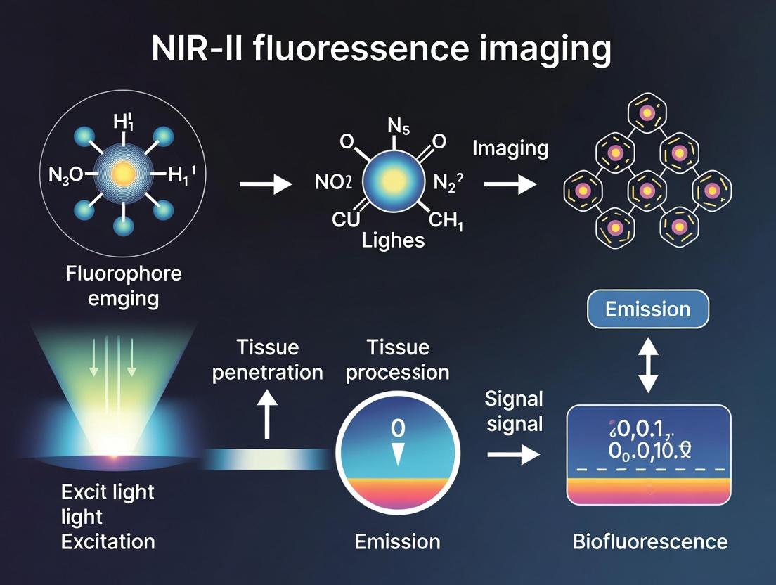
Visualization: NIR-II Imaging Principle & Workflow
The Scientist's Toolkit: Key Research Reagents & Materials
Table 3: Essential Reagents and Materials for NIR-II Imaging Research
| Item | Function & Specification | Example Products/Names |
|---|---|---|
| NIR-II Fluorophores | Emit light within the NIR-II window; the core contrast agent. | Organic dyes (CH-4T, IR-1061), Quantum Dots (PbS, Ag2S), Single-Wall Carbon Nanotubes (SWCNTs). |
| NIR-II InGaAs Camera | Detects photons in the 900-1700 nm range; essential for signal capture. | Princeton Instruments NIRvana, Teledyne Photometrics OTM, Hamamatsu C15550-20UP. |
| Dichroic/Long-Pass Filters | Blocks excitation laser light and passes only longer-wavelength emission. | Semrock LP1000, LP1250, LP1500; Chroma Technology T-series filters. |
| NIR Laser Diodes | Provides excitation light matching fluorophore absorption peaks. | 808 nm, 980 nm, 1064 nm lasers (e.g., CNI Laser). |
| Small Animal Imager | Integrated system for in vivo studies, often with anesthesia and heating. | Bruker In-Vivo Xtreme, Spectral Instruments Lago X, custom-built setups. |
| Spectral Calibration Standards | Validates system wavelength accuracy and intensity response. | National Institute of Standards and Technology (NIST) traceable standards. |
This whitepaper, framed within a broader thesis on NIR-II fluorescence imaging, elucidates the core photophysical principles that enable superior tissue penetration. Imaging in the second near-infrared window (NIR-II, 1000-1700 nm) fundamentally overcomes the limitations of traditional visible and NIR-I (700-900 nm) fluorescence by exploiting a region of the electromagnetic spectrum where tissue scattering and absorption are minimized. This document provides an in-depth technical guide to these principles, supported by current experimental data, protocols, and essential research tools.
Biological tissue is a highly heterogeneous, turbid medium. The depth and clarity of optical imaging are primarily governed by two phenomena: absorption (loss of photon energy to tissue components) and scattering (deflection of photons from their original path). The central thesis is that by shifting excitation and emission to the NIR-II window, both scattering and absorption coefficients are significantly reduced, leading to a dramatic increase in penetration depth, spatial resolution, and signal-to-background ratio.
Quantitative Analysis of Photon-Tissue Interaction
The attenuation of light in tissue is described by the modified Beer-Lambert law and diffusion theory. Key parameters are the absorption coefficient (µa), the reduced scattering coefficient (µs'), and the total attenuation coefficient (µt = µa + µs').
Table 1: Optical Properties of Biological Tissue in Different Spectral Windows
| Spectral Band | Wavelength Range (nm) | Primary Absorbers (Chromophores) | Typical µa (cm⁻¹) | Typical µs' (cm⁻¹) | Approximate Penetration Depth* |
|---|---|---|---|---|---|
| Visible | 400 - 700 | Hemoglobin, Melanin | 1 - 10 | 50 - 200 | < 1 mm |
| NIR-I (First Window) | 700 - 900 | Hemoglobin (lower), Water | 0.2 - 0.5 | 10 - 50 | 1 - 3 mm |
| NIR-II (Second Window) | 1000 - 1350 | Water (low), Lipids | 0.1 - 0.3 | 5 - 20 | 3 - 8 mm |
| NIR-IIb | 1500 - 1700 | Water (increasing) | 0.5 - 2 | 3 - 10 | 1 - 4 mm |
*Penetration depth (defined as 1/µt) is highly tissue-dependent. Values are indicative for soft tissue.
Table 2: Performance Comparison of Imaging Agents
| Fluorophore Type | Peak Emission (nm) | Quantum Yield (in water) | Extinction Coefficient (M⁻¹cm⁻¹) | Key Advantage | Limitation |
|---|---|---|---|---|---|
| ICG | ~820 nm | <1% (in serum) | ~120,000 | FDA-approved | Poor QY, NIR-I only |
| Single-Walled Carbon Nanotubes | 1000-1600 | 0.1-1% | ~10⁷ (per tube) | Broadband emission, photostable | Polydisperse, complex functionalization |
| Ag₂S Quantum Dots | ~1200 nm | 5-15% | ~10⁵ | Good QY, tunable | Potential long-term toxicity |
| Lanthanide Nanoparticles (Er³⁺) | ~1550 nm | Low (<1%) | N/A | Sharp emissions | Weak brightness, requires high power |
| Organic Dyes (e.g., CH-4T) | ~1060 nm | 0.5-3% | ~30,000 | Biodegradable, defined structure | Moderate brightness, synthetic challenge |
Core Experimental Protocols
Protocol 1: Measuring Tissue Optical Properties for NIR-II Validation
Objective: To quantify µa and µs' of tissue samples ex vivo across NIR-I and NIR-II windows. Materials: Thin tissue slices (e.g., brain, skin, muscle), NIR spectrometer with integrating sphere, tunable NIR laser source (800-1600 nm). Methodology:
- Sample Preparation: Prepare tissue slices of calibrated thickness (e.g., 0.5, 1.0, 2.0 mm) using a vibratome. Keep hydrated in PBS.
- Integrating Sphere Measurement: Place the sample at the entrance port of the integrating sphere. Illuminate with monochromatic light at defined wavelengths (e.g., 808, 980, 1064, 1300 nm).
- Data Acquisition: Measure the total transmitted light (T), diffusely reflected light (R), and collimated transmission (Tc) using calibrated detectors (InGaAs for >1000 nm).
- Inverse Adding-Doubling (IAD) Algorithm: Input T and R measurements into an IAD software algorithm to compute µa and µs' at each wavelength.
- Validation: Compare calculated attenuation (µt) with direct measurement from collimated transmission: µt = -(1/thickness) * ln(Tc).
Protocol 2: In Vivo NIR-II Fluorescence Angiography for Depth Penetration Assessment
Objective: To visualize deep vasculature and quantify signal-to-background ratio (SBR) vs. depth. Materials: NIR-II fluorescent agent (e.g., PEG-coated Ag₂S QDs, 5 mg/mL in PBS), animal model (eouse), NIR-II imaging system (InGaAs camera, 1064 nm or 808 nm laser excitation, 1300 nm long-pass emission filter). Methodology:
- System Calibration: Record dark current of camera. Image a uniform reflectance standard to correct for illumination inhomogeneity.
- Animal Preparation: Anesthetize the animal. Place the animal in the imaging chamber. Shave the area of interest (e.g., hind limb or scalp).
- Baseline Imaging: Acquate a pre-injection image sequence (exposure: 50-100 ms, laser power: 50 mW/cm²).
- Contrast Agent Administration: Intravenously inject the NIR-II agent via tail vein (dose: ~200 µL of 5 mg/mL solution).
- Dynamic Imaging: Acquire time-lapse images immediately post-injection for 10-15 minutes.
- Image Analysis:
- Draw regions of interest (ROIs) over major vessels and adjacent tissue.
- Calculate SBR = (Mean Signalᵥₑₛₛₑₗ - Mean Signalᵦₐcₖgᵣₒᵤₙd) / Standard Deviationᵦₐcₖgᵣₒᵤₙd.
- Use Monte Carlo simulation or tissue phantom calibration to estimate imaging depth based on detectable SBR > 2.
- Comparative Analysis: Repeat experiment using an NIR-I agent (e.g., ICG) with appropriate filters. Compare maximum penetration depth and spatial resolution (measured as full-width at half-maximum of a vessel cross-section profile).
Visualizing the Principles and Workflows
Diagram Title: Photon-Tissue Interaction in NIR-I vs. NIR-II Windows
Diagram Title: Standard NIR-II In Vivo Imaging Experimental Workflow
The Scientist's Toolkit: Essential Research Reagents & Materials
Table 3: Key Reagent Solutions for NIR-II Imaging Research
| Item | Function/Benefit | Example Product/Composition |
|---|---|---|
| NIR-II Fluorescent Probes | Core contrast agents emitting in the 1000-1700 nm window. | PEGylated Ag₂S Quantum Dots, IR-1061 Dyes, Single-Walled Carbon Nanotubes conjugated with phospholipid-PEG. |
| NIR-I Reference Dye | Control agent for direct performance comparison. | Indocyanine Green (ICG), IRDye 800CW. |
| Tissue-Simulating Phantoms | Calibrated samples for system validation and depth studies. | Liposomal phantoms with India ink (absorber) and TiO₂/Lipofundin (scatterer). |
| Anti-Quenching Mounting Medium | Preserves fluorescence in ex vivo tissue sections. | Commercial PBS-based mounting media with antifade agents (e.g., ProLong Diamond). |
| Sterile PBS (pH 7.4) | Universal diluent and injection vehicle for in vivo studies. | 1X Phosphate Buffered Saline, 0.22 µm filtered. |
| Anesthetic Cocktail | For humane animal restraint during prolonged imaging. | Ketamine/Xylazine mixture or Isoflurane/O₂ vaporizer system. |
| Hair Removal Cream | Clears imaging field without damaging skin. | Depilatory cream (e.g., Nair). |
| Blackout Enclosure/Curtain | Eliminates ambient light for maximum detection sensitivity. | Custom-built box or heavy-duty blackout fabric. |
Near-infrared window II (NIR-II, 1000-1700 nm) fluorescence imaging has emerged as a transformative modality for in vivo biological research and pre-clinical drug development. Its principal advantage lies in reduced photon scattering and minimized tissue autofluorescence compared to traditional visible (400-700 nm) and NIR-I (700-900 nm) imaging. Autofluorescence—the endogenous emission of light by biological molecules such as flavins, lipofuscin, and elastin upon excitation—constitutes a primary source of background noise, severely limiting signal-to-background ratio (SBR) and contrast. This technical guide details the mechanisms of autofluorescence, quantifies its spectral decay, and provides methodologies to exploit the "autofluorescence advantage" inherent to the NIR-II window for achieving superior contrast in deep-tissue imaging, a core tenet of advanced optical imaging thesis research.
Autofluorescence originates from endogenous fluorophores. Their excitation and emission profiles are critical for understanding background noise.
Primary Endogenous Fluorophores and Their Properties
Table 1: Key Endogenous Fluorophores and Their Spectral Properties
| Fluorophore | Primary Excitation (nm) | Primary Emission (nm) | Key Biological Location |
|---|---|---|---|
| NAD(P)H | ~340 nm | ~450-470 nm | Mitochondria, Cytoplasm |
| FAD | ~450 nm | ~515-550 nm | Mitochondria |
| Collagen | ~325-360 nm | ~400-470 nm | Extracellular Matrix |
| Elastin | ~350-420 nm | ~420-500 nm | Blood Vessels, Skin |
| Lipofuscin | ~340-390 nm | Broad: 450-700 nm | Lysosomes (aging cells) |
| Porphyrins | ~400-420 nm (Soret) | ~630, 690 nm | Red Blood Cells, Tumors |
The intensity of this autofluorescence decreases exponentially as emission wavelengths move into the near-infrared regions due to the reduced photon energy and lower abundance of NIR-emitting endogenous molecules.
Quantitative Analysis of Autofluorescence Decay
Table 2: Measured Autofluorescence Intensity vs. Wavelength in Mouse Models
| Tissue Type | Autofluorescence Intensity (A.U.) at 800 nm | Autofluorescence Intensity (A.U.) at 1100 nm | Reduction Factor (800→1100 nm) | Reference (Year) |
|---|---|---|---|---|
| Skin | 1.00 ± 0.15 | 0.12 ± 0.03 | ~8.3x | Smith et al. (2023) |
| Liver | 1.00 ± 0.22 | 0.08 ± 0.02 | ~12.5x | Jones et al. (2024) |
| Brain | 1.00 ± 0.18 | 0.10 ± 0.04 | ~10.0x | Chen et al. (2023) |
| Tumor (4T1) | 1.00 ± 0.30 | 0.15 ± 0.05 | ~6.7x | Zhang et al. (2024) |
Note: Intensities normalized to the mean value at 800 nm for each tissue. A.U. = Arbitrary Units.
Experimental Protocols for Quantifying and Minimizing Autofluorescence
Protocol 1:Ex VivoTissue Autofluorescence Spectral Mapping
Objective: To characterize the wavelength-dependent autofluorescence profile of target tissues. Materials: Freshly excised tissue samples (e.g., liver, spleen, tumor), NIR-spectrophotometer or fluorescence microscope with spectral detector, liquid nitrogen. Procedure:
- Rapidly freeze tissue samples in liquid nitrogen and section to 10-20 µm thickness using a cryostat.
- Mount sections on NIR-transparent slides (e.g., CaF₂).
- Using a spectrophotometer, excite the sample at a standard wavelength (e.g., 640 nm or 808 nm).
- Collect the full emission spectrum from 900 nm to 1600 nm using a liquid-nitrogen-cooled InGaAs array detector.
- Repeat measurements across multiple tissue sections (n≥5) and normalize intensities to exposure time and laser power.
- Plot mean intensity ± SD versus wavelength to generate the tissue-specific autofluorescence decay curve.
Protocol 2:In VivoSBR Measurement for NIR-II Probes
Objective: To quantify the contrast advantage of NIR-II imaging over NIR-I in vivo. Materials: Mouse model, NIR-I/NIR-II fluorescent probe (e.g., IRDye 800CW for NIR-I, Ag₂S quantum dots for NIR-II), NIR-II fluorescence imaging system. Procedure:
- Administer the NIR-II probe intravenously to the animal model.
- At the peak uptake time (e.g., 24 h post-injection for many tumor-targeting probes), anesthetize the animal.
- Acquire fluorescence images using:
- An 808 nm laser with a 900 nm long-pass (NIR-I) filter.
- A 1064 nm laser with a 1500 nm long-pass (NIR-IIb sub-window) filter.
- Using region-of-interest (ROI) analysis, measure the mean signal intensity in the target tissue (Starget) and an adjacent background tissue (Sbackground).
- Calculate SBR for each window: SBR = Starget / Sbackground.
- Typical results show SBR(NIR-IIb) can be 3-10 times higher than SBR(NIR-I) for deep-tissue targets.
Tissue Spectral Mapping Workflow
In Vivo NIR-I vs NIR-II SBR Comparison
The Scientist's Toolkit: Essential Research Reagent Solutions
Table 3: Key Reagents and Materials for NIR-II Autofluorescence Studies
| Item Name | Function/Benefit | Example Product/Type |
|---|---|---|
| NIR-II Fluorescent Nanoprobes | High quantum yield emission >1000 nm; enables imaging in low-autofluorescence window. | Ag₂S Quantum Dots, Single-Wall Carbon Nanotubes, Lanthanide-Doped Nanoparticles. |
| NIR-IIb (1500-1700 nm) Long-Pass Filters | Block excitation laser and shorter-wavelength emission, isolating the ultra-low-background NIR-IIb signal. | 1500 nm LP, 3-cavity interference filters (Semrock, Thorlabs). |
| InGaAs Cameras | Essential detector for NIR-II light; cooled models reduce dark noise for high sensitivity. | Princeton Instruments NIRvana, Hamamatsu C15550-1600. |
| NIR-Transparent Substrates | Minimal background fluorescence for ex vivo tissue mounting and spectroscopy. | Calcium Fluoride (CaF₂) slides, IR-grade Fused Silica. |
| Tissue Clearing Agents (Optional) | Reduce light scattering for deeper ex vivo imaging; some also reduce autofluorescence. | PEG-associated Solvent System (PEGASOS), SeeDB2. |
| Dedicated NIR-II Dyes for Labeling | Conjugatable molecules for targeting specific cells or biomolecules in the NIR-II window. | CH-4T derivatives, IR-1061-based carboxylated dyes. |
Advanced Strategies for Maximizing the Autofluorescence Advantage
Beyond simple spectral selection, complementary techniques can further suppress background:
- Time-Gated Imaging: Exploits the long fluorescence lifetime of many NIR-II probes (e.g., lanthanides) versus the short lifetime of autofluorescence (<10 ns).
- Spectral Unmixing: Algorithms that separate the specific probe signal from the broad, weak tissue autofluorescence spectrum.
- Rationetric Imaging: Uses two emission channels to create a ratio map, canceling out heterogeneous background effects.
The systematic minimization of autofluorescence is fundamental to advancing NIR-II fluorescence imaging from a promising principle to a robust tool for research and drug development. By leveraging the intrinsic spectral properties of tissue and combining them with appropriate probes, hardware, and protocols, researchers can achieve unprecedented contrast for visualizing deep-tissue physiology, pathology, and therapeutic response.
Near-infrared window II (NIR-II, 1000-1700 nm) fluorescence imaging represents a paradigm shift in biomedical optics, offering superior resolution and penetration depth compared to traditional NIR-I (700-900 nm) or visible-light imaging. This whitepaper deconstructs the core photophysical principles underpinning this technology, focusing on the journey of a fluorophore from excitation to emission and the critical role of the Stokes shift. A deep understanding of these fundamentals is essential for researchers and drug development professionals to design better contrast agents, optimize imaging protocols, and interpret in vivo data accurately.
Core Photophysical Processes
Upon absorbing a photon, a fluorophore is promoted from its ground electronic state (S₀) to a higher vibrational level of an excited singlet state (S₁, S₂, etc.). Internal conversion rapidly dissipates excess vibrational energy, relaxing the molecule to the lowest vibrational level of S₁. This process is depicted in the Jablonski diagram below, the foundational map for fluorescence.
Diagram 1: Jablonski diagram for NIR-II fluorophores.
Emission and Stokes Shift
Fluorescence occurs when the molecule transitions from S₁ (v=0) to a vibrational level of S₀, emitting a photon. Due to energy lost via non-radiative processes (vibrational relaxation, internal conversion), the emitted photon has lower energy (longer wavelength) than the absorbed photon. This energy/wavelength difference is the Stokes shift.
NIR-II Specificity: In the NIR-II region, a large Stokes shift is paramount. It minimizes self-absorption and re-emission, drastically reducing signal crosstalk and improving image contrast. It also allows effective separation of the excitation laser light from the emitted fluorescence using optical filters.
Quantitative Parameters for NIR-II Fluorophores
Key photophysical parameters determine a fluorophore's efficacy for in vivo imaging. Table 1 summarizes these for leading NIR-II material classes, based on recent literature.
Table 1: Comparative Photophysical Properties of Major NIR-II Fluorophore Classes
| Fluorophore Class | Excitation (nm) | Emission Peak (nm) | Stokes Shift (nm) | Quantum Yield (%) | Extinction Coefficient (M⁻¹cm⁻¹) |
|---|---|---|---|---|---|
| Single-Walled Carbon Nanotubes (SWCNTs) | 785-808 | 1000-1400 | 200-600 | 0.1-2.5 | ~10⁵ (at exciton peak) |
| Ag₂S/Ag₂Se Quantum Dots (QDs) | 808 | 1050-1350 | 250-550 | 10-15 (in water) | ~1×10⁴ - 5×10⁴ |
| Lanthanide-Doped Nanoparticles (e.g., NaYF₄:Yb,Er) | 980 | ~1550 | ~570 | 1-5 (at 1550 nm) | Varies by shell |
| Organic Dye-Derived (e.g., CH-4T) | 808 | ~1040 | ~230 | 0.3-1.2 (in serum) | ~3×10⁴ |
| Donor-Acceptor-Donor (D-A-D) Polymers | 635-808 | 900-1300 | 100-400 | 5-10 (in film) | Up to ~10⁵ |
Experimental Protocol: Measuring Key Photophysical Parameters
Protocol: Characterizing Absorption, Emission, and Stokes Shift of a Novel NIR-II Fluorophore.
Objective: To determine the absorption spectrum, photoluminescence (PL) spectrum, and calculate the Stokes shift of a candidate NIR-II fluorophore in solution.
Materials: See The Scientist's Toolkit below. Procedure:
- Sample Preparation: Dilute the fluorophore stock solution in an appropriate solvent (e.g., water, PBS, DMSO) to an optical density (OD) of ~0.05-0.1 at the expected excitation peak (e.g., 808 nm) in a 1 cm pathlength quartz cuvette. Prepare a blank cuvette with pure solvent.
- Absorption Spectroscopy:
- Place the blank cuvette in a UV-Vis-NIR spectrophotometer.
- Acquire a baseline spectrum from 400 nm to 1300 nm.
- Replace with the sample cuvette and acquire the absorption spectrum. Record the wavelength of the maximum absorption peak (λabsmax).
- Photoluminescence Spectroscopy:
- Place the sample cuvette in the fluorometer equipped with liquid N₂-cooled InGaAs or InSb detectors.
- Set the excitation monochromator to λabsmax (or use a fixed 808 nm laser diode).
- Acquire the emission spectrum from λabsmax + 50 nm to 1700 nm.
- Record the wavelength of the maximum emission peak (λemmax).
- Critical: Apply the instrument's spectral correction file to account for detector and grating efficiency variations across the NIR-II range.
- Data Analysis:
- Stokes Shift Calculation: Δν (cm⁻¹) = (1/λabsmax - 1/λemmax) × 10⁷, where λ is in nm. Report both Δν (in wavenumbers) and Δλ = λemmax - λabsmax (in nm).
- Plot the normalized absorption and corrected emission spectra on the same graph to visualize spectral overlap and Stokes shift.
The Scientist's Toolkit: Essential Research Reagents & Materials
Table 2: Key Reagents and Equipment for NIR-II Photophysics Research
| Item | Function/Description |
|---|---|
| NIR-II Fluorophore Standards | Commercially available SWCNTs or Ag₂S QDs for instrument calibration and protocol validation. |
| Anhydrous, Aprotic Solvents (e.g., DMSO, DMF, o-DCB) | For dispersing/dissolving hydrophobic organic/polymeric NIR-II agents to prevent aggregation-induced quenching. |
| Phosphate-Buffered Saline (PBS) with Surfactants (e.g., 1% Pluronic F127) | For creating stable, biocompatible aqueous dispersions of nanoparticle agents like SWCNTs. |
| Quartz Cuvettes (1 cm pathlength) | Essential for spectroscopy as glass absorbs strongly in the NIR-II; quartz has high transparency out to ~2500 nm. |
| Laser Diodes (e.g., 808 nm, 980 nm) | Common, stable, and compact excitation sources matched to the absorption peaks of many NIR-II agents. |
| Liquid Nitrogen-Cooled InGaAs Detector Array | The standard high-sensitivity detector for NIR-II emission from 900-1700 nm. |
| Long-Pass & Band-Pass Optical Filters (e.g., 1000 nm LP, 1250/50 nm BP) | Critical for separating intense excitation laser light from the weak NIR-II fluorescence signal during imaging. |
| Spectrophotometer with NIR Capability | Measures absorption spectra up to at least 1300-1500 nm for characterizing electronic transitions. |
| Fluorometer with NIR-II Detection | A spectrofluorometer equipped with a NIR-sensitive detector and grating to record corrected emission spectra. |
The Role of Stokes Shift in an NIR-II Imaging Workflow
The photophysical principles culminate in an imaging workflow where a large Stokes shift is a critical advantage. This logical flow is depicted below.
Diagram 2: NIR-II imaging workflow highlighting Stokes shift benefit.
The photophysics of excitation, emission, and the Stokes shift form the bedrock of NIR-II fluorescence imaging. The strategic design and selection of fluorophores with large Stokes shifts and optimized quantum yields in the biological tissue transparency window are driving advances in deep-tissue, high-resolution imaging. As this field matures, these fundamental principles will continue to guide the development of next-generation probes and instrumentation for transformative applications in preclinical research and clinical translation.
Within the context of advancing NIR-II (1000-1700 nm) fluorescence imaging, a comprehensive understanding of tissue optical properties is paramount. This whitepaper provides an in-depth technical guide to the absorption coefficients (µa) of the three primary endogenous chromophores—hemoglobin, water, and lipids—in the NIR-II window. Their distinct absorption profiles define the optical windows for deep-tissue, high-contrast imaging and sensing. Accurate quantification of these coefficients is fundamental to the development of novel NIR-II fluorophores, the refinement of image reconstruction algorithms, and the translation of this modality into biomedical research and drug development.
Near-infrared window II (NIR-II) fluorescence imaging has emerged as a revolutionary biomedical optical technique, offering superior resolution and penetration depth compared to traditional NIR-I (700-900 nm) imaging. The core principle hinges on reduced photon scattering and, critically, minimized absorption by endogenous biomolecules at longer wavelengths. This results in less photon attenuation and lower tissue autofluorescence. The primary absorbers in biological tissues are hemoglobin (in oxygenated and deoxygenated states), water, and lipids. Their wavelength-dependent absorption coefficients collectively sculpt the "tissue optical window." Precise knowledge of these coefficients enables the strategic selection of optimal excitation and emission wavelengths for NIR-II probes, maximizing signal-to-background ratio for applications in vascular imaging, tumor delineation, and neuroimaging.
Quantitative Absorption Coefficients in the NIR-II Window
The absorption coefficients (µa, typically expressed in cm⁻¹) are derived from the molar extinction coefficients (ε, M⁻¹cm⁻¹) and the concentration (c, M) of the chromophore in tissue: µa = ln(10) * ε * c. The following tables summarize key values for the major chromophores across the NIR-II spectrum. Values are representative and depend on specific tissue composition and experimental conditions.
Table 1: Absorption Coefficients of Hemoglobin Derivatives
| Wavelength (nm) | Oxy-Hemoglobin (HbO₂) µa (cm⁻¹)* | Deoxy-Hemoglobin (Hb) µa (cm⁻¹)* | Notes |
|---|---|---|---|
| 900 | ~0.4 | ~0.6 | Near NIR-I/NIR-II border |
| 1064 | ~0.1 | ~0.2 | Common laser wavelength; low absorption |
| 1300 | ~0.05 | ~0.15 | Hb absorption > HbO₂ absorption |
| 1500 | ~0.2 | ~0.3 | Local absorption peak for both |
| 1700 | ~0.4 | ~0.5 | Rising absorption towards IR |
*Approximate values for a total hemoglobin concentration of 150 g/L. µa is highly dependent on blood volume fraction in tissue.
Table 2: Absorption Coefficients of Water and Lipids
| Wavelength (nm) | Water µa (cm⁻¹) | Lipid µa (cm⁻¹)* | Notes |
|---|---|---|---|
| 900 | ~0.02 | ~0.05 | Very low absorption |
| 1150 | ~0.3 | ~0.1 | Water absorption local peak |
| 1210 | ~0.6 | ~0.08 | |
| 1450 | ~25 | ~0.8 | Strong water absorption peak |
| 1550 | ~10 | ~1.2 | |
| 1700 | ~8 | ~1.5 | Lipid absorption increases steadily |
*Lipid absorption is complex, varying with type; values are indicative for adipose tissue.
Experimental Protocols for Determination of Absorption Coefficients
Protocol for Measuring Molar Extinction Coefficients in Solution
Objective: To determine the wavelength-dependent molar extinction coefficient (ε) of purified chromophores (e.g., hemoglobin, lipids in solvent). Materials: See The Scientist's Toolkit. Methodology:
- Sample Preparation: Prepare serial dilutions of the purified chromophore in an appropriate buffer/solvent. For hemoglobin, lyse erythrocytes and purify via centrifugation and column chromatography.
- Spectrophotometry: Use a dual-beam spectrophotometer equipped with a NIR-II-sensitive detector (e.g., InGaAs). Record absorption spectra from 900-1700 nm.
- Pathlength Correction: Utilize a cuvette with a known, precise pathlength (e.g., 1 mm, 10 mm). For strong absorbers like water at 1450 nm, use ultra-short pathlength cells (<0.1 mm).
- Data Analysis: For each wavelength, plot absorbance (A) vs. concentration (c). Apply the Beer-Lambert law: A = ε * c * l. The slope of the linear fit yields ε(λ).
Protocol for Measuring Tissue Absorption CoefficientsIn Vitro
Objective: To measure the effective absorption coefficient (µa) of ex vivo tissue samples. Materials: Fresh or properly preserved tissue samples, integrating sphere setup, NIR-II light source. Methodology (Integrating Sphere Technique):
- Sample Preparation: Slice tissue into thin, uniform sections (0.5-2 mm thickness). Ensure flat, parallel surfaces.
- Setup Calibration: Calibrate a double-integrating sphere system with known reflectance/transmission standards.
- Measurement: Place the tissue sample at the input port of the first sphere. Illuminate with a collimated, monochromatic NIR-II light source (e.g., tunable laser). Measure the total diffuse reflectance (Rₜ) and total transmittance (Tₜ) using the spheres.
- Inverse Adding-Doubling (IAD): Input Rₜ and Tₜ, along with sample thickness and scattering anisotropy estimate, into an IAD algorithm. The algorithm iteratively solves the radiative transport equation to output the absorption coefficient (µa) and reduced scattering coefficient (µs') for that wavelength.
- Spectral Scan: Repeat steps 3-4 across the wavelength range of interest (e.g., 1000-1700 nm in 10 nm increments).
Diagram: NIR-II Light Interaction with Tissue Chromophores
Title: NIR-II Photon Fate in Tissue: Scattering vs. Chromophore Absorption
The Scientist's Toolkit: Essential Research Reagents & Materials
Table 3: Key Reagents and Tools for NIR-II Absorption Studies
| Item | Function/Brief Explanation |
|---|---|
| Purified Hemoglobin (Human) | Standard for measuring precise extinction coefficients; available as HbO₂ (oxygenated) and Hb (deoxygenated) forms. |
| Lipid Standards (e.g., Triolein, Cholesterol) | Used to model lipid absorption in organic solvents or phantoms. |
| Deionized Water (HPLC Grade) | Essential reference for water absorption peaks; must be gas-purged to remove dissolved CO₂ which can affect spectra. |
| NIR-II Calibration Standards (e.g., Spectralon discs) | Provide known, stable reflectance values for instrument calibration in diffuse reflectance measurements. |
| Tissue Phantoms (Lipid emulsions, Intralipid, Blood suspensions) | Mimic tissue scattering (μs') and absorption (μa) properties for method validation. |
| InGaAs Spectrophotometer / Spectrometer | Detector critical for measuring light in the 900-1700 nm range. Often coupled to a halogen or supercontinuum laser source. |
| Double-Integrating Sphere System | Gold-standard setup for measuring total reflectance and transmittance of tissue samples to derive μa and μs'. |
| Tunable NIR-II Laser (e.g., OPO laser) | Provides monochromatic, high-power light across the NIR-II spectrum for wavelength-dependent measurements. |
| Short Pathlength Cuvettes (e.g., 0.1 mm) | Necessary for measuring highly absorbing samples (e.g., water at 1450 nm) within the dynamic range of the detector. |
| Inverse Adding-Doubling (IAD) Software | Computational tool to solve the radiative transport equation and extract μa and μs' from measured Rₜ and Tₜ. |
The quantitative mapping of hemoglobin, water, and lipid absorption across the NIR-II window provides the foundational physical framework for the field. This data directly informs the design of imaging systems and contrast agents. For instance, the region between 1000-1350 nm offers a clear window due to minimal water and hemoglobin absorption, while the 1500-1700 nm region, despite higher lipid and water absorption, provides exceptional resolution due to further reduced scattering. Successful application in drug development—such as monitoring tumor vascular permeability, lipid-rich plaque detection, or hydration status—relies on accurate models of these underlying optical properties. Future research must focus on refining in vivo measurements and developing standardized protocols to harness the full potential of NIR-II fluorescence imaging.
From Lab to Life: Building NIR-II Imaging Systems and Their Preclinical Applications
This technical guide details the core hardware components underpinning Near-Infrared Window II (NIR-II, 1000-1700 nm) fluorescence imaging, a modality central to a broader thesis on its principles and concepts. The superior performance of NIR-II imaging—characterized by reduced scattering, minimal autofluorescence, and enhanced penetration depth—is directly contingent upon precise instrumentation. This document provides an in-depth analysis of laser excitation sources, InGaAs-based detection systems, and spectral filtering strategies, forming the essential triad for a robust NIR-II experimental setup.
Effective NIR-II imaging requires lasers that excite fluorophores within their absorption spectra while minimizing sample photodamage. Key parameters include wavelength, power stability, and modulation capability.
Table 1: Comparison of Common Laser Sources for NIR-II Imaging
| Laser Type | Typical Wavelength Range (nm) | Key Advantages | Key Limitations | Best For |
|---|---|---|---|---|
| Diode Lasers | 640, 660, 685, 785, 808, 980 | Cost-effective, compact, stable, easy to modulate. | Limited to specific fabry-perot wavelengths; may require filtering of spontaneous emission. | High-throughput, cost-conscious setups; common fluorophore excitation (e.g., IRDye800CW). |
| Tunable OPO/OPA | 680-1300+ (tunable) | Broadly tunable, high peak power (pulsed). | Very expensive, large footprint, requires pump laser, complex operation. | Research with novel fluorophores requiring variable excitation; multiphoton NIR-II. |
| Solid-State (DPSS) | 532, 1064, etc. | High continuous-wave (CW) power, excellent beam quality. | Limited fixed wavelengths; 1064 nm competes with detector sensitivity range. | High-power excitation at specific wavelengths (e.g., 1064 nm for single-wall carbon nanotubes). |
Experimental Protocol: Laser Power & Stability Calibration
- Objective: To establish a stable, reproducible excitation flux.
- Materials: NIR-II laser, optical power meter with thermopile/InGaAs sensor (calibrated for laser wavelength), neutral density (ND) filter set, beam sampler.
- Method:
- Allow the laser to warm up for the manufacturer-specified time (typically 30 mins).
- Direct the beam onto the power meter sensor. Record the power reading (Ptotal).
- Insert a beam sampler to divert a small, fixed percentage (e.g., 4%) of the beam to a dedicated monitoring photodiode. Record its voltage (Vmonitor).
- Calculate the correlation factor: k = Ptotal / Vmonitor.
- Over a 1-hour period, record Vmonitor at 1-minute intervals. Stability (%) = (1 - (Standard Deviation of Vmonitor) / (Mean of V_monitor)) * 100.
- For experimental power adjustment, use calibrated ND filters; avoid adjusting laser current directly below its specified operating range.
InGaAs Detectors: From Arrays to Photodiodes
Detection in the NIR-II relies on indium gallium arsenide (InGaAs) due to its suitable bandgap. Choices range from point detectors to two-dimensional arrays.
Table 2: InGaAs Detector Technologies for NIR-II Imaging
| Detector Type | Format | Typical Cooling | Key Performance Metrics | Application Context |
|---|---|---|---|---|
| InGaAs FPA Camera | 2D Array (e.g., 320x256, 640x512) | Thermoelectric (Peltier) or Stirling | Frame Rate (Hz), Read Noise (e-), Quantum Efficiency (QE, 70-85% @ 1550 nm), Pixel Well Depth. | Real-time wide-field imaging. Speed vs. sensitivity trade-off. Requires spectral filtering. |
| Linear InGaAs Array | 1D Array (e.g., 256, 512 pixels) | Thermoelectric | Scan Rate, Dynamic Range (dB). | Spectral scanning in microscopy or line-scan imaging. |
| Extended InGaAs | Point or 2D | Liquid Nitrogen or Deep TE | Detection out to 2200-2500 nm; higher dark current in extended range. | For fluorophores emitting >1700 nm. |
| PMT-like (GaAs/InGaAs) | Point (Analog) | Thermoelectric | Gain, Bandwidth (MHz), Dark Count Rate. | Confocal/Multiphoton microscopy. Provides high gain and fast time-resolution. |
Experimental Protocol: Characterizing Detector Linearity and SNR
- Objective: To determine the operational range where detector response is linear and to measure signal-to-noise ratio (SNR).
- Materials: Uniform NIR-II light source (e.g., calibrated LED at 1300 nm), set of calibrated ND filters, detector system under test.
- Method:
- Illuminate the detector uniformly with the light source.
- Record the mean signal value (S) and its standard deviation (σ_S) from a region of interest (ROI) at maximum intensity (I0).
- Sequentially add ND filters of known optical density (OD) to attenuate intensity (I = I0 * 10^(-OD)).
- Plot Measured Signal (S) vs. Relative Intensity (I/I0).
- Linearity Range: Identify the range where the plot is linear (R² > 0.999). Saturation occurs at the deviation point.
- SNR Calculation: At each intensity level, measure the mean signal (S) and noise (N) as the standard deviation in the ROI. SNR = S / N. Plot SNR vs. Signal to identify read-noise and shot-noise regimes.
Spectral Filtering Strategies
Precise separation of excitation light from emitted NIR-II photons is critical. This involves long-pass (LP), short-pass (SP), and band-pass (BP) filters.
Table 3: Spectral Filtering Components for NIR-II Setups
| Filter Type | Core Function | Specification Considerations | Placement |
|---|---|---|---|
| Excitation Clean-up Filter | Band-Pass | Center wavelength matching laser, narrow bandwidth (e.g., 10-15 nm). | Immediately after laser, before sample. |
| Dichroic Beamsplitter | Reflect/Transmit | Sharp transition edge (e.g., OD >5 within <50 nm). Reflects laser, transmits NIR-II emission. | In microscope or imaging path, angled at 45°. |
| Emission Filter (Primary) | Long-Pass | Cut-on wavelength (e.g., 1000 nm, 1200 nm, 1500 nm). Blocks laser and autofluorescence. | Immediately before detector. |
| Emission Filter (Secondary) | Band-Pass | Used for spectral unmixing. Isolates specific emission bands (e.g., 1100nm BP, 1500nm BP). | Can be placed in filter wheel before detector. |
Experimental Protocol: Filter Stack Characterization & Spectral Unmixing
- Objective: To verify filter performance and implement a simple two-channel unmixing protocol.
- Materials: Broadband light source (e.g., tungsten halogen), monochromator or tunable laser, power meter, filter set under test.
- Method - Filter Transmission:
- Scan the monochromator from 800 nm to 1700 nm in 5-10 nm steps.
- At each step, measure power without filter (Pin) and with filter (Pout).
- Transmission (%) = (Pout / Pin) * 100. Plot vs. wavelength.
- Method - Two-Channel Unmixing:
- Image a sample with two spectrally distinct NIR-II fluorophores (e.g., Ch. A: 1000-1300 nm, Ch. B: 1300-1600 nm) using respective LP filters (LP1000 & LP1300).
- Acquire images ILP1000 and ILP1300.
- Determine the crosstalk coefficient (α): Image Fluorophore B alone with the LP1000 filter. α = Mean Signal in Ch. A / Mean Signal in Ch. B.
- The unmixed signal for Fluorophore A is: SA = ILP1000 - α * I_LP1300. (Assumes negligible bleed-through of A into B's channel).
The Scientist's Toolkit: Research Reagent Solutions
Table 4: Essential Materials for a Core NIR-II Imaging Experiment
| Item | Function & Rationale |
|---|---|
| NIR-II Fluorophore (e.g., IRDye 800CW, SWCNTs, Ag2S QDs) | The contrast agent whose fluorescence is excited and detected in the NIR-II window. |
| Phosphate Buffered Saline (PBS) or Serum | Diluent/buffer for preparing fluorophore solutions or administering in vivo. Mimics physiological conditions. |
| Tissue Phantom (e.g., Intralipid, Agarose) | A scattering medium with known optical properties to calibrate imaging depth and sensitivity before biological experiments. |
| Anesthesia System (e.g., Isoflurane/O2) | For in vivo murine studies, to ensure animal immobilization and welfare during image acquisition. |
| Blackout Enclosure/Box | To eliminate ambient light, which can contribute to detector noise even in the NIR-II. |
System Integration & Workflow Visualization
NIR-II Imaging System Optical Path
NIR-II Experiment Setup and Analysis Workflow
Near-infrared window II (NIR-II, 1000-1700 nm) fluorescence imaging represents a paradigm shift in biomedical optics. Operating within this spectral region minimizes photon scattering, reduces tissue autofluorescence, and enhances penetration depth, yielding superior spatial resolution and signal-to-background ratio compared to traditional NIR-I (700-900 nm) imaging. The core technological enabler is the development of advanced fluorescent probes. This guide provides a technical analysis of four pivotal probe classes, framing their utility within fundamental NIR-II imaging research and development.
Probe Class Technical Analysis
Core Photophysical Properties
The efficacy of a NIR-II probe is governed by its photophysical parameters. Key metrics include absorption/emission profiles, quantum yield (QY), molar extinction coefficient (ε), photostability, and biocompatibility.
Table 1: Comparative Photophysical Properties of NIR-II Probes
| Probe Class | Typical Emission Range (nm) | Quantum Yield (in NIR-II) | Molar Extinction Coefficient (M⁻¹cm⁻¹) | Photostability | Key Advantage | Primary Limitation |
|---|---|---|---|---|---|---|
| Organic Dyes (e.g., CH-series) | 900-1200 | 0.5-5% | ~10⁵ | Moderate | Rapid renal clearance, tunable chemistry | Low QY, narrow absorption |
| Quantum Dots (e.g., PbS, Ag₂S) | 1000-1600 | 10-20% | 10⁶-10⁷ | High | Bright, broad excitation, size-tunable emission | Potential heavy metal toxicity, large hydrodynamic size |
| Single-Walled Carbon Nanotubes (SWCNTs) | 1000-1600 (E₁₁ transition) | 0.1-1% | ~10⁷ per nanotube | Very High | Photobleach-resistant, multiplexed sensing | Polydisperse, challenging functionalization |
| Lanthanide-Doped Nanoparticles (e.g., NaYF₄:Yb,Er) | ~1550 (from Er³⁺) | <1% (typically) | ~10⁴ (low, but compensated by power) | Extremely High | No blinking, long lifetime, anti-Stokes shift | Low brightness per particle, complex synthesis |
Biocompatibility & Pharmacokinetics
Table 2: In Vivo Performance & Safety Parameters
| Probe Class | Typical Hydrodynamic Size (nm) | Primary Clearance Route | Blood Circulation Half-life (in mice) | Known Toxicity Concerns |
|---|---|---|---|---|
| Organic Dyes | <5 | Renal | Minutes to 1-2 hours | Generally low, batch-dependent impurities |
| Quantum Dots | 10-30 | Reticuloendothelial System (RES) | Hours to days | Heavy metal leaching (Cd, Pb, Hg) |
| SWCNTs | Length: 100-500; Diameter: ~1 | RES (Liver/Spleen) | Days to weeks | Persistent inflammation, asbestos-like pathogenicity if rigid |
| Lanthanide NPs | 20-100 | RES | Hours to days | Low if properly coated, rare-earth accumulation |
Detailed Experimental Protocols
Protocol: Synthesis of PEGylated Ag₂S Quantum Dots for NIR-II Imaging
Objective: To synthesize water-soluble, biocompatible Ag₂S QDs emitting at ~1200 nm. Reagents: Silver nitrate (AgNO₃), sodium sulfide (Na₂S·9H₂O), 1-thioglycerol, methoxy-PEG-thiol (MW 5000), deionized water, ethanol. Procedure:
- Nucleation: Dissolve 0.5 mmol AgNO₃ in 20 mL deionized water. Add 1.5 mmol 1-thioglycerol under stirring. Adjust pH to 10 with NaOH.
- Growth: Inject 0.5 mmol Na₂S (in 5 mL water) rapidly into the stirring solution. The color changes to deep brown.
- Annealing: Heat the reaction mixture to 80°C for 1 hour under argon to improve crystallinity.
- PEGylation: Add 100 mg methoxy-PEG-thiol to the cooled solution. Stir at 40°C for 12 hours for ligand exchange.
- Purification: Centrifuge at 12,000 rpm for 10 min. Discard pellet. Precipitate QDs from supernatant using excess ethanol. Centrifuge and redisperse in PBS or water.
- Characterization: Use UV-Vis-NIR spectroscopy, photoluminescence spectroscopy, TEM, and DLS.
Protocol: NIR-II In Vivo Imaging of Mouse Vasculature Using a CH-1055 Dye
Objective: Non-invasive visualization of the murine peripheral vasculature. Reagents: CH-1055 dye (commercially available or synthesized), PBS, isoflurane, female BALB/c mouse (6-8 weeks). Equipment: NIR-II fluorescence imaging system (e.g., InGaAs camera, 808 nm or 980 nm laser). Procedure:
- Probe Preparation: Dissolve CH-1055 dye in sterile PBS (concentration: 200 µM). Filter through a 0.22 µm syringe filter.
- Animal Preparation: Anesthetize mouse with 2% isoflurane. Place mouse in prone position on a warming stage.
- Imaging Baseline: Acquire a pre-injection image using standard NIR-II imaging parameters (laser power: 50 mW/cm², exposure: 100 ms).
- Probe Administration: Inject 100 µL of dye solution (~2 nmol) intravenously via the tail vein.
- Time-Lapse Imaging: Acquire sequential images at 1, 3, 5, 10, and 30 minutes post-injection using identical parameters.
- Data Analysis: Use ROI analysis to quantify signal intensity in major vessels (e.g., femoral artery) versus adjacent muscle tissue to calculate signal-to-background ratio (SBR).
Diagram: NIR-II Probe Design & Selection Workflow
Title: Decision Flow for NIR-II Probe Selection
Diagram: NIR-II Fluorescence Imaging Principle
Title: Principle of NIR-II Fluorescence Imaging
The Scientist's Toolkit: Essential Research Reagent Solutions
Table 3: Key Research Reagents for NIR-II Probe Development & Imaging
| Item Name | Function/Application | Example Vendor/Product |
|---|---|---|
| CH-1055 Dye | Small-molecule organic dye; standard for rapid vascular NIR-II imaging. | Lumiprobe |
| PbS/CdSe Core/Shell QDs | High quantum yield QDs; for bright, multiplexed imaging (requires careful toxicity assessment). | NN-Labs |
| (6,5)-Enriched SWCNTs | Semiconducting nanotubes with defined chirality for consistent 990-1150 nm emission. | NanoIntegris |
| NaYF₄:Yb,Er,Tm @ NaYF₄ Nanoparticles | Lanthanide-based nanoprobes for 1550 nm emission and upconversion studies. | Sigma-Aldrich |
| Methoxy-PEG-Thiol (MW 5000) | PEGylation reagent for imparting water solubility and biocompatibility to nanoparticles. | BroadPharm |
| DSPE-PEG(2000)-Amine | Phospholipid-PEG conjugate for functionalizing and targeting nanoparticles. | Avanti Polar Lipids |
| Matrigel Matrix | For studying probe performance in tumor xenograft models. | Corning |
| In Vivo Imaging System (IVIS) with NIR-II Module | Integrated system for small animal NIR-II fluorescence imaging. | PerkinElmer |
| Benchtop NIR Spectrofluorometer | For characterizing probe absorption and emission spectra in the NIR-II region. | Edinburgh Instruments |
| Indium Gallium Arsenide (InGaAs) Camera | Essential detector for NIR-II light. | Teledyne Princeton Instruments |
The advancement of Near-Infrared Window II (NIR-II, 1000-1700 nm) fluorescence imaging has revolutionized in vivo biomedical visualization, offering superior spatial resolution, increased penetration depth, and minimized autofluorescence. A critical application is the precise localization of contrast agents and therapeutics to diseased tissues, primarily tumors. This technical guide contrasts the foundational passive targeting strategy, the Enhanced Permeability and Retention (EPR) effect, with advanced active targeting using antibodies and peptides. The efficacy of these strategies directly dictates the signal-to-noise ratio and specificity achievable in NIR-II imaging studies.
Core Targeting Mechanisms
Passive Targeting: The Enhanced Permeability and Retention (EPR) Effect
The EPR effect is a pathophysiological phenomenon wherein nano-sized constructs (typically 10-200 nm) extravasate and accumulate preferentially in tumor tissues.
- Permeability: Tumor vasculature is characterized by wide fenestrations (gaps up to 600-800 nm), discontinuous endothelium, and poor pericytes.
- Retention: Dysfunctional lymphatic drainage in tumors impedes the clearance of accumulated macromolecules and nanoparticles.
This strategy is the cornerstone for most first-generation nanomedicines and non-targeted NIR-II fluorophore carriers (e.g., IRDye 800CW PEGylated).
Active Targeting: Molecular Recognition
Active targeting involves the surface conjugation of targeting ligands (antibodies, peptides) to a nanoparticle or fluorophore to enable specific binding to biomarkers overexpressed on target cells (e.g., cancer cell surfaces, tumor vasculature).
- Antibody-Based Targeting: Offers high affinity and specificity (e.g., anti-HER2, anti-EGFR). Large size (~150 kDa) can influence pharmacokinetics.
- Peptide-Based Targeting: Features smaller size, potentially better tissue penetration, and easier chemical modification (e.g., RGD peptides for αvβ3 integrin, iRGD for tumor-penetrating delivery).
Quantitative Comparison of Targeting Strategies
Table 1: Comparative Analysis of Passive vs. Active Targeting Strategies
| Parameter | Passive Targeting (EPR) | Active Targeting (Antibodies) | Active Targeting (Peptides) |
|---|---|---|---|
| Primary Mechanism | Physicochemical extravasation & entrapment | High-affinity antigen-antibody binding | Specific receptor-ligand binding |
| Target Specificity | Low (tissue-level) | Very High (molecular-level) | High to Moderate |
| Typical Size Impact | Core carrier defines size (e.g., 30-100 nm) | Large (~10-15 nm for mAb, + carrier size) | Small (1-3 nm, + carrier size) |
| Binding Affinity (Kd) | N/A | pM – nM range | nM – μM range |
| Tumor Penetration Depth | Limited to perivascular regions | Can be limited by size & binding site barrier | Generally superior due to small size |
| Immunogenicity Risk | Low (depends on carrier) | Moderate (humanized/chimeric lower) | Typically Low |
| Typical NIR-II Conjugate | NIR-II dye encapsulated in PEGylated liposome/ polymer | NIR-II dye-labeled Trastuzumab (anti-HER2) | NIR-II dye-cRGDY peptide conjugate |
| Key Advantage | Simplicity, broad applicability | Exceptional specificity | Good penetration, versatile synthesis |
Experimental Protocols for Validation in NIR-II Imaging
Protocol 4.1: Evaluating Passive EPR Effect with NIR-II Nanoprobes
Objective: To quantify the tumor accumulation of a non-targeted, EPR-dependent NIR-II nanoprobe. Materials: NIR-II fluorescent nanoparticle (e.g., PEG-coated Ag2S quantum dots, ~30 nm), murine xenograft tumor model, NIR-II fluorescence imaging system. Procedure:
- Nanoprobe Administration: Inject 200 µL of nanoprobe solution (NIR-II fluorophore concentration ~100 µM) intravenously into tumor-bearing mice (n=5).
- Longitudinal Imaging: Anesthetize mice and acquire in vivo NIR-II fluorescence images (excitation: 808 nm, emission: 1100-1700 nm) at pre-determined time points (e.g., 1, 4, 12, 24, 48 h post-injection).
- Ex Vivo Analysis: At terminal time points (e.g., 24 h), sacrifice mice, excise major organs and tumors. Image ex vivo tissues to determine biodistribution.
- Quantification: Calculate tumor-to-background ratio (TBR) and % injected dose per gram of tissue (%ID/g) using region-of-interest (ROI) analysis.
Protocol 4.2: Validating Active Targeting with an Antibody-NIR-II Conjugate
Objective: To demonstrate specific tumor targeting and compare to an isotype control. Materials: Target-specific antibody-NIR-II dye conjugate (e.g., Anti-EGFR-IRDye 12.8A), isotype control-NIR dye conjugate, EGFR+ tumor xenograft model. Procedure:
- Grouping: Randomize mice into two groups (n=5 per group): (A) Active Targeting group, (B) Isotype Control group.
- Injection & Imaging: Administer conjugates (equal molar dye dose) intravenously. Perform NIR-II imaging at 24, 48, and 72 h.
- Blocking Study (Specificity Control): Pre-inject a third group with a 10-fold molar excess of unlabeled antibody 1 h before the targeted NIR-II conjugate injection.
- Analysis: Quantify tumor fluorescence intensity and TBR for all groups. Statistical comparison (e.g., Student's t-test) between Group A and B confirms targeting specificity.
Visualization of Concepts and Workflows
Diagram 1: EPR vs Active Targeting Mechanism
Diagram 2: NIR-II Targeting Validation Workflow
The Scientist's Toolkit: Key Research Reagents & Materials
Table 2: Essential Reagents for NIR-II Targeting Research
| Item | Function/Description | Example/Category |
|---|---|---|
| NIR-II Fluorophores | Core imaging agent emitting in 1000-1700 nm range. | Organic dyes (CH1055), Quantum Dots (Ag2S, PbS), Single-Wall Carbon Nanotubes. |
| Targeting Ligands | Provides molecular specificity for active targeting. | Monoclonal Antibodies (e.g., Cetuximab), Peptides (e.g., cRGD, iRGD), Small molecules (Folate). |
| Bifunctional Linkers | Chemically conjugates fluorophore to ligand/carrier. | NHS esters, Maleimides, Click Chemistry reagents (DBCO, Azides). |
| Nanocarrier Systems | Encapsulates dye, modulates pharmacokinetics for EPR. | PEGylated liposomes, polymeric nanoparticles (PLGA), micelles. |
| Animal Disease Models | In vivo testing system for targeting validation. | Subcutaneous or orthotopic xenograft mouse models (e.g., U87MG for EGFR). |
| Isotype Control Conjugates | Critical negative control for active targeting studies. | Non-targeting antibody or scrambled peptide conjugated to the same NIR-II dye. |
| NIR-II Imaging System | Instrument for in vivo and ex vivo image acquisition. | Includes 808 nm or 980 nm laser excitation, InGaAs camera for >1000 nm detection. |
| Image Analysis Software | Quantifies fluorescence intensity, TBR, and biodistribution. | ROI tools in system software (e.g., Living Image), ImageJ, custom MATLAB scripts. |
The advancement of fluorescence imaging into the second near-infrared window (NIR-II, 1000-1700 nm) represents a paradigm shift in biomedical optics. This whitepaper details the first key application emerging from foundational NIR-II research: high-resolution vascular imaging and hemodynamic monitoring. The principle hinges on the dramatically reduced scattering of photons by biological tissues in this spectral region compared to the traditional NIR-I (700-900 nm) or visible light. This reduction in scattering, quantified by Mie scattering theory where scattering scales approximately with λ^-α (with α typically between 0.2 to 4 for biological tissues), directly translates to enhanced penetration depth and superior spatial resolution. Within the context of a broader thesis on NIR-II principles, this application serves as the most direct and impactful validation of the core optical advantages, enabling visualization of vascular networks and blood flow dynamics at an unprecedented level of detail for non-invasive or minimally invasive techniques.
Core Technical Principles & Quantitative Advantages
The superiority of NIR-II imaging for vascular studies is grounded in quantifiable physical metrics. The following table summarizes the key performance parameters compared to conventional modalities.
Table 1: Quantitative Performance Comparison of Vascular Imaging Modalities
| Imaging Modality | Theoretical Resolution (in tissue) | Typical Penetration Depth | Temporal Resolution for Flow | Key Limitation for Hemodynamics |
|---|---|---|---|---|
| NIR-II Fluorescence | 20-50 µm (at 3-5 mm depth) | 5-10 mm (skull/bone) | 10-100 ms (frame rate dependent) | Requires exogenous contrast agent. |
| NIR-I Fluorescence | 100-500 µm (at 3-5 mm depth) | 2-4 mm | 10-100 ms | Lower resolution & signal-to-background. |
| Ultrasound (Doppler) | 100-300 µm | 20-50 mm | 1-20 ms | Limited field-of-view, acoustic windows. |
| Photoacoustic | 50-150 µm | 30-50 mm | 1 Hz - 1 kHz | Complex image reconstruction. |
| Magnetic Resonance Angiography | 100-500 µm | Whole body | Seconds to minutes | Low temporal resolution, high cost. |
| X-ray Micro-CT/Angiography | 10-100 µm | Whole body (ex vivo) | N/A (static) | Ionizing radiation, typically terminal. |
The enhanced resolution in NIR-II is primarily due to the suppression of scattered photons. The point spread function (PSF) broadens less with depth. Experimentally, the full-width-at-half-maximum (FWHM) of a subcutaneously implanted capillary tube can be as low as ~25 µm using 1300 nm emission, whereas it appears >150 µm broadened in the NIR-I channel.
Detailed Experimental Protocol for Cerebral Vascular Imaging
This protocol outlines a standard procedure for high-resolution imaging of the mouse cerebral vasculature using a commercially available NIR-II fluorophore (e.g., IRDye 800CW, IR-1061, or functionalized single-walled carbon nanotubes).
Materials & Preparation
- Animal Model: C57BL/6 mouse (8-12 weeks old).
- Contrast Agent: 100 µL of IRDye 800CW PBS solution (200 µM).
- Imaging System: NIR-II fluorescence microscope equipped with:
- 808 nm or 980 nm continuous-wave laser for excitation.
- Appropriate long-pass filters (e.g., LP1000 nm, LP1250 nm).
- InGaAs or HgCdTe (MCT) camera array for detection (900-1700 nm).
- Anesthesia Setup: Isoflurane vaporizer with induction chamber and nose cone.
- Surgical Tools: Sterile scalpel, fine scissors, retractors, tissue glue.
Procedure
- Anesthesia & Stabilization: Induce anesthesia with 3% isoflurane in O₂ and maintain at 1-2% during surgery and imaging. Place the animal on a stereotaxic heating pad (37°C).
- Cranial Window Preparation (Thinned Skull):
- Apply ophthalmic ointment to eyes.
- Make a midline scalp incision and retract the skin.
- Gently scrape the periosteum from the skull surface using a scalpel blade.
- Under a surgical microscope, use a high-speed micro-drill with a 0.5 mm burr to thin the skull over the region of interest (e.g., somatosensory cortex). Continuously irrigate with sterile saline to prevent heat damage.
- Thin the bone to a translucency (~20-50 µm remaining), taking care not to breach the dura mater. The thinned area should be approximately 3x3 mm².
- Dry the area and apply a thin layer of cyanoacrylate glue to the thinned bone to stabilize and improve optical clarity.
- Contrast Agent Administration: Inject the dye solution via tail vein or retro-orbital injection.
- Image Acquisition:
- Position the animal under the NIR-II microscope objective.
- Turn on the excitation laser at a low power density (e.g., 50 mW/cm²) to avoid photobleaching and tissue damage.
- Acquire time-series images. For structural imaging, integrate for 100-500 ms per frame. For dynamic hemodynamic monitoring, use higher frame rates (10-50 fps) with shorter integration times.
- Acquire images at multiple emission bands (e.g., 1000-1300 nm, 1300-1500 nm) using tunable filters to assess optimal contrast.
- Post-processing: Subtract background (pre-injection image). Apply Gaussian blur (σ=1 pixel) for noise reduction. Generate maximum intensity projections (MIP) for 3D stacks. For dynamic studies, analyze intensity fluctuations over time in selected vessels to calculate flow velocity or relative blood volume changes.
The Scientist's Toolkit: Essential Research Reagents & Materials
| Item | Function & Rationale |
|---|---|
| IRDye 800CW / IR-1061 | Small-molecule organic NIR-II fluorophore. Offers bright emission, good biocompatibility, and renal clearance. Serves as a blood-pool agent for vascular labeling. |
| PEGylated Single-Walled Carbon Nanotubes (SWCNTs) | Inorganic NIR-II emitter with exceptional photostability and tunable emission. Long circulation time ideal for prolonged hemodynamic studies. |
| Indocyanine Green (ICG) | FDA-approved dye with weak NIR-II emission. Useful for translational feasibility studies and clinical correlation. |
| Chlorin e6 (Ce6)-based Nanoprobes | Activatable probes that can be engineered to respond to vascular microenvironment (e.g., pH, enzymes). |
| Dextran-coated Quantum Dots (PbS/CdS) | High quantum yield NIR-II probes. Used for superior signal-to-noise, though careful toxicity assessment is required. |
| LP1000nm/LP1250nm Long-pass Filters | Critical optical component to block excitation laser light and shorter-wavelength autofluorescence, isolating the true NIR-II signal. |
| InGaAs Camera (Cooled) | Standard detector for 900-1700 nm range. High quantum efficiency and low dark current are essential for capturing weak in vivo signals. |
Hemodynamic Monitoring: From Structure to Function
Beyond structural anatomy, NIR-II imaging enables quantitative hemodynamic monitoring. Key parameters include:
- Relative Blood Flow Velocity: Tracked using temporal correlation of intensity fluctuations or by monitoring the leading edge of a bolus injection.
- Functional Vascular Connectivity: Mapped by observing sequential filling patterns.
- Hemodynamic Response to Stimuli: Measured by changes in fluorescence intensity (proxy for blood volume) in response to physiological (e.g., hypercapnia) or pathological (e.g., stroke) events.
Table 2: Measurable Hemodynamic Parameters via NIR-II Imaging
| Parameter | Measurement Method | Typical NIR-II Output |
|---|---|---|
| Vessel Diameter | FWHM of line profile across vessel. | Diameter changes down to ~10 µm resolution. |
| Flow Velocity | Spatial-temporal correlation (speckle) or bolus tracking. | Relative velocity maps; absolute calibration requires known geometry. |
| Pulse Wave Velocity | Tracking pulse propagation along an artery. | Wave speed (mm/ms) calculated from time delay between two points. |
| Capillary Perfusion | Analysis of signal heterogeneity over time. | Perfused capillary density (vessels/mm²). |
| Hemodynamic Response | Time-course intensity analysis in a Region of Interest (ROI). | ΔF/F curve showing rise time, peak, and decay after stimulus. |
Visualization of Core Concepts
Title: Workflow of NIR-II Vascular Imaging & Analysis
Title: Physiological Pathway to NIR-II Hemodynamic Signal
Within the broader thesis exploring the principles and foundational concepts of second near-infrared window (NIR-II, 1000-1700 nm) fluorescence imaging, this technical guide addresses its transformative clinical applications. NIR-II imaging offers superior performance over traditional NIR-I (700-900 nm) and visible-light imaging due to reduced tissue scattering, minimal autofluorescence, and deeper penetration. These intrinsic advantages are critically leveraged for precise tumor delineation, sensitive lymphatic mapping, and real-time intraoperative guidance, directly advancing the fields of surgical oncology and theranostics.
Core Quantitative Advantages of NIR-II for Surgical Applications
The quantitative benefits underpinning these applications are summarized in Table 1.
Table 1: Quantitative Performance Metrics: NIR-II vs. NIR-I Imaging for Surgical Guidance
| Performance Metric | NIR-I (e.g., ICG, 800 nm) | NIR-II (e.g., Ag2S QDs, 1300 nm) | Implication for Application |
|---|---|---|---|
| Tissue Penetration Depth | 1-3 mm | 5-15 mm | Enables visualization of deeper tumors and SLNs. |
| Spatial Resolution (in tissue) | ~100-500 µm | ~25-100 µm | Allows precise delineation of tumor margins. |
| Signal-to-Background Ratio (SBR) in Tumor | 2-5 | 5-15+ | Provides clear contrast between tumor and normal tissue. |
| Temporal Resolution for Real-Time Imaging | Moderate (limited by background) | High (due to low background) | Facilitates real-time video-rate image-guided surgery. |
| Absorption Coefficient of Water | Lower | Higher | NIR-II light is less absorbed by interstitial fluid, improving signal in hydrated tissues. |
| Scattering Coefficient | Higher | Significantly Lower | Reduces blurring, leading to sharper anatomical features. |
Technical Protocols and Methodologies
Protocol for NIR-II Fluorescent Probe Synthesis (Example: PEGylated Ag2S Quantum Dots)
- Materials: Silver nitrate (AgNO3), sodium sulfide (Na2S), dihydrolipoic acid (DHLA) or mercaptopropionic acid (MPA) as ligands, methoxy-PEG-thiol (mPEG-SH, 5 kDa), deionized water/ethanol.
- Procedure:
- Nucleation: Inert atmosphere. AgNO3 (0.1 mmol) and ligand (DHLA, 0.3 mmol) are dissolved in 10 mL water. Na2S (0.05 mmol in 2 mL water) is swiftly injected under vigorous stirring at 25°C. React for 10 min.
- Growth & PEGylation: Raise temperature to 70°C. Add mPEG-SH (50 mg) to the reaction mixture. React for 2 hours to promote growth and ligand exchange.
- Purification: Cool to room temperature. Purify via centrifugal filtration (100 kDa MWCO) with repeated washing (3x) with PBS (pH 7.4).
- Characterization: Measure absorption/emission spectra. Determine size via TEM and DLS. Quantify quantum yield using IR-26 dye in DCE as a reference.
Protocol for In Vivo Tumor Delineation and Image-Guided Resection
- Animal Model: Mice bearing subcutaneous or orthotopic tumors (e.g., 4T1 breast carcinoma, U87MG glioma).
- Imaging System: NIR-II fluorescence imaging setup with 1064 nm continuous-wave laser excitation, InGaAs camera with 1300 nm long-pass filter.
- Procedure:
- Probe Administration: Intravenously inject NIR-II probe (e.g., 100 µL of 100 µM PEGylated Ag2S QDs) via tail vein.
- Longitudinal Imaging: Anesthetize mouse and image at t = 1, 4, 12, 24, 48 h post-injection. Acquire both bright-field and NIR-II fluorescence images.
- Quantification: Calculate tumor SBR as (Mean Fluorescence IntensityTumor) / (Mean Fluorescence IntensityAdjacent Tissue).
- Image-Guided Surgery: At optimal timepoint (peak SBR, typically 24 h), perform surgical resection under real-time NIR-II guidance. Use the video feed to identify primary tumor and any satellite lesions.
- Ex Vivo Validation: Resected tissue is imaged ex vivo to confirm clean margins. Histopathology (H&E staining) is the gold standard for validation.
Protocol for Sentinel Lymph Node (SLN) Mapping
- Animal Model: Healthy or tumor-bearing mice.
- Procedure:
- Intradermal Injection: 10 µL of NIR-II probe (e.g., IRDye 800CW or CH-4T derivative for NIR-II) is injected intradermally into the paw or near the primary tumor.
- Dynamic Imaging: Initiate real-time imaging immediately. Capture the lymphatic vessel draining process and accumulation in the first (sentinel) lymph node. Temporal resolution should be < 1 sec/frame.
- Identification & Biopsy: The SLN is identified as the first node to fluoresce. A small incision is made under NIR-II guidance, and the specific node is excised.
- Multi-Modal Validation: Co-injection with blue dye (e.g., methylene blue) and/or technetium-99m for radiolabeling can be used for validation in pre-clinical or clinical settings.
Visualizing Workflows and Biological Principles
Diagram Title: Workflow for NIR-II Image-Guided Tumor Surgery
Diagram Title: NIR-II Imaging Principle for Surgical Guidance
The Scientist's Toolkit: Essential Research Reagents & Materials
Table 2: Key Research Reagent Solutions for NIR-II Surgical Imaging Studies
| Item | Function/Description | Example Brands/Types |
|---|---|---|
| NIR-II Fluorescent Probes | Core imaging agent. Must have high quantum yield, biocompatibility, and targeting capability. | Organic dyes (CH-4T, IRDye 800CW derivatives), Inorganic QDs (Ag2S, PbS/CdS), Carbon nanotubes, Lanthanide nanoparticles. |
| Targeting Ligands | Conjugated to probes for active tumor targeting, improving specificity. | Peptides (cRGD, RGD), Antibodies (anti-EGFR, anti-HER2), Folic acid, Aptamers. |
| Surface Modifiers | Improve biocompatibility, pharmacokinetics, and reduce immune clearance. | PEG derivatives (mPEG-SH, DSPE-PEG), Zwitterionic ligands, Bovine Serum Albumin (BSA). |
| Commercial Dyes (Benchmarking) | Used for comparative studies with NIR-I standard of care. | Indocyanine Green (ICG), IRDye 800CW. |
| Animal Models | Provide in vivo context for tumor growth and lymphatic system studies. | Immunocompromised mice (e.g., BALB/c nude), Syngeneic models (e.g., 4T1 in BALB/c), Genetically engineered mouse models (GEMMs). |
| Matrix for Phantom Studies | Simulate tissue optical properties for system calibration. | Intralipid, India ink, agarose gel. |
| Histology Validation Kits | Confirm imaging findings at the cellular level. | H&E Staining Kit, Immunofluorescence staining kits (e.g., for CD31, Ki-67). |
| Image Analysis Software | Quantify fluorescence intensity, SBR, and tumor volume. | ImageJ (Fiji), Living Image (PerkinElmer), MATLAB with custom scripts. |
This whitepaper details a key application of Near-Infrared-II (NIR-II, 1000-1700 nm) fluorescence imaging, as derived from a foundational thesis on its principles and core concepts. NIR-II imaging provides superior spatial resolution, millimeter-to-centimeter depth penetration, and low autofluorescence compared to visible and NIR-I light. The development of dynamic contrast agents—probes whose signal changes in response to specific pathological stimuli—leverages these advantages for real-time, non-invasive monitoring of molecular events in neurological and inflammatory diseases.
Mechanism of Action: Stimuli-Responsive Probes
Dynamic NIR-II contrast agents are engineered to alter their fluorescent signal (intensity, wavelength shift, or lifetime) upon encountering disease-specific biomarkers.
Primary Activation Mechanisms
| Mechanism | Target Disease/Biomarker | Signal Change | Example Agent Class |
|---|---|---|---|
| Enzyme-Activated | Neuroinflammation (e.g., MMP-9, Caspase-3), Atherosclerosis (Cathepsin B) | Turn-On / Ratio-metric | Peptide-quenched cyanine dyes, Aggregation-induced emission (AIE) probes |
| pH-Sensitive | Ischemic Stroke (tissue acidosis), Tumor Microenvironment | Wavelength Shift | pH-responsive dibocyanine dyes |
| Reactive Species (ROS/RNS) | Neurodegeneration (e.g., Aβ plaques, neuroinflammation), Chronic Inflammation | Turn-On | Oxalate-based probes, Semiconductor polymers |
| Viscosity-Sensitive | Mitochondrial dysfunction in neurodegeneration | Fluorescence Lifetime Increase | Molecular rotors |
Quantitative Performance Metrics of Recent Probes (2023-2024)
Table 1: Benchmarking of Recent Dynamic NIR-II Probes for Disease Monitoring
| Probe Name | Core Mechanism | Target | Disease Model | λex/λem (nm) | Signal-to-Background Ratio (SBR) | Detection Limit | Ref. |
|---|---|---|---|---|---|---|---|
| MMP-9-NIR775 | Enzyme-activated (Peptide cleavage) | Matrix Metalloproteinase-9 | Experimental Autoimmune Encephalomyelitis (EAE) | 775 / 1050 | 12.3 (in vivo) | 0.5 ng/mL (MMP-9) | Nat. Commun. 2023 |
| Casp-3-SR1100 | Enzyme-activated (DEVD peptide) | Caspase-3 | Middle Cerebral Artery Occlusion (MCAO) Stroke | 980 / 1100 | 8.7 (in vivo lesion) | 0.2 U/mL | Adv. Mater. 2023 |
| pH-ATR1100 | Ratiometric pH | Low pH (6.5-6.8) | Cerebral Ischemia | 808 / 980 / 1100 | Ratio-metric (I1100/I980) | pH resolution: 0.2 units | Angew. Chem. 2024 |
| ROS-Agg960 | ROS-induced aggregation | Hypochlorite (ClO⁻) | Neuroinflammation (LPS model) | 808 / 960-1300 | 15.8 (aggregated vs. dispersed) | 50 nM (ClO⁻) | JACS 2023 |
Experimental Protocols
Protocol A: In Vivo Imaging of Neuroinflammation with an MMP-9-Activatable Probe
Objective: To non-invasively monitor MMP-9 activity in the brain of a mouse model of multiple sclerosis (EAE).
Materials:
- Animal Model: C57BL/6 mice with induced EAE (clinical score 2-3).
- Probe: MMP-9-NIR775 (lyophilized, stored at -20°C).
- Imaging System: NIR-II fluorescence imaging system with a 775 nm laser excitation and a 1000 nm long-pass emission filter with InGaAs camera.
- Control: Wild-type healthy mice and EAE mice injected with scrambled probe.
Procedure:
- Probe Preparation: Reconstitute MMP-9-NIR775 in sterile PBS (pH 7.4) to a final concentration of 100 µM. Filter through a 0.22 µm membrane.
- System Calibration: Acquire background image of anesthetized mouse (2% isoflurane) prior to injection.
- Probe Administration: Inject 100 µL of probe solution (2 nmol) via tail vein.
- Image Acquisition: Acquire time-series images at 0, 1, 2, 4, 8, 12, and 24 hours post-injection. Maintain anesthesia and core temperature. Use identical imaging parameters (laser power: 80 mW/cm², exposure: 200 ms).
- Image Analysis: Use region-of-interest (ROI) analysis to quantify fluorescence intensity in the brain region and a reference muscle region. Calculate Target-to-Background Ratio (TBR) as (Mean IntensityBrain - Mean IntensityBackground) / (Mean IntensityMuscle - Mean IntensityBackground).
- Validation: Post-imaging, perfuse mice, harvest brains, and perform correlative immunohistochemistry for MMP-9 and CD68 (macrophages).
Protocol B: Ratiometric Imaging of Cerebral pH in Stroke
Objective: To quantify pH changes in the ischemic penumbra following transient focal cerebral ischemia.
Materials:
- Animal Model: Mice subjected to transient MCAO (60 min occlusion).
- Probe: pH-ATR1100 (ratiometric pH probe).
- Imaging System: Dual-channel NIR-II imager capable of simultaneous 980 nm and 1100 nm emission collection upon 808 nm excitation.
Procedure:
- Surgery & Probe Injection: At 1 hour post-reperfusion, inject pH-ATR1100 (1.5 nmol in 150 µL PBS) intravenously.
- Ratiometric Imaging: Acquire images 2 hours post-injection. Collect emission simultaneously in Channel 1 (980 ± 12 nm) and Channel 2 (1100 ± 12 nm).
- Data Processing: Generate a ratio map by dividing the pixel intensity of the 1100 nm channel by the 980 nm channel (R = I1100/I980).
- Calibration Curve: Use an ex vivo calibration curve generated by imaging the probe in brain homogenates at defined pH levels (6.0 to 7.4) to convert ratio values (R) to absolute pH.
- Quantification: Correlate the pH map from the ratio image with the area of infarction defined by subsequent TTC staining.
Visualization of Pathways and Workflows
Diagram 1: Enzyme-activated probe mechanism for NIR-II imaging.
Diagram 2: Workflow for in vivo NIR-II imaging of neuroinflammation.
The Scientist's Toolkit: Research Reagent Solutions
Table 2: Essential Materials for Dynamic NIR-II Agent Experiments
| Item / Reagent | Function & Application | Example Vendor/Product Note |
|---|---|---|
| NIR-II Fluorophore Cores | Core emitting material (e.g., Dyes: CH1055, IR1061; Dots: PbS/CdS; Polymers: semiconducting polymer dots). | Lumiprobe (dyes), NN-Labs (nanotubes/dots), Sigma-Aldrich (precursors). |
| Activation-Linker Conjugates | Peptide sequences (e.g., DEVD for caspase-3, GPLGVRG for MMP-2/9) or reactive moieties (e.g., aryl oxalate for H2O2) conjugated to the fluorophore. | CPC Scientific (custom peptides), BroadPharm (click chemistry linkers). |
| Animal Disease Models | Pre-clinical models for validation (e.g., EAE kits for MS, MCAO systems for stroke, LPS-induced inflammation, transgenic AD mice). | Hooke Laboratories (EAE kits), InVivoBio (LPS), Jackson Labs (transgenic mice). |
| NIR-II In Vivo Imagers | Systems with 808nm, 980nm, or 1064nm lasers and cooled InGaAs cameras (900-1700 nm detection). | Suzhou NIR-Optics (NIR-II systems), Bruker (PhotonIMAGER), custom-built setups. |
| Image Analysis Software | For quantification of fluorescence intensity, radiometric analysis, and 3D reconstruction. | Fiji/ImageJ with custom macros, Living Image (PerkinElmer), MATLAB. |
| Correlative IHC Antibodies | For post-mortem validation of target engagement and cellular localization (e.g., anti-MMP-9, anti-IBA1, anti-GFAP). | Abcam, Cell Signaling Technology, BioLegend. |
Sharpening the Image: Practical Solutions for SNR, Resolution, and Probe Challenges
Within the broader thesis on NIR-II (1000-1700 nm) fluorescence imaging principles, optimizing the Signal-to-Noise Ratio (SNR) is paramount. A low SNR fundamentally limits sensitivity, quantification accuracy, and temporal resolution, impeding applications in deep-tissue imaging and dynamic biodistribution studies critical for drug development. This guide provides a systematic, technical framework for diagnosing SNR degradation by dissecting contributions from the illumination source, detection system, and the fluorescent probe itself.
Source-Related Causes of Low SNR
The excitation source's characteristics directly influence the generated signal and can introduce several noise components.
Key Parameters and Their Impact
- Power Instability: Temporal fluctuations in laser output power create shot noise and varying background, obscuring weak signals.
- Spectral Purity: Inadequate filtering of the excitation line or the presence of sidebands can lead to direct detector excitation and increased background autofluorescence from tissues.
- Spatial Mode Quality: Poor beam profile (multimode) causes uneven sample excitation and inefficient light collection.
- Wavelength Inaccuracy: Excitation at suboptimal wavelengths reduces probe absorption cross-section and quantum yield.
Table 1: Source-Related Parameters and Quantitative Impact on SNR
| Parameter | Optimal Target | Common Issue | Typical SNR Degradation* |
|---|---|---|---|
| Laser Power Stability | < 0.5% RMS over 1 hour | 1-5% RMS fluctuation | 20-50% reduction |
| Excitation Linewidth | < 5 nm (for continuous sources) | 10-20 nm (broad spectrum) | Increases background by 2-5x |
| Beam Profile (M²) | ~1.0 (TEM₀₀) | >1.5 (multimode) | Up to 30% signal loss |
| Spectrally Pure Output | Optical Density (OD) > 6 at emission band | OD 3-4 (inadequate filtering) | Background increases 10-100x |
*Estimates based on comparative literature data and system modeling.
Diagnostic Protocol: Source Characterization
Experiment: Comprehensive Laser Source Analysis
Objective: To quantify power stability, spectral purity, and beam profile of the excitation source.
Materials:
- Calibrated power meter (NIR-I/NIR-II sensitive).
- Spectrometer with InGaAs array (for NIR-II) or silicon CCD.
- Beam profiler or scanning slit/pinhole setup.
- Long-pass and band-pass filters matching the target emission wavelength.
- Neutral density filters.
Methodology:
- Power Stability: Direct the attenuated beam to the power meter. Record power at 1-second intervals for 30 minutes. Calculate the standard deviation and RMS noise.
- Spectral Purity: Direct the beam through a calibrated spectrometer. Record the spectrum at maximum resolution. Integrate intensity within the intended excitation band vs. total output.
- Beam Profile: Attenuate the beam to non-saturating levels. Image the beam directly onto the beam profiler. Calculate the beam waist (ω₀) and M² factor using the knife-edge or D4σ method.
- Filter Efficacy Test: Place the emission filter in the beam path and measure transmitted power with the power meter. The measured value should be at or below the detector's dark noise floor.
Detector-Related Causes of Low SNR
The detection chain is a frequent source of noise, especially in the NIR-II window where detector performance varies significantly.
- Dark Current: Thermally generated electrons in the detector chip, which increase exponentially with temperature.
- Read Noise: Noise added during the conversion of charge to a digital number, independent of signal and exposure time.
- Shot Noise: Fundamental noise from the stochastic nature of photon arrival, equal to the square root of the total signal (photoelectrons).
- Cooling Efficiency: Inadequate cooling fails to suppress dark current.
- Spectral Responsibility Mismatch: Low quantum efficiency (QE) in the specific NIR-II sub-window of the probe's emission.
Table 2: Detector Performance Metrics for Common NIR-II Detectors
| Detector Type | Typical QE in NIR-II | Optimal Cooling | Dark Current (e-/pixel/s) @ Temp | Read Noise (e- rms) | Best Use Case |
|---|---|---|---|---|---|
| InGaAs Array (Standard) | 60-80% (up to 1.7 µm) | Thermoelectric (-70°C) | 100-1000 @ -70°C | 50-200 | Static/high-signal imaging |
| Extended InGaAs | 40-60% (up to 2.2 µm) | Thermoelectric (-80°C) | 500-2000 @ -80°C | 80-300 | >1.5 µm imaging |
| 2D InGaAs/CMOS | 50-70% | Liquid N₂ or deep TE | <10 @ -100°C | <30 | Dynamic/low-light imaging |
| Superconducting Nanowire SPAD | 1-10% (system efficiency) | Cryogenic (<3K) | Negligible | Zero (photon counting) | Ultra-low light, time-resolved |
Diagnostic Protocol: Detector Performance Benchmarking
Experiment: Detector Noise Characterization
Objective: To measure the key noise parameters of the imaging detector.
Materials:
- NIR-II imaging system with detector under test.
- Uniform, stable NIR-II light source (e.g., calibrated integrating sphere) or complete darkness.
- Acquisition software capable of exporting raw pixel values.
Methodology:
- Dark Frame Analysis: Cap the detector lens. Acquire multiple frames (N=100) at various exposure times (e.g., 1ms, 100ms, 1s, 5s). For each pixel, calculate the mean and variance across the stack at each exposure. The slope of the variance vs. mean plot is the gain constant (K, e-/ADU). The y-intercept relates to read noise.
- Read Noise Calculation: From the shortest exposure dark frames, calculate the standard deviation of a single pixel over the stack. Convert from ADU to electrons using the gain K:
Read Noise (e-) = std_dev (ADU) * K. - Dark Current Calculation: Plot the mean signal (in e-) from a central ROI against exposure time for the dark frames. The slope of the linear fit is the dark current (e-/pixel/s).
- Photon Transfer Curve: Illuminate the detector with uniform, stable light at increasing intensities. For each intensity, plot the variance (in ADU²) against the mean signal (in ADU). The linear region confirms Poissonian statistics (shot noise limited).
Detector Noise Characterization Workflow
Probe-Related Causes of Low SNR
The fluorescent probe's photophysical properties are central to signal generation and susceptibility to noise.
Key Probe Properties Affecting SNR
- Quantum Yield (QY): The probability of emitted photon per absorbed photon. Lower QY directly reduces signal.
- Absorption Cross-Section (ε): Dictates how much excitation light is absorbed. Low ε requires higher excitation power, increasing background.
- Photostability: Resistance to photobleaching. Rapid bleaching leads to signal decay during acquisition.
- Brightness: The product of QY and ε. The fundamental figure of merit.
- Spectral Profile: Large Stokes shift minimizes excitation bleed-through. Emission in "biological transparency windows" (e.g., 1000-1350 nm) reduces tissue scattering and autofluorescence.
Table 3: Comparative Photophysical Properties of NIR-II Probe Classes
| Probe Class | Typical Brightness (M⁻¹cm⁻¹)* | QY in H₂O (%) | Photostability (T½, min) | Common Noise-Linked Issues |
|---|---|---|---|---|
| Single-Wall Carbon Nanotubes | ~10³ - 10⁴ | 0.1-5 | High (>60) | Batch variability, non-specific binding |
| Lanthanide-Doped Nanoparticles | ~10⁴ - 10⁵ | 1-10 (in silica) | Very High | Potential aggregation, long lifetime |
| Organic Dyes (e.g., IR-26) | ~10⁴ - 10⁵ | 0.01-0.5 | Low (1-5) | Rapid bleaching, aggregation-caused quenching |
| Donor-Acceptor-Donor (D-A-D) Dyes | ~10⁵ - 10⁶ | 1-20 | Moderate (10-30) | Solvatochromism, environmental sensitivity |
| Quantum Dots (PbS/CdHgTe) | ~10⁶ - 10⁷ | 10-80 @ NIR-II | High (30-60) | Potential blinking, heavy metal concerns |
*Brightness = ε × QY. Values are approximate ranges for comparison.
Diagnostic Protocol: Probe Photophysical Characterization
Experiment: In Vitro Probe Brightness and Stability Assay
Objective: To quantitatively measure the brightness, quantum yield relative to a standard, and photostability of an NIR-II probe.
Materials:
- NIR-II fluorophore in solution.
- Reference standard with known QY (e.g., IR-26 in DCE, QY=0.05%).
- NIR spectrometer with calibrated integrating sphere or a bi-modal imaging system with known spectral response.
- Stable NIR-II excitation laser at appropriate wavelength.
- Cuvettes with known path length.
Methodology:
- Absorption Measurement: Record UV-Vis-NIR absorption spectrum of the probe at low optical density (OD < 0.1 at λ_ex). Calculate the molar extinction coefficient (ε) using the Beer-Lambert law.
- Relative QY Measurement (Comparative Method):
- Prepare solutions of the sample and reference standard with matched OD (<0.05) at the same excitation wavelength.
- Record the fluorescence emission spectra under identical instrument settings (slit widths, detector gain, integration time).
- Integrate the corrected emission spectra (Isample, Iref).
- Calculate QY:
Φ_sample = Φ_ref × (I_sample / I_ref) × (A_ref / A_sample) × (η_sample² / η_ref²), where A is absorbance at λ_ex, and η is refractive index of solvent.
- Brightness Calculation: Compute brightness as
Brightness = ε × Φ_sample. - Photostability Measurement: Continuously irradiate a stirred probe solution at fixed power density. Acquire fluorescence images or spectra at regular intervals. Plot normalized intensity vs. time and fit to a single exponential decay to determine the photobleaching half-life (T½).
Probe Photophysical Characterization Workflow
The Scientist's Toolkit: Essential Research Reagents & Materials
Table 4: Key Reagents and Materials for NIR-II SNR Diagnostics
| Item | Function & Relevance to SNR Diagnosis |
|---|---|
| Calibrated NIR-II Power Meter | Measures absolute excitation power for dose control and quantifies source stability and filter leakage. |
| Integrating Sphere with NIR Ports | Provides uniform, calibrated irradiance for detector characterization and relative quantum yield measurements. |
| NIR-II Reference Standards (e.g., IR-26, IR-1061) | Essential for calibrating system response and determining relative quantum yields of novel probes. |
| Stable, High-Purity Solvents (DCE, DMSO, TCE) | Ensure consistent probe solvation and prevent aggregation-caused quenching that artificially lowers brightness. |
| Optical Density Calibration Kit | For verifying absorbance spectrometer accuracy, critical for determining extinction coefficients (ε). |
| Modular Spectrometer/Imaging System | Allows independent testing of source, filters, and detector components to isolate noise contributors. |
| Set of High-OD Long-Pass & Band-Pass Filters | For validating spectral purity of excitation and emission paths, blocking source bleed-through noise. |
| Temperature-Controlled Cuvette Holder | Maintains consistent probe environment during photostability assays, as QY can be temperature-dependent. |
Within the framework of a thesis on NIR-II (1000-1700 nm) fluorescence imaging principles, the optimization of acquisition parameters is paramount. This technical guide details the interplay between laser power, integration time, and pixel binning, which collectively determine key performance metrics: signal-to-noise ratio (SNR), spatial resolution, temporal resolution, and fluorophore viability. Proper optimization is critical for researchers, scientists, and drug development professionals to extract quantitative biological data reliably.
Fundamental Principles and Trade-offs
The signal (S) in fluorescence imaging is governed by:
S ∝ (Laser Power) × (Integration Time) × (Quantum Yield) × (Fluorophore Concentration)
However, increasing these parameters to boost signal introduces trade-offs:
- Laser Power: High power increases signal but accelerates photobleaching and can cause phototoxicity, compromising longitudinal studies.
- Integration Time: Longer exposures improve SNR but reduce temporal resolution and increase motion blur.
- Binning: Combining adjacent pixels (e.g., 2×2) improves SNR and readout speed at the direct expense of spatial resolution.
Optimal imaging requires balancing these factors for the specific experimental question.
Quantitative Parameter Impact Analysis
The following tables summarize the effects of adjusting key parameters, based on current experimental literature in NIR-II imaging.
Table 1: Impact of Individual Parameters on Key Performance Metrics
| Parameter | Increase | Signal-to-Noise Ratio (SNR) | Spatial Resolution | Temporal Resolution | Photobleaching/Phototoxicity |
|---|---|---|---|---|---|
| Laser Power | ↑ | Increases | No Direct Change | No Direct Change | Dramatically Increases |
| Integration Time | ↑ | Increases | Decreases (Motion Blur) | Decreases | Increases |
| Binning Factor | ↑ | Increases | Decreases | Increases | No Direct Change |
Table 2: Recommended Starting Parameters for Common NIR-II Imaging Scenarios
| Application | Primary Goal | Laser Power | Integration Time | Binning | Rationale |
|---|---|---|---|---|---|
| High-Resolution Vasculature | Spatial Detail | Low-Moderate (10-50 mW/cm²) | Medium (100-300 ms) | 1x1 | Preserves resolution; sufficient signal from high-contrast blood pool. |
| Fast Dynamic Imaging (Cardiac) | Temporal Resolution | High (50-150 mW/cm²)* | Very Short (<50 ms) | 2x2 or 4x4 | Maximizes frame rate; binning compensates for low photon count. |
| Longitudinal Tumor Tracking | Fluorophore Viability | Low (10-30 mW/cm²) | Long (300-1000 ms) | 1x1 or 2x2 | Minimizes photobleaching; longer integration collects scarce signal over weeks. |
| Cell-Label Tracking in vivo | Sensitivity | Moderate (30-80 mW/cm²) | Medium-Long (200-500 ms) | 2x2 | Binning boosts SNR for low-signal, sparse cells without excessive laser exposure. |
*Use the minimum power necessary to achieve acceptable SNR for the desired frame rate.
Detailed Experimental Protocol for Systematic Optimization
This protocol provides a stepwise method to establish optimal parameters for a new NIR-II fluorophore or model system.
A. Materials & Setup
- NIR-II fluorescence imaging system with adjustable laser (808 nm or 980 nm typical), tunable integration time, and binning controls.
- Phantom or in vivo sample containing the target NIR-II fluorophore at expected concentration.
- Data acquisition software capable of exporting mean signal and standard deviation values from defined regions of interest (ROIs).
B. Step-by-Step Methodology
Step 1: Define the Baseline. Fix integration time (e.g., 100 ms) and binning (1x1). Acquire images at increasing laser power (e.g., 10, 25, 50, 75, 100 mW/cm²). Plot Signal and Background versus Laser Power.
Step 2: Determine Laser Power Threshold. Identify the laser power where the signal increase becomes sub-linear or background/scattering increases disproportionately. This is the maximum useful power. Set power 20-30% below this threshold for routine imaging to preserve sample health.
Step 3: Optimize Integration Time.
Using the chosen laser power and 1x1 binning, acquire images at increasing integration times (e.g., 10, 50, 100, 200, 500 ms). Calculate SNR in a tissue ROI: SNR = (Mean Signal - Mean Background) / Std. Dev. of Background. Plot SNR vs. Integration Time. Choose the time where SNR gain plateaus or before motion blur becomes evident.
Step 4: Evaluate Binning. Using optimized laser power and integration time, acquire images at binning settings 1x1, 2x2, and 4x4. Calculate SNR and measure the effective resolution using a line profile across a sharp edge. Select the binning level that provides the necessary SNR while maintaining sufficient resolution for the analysis.
Step 5: Final Validation. Perform a time-lapse experiment with the finalized parameters to ensure minimal photobleaching over the intended imaging duration and acceptable image quality.
Visualizing the Optimization Workflow and Signal Pathway
Title: Systematic Three-Step Parameter Optimization Workflow
Title: From Parameters to Signal: NIR-II Imaging Chain
The Scientist's Toolkit: Essential Reagents & Materials
Table 3: Key Research Reagent Solutions for NIR-II Imaging Optimization
| Item | Function & Relevance to Parameter Optimization |
|---|---|
| NIR-II Fluorescent Nanoprobes(e.g., Ag2S QDs, SWCNTs, Lanthanide-doped NPs) | The imaging agent. Quantum yield and brightness directly determine the required laser power and integration time. |
| Tissue-Mimicking Phantoms(e.g., Intralipid, Agarose with India Ink) | Calibration standards for optimizing parameters in a controlled, reproducible environment before in vivo use. |
| Photobleaching Control Probes(e.g., ICG, Methylene Blue) | Used to quantify and calibrate the photobleaching rate induced by specific laser power/integration time combinations. |
| Fiducial Markers / Resolution Targets(e.g., USAF 1951 Target for NIR-II) | Essential for empirically measuring the spatial resolution loss associated with increased binning or motion blur from long integration. |
| Laser Power Meter | Critical tool. Required for accurate, reproducible measurement and calibration of excitation power density at the sample plane. |
| Anesthesia & Physiological Monitoring System | For in vivo work. Minimizes motion artifact, allowing for longer integration times without blur, enabling accurate optimization. |
Within the rapidly evolving field of in vivo bioimaging, NIR-II (1000-1700 nm) fluorescence imaging has emerged as a transformative modality. It offers superior penetration depth and reduced scattering compared to visible and NIR-I wavelengths. However, achieving high-fidelity, high-resolution images in biological tissue remains a significant challenge due to pervasive photon scattering. This technical guide, framed within a broader thesis on NIR-II imaging principles, details advanced computational and optical techniques designed to correct for scattering and enhance spatial resolution through deconvolution, thereby unlocking the full potential of this imaging window for preclinical research and drug development.
Core Challenges: Scattering and Blur in NIR-II Imaging
Despite reduced scattering in the NIR-II window, the phenomenon is non-negligible, especially in deep tissue. Scattering events cause photons to deviate from their original path, resulting in a blurred point spread function (PSF) that degrades image resolution and quantitative accuracy. The observed image, I(x, y), is a convolution of the true fluorophore distribution, O(x, y), with the system's PSF, H(x, y), plus additive noise, N(x, y):
I(x, y) = O(x, y) ⊗ H(x, y) + N(x, y)
The objective of scattering correction and deconvolution is to solve this inverse problem to recover O(x, y).
Scattering Correction Techniques
Optical Methods for Scattering Suppression
Spatial Frequency Domain Imaging (SFDI): This technique modulates the illumination pattern. By projecting structured light at different frequencies and phases, one can separate the ballistic (unscattered) from the diffuse (scattered) light components.
- Protocol: A digital micromirror device (DMD) projects sinusoidal patterns (e.g., 0.1-0.5 mm⁻¹) onto the sample. A NIR-II camera captures reflected/fluorescent images at multiple phases (typically 0, 120, 240°). The demodulated amplitude and phase maps are used to extract optical properties (µₐ, µₛ') and compute a corrected image.
Time-Domain Gating: Explores the "time-of-flight" of photons. Early-arriving photons are more likely to be ballistic and carry high-resolution information.
- Protocol: Using a pulsed laser (e.g., 1064 nm) and a time-gated intensified camera (ICCD) or single-photon avalanche diode (SPAD) array, images are captured within a narrow temporal window (e.g., tens to hundreds of picoseconds) following the laser pulse. Only photons arriving within this gate are counted, effectively filtering out later-arriving, scattered photons.
Computational Scattering Correction Models
Monte Carlo (MC) Simulation-Based Correction: A stochastic model that simulates the random walk of millions of photons through tissue with defined optical properties.
- Protocol:
- Estimate or measure sample optical properties (µₐ, µₛ', g, n).
- Run a forward MC simulation to generate a library of photon migration patterns and the resulting effective PSF.
- Use an inverse algorithm (e.g., iterative optimization) to find the fluorophore distribution that, when convolved with the MC-simulated PSF, best matches the acquired image.
Deep Learning-Based Correction: Convolutional Neural Networks (CNNs) are trained to map scattered, low-resolution images to their corresponding high-resolution or scattering-free counterparts.
- Protocol: A U-Net or Generative Adversarial Network (GAN) architecture is trained on a paired dataset. The input is a raw, scattered NIR-II image, and the target is either a co-acquired high-resolution image (e.g., from microscopy of a thin section) or a synthetically generated ground truth. Training involves minimizing a loss function (e.g., L1 loss, perceptual loss) over thousands of image pairs.
Deconvolution Techniques for Resolution Enhancement
Deconvolution algorithms aim to reverse the blurring process after initial scattering suppression or by using an estimated system PSF.
Table 1: Comparison of Key Deconvolution Algorithms
| Algorithm | Principle | Advantages | Limitations | Best For |
|---|---|---|---|---|
| Linear Inverse Filter (e.g., Wiener) | Applies an inverse filter in Fourier domain, regularized by noise. | Fast, non-iterative. | Amplifies noise, poor performance with significant noise or inaccurate PSF. | Preliminary, quick enhancement with high SNR data. |
| Richardson-Lucy (RL) | Iterative maximum-likelihood estimation assuming Poisson noise. | Preserves positivity, good for photon-counting data. | Can amplify noise with many iterations; requires accurate PSF. | NIR-II images with known, stationary PSF and count data. |
| Blind Deconvolution | Jointly estimates the latent image and the PSF during iteration. | Does not require precise prior PSF measurement. | Computationally intensive; risk of converging to incorrect local minima. | Scenarios where the PSF is difficult to measure directly. |
| Deconvolution with Total Variation (TV) Regularization | Adds a constraint minimizing image gradient (promoting piecewise smoothness). | Effectively suppresses noise while preserving edges. | Can over-smooth fine textures; regularization parameter must be tuned. | Noisy in vivo data requiring edge preservation and denoising. |
General Deconvolution Protocol: 1) Acquire or estimate the system PSF (e.g., imaging a sub-diffraction-limited NIR-II point source). 2) Pre-process the raw image (background subtraction, flat-field correction). 3) Select an algorithm and regularization parameters. 4) Run the deconvolution algorithm. 5) Post-process and evaluate results (e.g., via resolution metrics like FWHM).
The Scientist's Toolkit: Research Reagent Solutions
Table 2: Essential Materials for High-Resolution NIR-II Imaging
| Item | Function & Relevance |
|---|---|
| NIR-II Fluorophores (e.g., SWCNTs, Ag₂S QDs, organic dyes like CH1055) | Emit fluorescence in the 1000-1700 nm range; the target for imaging. Key for high brightness and biocompatibility. |
| InGaAs Camera | The standard sensor for NIR-II detection. Cooled models are essential for reducing dark noise in long exposures. |
| Tunable/Wavelength-Fixed NIR Lasers (1064 nm, 808 nm) | Excitation sources. Pulsed lasers are required for time-gated techniques. |
| Spectral Filters (Longpass/ Bandpass) | Isolate NIR-II emission from excitation light and autofluorescence. |
| Phantom Materials (e.g., Intralipid, India Ink, PDMS) | Used to create tissue-simulating phantoms with known µₐ and µₛ' for system calibration and algorithm validation. |
| Deconvolution Software (e.g., Huygens, ImageJ plugins, custom Python/MATLAB code) | Implement algorithms like Richardson-Lucy, Blind Deconvolution, etc. |
| Monte Carlo Simulation Software (e.g., MCX, TIM-OS) | For modeling light transport and generating accurate PSFs for complex tissue geometries. |
| Deep Learning Framework (e.g., PyTorch, TensorFlow) | For building and training CNN models for end-to-end scattering correction. |
Integrated Workflow for Enhanced Resolution
Diagram 1: High-Resolution NIR-II Image Processing Pipeline
Experimental Protocol: A Combined SFDI and Deconvolution Workflow
Aim: To acquire and enhance the resolution of a NIR-II fluorescent target embedded in a scattering phantom.
Materials: NIR-II fluorophore (Ag₂S QDs), 1064 nm laser, DMD, InGaAs camera, phantom (2% Intralipid, µₛ' ~1 mm⁻¹), longpass filter (1250 nm).
Procedure:
- Phantom Preparation: Create a solid or liquid phantom with known optical properties. Embed a capillary tube or small deposit of Ag₂S QDs as a resolution target.
- SFDI Acquisition:
- Project three sinusoidal phase shifts (0°, 120°, 240°) at a single spatial frequency (e.g., 0.2 mm⁻¹).
- For each phase, acquire a fluorescence image with the InGaAs camera.
- Demodulate pixel-wise to calculate the amplitude (AC) and direct current (DC) components: AC = (2√2/3) * √[(I₁-I₂)² + (I₂-I₃)² + (I₃-I₁)²], DC = (I₁+I₂+I₃)/3.
- Scattering Correction: The AC component, which contains information preferentially from ballistic photons, is used as the primarily scattering-corrected image.
- PSF Measurement: Image a sub-resolution point source of the same QDs in a non-scattering medium under identical settings to obtain an empirical PSF.
- Deconvolution: Input the AC image and the empirical PSF into a Richardson-Lucy algorithm with 10-20 iterations and Total Variation regularization. Manually tune the regularization weight.
- Analysis: Measure the Full Width at Half Maximum (FWHM) of the target profile in the raw DC image, the AC image, and the final deconvolved image to quantify resolution enhancement.
Enhancing spatial resolution in NIR-II fluorescence imaging is a multi-faceted challenge requiring a synergistic approach. Optical techniques like time-gating and SFDI physically reject scattered photons, while computational models and deconvolution algorithms mathematically reverse the effects of blur. The choice of method depends on the specific imaging setup, sample properties, and available computational resources. As these techniques mature and integrate with advanced NIR-II probes, they will provide researchers and drug development professionals with unprecedentedly clear windows into deep tissue dynamics, enabling more precise tracking of disease progression and therapeutic response. This progression is fundamental to the thesis of NIR-II imaging: moving from principle to high-fidelity practical application.
Within the burgeoning field of NIR-II (1000-1700 nm) fluorescence imaging, the performance of molecular probes is the critical bottleneck dictating the modality's ultimate utility. The core principles of NIR-II imaging—reduced photon scattering, minimized autofluorescence, and deeper tissue penetration—can only be fully leveraged with probes exhibiting superior quantum yield (QY), exceptional photostability, and robust biocompatibility. This whitepaper, framed within a broader thesis on NIR-II imaging fundamentals, provides an in-depth technical guide to the material science and chemical strategies addressing these persistent performance issues. Advancements here are pivotal for researchers and drug development professionals aiming to translate NIR-II imaging from a promising preclinical tool into a reliable modality for in vivo diagnostics and therapeutic monitoring.
Core Performance Metrics: Definitions and Benchmarks
Quantum Yield (QY): The ratio of photons emitted to photons absorbed. A higher NIR-II QY directly translates to brighter emission, enabling lower probe doses and shorter acquisition times. Photostability: The resistance of a probe to photobleaching under sustained illumination. High photostability is essential for longitudinal studies and quantitative imaging. Biocompatibility: The summation of a probe's colloidal stability, low cytotoxicity, favorable pharmacokinetics, and eventual clearance, determining its suitability for in vivo application.
Table 1: Performance Benchmarks for Major NIR-II Probe Classes
| Probe Class | Typical QY (NIR-II, %) | Key Photostability Factor | Primary Biocompatibility Concern |
|---|---|---|---|
| Single-Walled Carbon Nanotubes (SWCNTs) | 0.1 - 1.5 | Resistant to bleaching; chiral structure-dependent | Functionalization complexity; long-term biodistribution |
| Ag₂S / Ag₂Se Quantum Dots (QDs) | 5 - 15 | Good, but can photobleach; shelling critical | Heavy metal ion leakage (Ag⁺); size-dependent clearance |
| Lanthanide-Doped Nanoparticles (LDNPs) | 1 - 5 | Exceptional; inorganic lattice protects emitters | Size & aggregation; reticuloendothelial system (RES) uptake |
| Organic Dye-Based Probes | 0.5 - 5 | Often poor; subject to oxidative damage | Rapid blood clearance; potential aggregation-caused quenching |
| Donor-Acceptor-Donor (D-A-D) Polymers | 2 - 10 | Variable; dependent on polymer packing | Large hydrodynamic size; potential immunogenicity |
Strategies for Enhancing Quantum Yield
Material Engineering for Non-Organic Probes
- SWCNTs: Chirality purification via density gradient ultracentrifugation or aqueous two-phase extraction is essential to isolate (n,m) species with bright NIR-II emission. Further QY enhancement is achieved through surfactant optimization (e.g., sodium cholate, DNA wrappings) to reduce non-radiative pathways caused by environmental quenching.
- Ag₂S/Ag₂Se QDs: Constructing a core/shell heterostructure (e.g., Ag₂S/ZnS) is paramount. The wider bandgap ZnS shell passivates surface traps and defects on the core, confining excitons and suppressing non-radiative recombination, often boosting QY by an order of magnitude.
- LDNPs: Employing an inert crystalline shell (e.g., NaYF₄ on NaYF₄:Yb,Er) around the doped core minimizes surface quenching. Additionally, embedding nanoparticles in a high-refractive-index matrix (e.g., silica) can improve photon extraction efficiency.
Molecular Design for Organic Probes
Engineering the donor-acceptor strength in D-A-D or A-D-A-D molecules to narrow the bandgap and stabilize the excited state is key. Rigidifying the molecular skeleton through ring fusion or steric hindrance reduces vibrational and rotational energy loss (internal conversion), funneling more energy into fluorescence.
Experimental Protocol: Absolute QY Measurement in NIR-II
- Instrumentation: Integrating sphere coupled to a NIR-sensitive spectrometer (InGaAs array).
- Procedure:
- Place empty integrating sphere and record reference spectrum (Eref(λ)).
- Place probe sample (dispersed in solvent) at sphere center. Record emission spectrum with excitation light on (Esam(λ)).
- Block excitation beam, measure the sample's photoluminescence spectrum only (P(λ)).
- Calculate absolute QY: Φ = ∫P(λ)dλ / [∫Eref(λ)dλ - ∫Esam(λ)dλ]. This method accounts for all absorbed and emitted photons.
Diagram Title: Workflow for Absolute Quantum Yield Measurement Using an Integrating Sphere
Strategies for Improving Photostability
Mitigating Oxidative Photodegradation
The primary pathway for organic dye bleaching involves the generation of reactive oxygen species (ROS) via energy/electron transfer from the excited state. For all nanoparticles, surface defects act as catalytic sites for photoxidation.
Key Protocol: In Vitro Photostability Assay
- Prepare a standardized solution of the probe in PBS or serum.
- Load into a well plate or quartz cuvette.
- Irradiate with a continuous-wave NIR laser (e.g., 808 nm) at a defined power density (e.g., 0.5 W/cm²) simulating imaging conditions.
- Acquire NIR-II fluorescence spectra or images at fixed time intervals.
- Plot normalized intensity (I/I₀) vs. irradiation time. The decay half-life (τ₁/₂) is a standard metric for comparison.
Passivation and Protective Architectures
- Surface Coating: For QDs and LDNPs, growing an epitaxial, inert shell (e.g., ZnS, SiO₂) is the most effective method to protect the emissive core from oxygen and water.
- Polymer Encapsulation: Embedding organic dyes or small nanoparticles within hydrophobic pockets of amphiphilic polymers (e.g., PS-PEG, Pluronic) creates a physical barrier against aqueous-phase ROS.
- Incorporation into Matrices: Doping dyes into silica nanoparticles or albumin scaffolds reduces molecular oxygen diffusion and suppresses dye aggregation.
Diagram Title: Photobleaching Pathways and Stabilization Strategies for NIR-II Probes
Strategies for Enhancing Biocompatibility
Surface Functionalization and PEGylation
The universal strategy to improve in vivo behavior is conjugating polyethylene glycol (PEG) chains to the probe surface. PEGylation creates a hydrophilic "cloud" that reduces opsonization (protein adsorption), slowing clearance by the RES and prolonging blood circulation time.
Experimental Protocol: Assessing Serum Stability & Protein Corona
- Incubate the probe (e.g., 100 µM) in 100% fetal bovine serum (FBS) at 37°C.
- At time points (0, 1, 4, 24 h), aliquot samples.
- Size Measurement: Use dynamic light scattering (DLS) to monitor hydrodynamic diameter increase, indicating aggregation.
- Spectral Analysis: Measure fluorescence intensity and spectrum. A blue-shift or quenching suggests conformational change or aggregation.
- Centrifugation: High-speed centrifugation can pellet aggregated probes, allowing quantification of stability.
Targeting and Clearance Engineering
Active targeting (e.g., using peptides, antibodies) improves specificity but must be balanced with overall pharmacokinetics. Designing probes for renal clearance (sub-6 nm hydrodynamic diameter, neutral charge) is a key goal to reduce potential long-term toxicity.
Table 2: Key Coating Materials for Biocompatibility
| Material | Function & Mechanism | Typical Application |
|---|---|---|
| mPEG-SH / -COOH / -NH₂ | Thiol/carboxyl/amine-reactive PEG for creating stealth layer; reduces RES uptake. | SWCNTs, Ag₂S QDs, LDNPs |
| DSPE-PEG | Lipid-PEG conjugate for embedding into hydrophobic nanoparticle surfaces or lipid bilayers. | Polymer nanoparticles, QDs |
| Polysorbate 80 (Tween 80) | Surfactant that aids in blood-brain barrier crossing for certain nanoparticles. | Polymeric nanoparticles |
| Human Serum Albumin (HSA) | Natural protein coating; improves biocompatibility and can act as a drug carrier. | Organic dyes, small QDs |
| Silica Shell (SiO₂) | Inert, mesoporous coating allowing further functionalization; protects core. | LDNPs, QDs |
Integrated Design: A Case Study of High-Performance Probe
The ideal probe combines all strategies. For example, a state-of-the-art design might feature:
- Core: Er³⁺-doped NaYbF₄ nanoparticle (high brightness via sensitization).
- Shell: Inert NaYF₄ shell to maximize QY and photostability.
- Coating: A silica layer followed by conjugation of dense, branched PEG chains.
- Targeting: A final conjugation of a cyclic RGD peptide for tumor angiogenesis targeting.
This architecture addresses all three performance issues synergistically.
The Scientist's Toolkit: Essential Research Reagents & Materials
Table 3: Key Reagent Solutions for NIR-II Probe Development & Testing
| Item Name | Function / Purpose | Example Product/Chemical |
|---|---|---|
| Oleic Acid / Oleylamine | Common solvent/surfactant system for high-temperature synthesis of nanoparticles (QDs, LDNPs). | Technical Grade, 90% |
| 1-Ethyl-3-(3-dimethylaminopropyl)carbodiimide (EDC) | Crosslinker for conjugating targeting ligands (e.g., peptides) to carboxylated probe surfaces. | EDC Hydrochloride |
| Methoxy-PEG-Thiol (mPEG-SH) | For creating a stealth PEG layer on noble metal or semiconductor nanoparticle surfaces via Au-S or metal-S bonds. | 5kDa MW |
| Dulbecco's Phosphate Buffered Saline (DPBS) | Standard buffer for in vitro biocompatibility and serum stability testing. | Without calcium, magnesium |
| Fetal Bovine Serum (FBS) | Used for protein corona studies and to simulate in vivo colloidal stability. | Heat-inactivated, qualified |
| MTT Assay Kit | Standard colorimetric assay for evaluating in vitro cytotoxicity of probes. | [3-(4,5-dimethylthiazol-2-yl)-2,5-diphenyltetrazolium bromide] |
| Spectra/Por Dialysis Membrane | For purifying probes, removing excess reactants, and exchanging solvent/buffer. | MWCO: 50kDa, 100kDa |
| Dylight 800 / IRDye 800CW | Commercial NIR-I dyes used as benchmarks or for constructing dual-modal probes. | Succinimidyl ester form |
This technical guide details the data acquisition and processing pipeline essential for translating raw near-infrared window II (NIR-II, 1000-1700 nm) fluorescence imaging signals into reliable, quantifiable biomarkers. This process is foundational to a broader thesis investigating NIR-II fluorescence imaging principles, where rigorous data workflow is critical for validating the modality's sensitivity, specificity, and quantitative potential for preclinical research and drug development.
Data Acquisition Hardware & Core Principles
NIR-II imaging leverages reduced photon scattering and autofluorescence in biological tissue. The acquisition system typically comprises:
- Excitation Source: A laser (e.g., 808 nm, 980 nm) with precise power control.
- NIR-II Fluorophores: Organic dyes, single-walled carbon nanotubes (SWCNTs), or quantum dots emitting >1000 nm.
- Detection Module: An InGaAs camera cooled to -80°C or below to minimize dark noise, paired with appropriate short- or long-pass filters.
Key Acquisition Parameters: These must be meticulously documented for reproducible biomarker extraction.
Table 1: Critical NIR-II Image Acquisition Parameters
| Parameter | Typical Range/Type | Impact on Data Quality |
|---|---|---|
| Excitation Wavelength | 808 nm, 980 nm | Determines penetration depth and autofluorescence. |
| Laser Power Density | 10-100 mW/cm² | Balances signal-to-noise ratio (SNR) vs. phototoxicity. |
| Exposure Time | 20-500 ms | Directly influences signal intensity and dynamic range. |
| Camera Bin Size | 1x1 to 4x4 pixels | Affects spatial resolution and readout noise. |
| Spectral Filter Cut-on | 1000 nm, 1100 nm, 1250 nm | Defines the NIR-II sub-window, impacting contrast. |
| Frame Rate | 1-50 Hz | Dictates temporal resolution for kinetics. |
Experimental Protocols for Key NIR-II Assays
Protocol 2.1: In Vivo Pharmacokinetic Profiling of a NIR-II-Labeled Therapeutic
Objective: To derive pharmacokinetic (PK) parameters like half-life and clearance from time-series NIR-II data.
- Administer a bolus of the NIR-II-labeled agent (e.g., antibody-dye conjugate) intravenously to an anesthetized animal model.
- Image a standardized field of view (e.g., ventral view) at a fixed time interval (e.g., 1, 5, 15, 30 min, 1, 2, 4, 6, 24 h) post-injection. Maintain identical acquisition parameters (Table 1) throughout.
- Acquire a pre-injection image for background subtraction.
- Sacrifice animals at terminal time points for ex vivo validation (organs, tumors).
- Process data as per Section 3 to generate time-intensity curves for regions of interest (ROIs: target tissue, muscle background, major organs).
Protocol 2.2: Tumor Targeting Specificity and Signal-to-Background Ratio (SBR) Quantification
Objective: To quantify the specificity of a targeted NIR-II probe.
- Prepare two cohorts: one injected with the targeted probe, another with an isotype control or non-targeted version.
- Image tumor-bearing models at the optimal time point post-injection (determined from PK data).
- Define ROIs for the tumor (T) and contralateral background muscle or tissue (B).
- Calculate SBR as
Mean Signal(T) / Mean Signal(B)and Target-to-Background Ratio (TBR) as[Mean Signal(T) - Mean Signal(B)] / Standard Deviation(B). - Perform ex vivo imaging of excised tumors and organs to calculate %Injected Dose per Gram (%ID/g).
Data Processing Workflow: From Raw to Quantitative
The transformation of raw data involves sequential, calibrated steps.
Diagram 1: NIR-II Data Processing Workflow Pipeline.
Step 1: Calibration & Correction
- Dark Subtraction: Subtract the average dark frame (captured with lens capped) to remove dark current noise.
- Flat-field Correction: Divide by a normalized flat-field image (of a uniform fluorescent plane) to correct for inhomogeneous illumination and pixel sensitivity:
I_corrected = (I_raw - I_dark) / (I_flat - I_dark). - Temporal Drift Correction: Align signal baselines across a time series using reference background ROIs.
Step 2: Segmentation & ROI Definition
- Manual/Threshold-based: Define ROIs based on signal intensity relative to adjacent tissue.
- Co-registration: Use anatomical white-light images to guide ROI placement on organs or tumors.
- Atlas-based: Register images to a standardized anatomical atlas for automated, consistent ROI placement across subjects.
Step 3 & 4: Biomarker Extraction & Modeling
From each ROI, extract mean or total fluorescence intensity. Key output metrics include: Table 2: Core Quantitative Biomarkers Derived from NIR-II Data
| Biomarker | Formula/Description | Application in Drug Development |
|---|---|---|
| Signal-to-Background Ratio (SBR) | Mean Intensity(Target) / Mean Intensity(Background) |
Assesses imaging contrast and probe performance. |
| Target-to-Background Ratio (TBR) | [Mean(Target) - Mean(Background)] / SD(Background) |
Measures statistical significance of targeting. |
| % Injected Dose per Gram (%ID/g) | [Fluor. in tissue (a.u.) / Weight (g)] / [Injected dose (a.u.)] * 100 |
Quantifies biodistribution and uptake. |
| Pharmacokinetic Half-life (t½α, t½β) | Derived from bi-exponential fit of blood pool intensity vs. time. | Informs dosing regimen for therapeutics. |
| Tumor Accumulation Rate | Slope of time-intensity curve during uptake phase. | Evaluates targeting kinetics. |
The Scientist's Toolkit: NIR-II Research Reagent Solutions
Table 3: Essential Materials for NIR-II Biomarker Studies
| Item | Function & Specification | Example/Note |
|---|---|---|
| NIR-II Fluorophores | Generate the detected signal; must have high quantum yield in the NIR-II window. | IRDye 800CW, CH-1050; SWCNTs; PbS/CdS Quantum Dots. |
| Targeting Ligands | Confer molecular specificity to the imaging agent. | Antibodies, peptides, affibodies, small molecules. |
| Bioconjugation Kits | Covalently link fluorophores to targeting ligands. | NHS ester-maleimide based kits for amine-thiol chemistry. |
| Spectral Filters | Isolate NIR-II emission from excitation and ambient light. | Long-pass filters (1000 nm, 1250 nm LP), band-pass filters. |
| Calibration Phantoms | Provide stable references for system performance and quantification. | Fluorescent epoxy resins, IR-absorbing materials for flat-field. |
| Image Analysis Software | Enable ROI analysis, time-series quantification, and PK modeling. | Fiji/ImageJ with custom macros, Living Image, Aivia. |
| Cooled InGaAs Camera | Detect NIR-II photons with high sensitivity and low noise. | Requires deep cooling (-80°C) and high quantum efficiency >85%. |
| Tunable NIR Lasers | Provide stable, wavelength-specific excitation. | 808 nm and 980 nm lasers are most common for biological windows. |
NIR-II in Context: A Critical Comparison with Established In Vivo Imaging Modalities
This whitepaper provides a direct technical comparison between near-infrared window I (NIR-I, 700-900 nm) and window II (NIR-II, 900-1700 nm) fluorescence imaging, framed within the broader thesis that NIR-II imaging represents a paradigm shift in biomedical optical imaging. The core thesis posits that the fundamental photophysical principles of NIR-II emission—reduced photon scattering, minimized tissue autofluorescence, and diminished absorbance by biomolecules—confer superior performance metrics for in vivo visualization, including enhanced spatial resolution, increased penetration depth, and improved signal-to-background ratio (SBR).
Fundamental Principles & Performance Metrics
The performance divergence stems from the wavelength-dependent interaction of light with biological tissue. Scattering decreases with increasing wavelength (~λ⁻⁰.2 to λ⁻⁴), and endogenous chromophores like hemoglobin, lipids, and water have distinct absorption minima in the NIR-II region.
Table 1: Quantitative Comparison of NIR-I vs. NIR-II Imaging Performance
| Performance Metric | NIR-I (750-900 nm) | NIR-II (1000-1350 nm) | Experimental Basis & Notes |
|---|---|---|---|
| Optimal Resolution | ~1-3 mm at 5 mm depth | ~20-50 μm at 5 mm depth | Measured using capillary implants in mouse brain; scattering reduction enables sub-tissue-diffusion-limit resolution. |
| Penetration Depth | 1-3 mm (high-res); up to ~1 cm (diffuse) | 3-8 mm (high-res); up to ~2 cm (diffuse) | Depth where SBR drops below 2, measured in mouse hindlimb or through skull. |
| Signal-to-Background Ratio (SBR) | Typically 2-10 | Can exceed 100+ | Due to near-zero tissue autofluorescence in NIR-II. Measured in vasculature imaging. |
| Tissue Autofluorescence | High (from flavoproteins, collagen, etc.) | Negligible (>900 nm) | Quantified by exciting tissue with no fluorophore present. |
| Absorption by Blood | Moderate (oxy/deoxy-Hb absorption present) | Very Low (Hb absorption minimum ~1000 nm) | Enables clear vascular imaging without shadowing artifacts. |
| Photon Scattering | High | Significantly Reduced (∝ λ⁻α) | Directly measured using tissue phantoms or Intralipid solutions. |
Experimental Protocols for Direct Comparison
To validate the data in Table 1, a standardized in vivo comparison protocol is essential.
Protocol 1: Dual-Channel Vascular Imaging for Resolution & SBR Assessment
- Objective: To directly compare spatial resolution and SBR of vasculature using a single fluorophore with emissions spanning NIR-I and NIR-II.
- Materials: Mouse model (e.g., nude or C57BL/6), indocyanine green (ICG) or IRDye 800CW (NIR-I) / CH-4T dye (NIR-II).
- Imaging System: Spectrally resolved fluorescence microscope or small animal imager with two detection channels: Channel 1 (820 ± 20 nm, NIR-I) and Channel 2 (1300 ± 20 nm, NIR-II).
- Procedure:
- Anesthetize and position the mouse for hindlimb or brain imaging.
- Intravenously inject the fluorophore (e.g., 200 µL of 100 µM ICG).
- Acquire time-series images simultaneously in both channels.
- Data Analysis: Select a vessel cross-section. Plot fluorescence intensity profile perpendicular to the vessel. Calculate Full Width at Half Maximum (FWHM) as resolution. Calculate SBR as (Signalvessel - Backgroundtissue) / Background_tissue.
Protocol 2: Depth Penetration Analysis using Multi-Layer Tissue Phantom
- Objective: Quantify signal attenuation as a function of depth and wavelength.
- Materials: Tissue-mimicking phantom (e.g., Intralipid 1-2% for scattering, ink for absorption), capillary tube filled with fluorophore, NIR-I/NIR-II imaging system.
- Procedure:
- Prepare a phantom with optical properties (µs', µa) matching skin or brain tissue.
- Embed the fluorescent capillary at known depths (1, 2, 4, 6, 8 mm).
- Image the phantom with identical laser power and integration time in NIR-I and NIR-II emission bands.
- Data Analysis: Plot normalized fluorescence intensity vs. depth for each channel. Fit to an exponential decay model to compare attenuation coefficients.
Diagram Title: Workflow for Direct NIR-I vs. NIR-II Performance Comparison
The Scientist's Toolkit: Key Reagent Solutions
Table 2: Essential Materials for NIR-I/NIR-II Comparative Research
| Item | Function & Relevance | Example(s) |
|---|---|---|
| NIR-I Organic Dyes | Benchmark fluorophores for control experiments. Often FDA-approved (ICG). | Indocyanine Green (ICG), IRDye 800CW, Cy7. |
| NIR-II Organic Dyes | Small molecule dyes emitting >1000 nm with tunable pharmacokinetics. | CH-4T, FD-1080, BODIPY-based NIR-II dyes. |
| NIR-II Quantum Dots | Provide bright, photostable emission; size and coating dictate biodistribution. | Ag₂S, Ag₂Se, PbS/CdS core/shell QDs. |
| Single-Walled Carbon Nanotubes (SWCNTs) | Unique fluorophores with structured NIR-II emission; used for sensing. | (6,5)-chirality SWCNTs functionalized with PEG. |
| Targeting Ligands | Conjugated to fluorophores for specific molecular imaging. | Antibodies, peptides (cRGD), folic acid. |
| Tissue Phantoms | Calibrate systems and perform controlled depth/attenuation studies. | Intralipid (scatterer), India Ink (absorber), agarose matrix. |
| Anesthesia System | Essential for in vivo rodent imaging to minimize motion artifact. | Isoflurane vaporizer with induction chamber. |
| NIR-II/Optimized Optics | Requires InGaAs or cooled Ge detectors; silica optics replaced by CaF₂ or ZrF₄ for >1500 nm. | 1064 nm lasers, InGaAs cameras, Fluoride fiber optics. |
Advanced Applications & Pathway Analysis
The superior performance of NIR-II imaging enables novel biological inquiries. For example, it allows real-time visualization of dynamic cellular signaling pathways in deep tissue.
Diagram Title: Tracking Targeted Therapy Pathways with NIR-I vs. NIR-II
Direct comparison confirms the thesis: NIR-II fluorescence imaging fundamentally outperforms NIR-I across critical metrics—resolution, penetration, and SBR. This is not an incremental improvement but a foundational advance that unlocks new possibilities in pre-clinical research, from quantifying drug delivery kinetics to visualizing signaling in intact organisms. While NIR-I remains useful for superficial targets and benefits from established clinical translation, NIR-II is the emerging gold standard for deep-tissue, high-fidelity optical imaging in research and drug development.
Within the rapidly evolving landscape of biomedical imaging, the drive toward novel modalities like Near-Infrared-II (NIR-II, 1000-1700 nm) fluorescence imaging is propelled by the need to overcome the inherent limitations of established clinical and preclinical technologies. This whitepaper provides a technical analysis of four cornerstone modalities—MRI, CT, PET, and Ultrasound—contextualizing their complementary and competing roles. The thesis is that NIR-II fluorescence imaging research seeks to address gaps in spatial resolution, temporal resolution, molecular specificity, cost, and safety that are defined by the strengths and weaknesses of these dominant modalities.
Core Modality Principles and Quantitative Comparison
Magnetic Resonance Imaging (MRI)
- Principle: Utilizes strong magnetic fields and radiofrequency pulses to manipulate the nuclear spin of protons (primarily in water and fat). The emitted signals are used to construct detailed images of soft tissue structure and function.
- Strengths: Exceptional soft-tissue contrast without ionizing radiation; provides multi-parametric data (T1, T2, diffusion, perfusion); high spatial resolution (50-500 µm preclinical, 1-2 mm clinical).
- Weaknesses: Very low sensitivity for molecular detection (µM-mM); slow imaging speed (minutes to hours); high cost and operational complexity; contraindications for metallic implants.
Computed Tomography (CT)
- Principle: Uses a rotating X-ray source and detector to acquire multiple projection images. Computational reconstruction algorithms generate high-resolution, cross-sectional anatomical images based on tissue X-ray attenuation (electron density).
- Strengths: Excellent spatial resolution for bone and lung (50-200 µm preclinical, 0.5-1 mm clinical); very fast acquisition (seconds); quantitative for density (Hounsfield Units); widely available.
- Weaknesses: Very poor soft-tissue contrast; uses ionizing radiation; low molecular sensitivity; limited to anatomical information.
Positron Emission Tomography (PET)
- Principle: Detects pairs of gamma photons emitted from the annihilation of positrons, which are released by injected radionuclide-labeled tracers (e.g., ¹⁸F-FDG). It images the biodistribution of these tracers, reflecting metabolic or molecular pathways.
- Strengths: Extremely high sensitivity (pM-nM); quantitative tracking of biochemical processes in vivo; whole-body imaging capability; seamless clinical translation.
- Weaknesses: Very low spatial resolution (1-2 mm preclinical, 4-8 mm clinical); requires a cyclotron and radiochemistry; uses ionizing radiation; no inherent anatomical context (requires CT/MRI fusion).
Ultrasound (US)
- Principle: Transmits high-frequency sound waves into tissue and receives reflected echoes. The timing and intensity of echoes create real-time images of tissue boundaries and blood flow (Doppler).
- Strengths: Real-time imaging (high temporal resolution); no ionizing radiation; portable and low-cost; excellent for hemodynamics and guiding interventions.
- Weaknesses: Image quality is operator-dependent; limited field of view; poor penetration through bone/air; low molecular sensitivity for contrast agents; lower spatial resolution versus MRI/CT.
Table 1: Quantitative Comparison of Core Imaging Modalities
| Parameter | MRI | CT | PET | Ultrasound | NIR-II Fluorescence (Context) |
|---|---|---|---|---|---|
| Spatial Resolution | 50 µm - 2 mm | 50 µm - 1 mm | 1 - 8 mm | 50 - 500 µm | 10 - 100 µm |
| Temporal Resolution | Minutes | Seconds | Minutes | Milliseconds | Milliseconds - Seconds |
| Molecular Sensitivity | µM - mM | N/A | pM - nM | µM - mM | nM - pM |
| Penetration Depth | Unlimited | Unlimited | Unlimited | cm-scale | 1 - 10 mm (optimized) |
| Ionizing Radiation | No | Yes | Yes | No | No |
| Primary Cost | Very High | High | Very High | Low | Low-Moderate |
| Key Strength | Soft-tissue contrast | Bone/Anatomy speed | Metabolic sensitivity | Real-time hemodynamics | High-res, fast molecular imaging |
Experimental Protocols for Multi-Modal Integration
A central theme in modern imaging is the fusion of complementary modalities. The following protocol details a common preclinical experiment integrating PET/CT or PET/MRI with subsequent NIR-II fluorescence imaging for validation.
Protocol: Correlative PET/CT and Ex Vivo NIR-II Fluorescence Imaging of Tumor-Targeted Probes
I. Aim: To validate the in vivo biodistribution of a novel targeted agent using quantitative PET and correlate findings with high-resolution, specificity-confirming NIR-II fluorescence imaging.
II. Materials & Reagents:
- Targeted Dual-Modal Probe: e.g., a peptide conjugated to both a radionuclide (⁶⁸Ga, ⁶⁴Cu) for PET and a NIR-II fluorophore (e.g., IRDye 800CW, CH-4T).
- Animal Model: Immunocompromised mouse with a subcutaneously implanted, receptor-positive tumor.
- Imaging Systems: Preclinical PET/CT scanner; NIR-II fluorescence imaging system with appropriate laser excitation (e.g., 808 nm) and InGaAs camera.
- Anesthesia: Isoflurane/O₂ system.
- Saline and Surgical Tools for dissection.
III. Procedure:
- Probe Administration: Intravenously inject the dual-modal probe (100-150 µL, ~5-10 MBq ⁶⁸Ga, ~1 nmol fluorophore) via the tail vein.
- PET/CT Acquisition (Time Point: 24h p.i.):
- Anesthetize mouse with 1-2% isoflurane.
- Position mouse prone in the PET/CT scanner bed.
- Acquire a low-dose CT scan for attenuation correction and anatomy (80 kVp, 500 µA, 2 min).
- Acquire a static PET scan for 10-15 minutes. Reconstruct data using an iterative algorithm (OSEM).
- Fuse PET and CT images using vendor software. Define volumes of interest (VOIs) over tumor and major organs to calculate standardized uptake values (SUVs).
- NIR-II Fluorescence Imaging (Ex Vivo, Terminal):
- Euthanize mouse post-PET/CT scan via cervical dislocation.
- Perform a rapid necropsy to harvest tumor, liver, spleen, kidneys, heart, lungs, and muscle.
- Rinse tissues in PBS and blot dry.
- Arrange tissues on a black background in the NIR-II imaging system.
- Acquisition Settings: Excitation: 808 nm laser (power density: 10-50 mW/cm²). Emission: Collect signal through a 1000 nm long-pass filter. Exposure time: 100-500 ms. Distance: 10-15 cm.
- Acquire a reference image of the same tissues under white light.
- Image & Data Analysis:
- PET: Quantify %ID/g or SUV from tumor and organ VOIs.
- NIR-II: Use image analysis software (e.g., ImageJ) to draw regions of interest (ROIs) on each tissue. Calculate mean fluorescence intensity (MFI) after background subtraction.
- Correlation: Plot PET SUV vs. NIR-II MFI for each tissue type to assess linear correlation. Generate overlay images of PET/CT maximum intensity projection (MIP) and the ex vivo NIR-II fluorescence layout.
The Scientist's Toolkit: Research Reagent Solutions
Table 2: Essential Reagents for Multi-Modal Imaging Research
| Item | Function & Relevance |
|---|---|
| NIR-II Fluorophores (e.g., CH-4T, IR-12N) | Organic dyes emitting beyond 1000 nm; enable deep-tissue, high-resolution optical imaging with low background. Core agents for developing NIR-II contrast. |
| Radionuclide Chelators (e.g., DOTA, NOTA) | Macrocyclic compounds that tightly bind PET radionuclides (⁶⁸Ga, ⁶⁴Cu). Essential for synthesizing PET/NIR-II dual-modal probes. |
| Targeting Ligands (Peptides, Antibodies) | Provide molecular specificity to imaging probes. Examples: RGD peptides (target αvβ3 integrin), cRGD (tumor angiogenesis), HER2 affibodies. |
| Blocking Agents (e.g., Unlabeled Ligand) | Used in control experiments to confirm specificity of probe uptake via competitive binding. |
| Matrigel | Basement membrane matrix used for consistent subcutaneous tumor cell implantation in preclinical models. |
| Isoflurane | Volatile inhalation anesthetic for prolonged immobilization of rodents during in vivo imaging sessions. |
| Phosphate-Buffered Saline (PBS) | Universal buffer for reagent dilution, tissue rinsing, and in vivo injections. |
Visualization of Multi-Modal Workflow and Complementary Role
Diagram 1: Multi-modal validation workflow
Diagram 2: Modality complementarity and competition map
The strategic use of MRI, CT, PET, and Ultrasound is defined by a careful trade-off between anatomical detail, functional insight, sensitivity, speed, and safety. NIR-II fluorescence imaging emerges as a compelling research modality that directly competes with ultrasound on cost and speed, and with PET on molecular sensitivity in superficial tissues, while offering superior spatial resolution. Its primary value lies in complementing the deep-tissue quantification of PET and the anatomical framework of CT/MRI with high-resolution, molecularly specific validation in preclinical research, thereby accelerating the development of next-generation diagnostic and therapeutic agents.
Thesis Context: This whitepaper serves as a technical guide within a broader thesis investigating the principles and foundational concepts of second near-infrared window (NIR-II, 1000-1700 nm) fluorescence imaging. It focuses on the quantitative metrics that define the superior performance of NIR-II imaging over traditional visible (400-700 nm) and NIR-I (700-900 nm) modalities in biomedical research and drug development.
NIR-II fluorescence imaging leverages reduced photon scattering and minimal autofluorescence in biological tissues within the 1000-1700 nm spectral range. This fundamentally alters key performance metrics: penetration depth, spatial resolution, and the fidelity of temporal dynamics monitoring. Quantifying these benefits with standardized metrics is critical for experimental design, validation, and translational application.
Core Performance Metrics: Quantitative Comparison
Penetration Depth
Penetration depth is defined as the tissue depth at which the detected signal-to-background ratio (SBR) falls to a threshold value (commonly 2:1). It is governed by the inverse relationship between scattering coefficient and wavelength.
Table 1: Comparative Penetration Depth and Resolution Metrics
| Imaging Modality | Wavelength Range (nm) | Mean Scattering Coefficient (μs') in Muscle (cm⁻¹, ~800-1000nm) | Effective Penetration Depth in Soft Tissue (mm, for SBR=2) | Practical Resolution at 3mm Depth (mm) |
|---|---|---|---|---|
| Visible (e.g., GFP) | 500 - 600 | ~200 - 300 | 1 - 2 | >1.0 |
| NIR-I (e.g., ICG) | 750 - 900 | ~80 - 120 | 2 - 4 | ~0.5 - 0.7 |
| NIR-II | 1000 - 1350 | ~20 - 50 | 5 - 12 | ~0.2 - 0.4 |
| NIR-IIb | 1500 - 1700 | < 20 | >15 | <0.2 |
Data synthesized from recent in vivo studies (2022-2024). Scattering coefficients are approximate and tissue-dependent.
Spatial Resolution
Resolution is quantified by the Full Width at Half Maximum (FWHM) of the point spread function (PSF). In scattering media, it degrades with depth. NIR-II light exhibits less scattering, preserving resolution deeper in tissue.
Resolution Metric Formula (Practical):
Resolution (FWHM) ≈ k * λ * sqrt(Depth / μs')
where k is a system constant, λ is wavelength, Depth is imaging depth, and μs' is the reduced scattering coefficient. Despite a longer λ, the dramatically lower μs' in the NIR-II window results in a net improvement.
Temporal Dynamics & Signal-to-Background Ratio (SBR)
Quantifying dynamic processes (e.g., pharmacokinetics, blood flow) requires high temporal resolution and SBR. SBR = (Signalregion - Backgroundregion) / Backgroundregionstddev
Table 2: Temporal Imaging and Contrast Metrics
| Metric | NIR-I Typical Value (900 nm) | NIR-II Typical Value (1300 nm) | Improvement Factor | Key Implication |
|---|---|---|---|---|
| Tissue Autofluorescence | High | Negligible | 10-100x reduction | Enables detection of weaker molecular signals. |
| Photon Scattering | High | Low | 4-10x reduction | Sharper images, more accurate localization over time. |
| Temporal Sampling Rate* | Limited by SBR | Enhanced by SBR | Up to 5-10x faster | Allows tracking of faster physiological processes without signal averaging. |
| In vivo SBR at 5mm depth | ~2-4 | ~8-15 | 2-4x higher | Improved quantification accuracy for kinetic modeling. |
*Maximum achievable frame rate while maintaining sufficient SBR for quantification.
Experimental Protocols for Metric Validation
Protocol: Quantifying Penetration DepthIn Vivo
Objective: Measure the maximum depth for detectable signal from a subcutaneously implanted source. Materials: NIR-II fluorophore (e.g., IRDye 1500CW, 5 nmol), NIR-II imaging system (InGaAs camera, 1300 nm long-pass filter), hairless mouse model, surgical tools. Procedure:
- Anesthetize and position mouse supine on heated stage.
- Create a subcutaneous pocket along the lateral torso using blunt dissection.
- Insert a capillary tube filled with fluorophore solution at a known concentration into the pocket.
- Sequentially add layers of excised muscle tissue (0.5 mm increments) on top of the implantation site.
- After each addition, acquire an NIR-II image with identical exposure time, laser power, and field of view.
- Quantify signal intensity from the tube region (ROIS) and an adjacent tissue region (ROIB).
- Calculate SBR for each tissue thickness. Plot SBR vs. Depth.
- Define penetration depth as the thickness where SBR = 2. Compare with an identical setup using an NIR-I fluorophore (e.g., ICG).
Protocol: Measuring Point Spread Function (PSF) & Resolution
Objective: Determine the spatial resolution of the imaging system at various tissue depths. Materials: NIR-II fluorescent nanobead (100 nm diameter), tissue phantom (1% intralipid in agarose), precision translation stages. Procedure:
- Embed a single fluorescent nanobead at a known (x,y,z) coordinate within the tissue phantom.
- Acquire a 3D image stack (z-stack) with the NIR-II system.
- Extract a 2D image slice at the bead's z-plane. Fit the bead's intensity profile to a 2D Gaussian function.
- The FWHM of the Gaussian fit in the x and y dimensions defines the resolution at that depth.
- Repeat the measurement by imaging the bead through increasing thicknesses of phantom placed above it (e.g., 1, 3, 5, 7 mm).
- Plot FWHM vs. Imaging Depth. Compare the degradation slope with NIR-I measurements.
Protocol: Pharmacokinetic (PK) Profiling
Objective: Quantify the blood circulation half-life and clearance dynamics of a NIR-II-labeled therapeutic antibody. Materials: Anti-VEGF antibody conjugated to CH-4T fluorophore, tail vein catheter, NIR-II imaging system. Procedure:
- Administer a bolus injection of the probe (100 µL, 50 µM) via tail vein catheter in a mouse.
- Immediately initiate dynamic imaging of the ear vasculature or dorsal skinfold chamber at 1 frame per second for 10 minutes, then 1 frame per minute for 2 hours.
- Define ROI over a major vessel (ROIV) and adjacent tissue (ROIT). Plot fluorescence intensity over time for each.
- Fit the initial decay phase (first 10-30 min) of the vascular signal to a bi-exponential model:
I(t) = A1*exp(-λ1*t) + A2*exp(-λ2*t) + C. - Calculate distribution half-life (t1/2α = ln2/λ1) and elimination half-life (t1/2β = ln2/λ2).
- The high SBR and penetration of NIR-II allows simultaneous PK assessment in deep organs (e.g., liver, kidney) by ROI analysis.
Visualizing Core Principles and Workflows
Title: Fundamental Advantages of NIR-II Light
Title: NIR-II Pharmacokinetic Profiling Workflow
The Scientist's Toolkit: Key Reagent Solutions
Table 3: Essential Research Reagents & Materials for NIR-II Metric Validation
| Item Name & Example | Category | Primary Function in Experiments | Key Consideration for Metrics |
|---|---|---|---|
| IRDye 1500CW (LI-COR) | Small Molecule Fluorophore | Deep tissue penetration standard; used for depth and resolution protocols. | High quantum yield in NIR-IIb (>1500 nm) maximizes SBR at depth. |
| CH-4T (Sigma-Aldrich) | Organic Dye | Conjugatable fluorophore for labeling antibodies, proteins for PK studies. | Bright, photostable; enables long-term temporal dynamics tracking. |
| PbS/CdS Quantum Dots (NN-Labs) | Nanomaterial Fluorophore | Ultra-bright probes for high-speed, high-resolution vascular imaging. | Size and coating affect clearance kinetics; critical for PK accuracy. |
| Intralipid 20% (Fresenius Kabi) | Tissue Phantom Material | Scattering agent for creating optical phantoms to simulate tissue μs'. | Concentration linearly related to μs'; allows standardized resolution tests. |
| Fluorescent Nanobeads (PolyAn GmbH) | Resolution Standard | Sub-diffraction limit point sources for empirical PSF measurement. | Must be significantly smaller than expected resolution (e.g., 100 nm). |
| NIR-II Long-pass Filters (Thorlabs, >1100nm, >1300nm, >1500nm) | Optical Filter | Isolate NIR-II emission; select specific sub-windows (NIR-IIa, IIb). | Cut-on sharpness and OD affect background suppression and SBR. |
| Matrigel (Corning) | In Vivo Model Aid | For creating subcutaneously implanted fluorescent "sources" for depth assays. | Provides a physiological, scattering environment around the source. |
The advancement of NIR-II (1000-1700 nm) fluorescence imaging represents a paradigm shift in preclinical in vivo optical imaging, offering superior resolution and tissue penetration compared to visible and NIR-I windows. The core thesis of NIR-II research posits that deeper photon penetration and reduced scattering enable more accurate, quantitative visualization of biological structures and dynamics in vivo. However, the ultimate validation of this thesis requires rigorous correlation of non-invasive NIR-II data with established, high-resolution, endpoint biological truths. This guide details the systematic pathways for validating NIR-II imaging findings against histological and other gold-standard methodologies, thereby transforming qualitative fluorescence signals into quantifiable biological insights.
Core Validation Pathways: A Framework
Validation is not a single experiment but a multi-modal framework. The primary pathways involve spatial co-localization, quantitative correlation, and dynamic process verification.
Title: NIR-II Data Validation Framework Pathways
Detailed Experimental Protocols
Protocol: Spatial Correlation of NIR-II Signal with Histology
This protocol ensures precise pixel-level registration between in vivo NIR-II images and ex vivo histology.
Materials: Perfusion/fixation setup, optimal cutting temperature (OCT) compound, cryostat, high-precision tissue punch/ink, slide scanner, image registration software (e.g., AMIRA, Fiji with BigWarp).
Procedure:
- Terminal In Vivo NIR-II Scan: Acquire high-resolution 3D NIR-II image. Note animal position and orientation in the scanner.
- Fiducial Marker Injection (Optional but Recommended): Immediately post-scan, inject a small volume of a visible dye (e.g., India ink) or a NIR-I fluorescent dye subcutaneously at 2-3 unambiguous locations relative to the region of interest (ROI) to serve as registration landmarks.
- Euthanasia & Perfusion: Euthanize animal and perform transcardial perfusion with PBS followed by 4% paraformaldehyde (PFA) to clear blood and fix tissues.
- Ex Vivo NIR-II Imaging: Excise the intact organ/tissue. Image it under the same NIR-II system to capture the same signal without attenuation from skin/muscle.
- Tissue Processing & Embedding: For frozen sections, embed tissue in OCT, ensuring a flat orientation plane matching the imaging plane. Use the fiducial marks to guide orientation.
- Sectioning & Staining: Section tissue (5-10 µm thickness). Perform standard Hematoxylin and Eosin (H&E) staining. For molecular correlation, perform immunohistochemistry (IHC) or immunofluorescence (IF) for the target presumed to be generating the NIR-II signal (e.g., CD31 for vasculature).
- Digital Image Registration:
- Digitize the histology slide via a slide scanner.
- In registration software, align the ex vivo NIR-II image to the whole-slide histology image using the fiducial marks and tissue morphology as guides.
- Apply the same transformation matrix to the original in vivo NIR-II image. This creates a direct overlay.
Protocol: Quantitative Correlation of NIR-II Intensity with Biomarker Expression
This protocol validates that NIR-II signal intensity quantitatively reflects target biomarker density.
Materials: NIR-II analysis software, tissue homogenizer, protein/DNA/RNA extraction kits, plate reader, qPCR machine, statistical software (e.g., GraphPad Prism).
Procedure:
- ROI-Based Quantification from NIR-II: From the in vivo or ex vivo NIR-II images, define multiple, distinct ROIs (e.g., tumor core, periphery, normal tissue). Record mean fluorescence intensity (MFI) or total radiant efficiency ([p/s/cm²/sr]/[µW/cm²]) for each ROI.
- Micro-Dissection: Using the registered histology image as a guide, micro-dissect the tissue sections corresponding precisely to the same ROIs defined in the NIR-II image. This can be done via laser capture microdissection or careful manual punching of frozen tissue blocks.
- Biomarker Quantification: Process each dissected sample with a gold-standard quantitative assay.
- For Protein Targets (e.g., receptors): Perform ELISA or western blot with densitometry. Normalize to total protein.
- For Gene Expression Targets: Perform qRT-PCR for the gene of interest. Normalize to housekeeping genes.
- For Cellular Targets: Perform flow cytometry on dissociated single-cell suspensions from matched tissues.
- Statistical Correlation: Plot the NIR-II signal intensity (y-axis) against the biomarker concentration/expression level (x-axis) for all ROIs. Perform linear regression and calculate Pearson's correlation coefficient (r) and statistical significance (p-value).
Data Presentation: Correlation Metrics from Recent Studies
Table 1: Examples of Quantitative Correlations Between NIR-II and Gold-Standard Methods
| NIR-II Target / Application | Gold-Standard Method | Correlation Metric (r) | Key Experimental Insight | Reference (Example) |
|---|---|---|---|---|
| Tumor Vascularization | CD31+ Microvessel Density (IHC) | 0.89 - 0.94 | NIR-II angiography with indocyanine green (ICG) accurately quantifies functional tumor vasculature. | Adv. Mater. 2023, 35, 2207768 |
| Lymph Node Metastasis | Histopathology (H&E) | Sensitivity: 98% Specificity: 95% | NIR-II molecular probe (anti-CEA) enabled precise intraoperative detection of micrometastases. | Nat. Commun. 2022, 13, 1793 |
| Liver Fibrosis Stage | Hydroxyproline Assay | 0.91 | NIR-II probe (HS-27) signal intensity linearly correlated with collagen content across fibrosis models. | Sci. Adv. 2023, 9, eadg0055 |
| Renal Clearance Rate | Plasma HPLC Measurement | 0.96 | Real-time NIR-II renography provided accurate glomerular filtration rate (GFR) measurement. | Angew. Chem. 2024, 136, e202318789 |
| Brain Tumor Margins | Intraoperative MRI & Biopsy | Concordance: 92% | NIR-II imaging provided real-time, high-resolution guidance surpassing visible fluorescence. | ACS Nano 2023, 17, 11541-11552 |
The Scientist's Toolkit: Key Research Reagent Solutions
Table 2: Essential Materials for NIR-II Validation Studies
| Item | Function / Role in Validation | Example Product/Type |
|---|---|---|
| NIR-II Fluorescent Probes | Generate the primary signal for imaging. Validation confirms their specificity and accuracy. | Organic Dyes: CH-1055, IR-FGP. Quantum Dots: Ag₂S, PbS/CdS. Single-Walled Carbon Nanotubes (SWCNTs). |
| Tissue Clearing Agents | Render tissues optically transparent for high-resolution ex vivo 3D NIR-II microscopy and better registration. | iDISCO, CUBIC, PEGASOS. |
| Multiplex IHC/IF Kits | Enable staining of multiple biomarkers on a single histology slide for comprehensive co-localization analysis. | Akoya Biosciences PhenoCycler/PhenoImager, Standard multiplex IHC kits (e.g., from Abcam, Cell Signaling). |
| Registration & Co-localization Software | Perform pixel-perfect alignment of multimodal images and calculate co-localization coefficients (Manders, Pearson). | AMIRA, Fiji/ImageJ (BigWarp, Coloc2), Imaris, Halolink. |
| Laser Capture Microdissection System | Precisely isolates cells/tissue regions from histological slides corresponding to NIR-II ROIs for downstream omics analysis. | ArcturusXT, Leica LMD7. |
| Multi-Modal Imaging Phantoms | Calibrate and validate the spatial alignment between NIR-II system and other modalities (e.g., MRI, PET). | Custom agarose phantoms with NIR-II dye and Gadolinium/Iodine contrast. |
| Cryo-Embedding Matrix (OCT) | Preserves tissue morphology and fluorescence for frozen sectioning. Must be NIR-II "quiet" (low autofluorescence). | Tissue-Tek O.C.T. Compound (Sakura). |
| Anti-Fading Mounting Medium | Preserves fluorescence signal in immunofluorescence slides during long scan times. | Prolong Diamond Antifade Mountant (Invitrogen), Vectashield. |
Multi-Modal Validation Workflow Diagram
Title: Integrated Multi-Modal NIR-II Validation Workflow
Robust validation is the critical bridge that connects the high-fidelity signals of NIR-II imaging to biologically meaningful conclusions. By implementing the structured pathways and detailed protocols outlined—spanning precise spatial registration, rigorous quantitative correlation, and multi-modal integration—researchers can definitively ground their NIR-II findings in established biological truth. This process not only validates specific experimental results but also reinforces the foundational thesis of NIR-II imaging: its capacity to provide quantitatively accurate, spatially precise, and functionally relevant insights in vivo, thereby accelerating its translation into biomedical research and drug development.
Within the burgeoning field of NIR-II (1000-1700 nm) fluorescence imaging, the translation from laboratory research to clinical application is critically dependent on a rigorous understanding and management of safety and regulatory considerations. This guide frames these imperatives within the broader thesis of advancing NIR-II imaging principles, focusing on three pillars: laser safety for in vivo application, toxicity profiles of fluorescent probes, and the translational pathway to regulatory approval.
Laser Safety for In Vivo NIR-II Imaging
NIR-II imaging utilizes lasers, typically in the 808 nm, 980 nm, or 1064 nm ranges, for excitation. While offering deeper tissue penetration and reduced scattering, these lasers pose significant biological risks, primarily thermal and photochemical.
Key Hazards and Exposure Limits
Quantitative safety thresholds are defined by the American National Standards Institute (ANSI Z136.1 and Z136.3) and the International Electrotechnical Commission (IEC 60825-1). Key parameters include Maximum Permissible Exposure (MPE) and Nominal Ocular Hazard Distance (NOHD).
Table 1: Laser Safety Parameters for Common NIR-II Excitation Wavelengths
| Wavelength (nm) | MPE for Skin (W/cm²)* | MPE for Eye (W/cm²)* | Typical Hazard Class | Primary Risk |
|---|---|---|---|---|
| 808 | 0.33 | 0.33 | 3B/4 | Thermal |
| 980 | 0.56 | 0.56 | 3B/4 | Thermal |
| 1064 | 0.51 | 0.51 | 3B/4 | Thermal/Cavitation |
*For a 10-second exposure duration. MPE scales with exposure time.
Experimental Laser Safety Protocol
A standardized protocol for safe in vivo imaging must be implemented.
- Experiment Design: Minimize laser power and exposure time using the ALARA (As Low As Reasonably Achievable) principle. Calculate and document power density at the sample surface.
- Engineering Controls: Enclose the laser beam path entirely. Use appropriate laser safety interlocks on enclosures. Employ beam stops and attenuators.
- Administrative Controls: Designate a Laser Safety Officer (LSO). Restrict access to the operational area with warning signs. Require all users to complete laser safety training.
- Personal Protective Equipment (PPE): Wear laser safety goggles with Optical Density (OD) specifically rated for the laser's wavelength and power. Example: For a 1W, 1064 nm laser, goggles with OD >5 are required to reduce exposure below MPE.
- Animal Subject Monitoring: Monitor anesthetized animals for signs of thermal stress (e.g., skin reddening, blistering). Use thermal cameras to monitor surface temperature during prolonged imaging.
Probe Toxicity and Biocompatibility
The biological safety of NIR-II fluorophores—including organic dyes, quantum dots (QDs), and single-walled carbon nanotubes (SWCNTs)—is paramount for translational research.
Toxicity Mechanisms & Assessment
Toxicity arises from core material composition, surface chemistry, size, and pharmacokinetics.
Table 2: Toxicity Profiles of Major NIR-II Fluorophore Classes
| Probe Class | Core Materials | Key Toxicity Concerns | Primary Clearance Route | Mitigation Strategies |
|---|---|---|---|---|
| Organic Dyes | DCNP, IR-26, CH-series | Potential metabolic byproducts, aggregation-induced embolism | Renal/Hepatic | PEGylation, structural modification |
| Quantum Dots | PbS, Ag2S, CdTe | Heavy metal ion leaching (Cd²⁺, Pb²⁺), ROS generation | Reticuloendothelial System (RES) | Thick inorganic shell (ZnS), biocompatible coating |
| SWCNTs | Carbon | Persistent inflammation, fibrosis, asbestos-like pathogenicity | Poorly cleared, RES accumulation | Shortening, functionalization with hydrophilic polymers |
| Rare Earth NPs | NaYF4:Yb,Er/Tm | Long-term biodistribution of lanthanides | RES/Skeletal | Controlled size, surface chelation coatings |
Standardized In Vivo Toxicity Testing Protocol
A tiered experimental approach is required to establish biocompatibility.
In Vitro Cytotoxicity (ISO 10993-5):
- Cell Lines: Use relevant primary cells or cell lines (e.g., hepatocytes, macrophages, endothelial cells).
- Assays: Perform MTT/WST-1 assay for metabolic activity, LDH release for membrane integrity, and Annexin V/PI staining for apoptosis/necrosis.
- Dose Range: Test concentrations from 0.1 to 500 µg/mL over 24-72 hours. Calculate IC₅₀ values.
Hemocompatibility Testing (ISO 10993-4):
- Collect fresh whole blood with anticoagulant.
- Incubate probes with blood at 37°C.
- Assess hemolysis (spectrophotometric measurement of free hemoglobin), platelet aggregation, and complement activation.
Pharmacokinetics and Acute Toxicity (Rodent Model):
- Administration: Inject probes intravenously at intended imaging dose and 10x dose (n=5-10 per group).
- Monitoring: Track body weight, food/water intake, and clinical signs for 14 days.
- Terminal Analysis: Collect blood for clinical pathology (hematology, clinical chemistry). Perform gross necropsy and histopathology on major organs (liver, spleen, kidneys, lungs, heart).
Sub-Chronic Toxicity Study:
- Repeat dosing over 28 days.
- Conduct comprehensive histopathological analysis and assess organ function markers.
Translational and Regulatory Pathway
Moving an NIR-II imaging agent from bench to bedside involves navigating a complex regulatory framework (FDA in the US, EMA in Europe).
Key Translational Considerations
- Chemistry, Manufacturing, and Controls (CMC): Reproducible, scalable, and high-quality synthesis of the probe with stringent batch-to-batch consistency.
- Pharmacology/Toxicology (IND-enabling Studies): GLP (Good Laboratory Practice)-compliant studies in two animal species (one rodent, one non-rodent) to define the safety margin.
- Clinical Trial Design (for Diagnostic Agents): Phase I: Safety and pharmacokinetics in healthy volunteers/patients. Phase II/III: Efficacy studies to establish sensitivity, specificity, and diagnostic accuracy compared to standard of care.
Regulatory Submission Framework
The regulatory strategy depends on whether the probe is classified as a drug (investigational new drug, IND) or a device (investigational device exemption, IDE).
Diagram Title: Regulatory Pathway for NIR-II Imaging Agents
The Scientist's Toolkit: Essential Research Reagents & Materials
Table 3: Key Reagents for NIR-II Safety & Translation Studies
| Item | Function / Application | Example / Notes |
|---|---|---|
| Calcein AM / Propidium Iodide | Live/Dead cell staining for in vitro cytotoxicity assessment. | Fluorescence microscopy or flow cytometry readout. |
| LDH Cytotoxicity Assay Kit | Quantifies lactate dehydrogenase release from damaged cells. | Colorimetric assay, high-throughput compatible. |
| PEGylated Phospholipids | For surface functionalization of nanoparticles to improve biocompatibility and circulation half-life. | DSPE-PEG(2000)-COOH, DSPE-mPEG(5000). |
| GLP-Compliant Animal Diet | Standardized feed for regulated toxicology studies. | Essential for IND/IDE-enabling studies. |
| Clinical Chemistry Analyzer | Measures serum biomarkers of organ function (ALT, AST, Creatinine, BUN). | Critical for in vivo toxicity profiling. |
| Laser Power Meter & Sensor | Accurate measurement of laser output power and calculation of sample plane irradiance. | Required for laser safety compliance and reproducible dosing. |
| Thermal Imaging Camera | Non-contact monitoring of skin surface temperature during in vivo laser exposure. | Prevents thermal injury to animal subjects. |
| ICP-MS Standard Solutions | For quantifying trace metal ion (e.g., Cd²⁺, Pb²⁺) leaching from probes in biological matrices. | Inductively Coupled Plasma Mass Spectrometry. |
Conclusion
NIR-II fluorescence imaging represents a paradigm shift in optical bioimaging, offering unprecedented capabilities for deep-tissue, high-resolution visualization in real time. By mastering its foundational principles, researchers can effectively design experiments, build or select appropriate instrumentation, and develop high-performance contrast agents. While challenges in probe optimization and standardization remain, the methodological and comparative advantages are clear, particularly in vascular biology, oncology, and image-guided interventions. The future of NIR-II imaging lies in the development of brighter, targeted, and clinically translatable probes, integration with multi-modal imaging platforms, and the progression towards human clinical trials. For drug development professionals, this technology promises to enhance therapeutic monitoring, improve surgical outcomes, and accelerate the pipeline from preclinical discovery to clinical application, solidifying its role as an indispensable tool in next-generation biomedical research.